
2017-03-12
1
2.X‐rayplanarradiographyandCT(1)
Lecture1,2
MedicalImagingSystems
JaeGwan Kim
[email protected] ,X2220
DepartmentofBioMedical ScienceandEngineering
Gwangju InstituteofSciencesandTechnologyCopyright.Mostfigures/tables/textsinthislecturearefromthetextbook“IntroductiontoMedicalImaging:Physics,EngineeringandClinicalApplicationsbyNadineBarrieSmithAndrewWebb2011”andthismaterialisonlyforthosewhotakethisclassandcannotbedistributedtoanyonewithoutthepermissionfromthelecturer.
Contents
1. Introduction
2. X‐rayenergyspectrum
3. InteractionofX‐rayswiththebody
1) Photoelectricattenuation
2) Comptonscattering
4. X‐raylinearandmassattenuationcoefficient
5. Instrumentationforplanarradiography
– X‐raytube

2017-03-12
2
Overview
Black Air
Dark Gray
Fat
Light Gray
Soft tissues/ Water
White Calcification/bone
Whiter Metal/constrast
Introduction
• X‐rayswerefirstobservedanddocumentedin1895byWilhelmConradRoentgen,aGermanscientistwhofoundthemquitebyaccidentwhenexperimentingwithvacuumtubes
• Aweeklater,hetookanX‐rayphotographofhiswife'shandwhichclearlyrevealedherweddingringandherbones.Thephotographelectrifiedthegeneralpublicandarousedgreatscientificinterestinthenewformofradiation.Roentgencalledit"X"toindicateitwasanunknowntypeofradiation.

2017-03-12
3
Introduction
• X‐rayplanarradiography– Bonefracture– Presenceofmassinlung– Presenceofkidneystones– Diseasesofthegastrointestinaltract
• ThebasisofX‐rayscancomesfromthedifferentialabsorptionofX‐raysbyvarioustissues.(highabsorptionbyboneandcalcifications)
• X‐raysdirectedtowardthepatientsanddetectedbyoppositepaneldetectorwhichisplacedbelowthepatient.
Introduction
• X‐raysaredetectedandconvertedintolight,andthenintovoltagewhichwillbedigitized.
• X‐raysarealsoscatteredastheypassthroughthebody,and‘anti‐scattergrid’canreducethebackgroundsignal
• Specializedapplications– X‐rayfluoroscopy(GItractwithcontrasts)– Digitalmammography(lowerdosethanstandardforhighsofttissuecontrast)
– Digitalsubtractionangiography(vasculatureimaging)

2017-03-12
4
Introduction
Planar radiography setup Anti‐scatter grid Chest X‐ray
Introduction
• Afull3Dimagesfromparticularregionofbodyarerequiredtodiagnosebetter.(Headimagingfortraumalocationandsize) ComputedTomography
• Problem:CTusesmuchhigherradiationdose

2017-03-12
5
X‐raySpectrum
HighEnergyElectronwithMetal
• Twopossibilitieswhenelectronsstrikethemetal
– Bremsstrahlung(brakingradiationinGerman):convertingkineticenergytoelectromagneticradiationduetodeceleration(byprotonsinnucleus)andchangeofmomentum,emitscontinuousenergyspectrumofX‐rayphotons
– Characteristicradiation(=X‐rayfluorescence):incidentelectronejectstheinnerelectron(K,L,M…shell)andoutershellelectrontransitionstotheinnershellandemitsanX‐ray

2017-03-12
6
HighEnergyElectronwithMetal
• TheenergyofX‐raybeam:weightedaverageofallthedifferentenergies,andistypically2/3ofthekVp value.
X‐ray energy
Relative number of X‐ray
Characteristic X‐rays
Low energy X‐rays are absorbed by X‐ray housing itself
PeriodicTable
http://en.wikipedia.org/wiki/Periodic_table

2017-03-12
7
HighEnergyElectronwithMetal
• Fortungsten,bindingenergiesoftheK,L,andMshellsare69.5,10.2~12.1,and1.9~2.8keV,respectively.
• ForMolybdenum,theyare20,2.5~2.8,and0.4~0.5keV,respectively.
Depends on the metal, therefore, it is characteristic
Refer to http://www.webelements.com/for the binding energy values
InteractionofX‐rayswiththebody
• ToacquirehighSNRandCNRimages,
– SufficientX‐rayspassthroughthebodyforhighSNR
– X‐rayabsorptionshouldbesufficientlydifferentamongtissuesforhighCNR
– BackgroundX‐raysscatteredfromunknownanglesneedtoberemoved
• TwomajormechanismsthatX‐rayinteractswithtissue
– PhotoelectricinteractionwithdifferentX‐rayattenuation
– Comptonscattering:X‐raysarebeingdeflectedfromitsoriginaltrajectory contributestorandombackgroundsignal
– Othermechanismsincludingcoherentscatteringareminorandnotconsideredforclinicalradiography

2017-03-12
8
InteractionofX‐rayswiththebody
• Photoelectriceffect(Hertzeffect,light emitselectroninamaterial)– TissueabsorbsX‐rays,providesthecontrastinX‐rayimages1. ElectronsatK‐ orL‐shellareemittedfrommatterafterabsorbing
energyfromX‐ray2. Electronsfromahigherenergylevelfillsthe‘hole’withtheemission
ofa‘characteristic’X‐raywhichisveryweak3. ThenetresultisthattheincidentX‐rayiscompletelyabsorbedand
doesnotreachthedetector
Electron binding energyCarbon(Z=6) 1s: 284.2eV, Nitrogen(Z=7) 1s: 409.9eV, 2s: 37.3eVOxygen(Z=8) 1s: 543.1eV, 2s: 41.6eV Calcium(Z=20) 1s: 4038.5eV, 2s: 438.4eV, 3s: 44.3eV
InteractionofX‐rayswiththebody
• Photoelectriceffect(Hertzeffect,light emitselectroninamaterial)– TissueabsorbsX‐rays,providesthecontrastinX‐rayimages
(photoelectron: energy is the difference between incident X-ray and binding energy of electron)
(1) (2) (3)
This characteristic X-ray energy is difference between binding energy of two electron, ~keV, and absorbed after travelling ~1mm in tissue

2017-03-12
9
InteractionofX‐rayswiththebody
• Photoelectriceffect– Theprobabilityofphotoelectricinteraction(Ppe)is
∝
• E:incidentX‐rayenergy• Zeff:theeffectiveatomicnumberofthetissue(tissue~7.4,lipid~6.9,bone
~13.8(duetoCalcium))• ρ:tissuedensity:(tissue:lipid:bone=1:0.9:1.85)• AsincidentX‐rayenergyincreases,thecontrastdecreases.
InteractionofX‐rayswiththebody
• Comptonscattering– InteractionbetweenanincidentX‐rayandalooselyboundelectroninanoutershellofanatomintissue
– ScatteringthatX‐rayorGammarayphotoninteractinmatter
– Inelasticscattering:energytransferstoejectedelectronandtherestenergycanbefoundinscatteredX‐ray

2017-03-12
10
InteractionofX‐rayswiththebody
• StandardComptonequation
– 1
– λistheinitialwavelength,λ'isthewavelengthafterscattering,h isthePlanckconstant,mo istherestmassoftheelectron,c isthespeedoflight,and isthescatteringangle. (assumption:eachX‐rayphotoninteractedwithonlyoneelectron)
λ
λ'
InteractionofX‐rayswiththebody
• Relevantlossinenergyis
∆ , ,
Therefore,theenergyofthescatteredX‐rayis
,,
1 , 1

2017-03-12
11
InteractionofX‐rayswiththebody
• Fromthisgraph,wecanseethatevenwith90degreescatteringangle,thescatteredX‐rayenergyreducesalittle.
• Andthus,mostofscatteredX‐raywillpassthroughthebody.• TheComptonscatteringprobabilityis
– Independentofatomicnumber– Proportionaltothetissueelectrondensity– WeaklydependentontheincidentX‐rayenergy
• LowX‐rayenergy:photoelectriceffectdominates
• HighX‐rayenergy:Comptonscatteringcontributesmore
X‐rayattenuationintissue
•– N:numberofX‐raystransmitted– No:incidentX‐rays– μ:tissuelinearattenuationcoefficient– x:tissuethickness
•
• Massattenuationcoefficient(μ/ρ)ismeasuredinunitsofcm2g‐1.
1⁄
g⁄
water

2017-03-12
12
• Atlowenergy,thereisabigdifferencebetweenboneandothers.
• However,thedifferencedecreasesasX‐rayenergyincreases.
• K‐edge:atanenergyjusthigherthantheK‐shellbindingenergyofacertainatom(calciumforbone=4keV),photoelectricinteractionincreasesgreatlybyafactorof5to8.
• Halfvaluelayer(HVL):
– ThetissuethicknessthatreducestheintensityofX‐raytothehalf
– HVL=(ln 2)/μ
– Ex)HVLformuscle:3.9cm,bone:2.3cmat100keVWith30keV(digitalmammography),muscleis1.8cmandboneis0.4cm.At100keV,3.9/2.3=1.69,andat30keV,1.8/0.4=4.5morecontrast
X‐rayattenuationintissue
LightMatterInteractions
• Lowenergyphenomena(afeweV~1MeV)
– Photoelectriceffect
• Midenergyphenomena(511keV)
– Thomsonscattering(elastic,when ≪ )
– Comptonscattering(inelastic)
• Highenergyphenomena(>1.022MeV)
– Pairproduction:thecreationofanelementaryparticleanditsantiparticle,normallyfromaphoton(oranotherneutralboson),ex)electronandpositron,muon andanti‐muon,tauandanti‐tau

2017-03-12
13
LightMatterInteractions
• DominatingtissueinteractionprocessdependingonX‐rayenergy
http://epswww.unm.edu/xrd/xrdclass/02‐Rad‐Safety.pdf
X‐RAYGENERATION

2017-03-12
14
X‐raytube
• Highenergyofelectronshitthesurfaceofametaltarget
• X‐rayproductionsteps
1) Anegativelychargedcathode:asmallhelixofthintungstenwire,~2200oCelectronsstarttoleave(thermionicemission)
• Anegativelycharged‘focusingcup’focuselectronbeamsfromcathodetoanode
Cathode:negativechargeflowsin
Anode:positivechargeflowsin
X‐raytube
• X‐rayproductionsteps
2) Anodeisapositivelychargedmetaltargetand25~140kVisappliedbetweencathodeandanode(acceleratingvoltage,kVp)
• Tubecurrent(no.ofelectronstravelingbetweenthecathodeandanode)is50~300mA(1mA=6.24x10¹⁵ electrons/s)
• Therefore,tubevoltage(kVp),tubecurrent(mA),andexposureduration(s)areparametersthatusercanselect
3) StrikingelectronsproduceX‐rayfromanodeandanodeshouldbeabletostandthehightemperature
2017-03-12
15
X‐raytube
• HigheratomicnumberofthemetalproduceX‐raymoreefficiently.
• Themostcommonmetalistungsten,atomicnumber74andmeltingpointis3370oC.
• Evenforthetungsten,only~1%ofelectronenergyconvertstoX‐ray andtherestgoesawayfortheheat.
• Tungstentargetisabout0.7mmthicknessandrotatesat~3000rpmtoreducethelocalheating.
• Inpractice,atungsten‐rhenium(Re)(2‐10%)alloyisusedforextrastability
• Forthedigitalmammography,molybdenumisusedasananodeinsteadoftungstensinceituseslowenergyX‐ray.
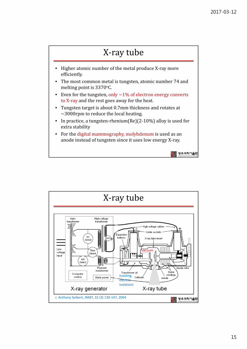
X‐raytube
vacuum
(cooling,electric isolation)
J. Anthony Seibert, JNMT, 32 (3) 139‐147, 2004

2017-03-12
16
X‐raytube
• X‐raygeneratorandx‐raytubecomponentsareillustrated.– Thex‐raygeneratorprovidesoperatorcontroloftheradiographictechniques,includingtubevoltage(kVp,kilovoltspeak),tubecurrent(mA),andexposureduration,anddeliverspowertothex‐raytube.
– Thex‐raytubeprovidestheenvironment(evacuatedx‐raytubeinsertandhigh‐voltagecablesockets),sourceofelectrons(cathode),sourceofx‐rays(anode),inductionmotortorotatetheanode(rotor/stator),transformeroilandexpansionbellowstoprovideelectricalandheatbuild‐upprotection,andthetubehousingtosupporttheinsertandprovideprotectionfromleakageradiation.
J. Anthony Seibert, JNMT, 32 (3) 139‐147, 2004
X‐raytube
http://www.waybuilder.net/sweethaven/MedTech/Dental/DentalRad/lessonMain.asp?iNum=fra0102
http://www.dentalxraywebsite.com/category/dental‐x‐ray‐tube/
Toshiba X‐ray Tube History 1915‐2005

2017-03-12
17
X‐raytube
• ManyX‐raytubeshavetwocathodefilamentsofdifferentlength
– alongone:highercurrent/lowerresolution
– ashortone:lowercurrent/higherresolution
Focusing cup
X‐raytube
• Anodeisbeveledatananglebetween8and17o (normally12‐15o)
• Thesmallerangle(θ) producesthesmallerfocalsize(f)
f =Fsin θ• frangesfrom0.3mmfordigitalmammographyandtobetween0.6and1.2mmforplanarX‐rayandCT
• BevelanglealsoaffectsthecoverageareaCoverage=2(source‐patientdistant)xtan θ(eg. 2(1m)*tan15o=0.53m)

2017-03-12
18
X‐raytube
• Heeleffect:X‐raybeamismoreintenseatthe‘cathode‐end’thanatthe‘anode‐end’
• ItisfromtheabsorptionofX‐raytothetargetitself.(moreX‐rayabsorptionattheanodeside)
• Therefore,signalintensityatcathodesideishigherthananodesideinplanarradiographyandcanbecorrectedbyimageprocessingalgorithms.
• However,inpracticeitdoesnotaffectthediagnosticqualityoftheimagessignificantly.
X‐raytube
• 3parameterstocontrol1. Acceleratingvoltage(kVp)
• ~25kVfordigitalmammographyand~140kVforboneandchestX‐ray
2. Tubecurrent(mA)• 50~400mAforplanarX‐ray• ~1000mAforCT
3. Exposuretime(sec)
• Powerrating:themaximumpowerdissipatedinanexposuretimeof0.1s
– Ex)10kWpowerrating:kVp of125kVwith1Atubecurrentfor~78ms
– X‐raytubeoutputismainlylimitedbyanodeheating

2017-03-12
19
Practice
• Ifthethicknessofthechestis20cm,whatpercentageofX‐raysaretransmittedthroughthechestatanincidentX‐rayenergyof70keVassumingHVLvaluesof3.5and1.8cmformuscleandbone,respectively,andthebonethicknesstobe4cmandthetissuethickness16cm?
Answer) Forthemuscle,μ =(ln 2)/3.5≈0.2cm‐1. For16cmoftissue:N/No =exp(‐0.2*16)=0.04. Forthebone,μ =(ln 2)/1.8≈0.4cm‐1. For4cmofbone:N/No =exp(‐0.4*4)=0.2. Therefore,theoverallpercentageis
100X(0.2X0.04)=0.8%


