Corneal Pathologies
Dr. Pradeep Bastola MD, Ophthalmologist
Assistant Professor 15th June, 2011
PERIPHERAL CORNEAL THINNING AND ULCERATION
1. Without systemic disease • Dellen • Terrien marginal degeneration • Mooren ulcer
2. With systemic disease • Rheumatoid arthritis • Wegener granulomatosis • Polyarteritis nodosa
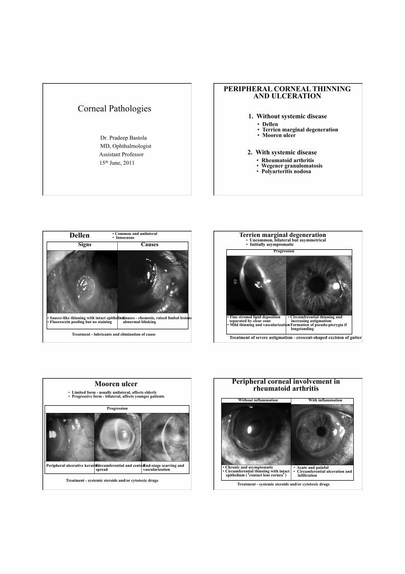
Dellen
• Saucer-like thinning with intact epithelium • Fluorescein pooling but no staining
• Causes - chemosis, raised limbal lesions, abnormal blinking
Treatment - lubricants and elimination of cause
Signs Causes
• Common and unilateral • Innocuous Terrien marginal degeneration
• Uncommon, bilateral but asymmetrical • Initially asymptomatic
• Fine stromal lipid deposition separated by clear zone • Mild thinning and vascularization
• Circumferential thinning and increasing astigmatism • Formation of pseudo-pterygia if longstanding
Progression
Treatment of severe astigmatism - crescent-shaped excision of gutter
Mooren ulcer • Limited form - usually unilateral, affects elderly • Progressive form - bilateral, affects younger patients
Peripheral ulcerative keratitis Circumferential and central spread
Progression
End-stage scarring and vascularization
Treatment - systemic steroids and/or cytotoxic drugs
Peripheral corneal involvement in rheumatoid arthritis
• Chronic and asymptomatic • Circumferential thinning with intact epithelium (‘contact lens cornea’)
• Acute and painful • Circumferential ulceration and infiltration
Treatment - systemic steroids and/or cytotoxic drugs
Without inflammation With inflammation
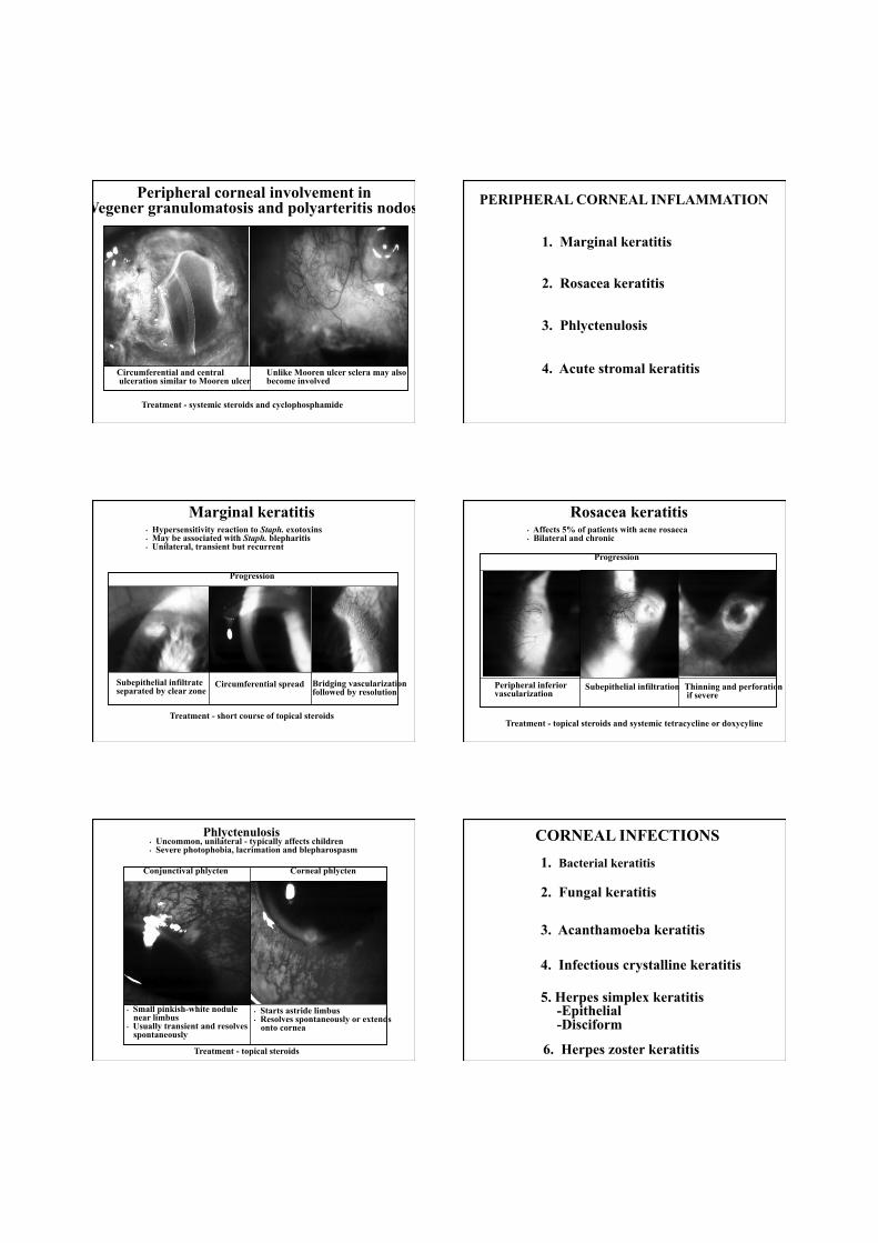
Peripheral corneal involvement in Wegener granulomatosis and polyarteritis nodosa
Circumferential and central ulceration similar to Mooren ulcer
Unlike Mooren ulcer sclera may also become involved
Treatment - systemic steroids and cyclophosphamide
PERIPHERAL CORNEAL INFLAMMATION
1. Marginal keratitis
2. Rosacea keratitis
3. Phlyctenulosis
4. Acute stromal keratitis
Marginal keratitis
Subepithelial infiltrate separated by clear zone
Circumferential spread Bridging vascularization followed by resolution
• Hypersensitivity reaction to Staph. exotoxins • May be associated with Staph. blepharitis • Unilateral, transient but recurrent
Progression
Treatment - short course of topical steroids
Rosacea keratitis
Peripheral inferior vascularization
Subepithelial infiltration Thinning and perforation if severe
• Affects 5% of patients with acne rosaeca • Bilateral and chronic
Progression
Treatment - topical steroids and systemic tetracycline or doxycyline
Phlyctenulosis
• Small pinkish-white nodule near limbus • Usually transient and resolves spontaneously
• Starts astride limbus • Resolves spontaneously or extends onto cornea
• Uncommon, unilateral - typically affects children • Severe photophobia, lacrimation and blepharospasm
Conjunctival phlycten
Treatment - topical steroids
Corneal phlycten
CORNEAL INFECTIONS 1. Bacterial keratitis
2. Fungal keratitis
3. Acanthamoeba keratitis
4. Infectious crystalline keratitis
5. Herpes simplex keratitis -Epithelial -Disciform
6. Herpes zoster keratitis
Bacterial keratitis Predisposing factors • Contact lens wear • Chronic ocular surface disease • Corneal hypoaesthesia
Expanding oval, yellow-white, dense stromal infiltrate
Stromal suppuration and hypopyon
Treatment - topical ciprofloxacin 0.3% or ofloxacin 0.3%
Fungal keratitis Frequently preceded by ocular trauma with organic matter
Greyish-white ulcer which may be surrounded by feathery infiltrates
Slow progression and occasionally hypopyon
• Topical antifungal agents • Systemic therapy if severe • Penetrating keratoplasty if unresponsive
Treatment
Acanthamoeba keratitis • Contact lens wearers at particular risk • Symptoms worse than signs
Small, patchy anterior stromal infiltrates
Perineural infiltrates (radial keratoneuritis)
Ulceration, ring abscess & small, satellite lesions
- chlorhexidine or polyhexamethylenebiguanide
Stromal opacification
Treatment
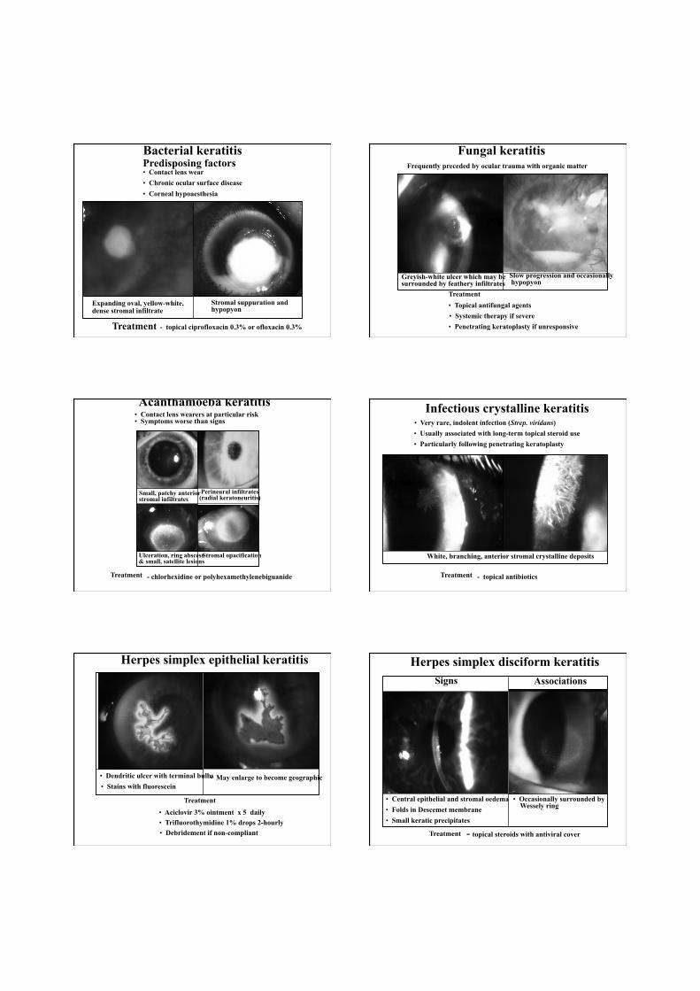
Infectious crystalline keratitis • Very rare, indolent infection (Strep. viridans)
• Particularly following penetrating keratoplasty
White, branching, anterior stromal crystalline deposits
- topical antibiotics Treatment
• Usually associated with long-term topical steroid use
Herpes simplex epithelial keratitis
• Dendritic ulcer with terminal bulbs • Stains with fluorescein
• May enlarge to become geographic
• Aciclovir 3% ointment x 5 daily • Trifluorothymidine 1% drops 2-hourly • Debridement if non-compliant
Treatment
Herpes simplex disciform keratitis
• Central epithelial and stromal oedema • Folds in Descemet membrane • Small keratic precipitates
- topical steroids with antiviral cover
• Occasionally surrounded by Wessely ring
Treatment
Signs Associations

Herpes zoster keratitis
• Develops in about 50% within 2 days of rash • Small, fine, dendritic or stellate epithelial lesions • Tapered ends without bulbs • Resolves within a few days
• Develops in about 30% within 10 days of rash • Multiple, fine, granular deposits just beneath Bowman membrane • Halo of stromal haze
Nummular keratitis Acute epithelial keratitis
• May become chronic
Treatment - topical steroids, if appropriate
Acute stromal keratitis
Superficial or mid-stromal infiltration Opacification and vascularization
• Uncommon, usually unilateral • Associated with non-necrotizing scleritis
Progression
Treatment - topical steroids and systemic NSAIDs
CORNEAL DEGENERATIONS 1. Age-related
• Arcus senilis • Vogt white limbal girdle
2. Lipid keratopathy
• Crocodile shagreen • Cornea guttata
• Primary • Secondary
3. Band keratopathy 4. Spheroidal degeneration 5. Salzmann nodular degeneration
Arcus senilis • Innocuous and extremely common in elderly • Occasionally associated with hyperlipoproteinaemia
• Bilateral, circumferential bands of lipid deposits • Diffuse central and sharp peripheral border
• Peripheral border separated from limbus by clear zone • Clear zone may be thinned ( senile furrow)
Vogt white limbal girdle • Innocuous and very common in elderly • Bilateral
• White, crescentic line along nasal and temporal limbus • Type 1 - separated from limbus by clear zone • Type 2 - not separated by clear zone
Crocodile shagreen • Uncommon and innocuous • Usually bilateral
• Polygonal stromal opacities separated by clear space • Most frequently involve anterior stroma (anterior crocodile shagreen) • Occasionally involve posterior stroma (posterior crocodile shagreen)

Cornea guttata • Common, bilateral and usually innocuous • Rarely progression to Fuchs dystrophy
• Tiny dark spots on central endothelium • Similar peripheral lesions are Hassell-Henle bodies
Causes of Band Keratopathy
1. Ocular (common)
• Silicone oil in anterior chamber
• Increased serum calcium and phosphorus
• Chronic iridocyclitis, particularly in children
• Hyperuricaemia
• Associated with phthisis bulbi
3. Hereditary (rare) • Chronic renal failure
• Familial band keratopathy • Hereditary ichthyosis
2. Metabolic (rare)
4. Age-related (uncommon)
Removal of corneal epithelium Application of sodium versenate
Chelation of band keratopathy Spheroidal degeneration
Treatment Central spread, coalescence and opacification
Advanced lesions become nodular and elevated
• Rare, typically affects outdoor workers
• Starts with peripheral, interpalpebral, small amber-coloured granules in superficial stroma
• Debridement or superficial keratotomy if mild • Keratoplasty if severe
Progression
Salzmann nodular degeneration • Uncommon, unilateral or bilateral • Secondary to chronic keratitis
• Discrete superficial stromal opacities and nodules • Base of nodule may be surrounded by iron deposits
Treatment - similar to spheroid degeneration
Spheroidal degeneration
Treatment Central spread, coalescence and opacification
Advanced lesions become nodular and elevated
• Rare, typically affects outdoor workers
• Starts with peripheral, interpalpebral, small amber-coloured granules in superficial stroma
• Debridement or superficial keratotomy if mild • Keratoplasty if severe
Progression
CORNEAL DYSTROPHIES 1. Anterior
• Cogan microcystic • Reis-Bucklers • Meesmann • Schnyder
2. Stromal • Lattice I, II, III • Granular I, II, III (Avellino) • Macular
3. Posterior • Fuchs endothelial • Posterior polymorphous
..
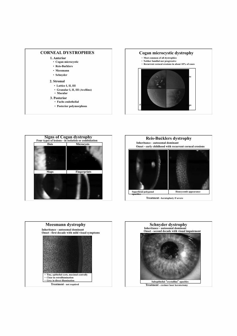
Cogan microcystic dystrophy • Most common of all dystrophies • Neither familial nor progressive • Recurrent corneal erosions in about 10% of cases
Dots
Fingerprints
Cysts
Maps
Four types of lesions - in isolation or combination Microcysts Dots
Maps Fingerprints
Signs of Cogan dystrophy Inheritance - autosomal dominant Onset - early childhood with recurrent corneal erosions
Superficial polygonal opacities
Honeycomb appearance
Treatment - keratoplasty if severe
Reis-Bucklers dystrophy ..
Meesmann dystrophy Inheritance - autosomal dominant Onset - first decade with mild visual symptoms
Treatment - not required
• Clear in retroillumination • Grey in direct illumination
• Tiny, epithelial cysts, maximal centrally
Inheritance - autosomal dominant Onset - second decade with visual impairment
Treatment - excimer laser keratectomy
Schnyder dystrophy
Subepithelial ‘crystalline’ opacities

Inheritance - autosomal dominant Onset - late first decade with recurrent corneal erosions
Treatment - penetrating keratoplasty if severe
Lattice dystrophy type I
Fine, spidery, branching lines within stroma
Later general haze may submerge lesions
Progression Inheritance Autosomal dominant
Onset Middle age with progressive facial palsy and lattice dystrophy identical to type I
Systemic features • Cranial and peripheral neuropathy • Skin laxity • Renal and cardiac failure
Treatment Penetrating keratoplasty if severe
Lattice dystrophy type II (Familial amyloidosis with lattice dystrophy, Meretoja syndrome)
Mask-like facies
Inheritance - autosomal dominant Onset - fourth decade
• Thick, ropey lines and minimal intervening haze • May be asymmetrical and initially unilateral
Treatment - penetrating keratoplasty if severe
Lattice dystrophy type III
Inheritance - autosomal dominant Onset - first decade with recurrent corneal erosions
Treatment - penetrating keratoplasty if severe
Granular dystrophy type I
Eventual confluence Initial superficial and central crumb-like opacities
Later deeper and peripheral spread but limbus spared
Progression
Inheritance - autosomal dominant Onset - fourth or fifth decade with mild recurrent erosions
Superficial, discrete crumb-like opacities Treatment - penetrating keratoplasty if severe
Granular dystrophy type II Inheritance - autosomal dominant Onset - late in life; frequently asymptomatic
• Few, superficial, discrete, ring-shaped lesions • Increase in density and size with time
Treatment - not required
Granular dystrophy type III (Avellino)

Inheritance - autosomal recessive Onset - second decade with painless visual loss
Macular dystrophy
Treatment - penetrating keratoplasty
Initial dense, poorly delineated opacities
Later generalized opacification
Thinning
Progression
Inheritance - occasionally autosomal dominant Onset - old age
Treatment - penetrating keratoplasty if advanced
Fuchs endothelial dystrophy
Eventually bullous keratopathy
Later central stromal oedema
Gradual increase in cornea guttata with peripheral spread
Progression
Inheritance - usually autosomal dominant Onset - difficult to determine because asymptomatic
• Subtle, vesicular, geographic, or band-like lesions • Frequently asymmetrical
Treatment - not required
Posterior polymorphous dystrophy
1. Keratoconus
2. Keratoglobus
3. Pellucid marginal degeneration
CORNEAL ECTASIAS
Morphological classification of keratoconus
Nipple cone Oval cone Globus cone
Small and steep curvature Larger and ellipsoidal Largest
Signs of keratoconus Bilateral in 85% but asymmetrical
Oil droplet reflex Prominent corneal nerves Vogt striae
Acute hydrops
Munson sign Fleischer ring & scarring
Bulging of lower lids on downgaze

Systemic associations of keratoconus
Crouzon syndrome Marfan syndrome Osteogenesis imperfecta
Atopic dermatitis Down syndrome Ehlers-Danlos syndrome
Keratoglobus
• Bilateral protrusion and thinning of entire cornea • Associations - Leber congenital amaurosis and blue sclera
• Onset usually at birth
Pellucid marginal degeneration
• Bilateral crescent-shaped inferior corneal thinning • Onset between 20 and 40 years
1. Microcornea
2. Megalocornea
3. Sclerocornea
4. Cornea plana
5. Keratectasia
CONGENITAL CORNEAL ANOMALIES
Microcornea • Very rare, hereditary, unilateral or bilateral • Corneal diameter is 10 mm or less • Shallow anterior chamber but other dimensions are normal
Associated systemic syndromes Turner, Ehlers-Danlos, Weill-Marchesani and Waardenburg
Ocular associations Glaucoma, cataract, cornea plana, leukoma and iris abnormalities
Megalocornea
• Renal carcinoma and mental handicap
Systemic associations • Marfan, Apert, Ehlers-Danlos and Down syndromes
• Osteogenesis imperfecta
• Very rare, hereditary, bilateral • Corneal diameter 13 mm or more • Very deep anterior chamber
• High myopia and astigmatism • Occasionally lens subluxation

Sclerocornea • Very rare, usually bilateral • Peripheral opacification and vascularization of cornea • ‘Scleralization’ makes cornea appear smaller
Cornea plana • Very rare, bilateral severe decrease in corneal curvature • Hypermetropia and shallow anterior chamber
Ocular associations Glaucoma, microcornea, microphthalmos and Peters anomaly
Keratectasia • Very rare, usually unilateral • Severe corneal opacification and protruberance • Probably caused by intrauterine keratitis
CORNEAL SURGERY 1. Penetrating keratoplasty
2. Keratoprosthesis
3. Refractive surgery • Radial keratotomy • Photorefractive keratectomy (PRK) • Laser in-situ keratomileusis (LASIK) • Non-contact laser thermal keratoplasty
Penetrating Keratoplasty
1. Indications • Optical (e.g. bullous keratopathy, dystrophies) • Tectonic (e.g. severe stromal thinning, descemetocele) • Therapeutic (e.g. severe keratitis) • Cosmetic
• Severe stromal vascularization • Absence of corneal sensation • Progressive conjunctival inflammation (e.g. pemphigoid) • Tear film dysfunction • Glaucoma
2. Adverse prognostic factors
Technique of penetrating keratoplasty
Excision of donor tissue a, b - Excision of host tissue c - Fixation of donor tissue
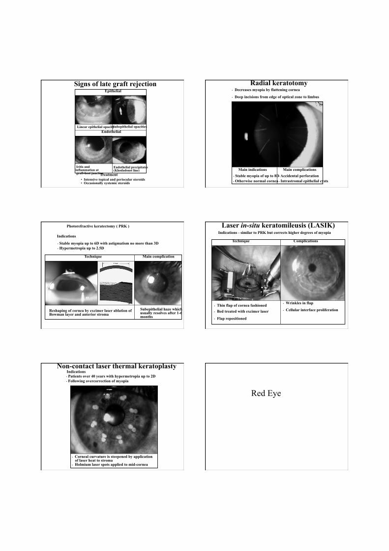
Signs of late graft rejection Epithelial
• Intensive topical and periocular steroids • Occasionally systemic steroids
Endothelial
Treatment
Iritis and inflammation at graft-host junction
Endothelial precipitates (Khodadoust line)
Linear epithelial opacity Subepithelial opacities
Radial keratotomy
Main indications • Stable myopia of up to 8D • Otherwise normal cornea
• Accidental perforation • Intrastromal epithelial cysts
Main complications
• Decreases myopia by flattening cornea
• Deep incisions from edge of optical zone to limbus
Photorefractive keratectomy ( PRK )
Indications
• Stable myopia up to 6D with astigmatism no more than 3D • Hypermetropia up to 2.5D
Main complication
Subepithelial haze which usually resolves after 1-6 months
Reshaping of cornea by excimer laser ablation of Bowman layer and anterior stroma
Technique
Laser in-situ keratomileusis (LASIK) Indications - similar to PRK but corrects higher degrees of myopia
• Thin flap of cornea fashioned • Bed treated with excimer laser
• Flap repositioned
Complications
• Wrinkles in flap
• Cellular interface proliferation
Technique
Non-contact laser thermal keratoplasty Indications • Patients over 40 years with hypermetropia up to 2D • Following overcorrection of myopia
• Corneal curvature is steepened by application of laser heat to stroma • Holmium laser spots applied to mid-cornea
Red Eye
Introdution
• Relevance – Red Eye
• Frequent presentation to GP • Must be able to differentiate between serious vision
threatening conditions and simple benign conditions
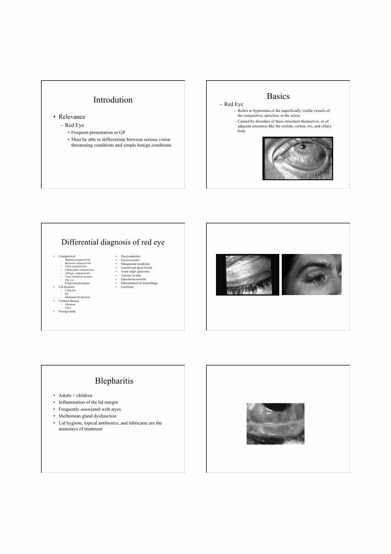
Basics – Red Eye
– Refers to hyperemia of the superficially visible vessels of the conjunctiva, episclera, or the sclera
– Caused by disorders of these structures themselves, or of adjacent structures like the eyelids, cornea, iris, and ciliary body
Differential diagnosis of red eye • Conjunctival
– Blepharoconjunctivitis – Bacterial conjunctivitis – Viral conjunctivitis – Chlamydial conjunctivitis – Allergic conjunctivitis – Toxic/chemical reaction – Dry eye – Pinguecula/pteyrgium
• Lid diseases – Clalazion – Sty – Abnormal lid function
• Corneal disease – Abrasion – Ulcer
• Foreign body
• Dacryoadenitis • Dacryocystitis • Masquerade syndrome • Carotid and dural fistula • Acute angle glaucoma • Anterior uveitis • Episcleritis/scleritis • Subconjunctival hemorrhage • Factitious
Blepharitis • Adults > children • Inflammation of the lid margin • Frequently associated with styes • Meibomian gland dysfunction • Lid hygiene, topical antibiotics, and lubricants are the
mainstays of treatment

Bacterial Conjunctivitis • Both adults and children • Tearing, foreign body sensation, burning, stinging and
photophobia • Mucopurulent or purulent discharge • Lid and conjunctiva maybe edematous • Streptococcus pneumoniae, Haemophilus influenzae, and
staphylococcus aureus and epidermidis • Conjunctival swab for culture • Topical broad spectrum antibiotics
Viral Conjunctivitis – Acute, watery red eye with soreness, foreign body sensation and
photophobia – Conjunctiva is often intensely hyperaemic and there maybe
follicles, haemorrhages, inflammatory membranes and a pre-auricular node
– The most common cause is an adenoviral infection – No specific therapy but cold compresses are helpful
Allergic Conjunctivitis – Encompasses a spectrum of clinical condition – All associated with the hallmark symptom of itching – There is often a history of rhinitis, asthma and family history of
atopy – Signs may include mildly red eyes, watery discharge, chemosis,
papillary hypertrophy and giant papillae – Treatment consist of cold compresses, antihistamines,
nonsteroidals, mast cells stabilizers, topical corticosteroids and cyclosporine
Chlamydial Conjunctivitis
– Usually occur in sexually active individuals with or without an associated genital infection
– Conjunctivitis usually unilateral with tearing, foreign body sensation, lid crusting, conjunctival discharge and follicles
– There is often non-tender preauricular node – Treatments requires oral tetracycline or azithromycin
Dry Eye • Symptoms
– Burning or foreign body sensation – Tearing – Usually bilateral
• Etiology – Idiopathic – Collagen vascular diseases – Conjunctival scarring – Infiltration of the lacrimal gland – Vitamin A deficiency
• Treatment – Artificial tears
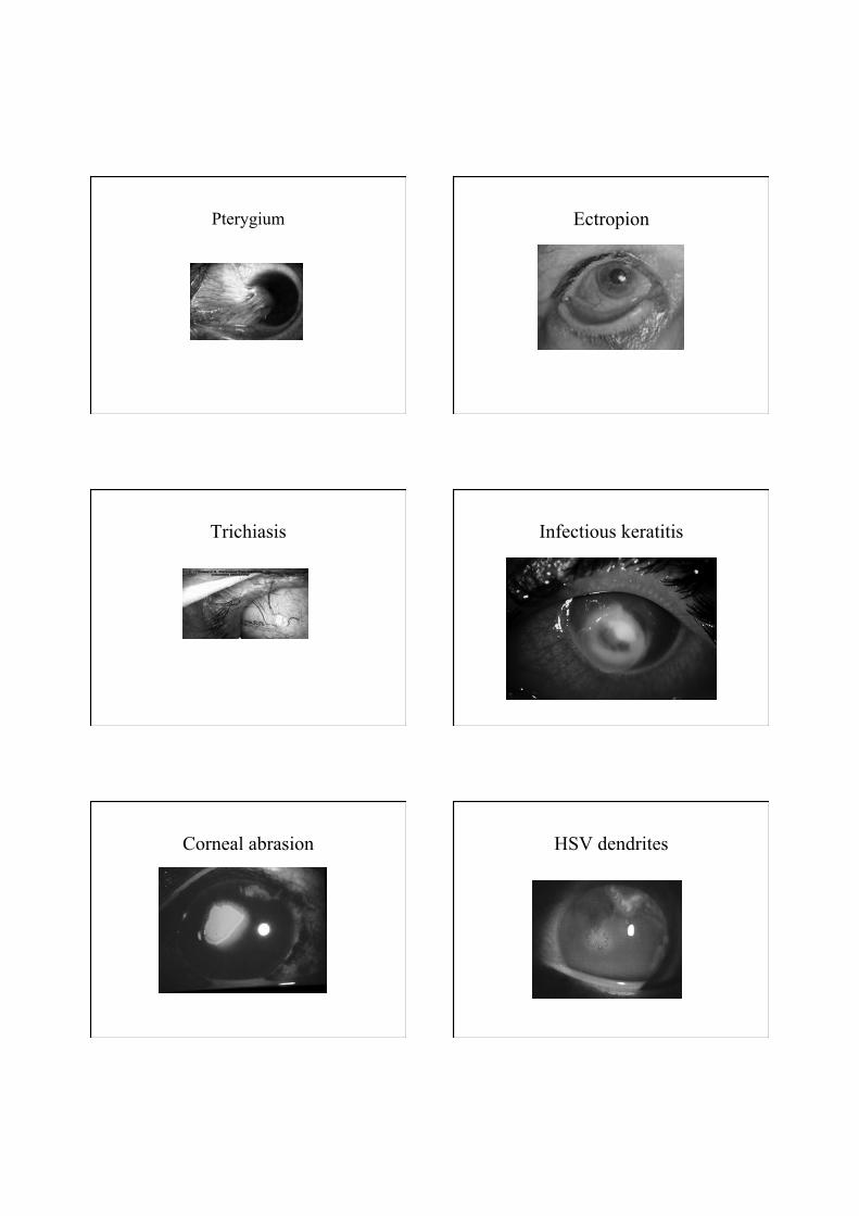
Pterygium
Ectropion
Trichiasis Infectious keratitis
Corneal abrasion HSV dendrites
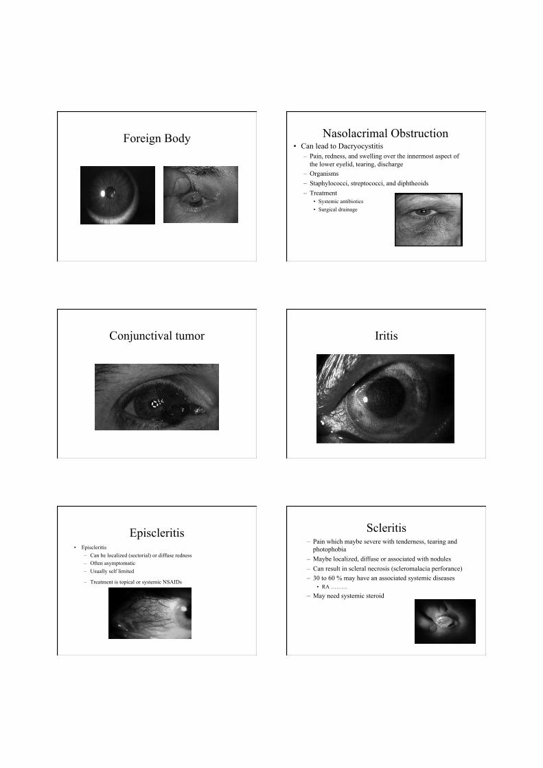
Foreign Body Nasolacrimal Obstruction • Can lead to Dacryocystitis
– Pain, redness, and swelling over the innermost aspect of the lower eyelid, tearing, discharge
– Organisms – Staphylococci, streptococci, and diphtheoids – Treatment
• Systemic antibiotics • Surgical drainage
Conjunctival tumor Iritis
Episcleritis • Episcleritis
– Can be localized (sectorial) or diffuse redness – Often asymptomatic – Usually self limited
– Treatment is topical or systemic NSAIDs
Scleritis – Pain which maybe severe with tenderness, tearing and
photophobia – Maybe localized, diffuse or associated with nodules – Can result in scleral necrosis (scleromalacia perforance) – 30 to 60 % may have an associated systemic diseases
• RA ………
– May need systemic steroid

Subconjunctival Hemorrhage • Usually asymptomatic • Blood underneath the conjunctiva, often in a sector of the eye • Etiology
– Valsalva (coughing or straining) – Traumatic – Hypertension – Bleeding disorder – idiopathic
Red Eye Treatment Algorithm • History
– Trauma – Contact lens wearer – Severe pain/photophobia – Significant vision changes – History of prior ocular diseases
• Exam – Abnormal pupil – Ocular tenderness – White corneal opacity – Increased intraocular pressure
YES
Refer urgently to ophthalmologist
Is it conjunctivitis?
• History – Itching – Exposure to person
with red eye – URTI – Past history of
conjunctivitis – Discharge with
morning crusting – Exposure to drugs
• Signs – Discharge – Lid and conjunctival
edema – Conjunctival redness – Preauricular lymph
node – Facial or eye lid
vesicles
Thank you


