Download - 8 gastric ulceration

8 Gastric Ulceration
CLINICAL IMAGAGINGAN ATLAS OF DIFFERENTIAL DAIGNOSIS
EISENBERG
DR. Muhammad Bin Zulfiqar PGR-FCPS III SIMS/SHL

• Fig GI 8-1 Fold patterns in gastric ulcers (arrow). (A) Small, slender folds radiating to the edge of a benign ulcer. (B) Thick folds radiating to an irregular mound of tissue surrounding a malignant gastric ulcer (arrow).
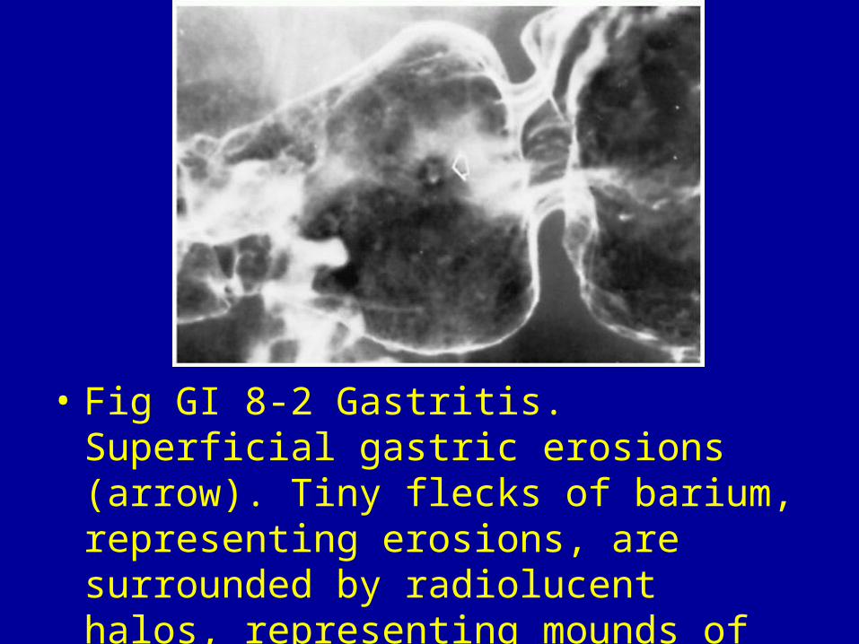
• Fig GI 8-2 Gastritis. Superficial gastric erosions (arrow). Tiny flecks of barium, representing erosions, are surrounded by radiolucent halos, representing mounds of edematous mucosa.

• Fig GI 8-3 MALT lymphoma. Greater curvature ulcer (arrow) surrounded by a soft-tissue mass and associated with regional enlargement of rugal folds.

• Fig GI 8-4 Carcinoma of the stomach. Carman's meniscus sign in malignant gastric ulcer. The huge ulcer has a semicircular configuration with its inner margin convex toward the lumen. The ulcer is surrounded by the radiolucent shadow of an elevated ridge of neoplastic tissue (arrows).
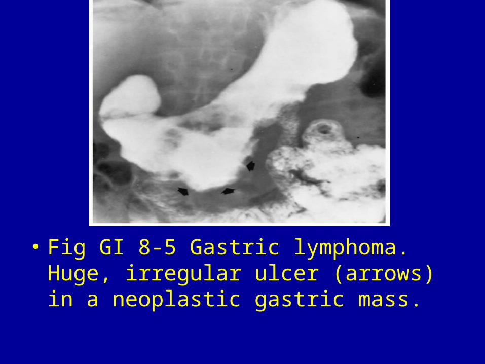
• Fig GI 8-5 Gastric lymphoma. Huge, irregular ulcer (arrows) in a neoplastic gastric mass.

Fig GI 8-6 Leiomyosarcoma of the stomach. The large fundal mass (arrows) shows exophytic extension and ulceration.
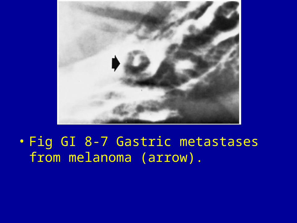
• Fig GI 8-7 Gastric metastases from melanoma (arrow).


