ORIGINAL PAPER
A comparative morphology of the male genitalia of Aphididae(Insecta, Hemiptera): part 2
Karina Wieczorek • Bartosz J. Płachno •
Piotr Swiatek
Received: 30 November 2011 / Revised: 24 May 2012 / Accepted: 29 May 2012 / Published online: 26 June 2012
� The Author(s) 2012. This article is published with open access at Springerlink.com
Abstract The present study provides new data related to
the morphology of the male genitalia of Aphididae. The
structure of the male genitalia of 39 species from 23 genera
of Aphididae was studied using light and scanning electron
microscopy. In the species studied, the genitalia of males
consist of a phallus composed of the sclerotized basal part
with its articulation and a membranous apical part—an
aedeagus as well as parameres. This state probably repre-
sents the hypothetical plesiomorphic condition of the
external male genitalia of aphids. According to the results
of the present study, the male genitalia vary among sub-
families (the most varied in Lachninae). Both the phallus
and parameres show great variability in their form and the
number of setae and may provide characters of taxonomic
and diagnostic importance. The shape, size, and modifi-
cation of parameres are considered in conjunction with the
phylogenetic relationships among the studied taxa. Com-
pared with Lachninae, Greenideinae, Aiceoninae, the
external genitalia of Aphidinae are less specialized, having
many features in common with those of drepanosiphine
aphids and differing little from the hypothetical condition.
In dwarfish males of Anoeciinae, Thelaxinae, Hormaph-
idinae, and Eriosomatinae, the miniaturization of the body
size affects on the modification of genitalia, mostly para-
meres. However, the homology of non-modified and
modified structures of parameres is not clear.
Keywords External reproductive system � Parameres �Phallus � Insects � SEM
Introduction
The male genitalia have proven to be of considerable tax-
onomic value for various groups of insects. This paper
continues our studies into comparative morphology and
ultrastructure of the internal and external male genitalia of
Aphididae (Insecta, Hemiptera) (Wieczorek 2006, 2008;
Wieczorek and Swiatek 2008, 2009; Wieczorek et al. 2011).
In comparison with other hemipterans, the external male
genitalia of aphid species are characterized by simplicity;
however, they are well developed and typically consist of
the phallus composed of the sclerotized basal part with its
articulation and a membranous apical part—the aedeagus.
Laterally and above the phallus, there is a pair of setose
parameres. In the first part of our studies devoted to external
male genitalia (Wieczorek et al. 2011), the general mor-
phologies of these structures were described and compared
in those representatives of the Aphididae in which the
genitalia are not modified—Drepanosiphinae, Chaitophor-
inae, Calaphidinae, Phyllaphidinae, Saltusaphidinae, Lize-
riinae, Spicaphidinae, Tamalinae, Parachaitophorinae,
Phloeomyzinae, and Aphidinae.
In the present study, strongly modified structures of the
male genitalia in dwarfish males of the Mindarinae,
Communicated by T. Bartolomaeus.
K. Wieczorek (&)
Department of Zoology, University of Silesia,
Bankowa 9, 40-007 Katowice, Poland
e-mail: [email protected]
B. J. Płachno
Department of Plant Cytology and Embryology,
Jagiellonian University, Grodzka 52, 31-044 Cracow, Poland
e-mail: [email protected]
P. Swiatek
Department of Animal Histology and Embryology,
University of Silesia, Bankowa 9, 40-007 Katowice, Poland
e-mail: [email protected]
123
Zoomorphology (2012) 131:303–324
DOI 10.1007/s00435-012-0163-2

Hormaphidinae, Anoeciinae, Thelaxinae, and Eriosomati-
nae as well as in normal-sized males of the Greenideinae,
Aiceoninae, and Lachninae are described. To date, the
external male genitalia of species belonging to these groups
have only been studied in a limited number of species [e.g.,
Eriosoma lanigerum (Hausmann, 1802) (Baker 1915; de
Fluiter 1931); Thelaxes dryophila (Schrank, 1801) (Pola-
szek 1987a); Greenidea (Trichosiphum) anonae (Pergande,
1906) (Chaudhuri 1956); Aiceona japonica Takahashi,
1960 (Takahashi 1960); Stomaphis species (Sorin 1965);
Cinara species (Eastop 1972); Eutrichosiphum assamense
A. K. Ghosh, R. C Basu & D. N. Raychaudhuri, 1969
(Chakrabarti and Maity 1980)]. They have usually been
poorly described or simply illustrated. Only Polaszek
(1987b in lit.) provided a comprehensive study of the male
genitalia of the entire family Aphididae, using numerous
examples from most of these subfamilies.
In this study, we present the general morphologies of the
modified male genitalia of selected species of Aphididae,
mainly the variability of parameres in their form and
location, and consider these structures in conjunction with
the phylogenetic relationships among the entire family.
Materials and methods
Taxon sampling
The structure of the male genitalia of 39 species belonging to
the Mindarinae, Hormaphidinae, Anoeciinae, Thelaxinae,
Eriosomatinae, Greenideinae, Aiceoninae, and Lachninae
was studied using light (LM) and scanning electron micros-
copy (SEM). All of the species studied were borrowed from
the Museum national d’Histoire naturelle, Paris, France, with
the exception of Pachypappa tremulae (Linne, 1761), Proc-
iphilus fraxini (Fabricius, 1777), Anomalosiphum mendeli
Quednau & Martin 2006, and Schoutedenia ralumensis
Rubsaamen, 1905, which were borrowed from the Natural
History Museum, London, United Kingdom. The adult males
of Glyphina betulae (Linne, 1758), Lachnus roboris (Linne,
1758) and nymphs of Stomaphis quercus (Linne, 1758) were
collected in Poland in the years 2008–2011. Because of the
rarity of males, sample sizes consisted of 1–10 individuals.
The genitalia of 39 species were studied from aphid material
preserved in alcohol and slide-mounted specimens (LM); 13
species were also observed using SEM techniques. The col-
lection data and the microscopic techniques used are sum-
marized in Table 1.
Light and electron microscopy
Alcohol-preserved specimens and slides were examined
and photographed using Nikon Eclipse 600 and Leica
DMR light microscopes. Drawings were made with a
camera lucida. A magnified view is provided for each of
the photographs and drawings.
The procedure for preparing samples for SEM was as
described earlier (Płachno and Swiatek 2009, 2010).
Briefly, whole specimens were fixed with 2.5 % glutaral-
dehyde in a 0.1 M phosphate buffer (pH 7.4) for several
days or fixed in 70 % ethanol. Later, the samples, which
had been dehydrated in ethanol as well as an acetone series,
were critical-point dried in liquid CO2 and coated with gold
using a JEOL-JFC 1100E sputter coater. The specimens
were viewed using a HITACHI S-4700 microscope
(Scanning Microscopy Laboratory of Biological and Geo-
logical Sciences, Jagiellonian University) at 20 kV.
Results
Morphology
The external male genitalia of Aphididae consist of the
phallus, which is composed of the partially sclerotized
basal part with its articulation (sclerotized arms) and a
membranous apical part, the aedeagus, as well as para-
meres. These structures are defined in the following way.
The phallus is the median intromittent organ as a whole.
The basal part of the phallus is a pair of partially sclerotized,
various-shaped lobes which enfold the aedeagus and take part
in everting and maintaining it in position during copulation.
The sclerotized arms of the basal part of the phallus are a pair of
sclerotized projections divided into distal and proximal part,
giving support for the basal part of the phallus.
The aedeagus is a membranous, copulatory part of the
phallus, which is withdrawn within the body and everted
during copulation.
The parameres is usually a pair of various-shaped, setose
ventral processes positioned at the anterior end of the genital
area. In some species of Aphididae, the parameres are
strongly modified: completely fused into a single, sharply
pointed structure (Anoeciinae, Thelaxinae), reduced to
elongated projections supported by additional sclerotization
(Eriosomatinae), divided into lobate parts which arise into
various-shaped projections toward base of the phallus
(Greenideinae, Aiceoninae and partially Lachninae).
The general morphologies of these structures of selected
species of Aphididae were described and individual species
were then studied systematically (the classification of
Aphidoidea is after Nieto Nafria et al. 1998)
Mindarinae
Mindarus abietinus Koch, 1857 (male wingless, dwarf-
ish)—parameres are present, located above the basal part of
304 Zoomorphology (2012) 131:303–324
123
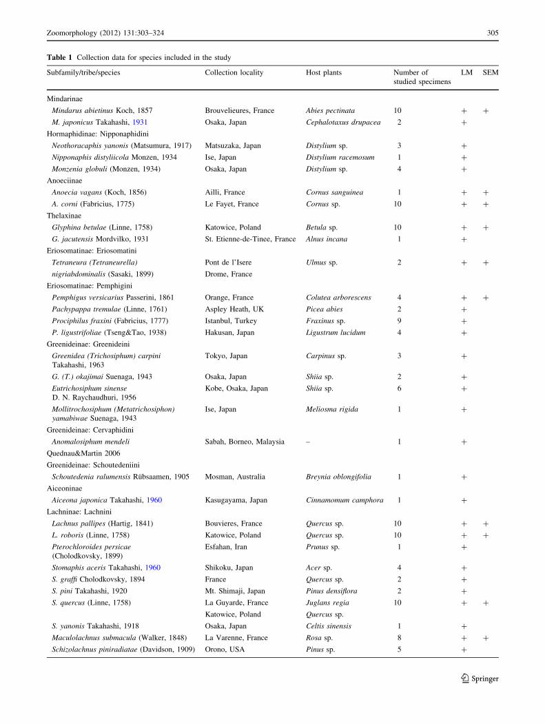
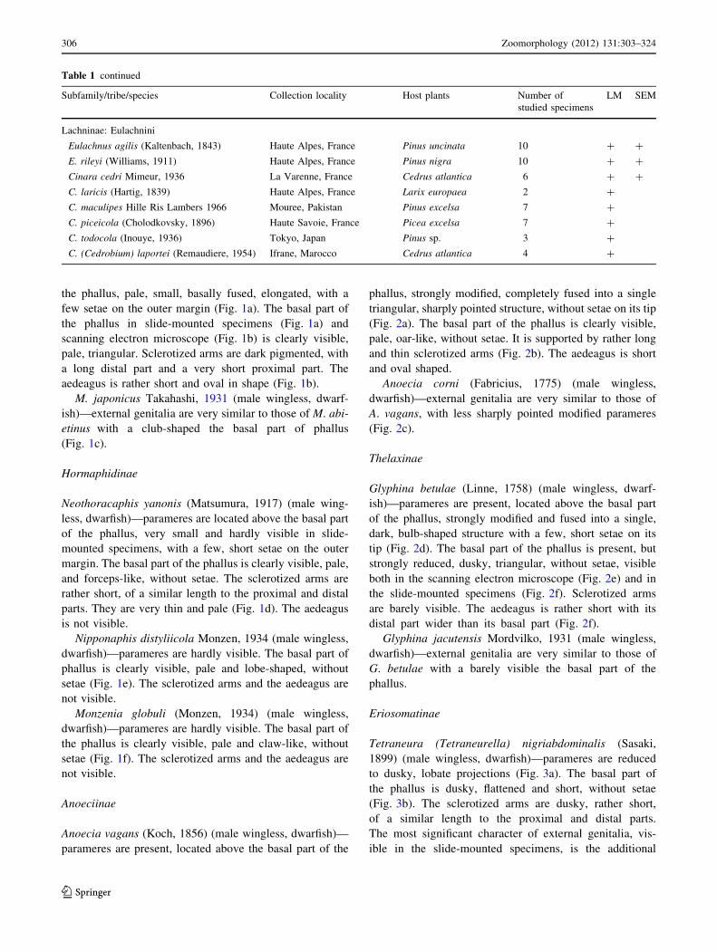
Table 1 Collection data for species included in the study
Subfamily/tribe/species Collection locality Host plants Number of
studied specimens
LM SEM
Mindarinae
Mindarus abietinus Koch, 1857 Brouvelieures, France Abies pectinata 10 ? ?
M. japonicus Takahashi, 1931 Osaka, Japan Cephalotaxus drupacea 2 ?
Hormaphidinae: Nipponaphidini
Neothoracaphis yanonis (Matsumura, 1917) Matsuzaka, Japan Distylium sp. 3 ?
Nipponaphis distyliicola Monzen, 1934 Ise, Japan Distylium racemosum 1 ?
Monzenia globuli (Monzen, 1934) Osaka, Japan Distylium sp. 4 ?
Anoeciinae
Anoecia vagans (Koch, 1856) Ailli, France Cornus sanguinea 1 ? ?
A. corni (Fabricius, 1775) Le Fayet, France Cornus sp. 10 ? ?
Thelaxinae
Glyphina betulae (Linne, 1758) Katowice, Poland Betula sp. 10 ? ?
G. jacutensis Mordvilko, 1931 St. Etienne-de-Tinee, France Alnus incana 1 ?
Eriosomatinae: Eriosomatini
Tetraneura (Tetraneurella) Pont de l’Isere Ulmus sp. 2 ? ?
nigriabdominalis (Sasaki, 1899) Drome, France
Eriosomatinae: Pemphigini
Pemphigus versicarius Passerini, 1861 Orange, France Colutea arborescens 4 ? ?
Pachypappa tremulae (Linne, 1761) Aspley Heath, UK Picea abies 2 ?
Prociphilus fraxini (Fabricius, 1777) Istanbul, Turkey Fraxinus sp. 9 ?
P. ligustrifoliae (Tseng&Tao, 1938) Hakusan, Japan Ligustrum lucidum 4 ?
Greenideinae: Greenideini
Greenidea (Trichosiphum) carpiniTakahashi, 1963
Tokyo, Japan Carpinus sp. 3 ?
G. (T.) okajimai Suenaga, 1943 Osaka, Japan Shiia sp. 2 ?
Eutrichosiphum sinenseD. N. Raychaudhuri, 1956
Kobe, Osaka, Japan Shiia sp. 6 ?
Mollitrochosiphum (Metatrichosiphon)yamabiwae Suenaga, 1943
Ise, Japan Meliosma rigida 1 ?
Greenideinae: Cervaphidini
Anomalosiphum mendeli Sabah, Borneo, Malaysia – 1 ?
Quednau&Martin 2006
Greenideinae: Schoutedeniini
Schoutedenia ralumensis Rubsaamen, 1905 Mosman, Australia Breynia oblongifolia 1 ?
Aiceoninae
Aiceona japonica Takahashi, 1960 Kasugayama, Japan Cinnamomum camphora 1 ?
Lachninae: Lachnini
Lachnus pallipes (Hartig, 1841) Bouvieres, France Quercus sp. 10 ? ?
L. roboris (Linne, 1758) Katowice, Poland Quercus sp. 10 ? ?
Pterochloroides persicae(Cholodkovsky, 1899)
Esfahan, Iran Prunus sp. 1 ?
Stomaphis aceris Takahashi, 1960 Shikoku, Japan Acer sp. 4 ?
S. graffi Cholodkovsky, 1894 France Quercus sp. 2 ?
S. pini Takahashi, 1920 Mt. Shimaji, Japan Pinus densiflora 2 ?
S. quercus (Linne, 1758) La Guyarde, France Juglans regia 10 ? ?
Katowice, Poland Quercus sp.
S. yanonis Takahashi, 1918 Osaka, Japan Celtis sinensis 1 ?
Maculolachnus submacula (Walker, 1848) La Varenne, France Rosa sp. 8 ? ?
Schizolachnus piniradiatae (Davidson, 1909) Orono, USA Pinus sp. 5 ?
Zoomorphology (2012) 131:303–324 305
123
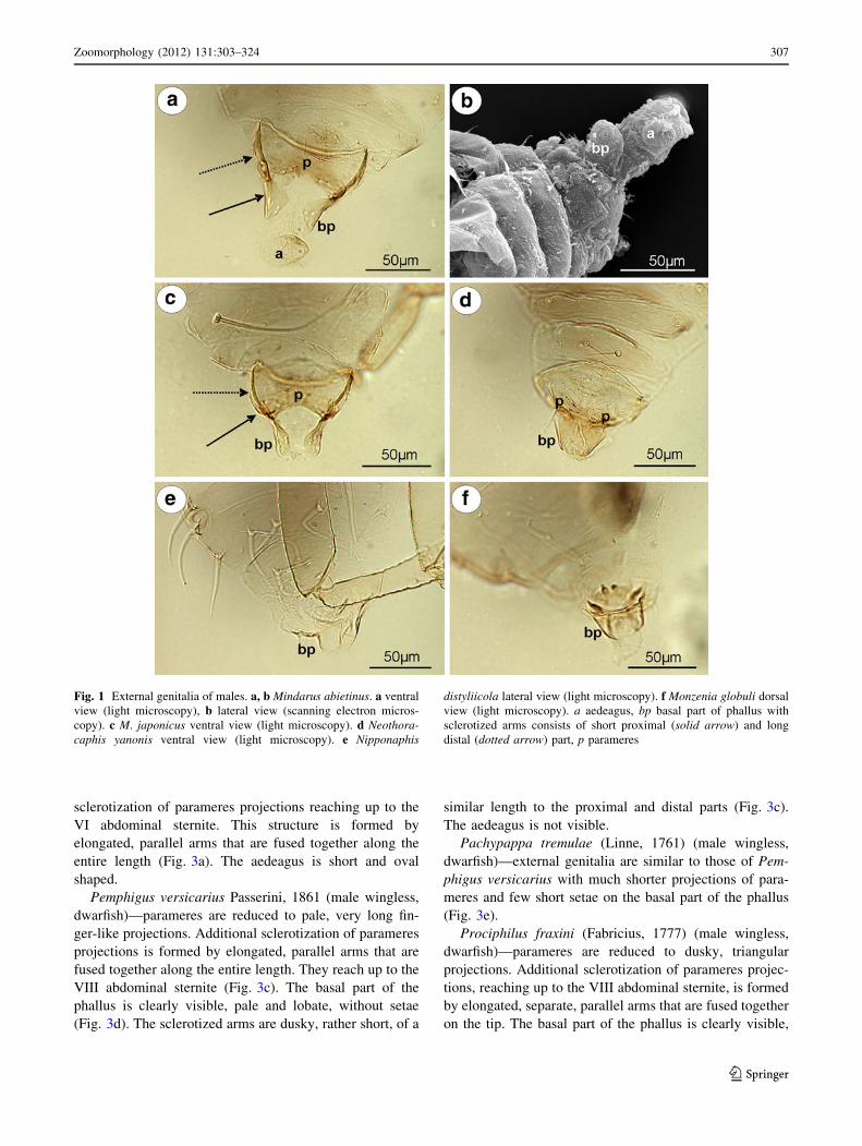
the phallus, pale, small, basally fused, elongated, with a
few setae on the outer margin (Fig. 1a). The basal part of
the phallus in slide-mounted specimens (Fig. 1a) and
scanning electron microscope (Fig. 1b) is clearly visible,
pale, triangular. Sclerotized arms are dark pigmented, with
a long distal part and a very short proximal part. The
aedeagus is rather short and oval in shape (Fig. 1b).
M. japonicus Takahashi, 1931 (male wingless, dwarf-
ish)—external genitalia are very similar to those of M. abi-
etinus with a club-shaped the basal part of phallus
(Fig. 1c).
Hormaphidinae
Neothoracaphis yanonis (Matsumura, 1917) (male wing-
less, dwarfish)—parameres are located above the basal part
of the phallus, very small and hardly visible in slide-
mounted specimens, with a few, short setae on the outer
margin. The basal part of the phallus is clearly visible, pale,
and forceps-like, without setae. The sclerotized arms are
rather short, of a similar length to the proximal and distal
parts. They are very thin and pale (Fig. 1d). The aedeagus
is not visible.
Nipponaphis distyliicola Monzen, 1934 (male wingless,
dwarfish)—parameres are hardly visible. The basal part of
phallus is clearly visible, pale and lobe-shaped, without
setae (Fig. 1e). The sclerotized arms and the aedeagus are
not visible.
Monzenia globuli (Monzen, 1934) (male wingless,
dwarfish)—parameres are hardly visible. The basal part of
the phallus is clearly visible, pale and claw-like, without
setae (Fig. 1f). The sclerotized arms and the aedeagus are
not visible.
Anoeciinae
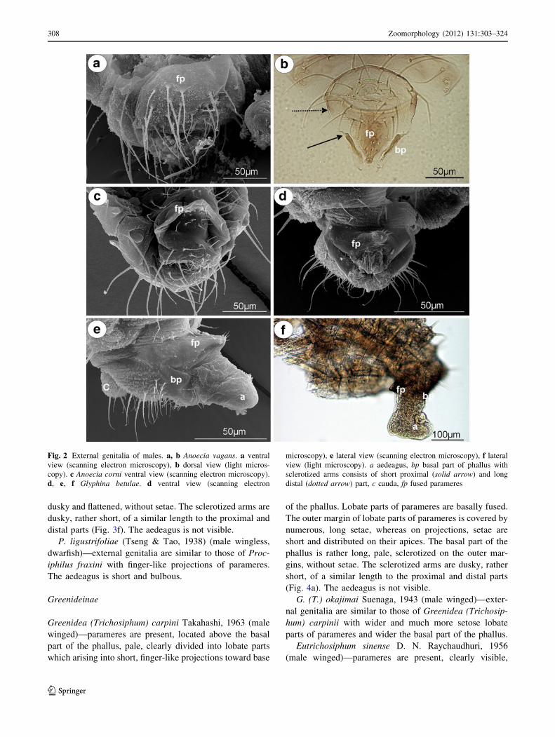
Anoecia vagans (Koch, 1856) (male wingless, dwarfish)—
parameres are present, located above the basal part of the
phallus, strongly modified, completely fused into a single
triangular, sharply pointed structure, without setae on its tip
(Fig. 2a). The basal part of the phallus is clearly visible,
pale, oar-like, without setae. It is supported by rather long
and thin sclerotized arms (Fig. 2b). The aedeagus is short
and oval shaped.
Anoecia corni (Fabricius, 1775) (male wingless,
dwarfish)—external genitalia are very similar to those of
A. vagans, with less sharply pointed modified parameres
(Fig. 2c).
Thelaxinae
Glyphina betulae (Linne, 1758) (male wingless, dwarf-
ish)—parameres are present, located above the basal part
of the phallus, strongly modified and fused into a single,
dark, bulb-shaped structure with a few, short setae on its
tip (Fig. 2d). The basal part of the phallus is present, but
strongly reduced, dusky, triangular, without setae, visible
both in the scanning electron microscope (Fig. 2e) and in
the slide-mounted specimens (Fig. 2f). Sclerotized arms
are barely visible. The aedeagus is rather short with its
distal part wider than its basal part (Fig. 2f).
Glyphina jacutensis Mordvilko, 1931 (male wingless,
dwarfish)—external genitalia are very similar to those of
G. betulae with a barely visible the basal part of the
phallus.
Eriosomatinae
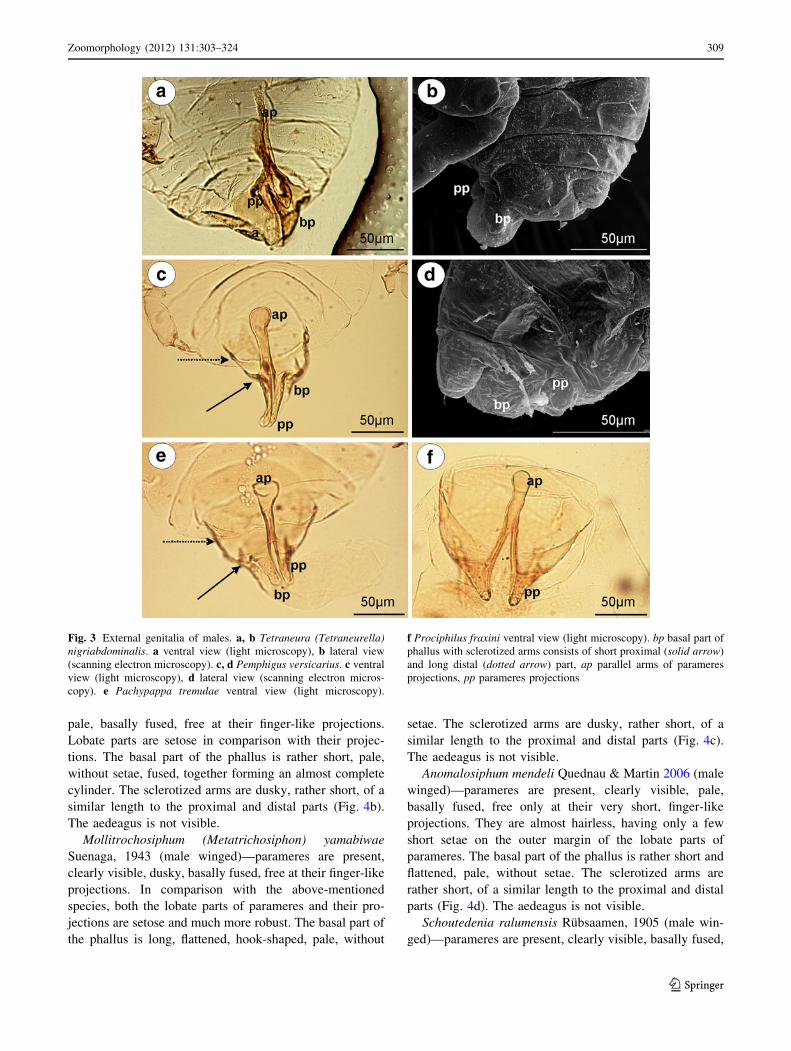
Tetraneura (Tetraneurella) nigriabdominalis (Sasaki,
1899) (male wingless, dwarfish)—parameres are reduced
to dusky, lobate projections (Fig. 3a). The basal part of
the phallus is dusky, flattened and short, without setae
(Fig. 3b). The sclerotized arms are dusky, rather short,
of a similar length to the proximal and distal parts.
The most significant character of external genitalia, vis-
ible in the slide-mounted specimens, is the additional
Table 1 continued
Subfamily/tribe/species Collection locality Host plants Number of
studied specimens
LM SEM
Lachninae: Eulachnini
Eulachnus agilis (Kaltenbach, 1843) Haute Alpes, France Pinus uncinata 10 ? ?
E. rileyi (Williams, 1911) Haute Alpes, France Pinus nigra 10 ? ?
Cinara cedri Mimeur, 1936 La Varenne, France Cedrus atlantica 6 ? ?
C. laricis (Hartig, 1839) Haute Alpes, France Larix europaea 2 ?
C. maculipes Hille Ris Lambers 1966 Mouree, Pakistan Pinus excelsa 7 ?
C. piceicola (Cholodkovsky, 1896) Haute Savoie, France Picea excelsa 7 ?
C. todocola (Inouye, 1936) Tokyo, Japan Pinus sp. 3 ?
C. (Cedrobium) laportei (Remaudiere, 1954) Ifrane, Marocco Cedrus atlantica 4 ?
306 Zoomorphology (2012) 131:303–324
123
sclerotization of parameres projections reaching up to the
VI abdominal sternite. This structure is formed by
elongated, parallel arms that are fused together along the
entire length (Fig. 3a). The aedeagus is short and oval
shaped.
Pemphigus versicarius Passerini, 1861 (male wingless,
dwarfish)—parameres are reduced to pale, very long fin-
ger-like projections. Additional sclerotization of parameres
projections is formed by elongated, parallel arms that are
fused together along the entire length. They reach up to the
VIII abdominal sternite (Fig. 3c). The basal part of the
phallus is clearly visible, pale and lobate, without setae
(Fig. 3d). The sclerotized arms are dusky, rather short, of a
similar length to the proximal and distal parts (Fig. 3c).
The aedeagus is not visible.
Pachypappa tremulae (Linne, 1761) (male wingless,
dwarfish)—external genitalia are similar to those of Pem-
phigus versicarius with much shorter projections of para-
meres and few short setae on the basal part of the phallus
(Fig. 3e).
Prociphilus fraxini (Fabricius, 1777) (male wingless,
dwarfish)—parameres are reduced to dusky, triangular
projections. Additional sclerotization of parameres projec-
tions, reaching up to the VIII abdominal sternite, is formed
by elongated, separate, parallel arms that are fused together
on the tip. The basal part of the phallus is clearly visible,
Fig. 1 External genitalia of males. a, b Mindarus abietinus. a ventral
view (light microscopy), b lateral view (scanning electron micros-
copy). c M. japonicus ventral view (light microscopy). d Neothora-caphis yanonis ventral view (light microscopy). e Nipponaphis
distyliicola lateral view (light microscopy). f Monzenia globuli dorsal
view (light microscopy). a aedeagus, bp basal part of phallus with
sclerotized arms consists of short proximal (solid arrow) and long
distal (dotted arrow) part, p parameres
Zoomorphology (2012) 131:303–324 307
123
dusky and flattened, without setae. The sclerotized arms are
dusky, rather short, of a similar length to the proximal and
distal parts (Fig. 3f). The aedeagus is not visible.
P. ligustrifoliae (Tseng & Tao, 1938) (male wingless,
dwarfish)—external genitalia are similar to those of Proc-
iphilus fraxini with finger-like projections of parameres.
The aedeagus is short and bulbous.
Greenideinae
Greenidea (Trichosiphum) carpini Takahashi, 1963 (male
winged)—parameres are present, located above the basal
part of the phallus, pale, clearly divided into lobate parts
which arising into short, finger-like projections toward base
of the phallus. Lobate parts of parameres are basally fused.
The outer margin of lobate parts of parameres is covered by
numerous, long setae, whereas on projections, setae are
short and distributed on their apices. The basal part of the
phallus is rather long, pale, sclerotized on the outer mar-
gins, without setae. The sclerotized arms are dusky, rather
short, of a similar length to the proximal and distal parts
(Fig. 4a). The aedeagus is not visible.
G. (T.) okajimai Suenaga, 1943 (male winged)—exter-
nal genitalia are similar to those of Greenidea (Trichosip-
hum) carpinii with wider and much more setose lobate
parts of parameres and wider the basal part of the phallus.
Eutrichosiphum sinense D. N. Raychaudhuri, 1956
(male winged)—parameres are present, clearly visible,
Fig. 2 External genitalia of males. a, b Anoecia vagans. a ventral
view (scanning electron microscopy), b dorsal view (light micros-
copy). c Anoecia corni ventral view (scanning electron microscopy).
d, e, f Glyphina betulae. d ventral view (scanning electron
microscopy), e lateral view (scanning electron microscopy), f lateral
view (light microscopy). a aedeagus, bp basal part of phallus with
sclerotized arms consists of short proximal (solid arrow) and long
distal (dotted arrow) part, c cauda, fp fused parameres
308 Zoomorphology (2012) 131:303–324
123
pale, basally fused, free at their finger-like projections.
Lobate parts are setose in comparison with their projec-
tions. The basal part of the phallus is rather short, pale,
without setae, fused, together forming an almost complete
cylinder. The sclerotized arms are dusky, rather short, of a
similar length to the proximal and distal parts (Fig. 4b).
The aedeagus is not visible.
Mollitrochosiphum (Metatrichosiphon) yamabiwae
Suenaga, 1943 (male winged)—parameres are present,
clearly visible, dusky, basally fused, free at their finger-like
projections. In comparison with the above-mentioned
species, both the lobate parts of parameres and their pro-
jections are setose and much more robust. The basal part of
the phallus is long, flattened, hook-shaped, pale, without
setae. The sclerotized arms are dusky, rather short, of a
similar length to the proximal and distal parts (Fig. 4c).
The aedeagus is not visible.
Anomalosiphum mendeli Quednau & Martin 2006 (male
winged)—parameres are present, clearly visible, pale,
basally fused, free only at their very short, finger-like
projections. They are almost hairless, having only a few
short setae on the outer margin of the lobate parts of
parameres. The basal part of the phallus is rather short and
flattened, pale, without setae. The sclerotized arms are
rather short, of a similar length to the proximal and distal
parts (Fig. 4d). The aedeagus is not visible.
Schoutedenia ralumensis Rubsaamen, 1905 (male win-
ged)—parameres are present, clearly visible, basally fused,
Fig. 3 External genitalia of males. a, b Tetraneura (Tetraneurella)nigriabdominalis. a ventral view (light microscopy), b lateral view
(scanning electron microscopy). c, d Pemphigus versicarius. c ventral
view (light microscopy), d lateral view (scanning electron micros-
copy). e Pachypappa tremulae ventral view (light microscopy).
f Prociphilus fraxini ventral view (light microscopy). bp basal part of
phallus with sclerotized arms consists of short proximal (solid arrow)
and long distal (dotted arrow) part, ap parallel arms of parameres
projections, pp parameres projections
Zoomorphology (2012) 131:303–324 309
123
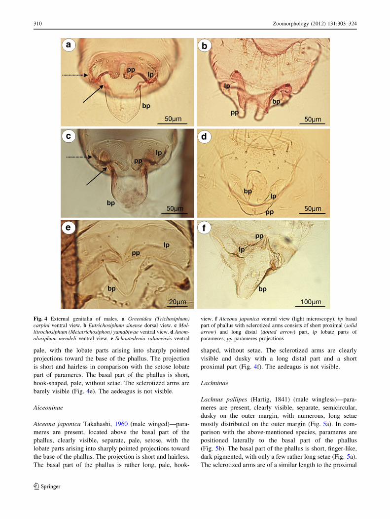
pale, with the lobate parts arising into sharply pointed
projections toward the base of the phallus. The projection
is short and hairless in comparison with the setose lobate
part of parameres. The basal part of the phallus is short,
hook-shaped, pale, without setae. The sclerotized arms are
barely visible (Fig. 4e). The aedeagus is not visible.
Aiceoninae
Aiceona japonica Takahashi, 1960 (male winged)—para-
meres are present, located above the basal part of the
phallus, clearly visible, separate, pale, setose, with the
lobate parts arising into sharply pointed projections toward
the base of the phallus. The projection is short and hairless.
The basal part of the phallus is rather long, pale, hook-
shaped, without setae. The sclerotized arms are clearly
visible and dusky with a long distal part and a short
proximal part (Fig. 4f). The aedeagus is not visible.
Lachninae
Lachnus pallipes (Hartig, 1841) (male wingless)—para-
meres are present, clearly visible, separate, semicircular,
dusky on the outer margin, with numerous, long setae
mostly distributed on the outer margin (Fig. 5a). In com-
parison with the above-mentioned species, parameres are
positioned laterally to the basal part of the phallus
(Fig. 5b). The basal part of the phallus is short, finger-like,
dark pigmented, with only a few rather long setae (Fig. 5a).
The sclerotized arms are of a similar length to the proximal
Fig. 4 External genitalia of males. a Greenidea (Trichosiphum)carpini ventral view. b Eutrichosiphum sinense dorsal view. c Mol-litrochosiphum (Metatrichosiphon) yamabiwae ventral view. d Anom-alosiphum mendeli ventral view. e Schoutedenia ralumensis ventral
view. f Aiceona japonica ventral view (light microscopy). bp basal
part of phallus with sclerotized arms consists of short proximal (solidarrow) and long distal (dotted arrow) part, lp lobate parts of
parameres, pp parameres projections
310 Zoomorphology (2012) 131:303–324
123

and distal parts, robust and clearly visible. Distal part ends
in forked apices (Fig. 5c). The aedeagus is short and bul-
bous (Fig. 5a).
Lachnus roboris (Linne, 1758) (male winged)—para-
meres are present, located laterally to the basal part of the
phallus, clearly visible, separate, semicircular, pale, with
numerous, long setae on the entire surface (Fig. 5d). The
basal part of the phallus is wide, pale, without setae
(Fig. 5e). Sclerotized arms are similar to those of the
above-mentioned species but much wider. The aedeagus is
short and bulbous (Fig. 5d).
Pterochloroides persicae (Cholodkovsky, 1899) (male
winged)—parameres are present, located above the basal
part of the phallus, clearly visible, separate, lobe-shaped,
dusky on the outer margin, with numerous, rather short
setae on the entire surface. The basal part of the phallus is
short, flattened, oval paddle-shaped, dusky, with setae. The
sclerotized arms are clearly visible, very short, of a similar
length to the proximal and distal parts (Fig. 5f). The ade-
agus is not visible.
Stomaphis aceris Takahashi, 1960 (male wingless,
dwarfish)—parameres are present, located above the basal
part of the phallus, clearly visible, separate, pale, divided
into lobate parts arising into a finger-like projection toward
the base of the phallus. The projection is very thin and long
(the longest among the species of the genus Stomaphis
studied and twice as long as the basal part of the phallus)
and almost hairless in comparison with the lobate part of
Fig. 5 External genitalia of males. a, b, c Lachnus pallipes. a lateral
view (scanning electron microscopy), b dorsal view (scanning
electron microscopy), c ventral view (light microscopy). d, e Lachnusroboris. d ventral view (scanning electron microscopy), e ventral
view (light microscopy). f Pterochloroides persicae ventral view
(light microscopy). a aedeagus, bp basal part of phallus with
sclerotized arms consists of short proximal (solid arrow) and long
distal (dotted arrow) part, p parameres
Zoomorphology (2012) 131:303–324 311
123
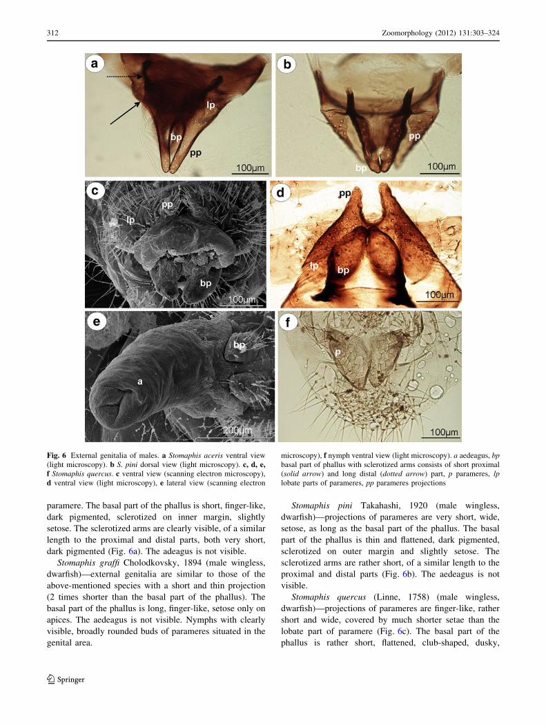
paramere. The basal part of the phallus is short, finger-like,
dark pigmented, sclerotized on inner margin, slightly
setose. The sclerotized arms are clearly visible, of a similar
length to the proximal and distal parts, both very short,
dark pigmented (Fig. 6a). The adeagus is not visible.
Stomaphis graffi Cholodkovsky, 1894 (male wingless,
dwarfish)—external genitalia are similar to those of the
above-mentioned species with a short and thin projection
(2 times shorter than the basal part of the phallus). The
basal part of the phallus is long, finger-like, setose only on
apices. The aedeagus is not visible. Nymphs with clearly
visible, broadly rounded buds of parameres situated in the
genital area.
Stomaphis pini Takahashi, 1920 (male wingless,
dwarfish)—projections of parameres are very short, wide,
setose, as long as the basal part of the phallus. The basal
part of the phallus is thin and flattened, dark pigmented,
sclerotized on outer margin and slightly setose. The
sclerotized arms are rather short, of a similar length to the
proximal and distal parts (Fig. 6b). The aedeagus is not
visible.
Stomaphis quercus (Linne, 1758) (male wingless,
dwarfish)—projections of parameres are finger-like, rather
short and wide, covered by much shorter setae than the
lobate part of paramere (Fig. 6c). The basal part of the
phallus is rather short, flattened, club-shaped, dusky,
Fig. 6 External genitalia of males. a Stomaphis aceris ventral view
(light microscopy). b S. pini dorsal view (light microscopy). c, d, e,f Stomaphis quercus. c ventral view (scanning electron microscopy),
d ventral view (light microscopy), e lateral view (scanning electron
microscopy), f nymph ventral view (light microscopy). a aedeagus, bpbasal part of phallus with sclerotized arms consists of short proximal
(solid arrow) and long distal (dotted arrow) part, p parameres, lplobate parts of parameres, pp parameres projections
312 Zoomorphology (2012) 131:303–324
123
sclerotized on inner margin, with numerous, long setae
(Fig. 6c). The sclerotized arms are dark pigmented and
much more robust than in the above-mentioned species
(Fig. 6d). The aedeagus is short (Fig. 6e). Nymphs with
clearly visible, broadly rounded buds of parameres situated
in the genital area (Fig. 6f).
Stomaphis yanonis Takahashi, 1918 (male wingless,
dwarfish)—external genitalia are similar to those of
S. aceris with shorter, paler and much more setose pro-
jections of parameres.
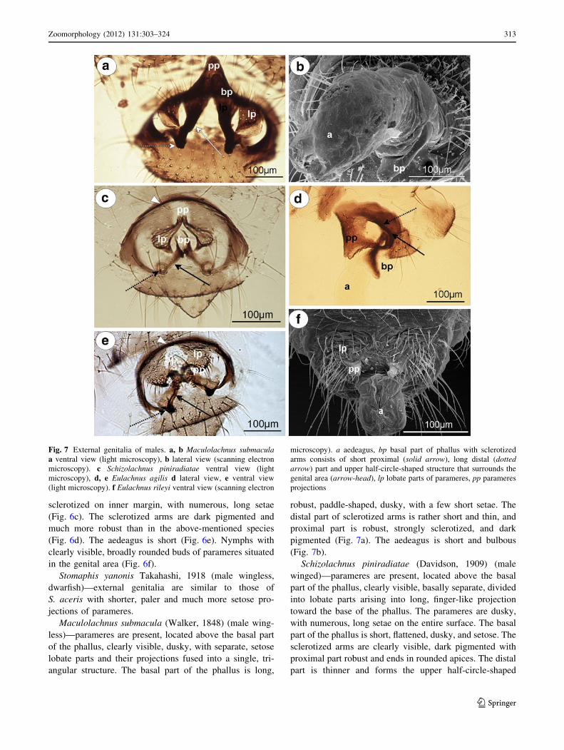
Maculolachnus submacula (Walker, 1848) (male wing-
less)—parameres are present, located above the basal part
of the phallus, clearly visible, dusky, with separate, setose
lobate parts and their projections fused into a single, tri-
angular structure. The basal part of the phallus is long,
robust, paddle-shaped, dusky, with a few short setae. The
distal part of sclerotized arms is rather short and thin, and
proximal part is robust, strongly sclerotized, and dark
pigmented (Fig. 7a). The aedeagus is short and bulbous
(Fig. 7b).
Schizolachnus piniradiatae (Davidson, 1909) (male
winged)—parameres are present, located above the basal
part of the phallus, clearly visible, basally separate, divided
into lobate parts arising into long, finger-like projection
toward the base of the phallus. The parameres are dusky,
with numerous, long setae on the entire surface. The basal
part of the phallus is short, flattened, dusky, and setose. The
sclerotized arms are clearly visible, dark pigmented with
proximal part robust and ends in rounded apices. The distal
part is thinner and forms the upper half-circle-shaped
Fig. 7 External genitalia of males. a, b Maculolachnus submaculaa ventral view (light microscopy), b lateral view (scanning electron
microscopy). c Schizolachnus piniradiatae ventral view (light
microscopy), d, e Eulachnus agilis d lateral view, e ventral view
(light microscopy). f Eulachnus rileyi ventral view (scanning electron
microscopy). a aedeagus, bp basal part of phallus with sclerotized
arms consists of short proximal (solid arrow), long distal (dottedarrow) part and upper half-circle-shaped structure that surrounds the
genital area (arrow-head), lp lobate parts of parameres, pp parameres
projections
Zoomorphology (2012) 131:303–324 313
123

structure that surrounds the genital area (Fig. 7c). The
aedeagus is not visible.
Eulachnus agilis (Kaltenbach, 1843) (male winged)—
parameres are present, located above the basal part of the
phallus, clearly visible, basally fused. Their lobate parts
(triangular in lateral view Fig. 7d) arise into a distinct,
finger-like projection toward the base of the phallus. The
parameres are dark pigmented on the outer margin, with
numerous, long setae on the entire surface with the
exception of a few, short setae distributed on the projec-
tions. The basal part of the phallus is short, flattened, pale,
with a few short setae. The sclerotized arms are clearly
visible, strongly sclerotized, and dark pigmented. The
proximal part is robust and ends in oval apex. The distal
part is thinner and forms the upper half-circle-shaped
structure that surrounds the genital area (Fig. 7e). The
aedeagus is not visible.
Eulachnus rileyi (Williams, 1911) (male winged)—
external genitalia are similar to those of E. agilis with a
thinner basal part of the phallus. The aedeagus is short and
bulbous (Fig. 7f).
Cinara cedri Mimeur, 1936 (male winged)—parameres
are present, located above the basal part of the phallus,
clearly visible, dark pigmented, with setose lobate parts
fused and arising into a finger-like projections toward base
of the phallus (Fig. 8a). The projection is long (twice as
Fig. 8 External genitalia of males. a, b Cinara cedri a ventral view
(scanning electron microscopy), b dorsal view (light microscopy).
c Cinara maculipes ventral view (light microscopy). d Cinarapiceicola ventral view (light microscopy). e Cinara todocola ventral
view (light microscopy). f Cinara (Cedrobium) laportei ventral view
(light microscopy). bp basal part of phallus with sclerotized arms
consists of short proximal (solid arrow), long distal (dotted arrow)
part and upper half-circle-shaped structure that surrounds the genital
area (arrow-head), lp lobate parts of parameres, pp parameres
projections
314 Zoomorphology (2012) 131:303–324
123
long as the basal part of the phallus) and covered with
numerous, long setae. The basal part of the phallus is rather
short, flattened, dark pigmented, and slightly setose. The
sclerotized arms are clearly visible, strongly sclerotized,
and dark pigmented. The proximal part is robust and ends
in oval apex. The distal part is thinner and forms the upper
half-circle-shaped structure that surrounds the genital area
(Fig. 8b). The aedeagus is not visible.
Cinara laricis (Hartig, 1839) (male winged)—external
genitalia are similar to those of C. cedri with shorter and
paler projections of parameres.
Cinara maculipes Hille Ris Lambers 1966 (male win-
ged)—projections of parameres are sharply pointed, trian-
gular, setose, dark pigmented (Fig. 8c). The basal part of
the phallus is slightly visible, in lateral view almost roun-
ded with more sclerotized outer margin. The aedeagus is
not visible.
Cinara piceicola (Cholodkovsky, 1896) (male wing-
less)—lobate parts of parameres are broadly rounded,
setose, and the projections of parameres are very long,
finger-like, setose and dark pigmented. The basal part of
the phallus is clearly visible, very thin and long, almost
parallel, dark pigmented, setose. The sclerotized arms are
clearly visible. The proximal part is robust, dark pigmented
and ends in spiniform apices. The distal part is much
thinner and forms the upper half-circle-shaped structure
that surrounds the genital area (Fig. 8d). The aedeagus is
not visible.
Cinara todocola (Inouye, 1936) (male winged)—lobate
parts of parameres are crescent-shaped, and the projections
of parameres are short and sharply pointed, dark pig-
mented, setose on the entire surface. The basal part of the
phallus is hook-shaped, short, pale, without setae. The
proximal part of sclerotized arms is robust, dark pigmented
and ends in a spiniform apices. The distal part is much
thinner and forms the upper half-circle-shaped structure
that surrounds the genital area. (Fig. 8e). The aedeagus is
not visible.
Cinara (Cedrobium) laportei (Remaudiere, 1954) (male
winged)—external genitalia are similar to those of C. cedri
with slight projections of parameres and less robust scler-
otized arms (Fig. 8f).
Discussion
The previous study of the comparative morphology of the
external male genitalia of Aphididae (Wieczorek et al.
2011) showed that these structures are well developed and
typically consist of the phallus, which is composed of the
sclerotized basal part with its articulation and a membra-
nous apical part, the aedeagus. Above the phallus, there is a
pair of setose parameres. However, this scheme was
characteristic mainly for taxa of a normal size (i.e., not
dwarfish) and mostly for winged males. The present study
shows that in almost all of the species examined (repre-
sentatives of eight taxa traditionally classified as subfami-
lies within 24 subfamilies of the Aphididae), the external
male genitalia are modified in both dwarfish and normal-
sized males (winged and wingless).
The external genitalia of males—the phallus
In Aphididae, the phallus is composed of a membranous
part—the aedeagus and the sclerotized basal part supported
by its articulations. The aedeagus is entirely unsclerotized
and its shape is estimated mostly based on material pre-
served in alcohol or with scanning electron microscope. In
most of the species studied, it is clearly visible, although it
is rather short and oval shaped (Lachnus pallipes Fig. 5a
and Stomaphis quercus Fig. 6e). Only in Glyphina betulae
is the distal part of the aedeagus bulbous and wider than its
proximal part (Fig. 2f). Polaszek (1987b in lit.), according
to the terminology of Bonhag and Wick (1953), distin-
guished the basal part of the aedeagus—a conjuctiva and its
distal part—a vesica in some species. The present study
does not support this division. Generally, in the species
studied, the aedeagus (when visible) is characterized by a
small amount of variation in its shape in comparison with
previously studied species of Drepanosiphinae, Calaphidi-
nae, Chaitophorinae or Aphidinae.
The basal part of the phallus (the so-called penis valves
by some authors), which is composed of the partially
sclerotized lobes, is present and clearly visible in all of the
species studied. Only in the dwarfish males of Glyphina
betulae is it reduced to small, triangular plates (Fig. 2e).
Polaszek (1987b in lit.) stated that in representatives of the
genera Anoecia, Glyphina, Thelaxes, and Pemphigus (all
with dwarfish males), the basal part of the phallus is either
absent, modified and fused or is replaced by an extension of
the anal plate. The present study does not support this
observation. In all of the species with dwarfish males
studied, representatives of the Mindarinae, Hormaphidinae,
Anoeciinae, Thelaxinae and Eriosomatinae as well as
dwarf males of the genus Stomaphis (Lachninae), the basal
part of the phallus is present and visible in the scanning
electron microscope and the slide-mounted specimens. In
most cases, the lobes are rather short and flattened (Nipp-
onaphis distyliicola Fig. 1e, Pemphigus versicarius
Fig. 3d), forceps-like (Neothoracaphis yanonis Fig. 1d,
Monzenia globuli Fig. 1f), triangular (Mindarus abietinus
Fig. 1a, Anoecia vagans Fig. 2b) or finger-like (Stomaphis
aceris Fig. 6a). Representatives of the Greenideinae,
Aiceoninae, and Lachninae (in exception of the genus
Stomaphis) are characterized by normal-sized males. In
most of species of the Greenideinae studied, the basal part
Zoomorphology (2012) 131:303–324 315
123
of the phallus is rather short and flattened (Anomalosiphum
mendeli Fig. 4d) or hook-shaped (Mollitrochosiphum
(Metatrichosiphon) yamabiwae Fig. 4c), usually without
setae. However, the shape of basal part of the phallus in
Eutrichosiphum sinense differs from these structures in
other aphid genera. The lobes are fused, free only at their
tips and together form an almost complete cylinder
(Fig. 4b). Unfortunately, only slide-mounted males were
available for Greenideinae so it was not possible to perform
a detailed electron microscopy study of these structures.
The basal part of the phallus of Aiceona japonica is similar
to the hook-shape of the above-mentioned species of
Greenideinae; however, it is much longer (Fig. 4f). Lach-
ninae are characterized by the most various shapes of the
basal part of the phallus. In general, the most common is
the finger-like shape (Lachnus roboris Fig. 5e, Cinara
piceicola Fig. 8d) and rarely club-shaped (Stomaphis
quercus Fig. 6d) or paddle-shaped (Maculolachnus sub-
macula Fig. 7a). It is joined laterally with the aedeagus and
attached to it (Figs. 5b, 6e). Moreover, the basal part of the
phallus is more setose in Lachninae than in representatives
of the other subfamilies.
In all of the species studied, the basal part of the phallus
is additionally fortified by sclerotized arms. These struc-
tures are not uniform and vary in form and length. In rep-
resentatives of the Hormaphidinae, Anoeciinae (Anoecia
vagans Fig. 2b), Thelaxinae, Eriosomatinae (Pemphigus
versicarius Fig. 3c), and Greenideinae (Mollitrochosiphum
(Metatrichosiphon) yamabiwae Fig. 4c), the sclerotized
arms are barely visible, pale, rather short and of a similar
length to the proximal and distal parts. In Mindarinae
(Mindarus japonicus Fig. 1c) and Aiceoninae, the sclero-
tized arms are clearly visible and dusky with a long distal
part and a short proximal part. In Lachninae, the sclerotized
arms are the most variable: of a similar length to the
proximal and distal parts in the genera Lachnus (Fig. 5c),
Pterochloroides and Stomaphis (Fig. 6a) with a rather
short and thin distal part and a robust proximal part in
Maculolachnus submacula (Fig. 7a), or with a robust
proximal part ending in an oval apices and a much thinner
distal part, which forms the upper half-circle-shaped
structure that surrounds the genital area in the genera
Schizolachnus (Fig. 7c), Eulachnus (Fig. 7e) and Cinara
(Fig. 8b–f). Additionally, in all of the species of this
subfamily studied, the arms are heavily sclerotized, robust
and dark pigmented.
The external genitalia of males—parameres
Parameres are the most variable part of the male external
genitalia of species studied. In Mindarus (Fig. 1a), which
has dwarfish males, parameres are rather small, fused
basally and elongated, similar to those of previously
studied species of Drepanosiphinae, Calaphidinae, Chai-
tophorinae, or Aphidinae. The dwarfish males of Hor-
maphidinae (Neothoracaphis yanonis Fig. 1d) have a
similar type of broadly rounded and barely visible para-
meres. On the other hand, parameres appear to be signifi-
cantly modified in the dwarfish males of the Anoeciinae,
Thelaxinae, and Eriosomatinae. In representatives of the
genera Anoecia and Glyphina, parameres are fused into a
single, sharply pointed structure that is triangular without
setae on the tip in Anoecia (Fig. 2a–c) or bulb-shaped with
few, short setae on the tip in Glyphina (Fig. 2d). In the
species of Eriosomatinae studied, parameres are reduced to
variable-shaped projections—mostly finger-like (Pemphi-
gus versicarius Fig. 3c) but also lobate (Tetraneura
(Tetraneurella) nigriabdominalis Fig. 3a) or triangular
(Prociphilus fraxini Fig. 3f)—and are supported by addi-
tional sclerotization that reaches up to the VI-VIII
abdominal sternite. This structure has never been described
before and is formed by elongated, parallel arms fused
together along the entire length (Fig. 3a) or fused only on
the tip (Fig. 3f). Only de Fluiter (1931) called it a ‘‘ventrale
chitinestaaf’’ (a chitinized rod) and figured it in Eriosoma
lanigerum. As in a case of the sclerotized arms of basal part
of the phallus, they are probably apophyses that provide
support for muscle attachment. In male insects, parameres
are the grasping apparatus, which play an important role
during copulation. It is possible that the modified para-
meres of Eriosomatinae need additional support; however,
the mating behavior of these aphids has never been
observed. Greenideinae are also characterized by modified
parameres. They are clearly divided into lobate parts arise
into short, usually finger-like projections toward the base of
the phallus. The lobate parts of parameres are fused basally
and are free only at their projections. Polaszek (1987b in
lit.) stated that parameres of Greenideinae are reduced and
only the structure he called the anterior genital process,
which forms a pair of finger-like processes, is developed.
The present study does not support this observation. All of
the species studied, especially representatives of the tribe
Greneeidini (Fig. 4a–c), have large, well-developed para-
meres that are clearly visible in the slide-mounted speci-
mens. Chaudhuri (1956) and Chakrabarti and Maity (1980)
also described and illustrated parameres of Greneeidini as
composed of the base with lateral projections. Moreover,
scanning electron micrographs of the external genitalia of
males of Schoutedenia ralumensis presented by Polaszek
(1987b in lit.) show that these structures are well devel-
oped. Aiceona japonica (Aiceoninae) has a similar type of
parameres as those found in Greenideinae (Fig. 4f). All of
the mentioned species have parameres positioned above the
basal part of the phallus. The most diverse forms of para-
meres are characterized for Lachninae. They are separate,
semicircular or lobate without a finger-like projection in
316 Zoomorphology (2012) 131:303–324
123
the genera Lachnus (Fig. 5c, e) and Pterochloroides
(Fig. 5f) (Lachnini) or divided into lobate parts (separate or
fused) arising into variable-shaped projections toward the
base of the phallus in the remaining species of this sub-
family that were studied. The projections of the lobate parts
of parameres (called the anterior genital process by Pola-
szek 1987b in lit.) are variable between different genera
and species, but they vary very little within each species.
Their shape and length is a very good diagnostic character
for distinguishing males belonging to the different species
of this subfamily. Those observed in the genera Stomaphis
(Lachnini—Fig. 6a, b, d) and Cinara (Eulachnini—
Fig. 8b–f) are the most variable. In Maculolachnus sub-
macula (Lachnini—Fig. 7a), the projections of the lobate
parts of parameres are fused into a single, large, triangular
structure. Moreover, nymphs of the genus Stomaphis have
clearly visible, broadly rounded buds of parameres situated
in the genital area (Fig. 6f). Parameres of Lachninae are
more setose than in the other subfamilies. In the genus
Lachnus they are positioned laterally to the basal part of the
phallus (Lachnus pallipes Fig. 5b), whereas in the
remaining species of this subfamily, parameres are located
above the basal part of the phallus.
The external genitalia of males—phylogenetic
implications
Comparative studies of male genitalia have formed the
basis of the taxonomic and phylogenetic studies of many
groups of insects (e.g., Zumpt and Heinz 1950; Eades
2000). Males of most Aphididae, unlike males of other
Hemiptera, do not appear until autumn and have to mature
quickly. This is why the taxonomy and construction of the
higher classifications of this group of insects have gener-
ally been based on the morphology and the characters of
the females. However, a comparative, systematic study
of the male genitalia of Aphididae has revealed a number
of characters that are of potential use in discussions of the
phylogenetic relationships of this group of Hemiptera.
Phylogenetic implications—why the phallus presents some
stable characters.
The phallus, which is composed of the sclerotized basal
part with its articulation and a membranous apical part—
the aedeagus, presents some stable characters among all of
the subfamilies studied, that is, with dwarfish, normal-
sized, winged or wingless males. The stable character of
the components of the phallus is a result of their functions:
the lateral sclerotization of the otherwise membranous
aedeagus enfolds it and supported by the sclerotized arms
takes part in everting the aedeagus and maintaining it in the
right position during copulation. The shape and size of
the aedeagus and the basal part of the phallus as well as the
length of the sclerotized arms is variable between different
genera and species, but varies very little within each species.
Thus, this characteristic is valuable for diagnosing species
but is less important in phylogenetic considerations.
Phylogenetic implications—what is important parameres
not modified or modified?
On the other hand, parameres show the most striking
differences and appear to be highly variable among the
different subfamilies studied. In Drepanosiphinae, Chaito-
phorinae, Calaphidinae, Phyllaphidinae, Saltusaphidinae,
Lizeriinae, Spicaphidinae, Tamalinae, Parachaitophorinae,
Phloeomyzinae, and Aphidinae (Fig. 9a), parameres are
large and positioned at the anterior end of the genital area,
above the basal part of the phallus (Wieczorek et al. 2011).
Similar types of parameres have been found in the dwarfish
males of the Mindarinae (Fig. 9b) and Hormaphidinae. The
Mindarinae, represented by only one extant genus Minda-
rus Koch, are considered to be the most primeval drepa-
nosiphine aphids by some authors (Takahashi 1931;
Mordvilko 1934; Heie and Wegierek 2009b; Quednau
2010) or are recognized as an independent subfamily (Heie
1980; Remaudiere and Stroyan 1984). In comparison with
other taxa with dwarfish males, parameres of M. abietinus
and M. japonicus are not modified and are similar to those
observed in drepanosiphine aphids. Thus, our study cor-
roborates Quednau’s (2010) point of view, who claims that
this genus represents the most primordial group of the
drepanosiphine aphids. The dwarfish males of Hormaph-
idinae that were studied represent the same type of para-
meres, although they are very small. According to Quednau
(2010), the Hormaphidinae together with Eriosomatinae
and Anoeciinae form the common lineage. However,
parameres of studied species belonging to these subfami-
lies differ significantly. In Eriosomatinae, parameres are
reduced to rather long projections, which are supported by
additional sclerotization reaching up to the VI–VIII
abdominal sternite (Fig. 9c). This unusual shape of para-
meres may be regarded as the autapomorphy of this sub-
family. In Anoeciinae, on the other hand, parameres are
fused into a single, sharply pointed structure, similar to that
in Thelaxinae (Fig. 9d). It is not clear whether this for-
ward-pointing structure at the anterior end of the genital
area actually represents the parameres that have become
fused, or whether it is derived from the fusion of para-
meres’ projections or whether it has arisen in some other
way. According to the molecular studies of Ortiz-Rivas and
Martinez-Torres (2010), Anoeciinae, Eriosomatinae, Hor-
maphidinae, Mindarinae, and Thelaxinae are grouped
together. Moreover, these subfamilies share a common
feature—the presence of a wingless, dwarfish sexual
Zoomorphology (2012) 131:303–324 317
123

generation (feeding—Anoeciinae, Mindarinae, Thelaxinae,
Hormaphidinae or non-feeding—Eriosomatinae). How-
ever, they do not share a common origin of host alternation
(dioecious—pemphigid host alternation—return of sexu-
parae to the primary host in Anoeciinae, Eriosomatinae,
Hormaphidinae, and monoecious in Mindarinae and Thel-
axinae). Von Dohlen and Moran (2000) stated that host
alternation originated independently in the ancestors of
these aphids. It is probable that the modifications in para-
meres also arose independently. Moreover, reduction in the
size of sexual morphs may then have a direct effect on
characters associated with external genitalia, that is, very
small and barely visible parameres in Hormaphidinae and
very small the basal part of the phallus in Thelaxinae.
Greenideinae (Fig. 10a) and Aiceoninae (Fig. 10b) are
characterized by normal-sized males and large parameres
divided into lobate parts arising into short, usually finger-
like projections toward the base of the phallus. The close
relations between these subfamilies were pointed out by
Heie and Wegierek (2009b); however, they remain un-
sampled for molecular data (von Dohlen 2009). According
to Quednau and Martin (2006), the genus Aiceona Takah-
ashi may have evolved from Neophyllaphis-like ancestors,
which may also be the ancestors of primitive Lachninae. In
Fig. 9 External genitalia of males. a Not modified in the Drepanos-
iphinae, Chaitophorinae, Calaphidinae, Phyllaphidinae, Saltusaphid-
inae, Lizeriinae, Spicaphidinae, Tamalinae, Parachaitophorinae,
Phloeomyzinae and Aphidinae. b Not modified in the Mindarinae
and Hormaphidinae. c Modified with parameres reduced to variable-
shaped projections in the Eriosmatinae. d Modified with parameres
fused into a single, sharply pointed structure in the Anoeciinae and
Thelaxinae. a aedeagus, bp basal part of phallus with sclerotized arms
consists of short proximal (solid arrow) and long distal (dotted arrow)
part, ap parallel arms of parameres projections, fp fused parameres,
p parameres, pp parameres projections. Drawings according to light
microscopical preparations
318 Zoomorphology (2012) 131:303–324
123

fact, parameres of A. japonica are also very similar to those
of some Cinara species.
The most diverse forms of parameres, similar to almost
all of the types described above, are characterized for
Lachninae. Parameres of the genera Lachnus (Fig. 10c) and
Pterochloroides resemble those found in Aphidinae and
drepanosiphine aphids in shape and size; however, in the
genus Lachnus, they are positioned laterally to the basal
part of the phallus. In Maculolachnus submacula
(Fig. 10d), projections fused into a single structure,
resemble those found in Anoeciinae and Thelaxinae, but
the lobate parts of parameres are large and well developed.
In the genera Cinara (Fig. 10e), Eulachnus, Schizolachnus,
and Stomaphis (Fig. 10f) (and in representatives of the
Fig. 10 External genitalia of males. a Modified with parameres
divided into lobate parts arising into finger-like projections in the
Greenideinae. b Modified with parameres divided into lobate parts
arising into finger-like projections in the Aiceoninae. c Not modified
in genera Lachnus and Pterochloroides (Lachninae). d Modified in
the genus Maculolachnus submacula (Lachninae) with parameres
projections fused into single structure. e Modified in the genera
Cinara, Eulachnus, Schizolachnus and f Stomaphis (Lachninae) with
parameres divided into lobate parts arising into finger-like projec-
tions. bp basal part of phallus with sclerotized arms consists of short
proximal (solid arrow) and long distal (dotted arrow) part, lp lobate
parts of parameres, p parameres, pp parameres projections. Drawings
according to light microscopical preparations
Zoomorphology (2012) 131:303–324 319
123
tribe Tramini Trama troglodytes von Heyden not studied
here (Blackman et al. 2001)), parameres divided into lobate
parts arising into projections, resemble those found in
Greenideinae and Aiceoninae; however, they are highly
diverse in the shape and size of their projections. Among
Aphididae, Lachninae are one of the most diverse groups in
host–plant association, ecological and life-history charac-
ters (Normark 2000) with males that are relatively small,
but not dwarfish (in exception of the genus Stomaphis).
Whether the parameres of Lachninae are homologous with
those found in the remaining subfamilies is not clear.
Because external male genitalia are composed of func-
tionally different components (the phallus—a copulatory
organ and parameres—the grasping apparatus), any unique
combination of these components should be species-spec-
ificity (Song 2009). Lachninae differ from other aphids, as
many species may infest the same host plant. Thus, the
highly variable genitalia of Lachninae could play an
important role in the context of sexual selection. According
to the molecular studies of Ortiz-Rivas and Martinez-
Torres (2010), Lachninae constitute an independent and
basal lineage in Aphididae. However, both Heie and
Wegierek (2009a) and Quednau (2010) stated that Aice-
oninae, Greenideinae, Aphidinae, and Lachninae constitute
a complex of modern aphids. Our previous study (Wieczorek
et al. 2011) showed that Aphidinae are characterized by
parameres that are not modified and are similar to those in the
drepanosiphine aphids, whereas parameres of Lachninae
(except for the genera Lachnus and Pterochloroides)
resemble those in Aiceoninae and Greenideinae.
External genitalia—hypothetical plesiomorphic condition
and its modifications
The contribution of comparative morphology of male
external genitalia, if studied alone, is limited and needs
further morphological characters, when reconstructing the
phylogeny. In addition, in the absence of data in the fossil
material, it is difficult to speculate which external genitalia
characters should be interpreted as primitive (plesiomor-
phic) and which are derived. Aphids are characterized by
the ‘mosaic’ type of evolution—some features have
evolved independently, several times, in various evolu-
tionary lines, which makes the interpretation of data
complicated (Heie and Wegierek 2009a; Quednau 2010).
Probably, the hypothetical plesiomorphic condition of the
external male genitalia of aphids is characterized by the
rather large, lobate and setose parameres positioned at the
anterior end of the genital area as well as well developed
the basal part of the phallus which protects the aedeagus
and is supported by the sclerotization. This type of external
genitalia is found in most modern taxa of drepanosiphine
aphids and Aphidinae. In dwarfish males of Anoeciinae,
Thelaxinae, Hormaphidinae and Eriosomatinae the minia-
turization of the body size affects on the modification of
genitalia—mostly parameres. Various modified parameres
found in males of these four subfamilies do not show any
strong similarites with each other (single structure in
Anoeciinae and Thelaxinae, very small and barely visible
in Hormaphidinae or reduced to elongated projections
supported by additional sclerotization in Eriosomatinae)
and have developed in different way. It suggests that
reduction in the size of sexual generation has occurred
independently in a number of distinct lineages within
Aphididae. Whether the parameres of mentioned dwarf
males are homologus with those found in the drepanosi-
phine aphids and Aphidinae is not clear. The question,
whether they function as the grasping apparatus, is also
open. It may therefore be that the parameres are non-
functional. However, the mating behavior of these aphids
has never been observed. Compared with the drepanosi-
phine aphids and Aphidinae, the external genitalia of
Lachninae (except for the genera Lachnus and Pterochlo-
roides), Aiceoninae, and Greenideinae are more special-
ized, that is, parameres are divided into lobate parts ended
by various-shaped projections. It is possible that the
extended region of parameres perform the same function,
presumably that of gripping the female, as the parameres
do in other aphids. Although the character of parameres
appears similar in Lachninae, Aiceoninae, and Greenidei-
nae, these subfamilies differ greatly otherwise. It seems
likely that the modification of parameres have evolved
independently in Lachninae as well as Aiceoninae and
Greenideinae on the other hand.
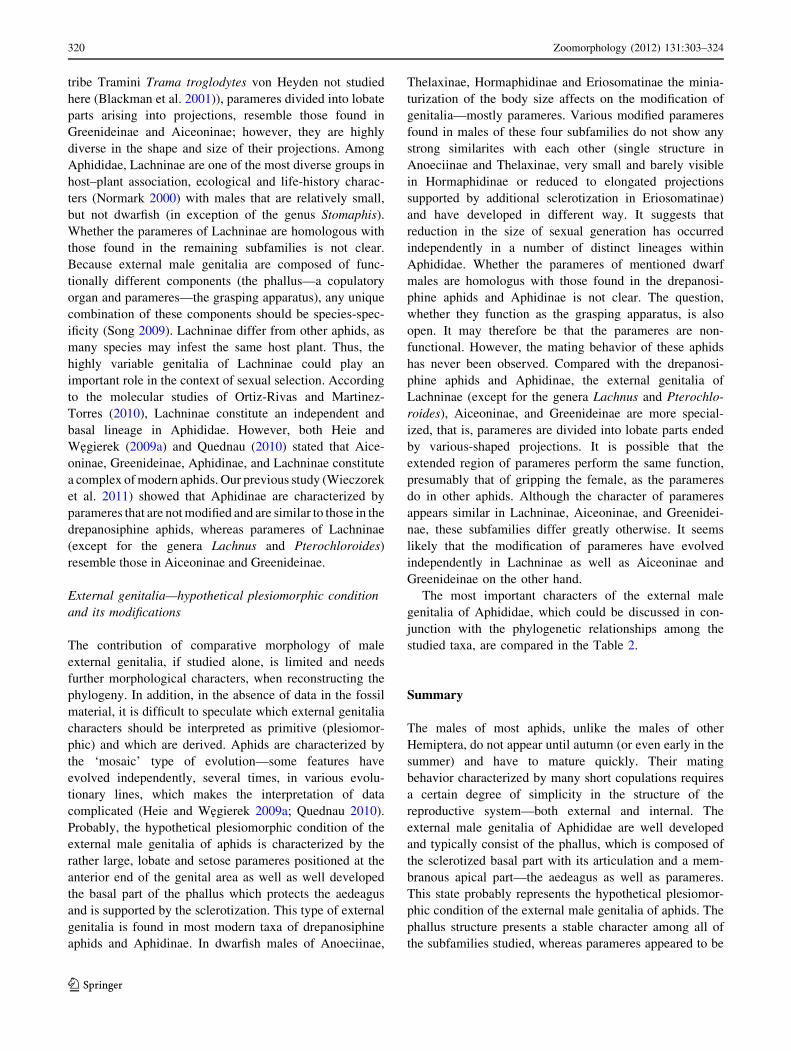
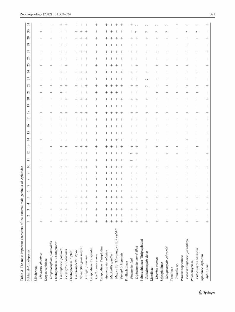
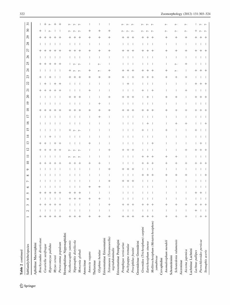
The most important characters of the external male
genitalia of Aphididae, which could be discussed in con-
junction with the phylogenetic relationships among the
studied taxa, are compared in the Table 2.
Summary
The males of most aphids, unlike the males of other
Hemiptera, do not appear until autumn (or even early in the
summer) and have to mature quickly. Their mating
behavior characterized by many short copulations requires
a certain degree of simplicity in the structure of the
reproductive system—both external and internal. The
external male genitalia of Aphididae are well developed
and typically consist of the phallus, which is composed of
the sclerotized basal part with its articulation and a mem-
branous apical part—the aedeagus as well as parameres.
This state probably represents the hypothetical plesiomor-
phic condition of the external male genitalia of aphids. The
phallus structure presents a stable character among all of
the subfamilies studied, whereas parameres appeared to be
320 Zoomorphology (2012) 131:303–324
123
Ta
ble
2T
he
mo
stim
po
rtan
tch
arac
ters
of
the
exte
rnal
mal
eg
enit
alia
of
Ap
hid
idae
Su
bfa
mil
y/t
rib
e/sp
ecie
s1
23
45
67
89
10
11
12
13
14
15
16
17
18
19
20
21
22
23
24
25
26
27
28
29
30
31
Min
dar
inae
Min
da
rus
ab
ieti
nu
s?
--
??
--
--
??
--
?-
--
?-
-?
?-
-?
-?
-?
?-
Dre
pan
osi
ph
inae
Dre
pa
no
sip
hu
mp
lata
no
idis
?-
?-
?-
--
-?
?-
?-
--
-?
--
??
--
?-
?-
?-
?
Ch
aito
ph
ori
nae
Ch
aito
ph
ori
ni
Ch
ait
op
ho
rus
po
pu
leti
?-
?-
?-
--
-?
?-
?-
--
-?
--
??
-?
--
??
--
?
Per
iph
yllu
sco
raci
nu
s?
-?
-?
--
--
??
-?
--
--
?-
?-
?-
-?
-?
?-
-?
Ch
aito
ph
ori
nae
Sip
hin
i
Ch
aet
osi
ph
ella
stip
ae
?-
-?
?-
--
-?
?-
?-
--
-?
-?
-?
-?
--
?-
??
-
Sip
ha
(Ru
ng
sia
)m
ayd
is?
--
??
--
--
??
-?
--
--
?-
?-
-?
?-
-?
-?
?-
La
ing
iap
sam
ma
e?
--
??
--
--
??
-?
--
--
?-
?-
?-
?-
-?
-?
?-
Cal
aph
idin
aeC
alap
hid
ini
Cle
thro
biu
sco
mes
?-
?-
?-
--
-?
?-
?-
--
-?
--
??
--
?-
?-
?-
?
Cal
aph
idin
aeP
anap
hid
ini
Ap
pen
dis
eta
rob
inia
e?
-?
-?
--
--
??
-?
--
--
?-
-?
?-
?-
-?
-?
-?
Mex
ica
llis
spin
ifer
?-
?-
?-
--
-?
?-
?-
--
-?
--
??
--
?-
?-
??
-
Myz
oca
llis
(Lin
eom
yzo
call
is)
wa
lsh
ii?
-?
-?
--
--
??
--
?-
--
?-
-?
?-
-?
-?
-?
-?
Pa
na
ph
isju
gla
nd
is?
-?
-?
--
--
??
-?
--
--
?-
?-
?-
?-
-?
-?
-?
Ph
yll
aph
idin
ae
Ph
ylla
ph
isfa
gi
?-
?-
?-
--
-?
??
?-
--
-?
-?
-?
-?
--
?-
?-
?
Dip
hyl
lap
his
mo
rdvi
lko
i?
-?
-?
--
--
?-
??
--
--
?-
?-
?-
-?
-?
-?
??
Sal
tusa
ph
idin
aeT
hri
psa
ph
idin
i
Su
bsa
ltu
sap
his
fla
va?
-?
-?
--
--
??
--
?-
--
?-
?-
??
?-
-?
-?
??
Liz
erii
nae
Liz
eriu
so
cote
ae
?-
?-
?-
--
-?
?-
?-
--
-?
--
?-
?-
?-
?-
??
?
Sp
icap
hid
inae
Neu
qu
ena
ph
ised
wa
rdsi
?-
?-
?-
--
-?
-?
?-
--
-?
--
??
--
?-
?-
??
?
Tam
alin
ae
Ta
ma
lia
sp.
?-
?-
?-
--
-?
-?
?-
--
-?
--
?-
?-
?-
?-
?-
?
Par
ach
aito
ph
ori
nae
Pa
rach
ait
op
ho
rus
yam
ash
ita
i?
-?
-?
--
--
??
--
?-
--
?-
-?
?-
-?
-?
?-
??
Ph
loeo
my
zin
ae
Ph
loeo
myz
us
pa
sser
ini
?-
?-
?-
--
-?
?-
?-
--
-?
--
??
-?
--
?-
??
?
Ap
hid
inae
Ap
hid
ini
Ap
his
po
mi
?-
?-
?-
--
-?
-?
--
?-
-?
-?
-?
--
?-
?-
?-
?
Zoomorphology (2012) 131:303–324 321
123
Ta
ble
2co
nti
nu
ed
Su
bfa
mil
y/t
rib
e/sp
ecie
s1
23
45
67
89
10
11
12
13
14
15
16
17
18
19
20
21
22
23
24
25
26
27
28
29
30
31
Ap
hid
inae
Mac
rosi
ph
ini
Bra
chyc
au
du
sd
iva
rica
tae
?-
?-
?-
--
-?
-?
?-
--
-?
--
?-
?-
?-
?-
??
-
Ca
vari
ella
saxi
fra
ga
e?
-?
-?
--
--
?-
??
--
--
?-
-?
?-
-?
-?
-?
-?
Hyp
ero
myz
us
pa
llid
us
?-
?-
?-
--
-?
-?
-?
--
-?
--
?-
?-
?-
?-
??
?
Myz
us
cera
si?
-?
-?
--
--
?-
??
--
--
?-
-?
?-
-?
-?
-?
-?
Pte
roco
mm
ap
op
ule
um
?-
?-
?-
--
-?
-?
?-
--
-?
--
??
--
?-
?-
?-
?
Ho
rmap
hid
inae
Nip
po
nap
hid
ini
Neo
tho
raca
ph
isya
no
nis
?-
-?
?-
--
-?
??
--
?-
-?
-?
--
??
--
?-
??
?
Nip
po
na
ph
isd
isty
liic
ola
?-
-?
?-
--
-?
??
--
?-
-?
-?
--
??
?-
?-
??
?
Mo
nze
nia
glo
bu
li?
--
??
--
--
??
?-
-?
--
?-
?-
-?
??
-?
-?
??
An
oec
iin
ae
An
oec
iava
ga
ns
?-
-?
-?
--
-?
--
?-
--
-?
--
?-
??
--
?-
??
-
Th
elax
inae
Gly
ph
ina
bet
ula
e?
--
?-
?-
--
?-
--
--
-?
??
--
-?
??
-?
-?
?-
Eri
oso
mat
inae
Eri
oso
mat
ini
Tet
ran
eura
(Tet
ran
eure
lla
)n
igri
ab
do
min
ali
s?
-?
--
-?
-?
--
?-
--
--
?-
?-
-?
?-
-?
-?
?-
Eri
oso
mat
inae
Pem
ph
igin
i
Pem
ph
igu
sve
rsic
ari
us
?-
?-
--
?-
?-
-?
--
--
-?
-?
--
??
--
?-
??
?
Pa
chyp
ap
pa
trem
ula
e?
-?
--
-?
-?
--
?-
--
--
?-
?-
?-
?-
-?
-?
??
Pro
cip
hil
us
fra
xin
i?
-?
--
-?
-?
--
?-
--
--
?-
?-
-?
?-
-?
-?
??
Gre
enid
ein
aeG
reen
idei
ni
Gre
enid
ea(T
rich
osi
ph
um
)ca
rpin
i?
-?
--
--
?-
??
--
-?
--
?-
-?
-?
?-
-?
-?
??
Eu
tric
ho
sip
hu
msi
nen
se?
-?
--
--
?-
??
--
--
?-
?-
?-
-?
?-
-?
-?
??
Mo
llit
roch
osi
ph
um
(Met
atr
ich
osi
ph
on
)ya
ma
biw
ae
?-
?-
--
-?
-?
?-
?-
--
-?
--
?-
??
--
?-
??
?
Cer
vap
hid
ini
An
om
alo
sip
hu
mm
end
eli
?-
?-
--
-?
-?
?-
--
?-
-?
-?
--
??
--
?-
??
?
Sch
ou
ted
enii
ni
Sch
ou
ted
enia
ralu
men
sis
?-
?-
--
-?
-?
?-
--
-?
-?
-?
--
??
?-
?-
??
?
Aic
eon
inae
Aic
eon
aja
po
nic
a?
-?
--
--
?-
?-
?-
--
?-
?-
-?
-?
-?
-?
-?
??
Lac
hn
inae
Lac
hn
ini
La
chn
us
pa
llip
es-
??
-?
--
--
?-
?-
-?
--
?-
?-
?-
?-
-?
-?
?-
Pte
roch
loro
ides
per
sica
e?
-?
-?
--
--
?-
??
--
--
?-
?-
?-
?-
-?
-?
??
Sto
ma
ph
isa
ceri
s?
-?
--
--
?-
?-
?-
--
?-
?-
?-
?-
?-
-?
-?
??
322 Zoomorphology (2012) 131:303–324
123
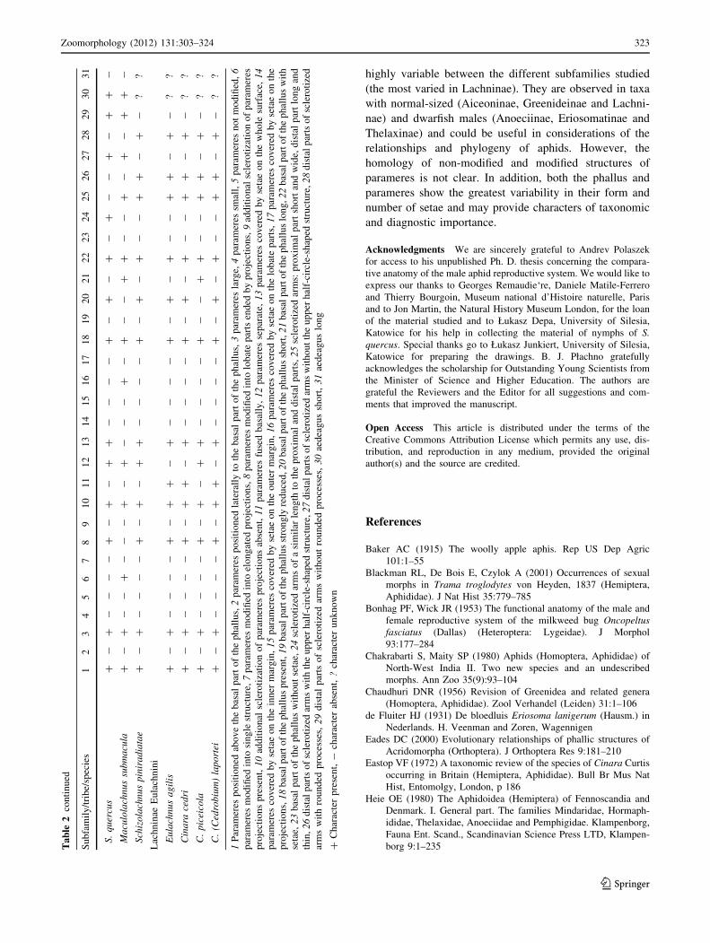
highly variable between the different subfamilies studied
(the most varied in Lachninae). They are observed in taxa
with normal-sized (Aiceoninae, Greenideinae and Lachni-
nae) and dwarfish males (Anoeciinae, Eriosomatinae and
Thelaxinae) and could be useful in considerations of the
relationships and phylogeny of aphids. However, the
homology of non-modified and modified structures of
parameres is not clear. In addition, both the phallus and
parameres show the greatest variability in their form and
number of setae and may provide characters of taxonomic
and diagnostic importance.
Acknowledgments We are sincerely grateful to Andrev Polaszek
for access to his unpublished Ph. D. thesis concerning the compara-
tive anatomy of the male aphid reproductive system. We would like to
express our thanks to Georges Remaudie‘re, Daniele Matile-Ferrero
and Thierry Bourgoin, Museum national d’Histoire naturelle, Paris
and to Jon Martin, the Natural History Museum London, for the loan
of the material studied and to Łukasz Depa, University of Silesia,
Katowice for his help in collecting the material of nymphs of S.quercus. Special thanks go to Łukasz Junkiert, University of Silesia,
Katowice for preparing the drawings. B. J. Płachno gratefully
acknowledges the scholarship for Outstanding Young Scientists from
the Minister of Science and Higher Education. The authors are
grateful the Reviewers and the Editor for all suggestions and com-
ments that improved the manuscript.
Open Access This article is distributed under the terms of the
Creative Commons Attribution License which permits any use, dis-
tribution, and reproduction in any medium, provided the original
author(s) and the source are credited.
References
Baker AC (1915) The woolly apple aphis. Rep US Dep Agric
101:1–55
Blackman RL, De Bois E, Czylok A (2001) Occurrences of sexual
morphs in Trama troglodytes von Heyden, 1837 (Hemiptera,
Aphididae). J Nat Hist 35:779–785
Bonhag PF, Wick JR (1953) The functional anatomy of the male and
female reproductive system of the milkweed bug Oncopeltusfasciatus (Dallas) (Heteroptera: Lygeidae). J Morphol
93:177–284
Chakrabarti S, Maity SP (1980) Aphids (Homoptera, Aphididae) of
North-West India II. Two new species and an undescribed
morphs. Ann Zoo 35(9):93–104
Chaudhuri DNR (1956) Revision of Greenidea and related genera
(Homoptera, Aphididae). Zool Verhandel (Leiden) 31:1–106
de Fluiter HJ (1931) De bloedluis Eriosoma lanigerum (Hausm.) in
Nederlands. H. Veenman and Zoren, Wagennigen
Eades DC (2000) Evolutionary relationships of phallic structures of
Acridomorpha (Orthoptera). J Orthoptera Res 9:181–210
Eastop VF (1972) A taxonomic review of the species of Cinara Curtis
occurring in Britain (Hemiptera, Aphididae). Bull Br Mus Nat
Hist, Entomolgy, London, p 186
Heie OE (1980) The Aphidoidea (Hemiptera) of Fennoscandia and
Denmark. I. General part. The families Mindaridae, Hormaph-
ididae, Thelaxidae, Anoeciidae and Pemphigidae. Klampenborg,
Fauna Ent. Scand., Scandinavian Science Press LTD, Klampen-
borg 9:1–235Ta
ble
2co
nti
nu
ed
Su
bfa
mil
y/t
rib
e/sp
ecie
s1
23
45
67
89
10
11
12
13
14
15
16
17
18
19
20
21
22
23
24
25
26
27
28
29
30
31
S.
qu
ercu
s?
-?
--
--
?-
?-
??
--
--
?-
?-
?-
?-
-?
-?
?-
Ma
culo
lach
nu
ssu
bm
acu
la?
-?
--
?-
--
?-
?-
--
?-
?-
-?
?-
-?
-?
-?
?-
Sch
izo
lach
nu
sp
inir
ad
iata
e?
-?
--
--
?-
?-
??
--
--
?-
?-
?-
-?
?-
?-
??
Lac
hn
inae
Eu
lach
nin
i
Eu
lach
nu
sa
gil
is?
-?
--
--
?-
??
-?
--
--
?-
?-
?-
-?
?-
?-
??
Cin
ara
ced
ri?
-?
--
--
?-
??
-?
--
--
?-
?-
?-
-?
?-
?-
??
C.
pic
eico
la?
-?
--
--
?-
?-
??
--
--
?-
-?
?-
-?
?-
?-
??
C.
(Ced
rob
ium
)la
po
rtei
?-
?-
--
-?
-?
?-
?-
--
-?
-?
-?
--
??
-?
-?
?
1P
aram
eres
po
siti
on
edab
ov
eth
eb
asal
par
to
fth
ep
hal
lus,
2p
aram
eres
po
siti
on
edla
tera
lly
toth
eb
asal
par
to
fth
ep
hal
lus,
3p
aram
eres
larg
e,4
par
amer
essm
all,
5p
aram
eres
no
tm
od
ified
,6
par
amer
esm
od
ified
into
sin
gle
stru
ctu
re,
7p
aram
eres
mo
difi
edin
toel
on
gat
edp
roje
ctio
ns,
8p
aram
eres
mo
difi
edin
tolo
bat
ep
arts
end
edb
yp
roje
ctio
ns,
9ad
dit
ion
alsc
lero
tiza
tio
no
fp
aram
eres
pro
ject
ion
sp
rese
nt,
10
add
itio
nal
scle
roti
zati
on
of
par
amer
esp
roje
ctio
ns
abse
nt,
11
par
amer
esfu
sed
bas
ally
,1
2p
aram
eres
sep
arat
e,1
3p
aram
eres
cov
ered
by
seta
eo
nth
ew
ho
lesu
rfac
e,1
4p
aram
eres
cov
ered
by
seta
eo
nth
ein
ner
mar
gin
,1
5p
aram
eres
cov
ered
by
seta
eo
nth
eo
ute
rm
arg
in,
16
par
amer
esco
ver
edb
yse
tae
on
the
lob
ate
par
ts,
17
par
amer
esco
ver
edb
yse
tae
on
the
pro
ject
ion
s,1
8b
asal
par
to
fth
ep
hal
lus
pre
sen
t,1
9b
asal
par
to
fth
ep
hal
lus
stro
ng
lyre
du
ced
,2
0b
asal
par
to
fth
ep
hal
lus
sho
rt,2
1b
asal
par
to
fth
ep
hal
lus
lon
g,2
2b
asal
par
to
fth
ep
hal
lus
wit
h
seta
e,2
3b
asal
par
to
fth
ep
hal
lus
wit
ho
ut
seta
e,2
4sc
lero
tize
dar
ms
of
asi
mil
arle
ng
thto
the
pro
xim
alan
dd
ista
lp
arts
,2
5sc
lero
tize
dar
ms:
pro
xim
alp
art
sho
rtan
dw
ide,
dis
tal
par
tlo
ng
and
thin
,2
6d
ista
lp
arts
of
scle
roti
zed
arm
sw
ith
the
up
per
hal
f-ci
rcle
-sh
aped
stru
ctu
re,
27
dis
tal
par
tso
fsc
lero
tize
dar
ms
wit
ho
ut
the
up
per
hal
f-ci
rcle
-sh
aped
stru
ctu
re,
28
dis
tal
par
tso
fsc
lero
tize
d
arm
sw
ith
rou
nd
edp
roce
sses
,2
9d
ista
lp
arts
of
scle
roti
zed
arm
sw
ith
ou
tro
un
ded
pro
cess
es,
30
aed
eag
us
sho
rt,
31
aed
eag
us
lon
g
?C
har
acte
rp
rese
nt,
-ch
arac
ter
abse
nt,
?ch
arac
ter
un
kn
ow
n
Zoomorphology (2012) 131:303–324 323
123
Heie OE, Wegierek P (2009a) A classification of the Aphidomorpha
(Hemiptera: Sternorrhyncha) under consideration of the fossil
taxa. Redia 92:69–77
Heie OE, Wegierek P (2009b) Diagnoses of the higher taxa of
Aphidomorpha (Hemiptera: Sternorrhyncha). Redia 92:261–269
Mordvilko AK (1934) On the evolution of Aphids. Arch Naturgesch
N S 3:1–60
Nieto Nafria JM, Mier Durante MP, Remaudiere G (1998) Les noms
des taxa du group-famille chez les Aphididae (Hemiptera). Rev
Fr Entomol (NS) 19:77–92
Normark BB (2000) Molecular systematics and evolution of the3
aphid family Lachnidae. Mol Phylogenet Evol 14:131–140
Ortiz-Rivas B, Martinez-Torres D (2010) Combination of molecular
data support the existence of three main lineages in the
phylogeny of aphids (Hemiptera: Aphididae) and the basal
position of the subfamily Lachninae. Mol Phylogenet Evol
55(1):305–317
Płachno BJ, Swiatek P (2009) Functional anatomy of the ovule in
Genlisea with remarks on ovule evolution in Lentibulariaceae.
Protoplasma 236:39–48. doi:10.1007/s00709-009-0045-8
Płachno BJ, Swiatek P (2010) Unusual embryo structure in viviparous
Utricularia nelumbifolia with remarks on embryo evolution in
genus Utricularia. Protoplasma 239:69–80. doi:10.1007/s00709-
009-0084-1
Polaszek A (1987a) Studies on the comparative anatomy of aphid
reproductive systems. In: Population, structure, genetics and
taxonomy of Aphids and Thysanoptera. Proceedings of the
international symposium, Smolenice, Czechoslovakia, 1985.
SPB Acad Publ, The Hague, pp 261–266
Polaszek A (1987b) Comparative anatomy of the male aphid
reproductive system. Unpublished Ph. D. Thesis
Quednau FW (2010) Atlas of the Drepanosiphine aphids of the world.
Part III: Mindarinae Tullgren 1909 to Saltusaphidinae Baker
1920 (Hemiptera: Sternorrhyncha, Aphididae). Mem Am Ent
Inst 83:1–361
Quednau FW, Martin JH (2006) Descriptions of two new species of
Anomalosiphum (Hemiptera: Aphididae, Greenideinae), includ-
ing a winged ovipara with pedunculate eggs. Zool J Linn Soc
146:239–249
Remaudiere G, Stroyan HLG (1984) Un Tamalia nouveau de
Californie (USA). Discussion sur les Tamaliinae subfam. Nov.
(Hom. Aphididae). Annls Soc entomol Fr N. S. 20:93–103
Song H (2009) Species-specificity of male genitalia is characterized
by shape, size and complexity. Insect Syst Evol 40:150–170
Sorin M (1965) Three new species of the genus Stomaphis in Japan
with a redescription of S. yanonis Takahashi (Aphididae,
Homoptera). Bull Univ Osaka Prefect 16:81–88
Takahashi R (1931) Aphididae of Formosa. Part 6. Rept Dept Agr
Govt Res Inst Formosa 53:1–127
Takahashi R (1960) Kurisakia and Aiceona of Japan (Homoptera,
Aphididae). Insecta Matsumurana 23(1):1–10
Von Dohlen CD (2009) Aphid molecular systematics: history,
progress and prospects. Redia 92:34–45
Von Dohlen CD, Moran NA (2000) Molecular data support a rapid
radiation of aphids in the Cretaceous and multiple origins of host
alternation. Biol J Linnean Soc 71:689–717
Wieczorek K (2006) Anatomical investigations of the male repro-
ductive system of five species of Calaphidinae (Hemiptera,
Ahidoidea). Insect Syst Evol 37:457–465
Wieczorek K (2008) Structure of the male reproductive system of
Anoecia (Anoecia) corni Fabricius, 1775 (Hemiptera, Aphidoi-
dea) a representative of the family Anoeciidae. Acta Zool
89:163–167
Wieczorek K, Swiatek P (2008) Morphology and ultrastructure of the
male reproductive system of the woolly beech aphid Phyllaphisfagi (Hemiptera, Aphidoidea: Phyllaphidinae). Eur J Entomol
105:707–712
Wieczorek K, Swiatek P (2009) Comparative study of the structure of
the reproductive system of dwarfish males of Glyphina betulae(Linnaeus, 1758) and Anoecia (Anoecia) corni (Fabricius, 1775)
(Hemiptera, Aphididae). Zoologischer Anzeiger J Comp Zool
248:153–159
Wieczorek K, Płachno BJ, Swiatek P (2011) Comparative morphol-
ogy of the male genitalia of Aphididae (Insecta, Hemiptera)—
part 1. Zoomorphology 130:289–303
Zumpt F, Heinz HJ (1950) Studies on the sexual armature of Diptera
II. A contribution to the study of the morphology and homology
of the male terminalia of Calliphora and Sarcophaga. Ent Mon
Mag 86:207–216
324 Zoomorphology (2012) 131:303–324
123


