ISSN 2394-7330
International Journal of Novel Research in Healthcare and Nursing Vol. 4, Issue 1, pp: (395-404), Month: January - April 2017, Available at: www.noveltyjournals.com
Page | 395 Novelty Journals
A Five Year Experience with Cervical Disc
Prosthesis and Review of the Literature
1Munthir Al-Zabin,
2Dieter Hoffmann
1Corresponding Author: MD, PhD, Senior Specialist Neurosurgey, Neurosurgical Department, Khoula Hospital, Muscat,
Sultanate of Oman 2MD, PhD, Klinik Breslauer Berg, Berlin, Germany
Abstract: Patients with cervicobrachialgia and cervical spine stenosis can be treated by a ventral discectomy and
implantation of prosthesis. Retrospectively, a study of the cases between 2003 and 2013 was performed with an
evaluation of the clinical course and follow-up.
Materials and Methods: Between May 2002 and August 2003, 50 cases were treated, in 2008 there were 248 cases,
whereas a ventral discectomy was performed with implantation of a Bryan cervical prosthesis. Only 50 operated
cases were followed up until January 2013. The mean age was 45 years (range 31 – 64 years), 25 males, and 25
females.
Results: Prior to surgery 20 Patients had pain > 50 on VAS, 5 years later about 4 patients. Motor deficits were in
11 patients prior and in 5 patients after the surgery, but in all cases there was a significant improvement. 8
Patients had sensory deficits prior and 8 after the surgery. No myelopathy has been occurred after 5 years, prior to
surgery in 3 cases this was observed. Ability to work: 11 full time, 4 part time, 1 unable to work and 4 retired.
Back to work ratio 85%. About 20 out of 50 patients were available for reexamination and interview after the
implantation of the prostheses. 18 were very satisfied with the outcome, 19 would do the surgery again and 19
would recommend this operation to a friend. Radiological findings: ratio of motion > 2° in 33 patients, < 2° in 11
patients and in 6 patients heterotopic ossification had occurred. There is no statistical correlation between clinical
outcome and rate of motion. Correlation after Pearson: ODOM p= 0.27 and 0.247 respectively. Complications:
Voice: One permanent paralysis of recurrent nerve. Bleeding: In 2 patients with re-bleeding a surgical evacuation
was required on the day of surgery with uneventful further recovery. Dislocation of prosthesis: one patient with
uneventful complete recovery after surgical replacement. So far there was no single case of infection or removal of
prosthesis.
Conclusions: Implantation of cervical disc prostheses is not associated with an increased risk for the patient as
compared to conventional fusion. A preservation of motion for several years in 70% to 80% may be expected.
Cervical disc prosthesis appears to be particularly practicable in younger age groups up to the age of 65 years. The
long term effect of the prosthesis on adjacent level disease in comparison to fusion is unclear as of yet.
Keywords: Cervicalgia, cervical disc prosthesis, cervical spinal canal stenosis.
1. INTRODUCTION
Cervical spondylosis is a common cause of neck pain, radiculopathy and myelopathy. Degenerative changes in a disc can
cause it to prolapse or osteophytes to be formed. Each of these can cause pressure on the spinal cord leading to
myelopathy or radiculopathy. The traditional treatment for this condition consists of an anterior cervical discectomy with
a bridging bone graft, originally described by Cloward (1) and Smith and Robinson (2). Recent alternatives to bone
grafting have been described including interbody cages (3), bone substitutes or spacers (4), in order to avoid the
complications of grafting (5). Cervical discectomy and fusion has provided good clinical results for radiculopathy and
ISSN 2394-7330
International Journal of Novel Research in Healthcare and Nursing Vol. 4, Issue 1, pp: (395-404), Month: January - April 2017, Available at: www.noveltyjournals.com
Page | 396 Novelty Journals
acceptable ones for cervical spondylotic myelopathy. The short-term clinical results (12 months) published by several
authors report a good outcome in 70% to 90% of cases (6-10). However, some patients return after few years with similar
symptoms associated with degenerative changes affecting an adjacent segment.
The concept that arthrodesis causes an increased biomechanical stress in adjacent segments has been widely postulated
(11-14). A recent publication by Hilibrand et al(15) reported that up to one third of their patients suffered from
degenerative changes in an adjacent segment at ten years and further surgery was required in two thirds of this group (58
procedures in 374 fused patients). Many suggest that the adjacent segments are degenerative at the time of the first
procedure and that the requirement of further surgery reflects the chronic nature of the disease process. The disc
arthroplasty, in preserving the motion segment, might prevent degenerative changes in adjacent segments and therefore
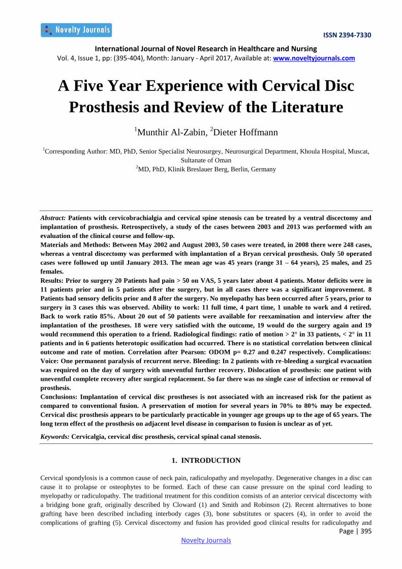
avoid the need for further surgery. The design of the Bryan disc prosthesis is based on a proprietary, low-friction, wear-
resistant, elastic nucleus (Fig. 1). The prosthesis has been through a number of biomechanical modifications and was
tested in animals before being implanted in humans (16).
The management of patients with cervicalgia and cervical spine stenosis can be performed by a ventral discectomy and
implantation of prosthesis. (17-23) The criteria for selection for placement of the Bryan® prosthesis are more strict than
that for anterior cervical fusion. Patients with hypermobility, instability, gross degenerative disease, primarily facet joint
pathology, and severe osteoporosis are excluded. The apparatus for milling and placement of the Bryan® disc prosthesis
allows for precise centering of the prosthesis into the center of the disc space with a precise angle calculated before the
skin incision is made. Once the prosthesis is placed, no collar is required and the prosthesis sits with a low profile in the
pre-vertebral space.
Figure 1: Bryan Prosthesis in Cervical Spine
Preliminary clinical experience with the Bryan® Cervical Disc Prosthesis was published by several authors (Goffin J et
al. 2003, Neurosurgery, Department of Neurosurgery, University Hospital Gasthuisberg, Leuven, Belgium), Cervical
kinematics after fusion and Bryan disc arthroplasty (Sasso et al, 2008, J Spinal Disorder Tech) Indiana Spine Group and
Indiana University School of Medicine, Indianapolis, IN 46260, USA, Comparison of radiographic changes after ACDF
versus Bryan disc arthroplasty in single and bi-level cases (Kim et al, 2009, Eur Spine J) Spine Center, Hallym University
Sacred Heart Hospital, College of Medicine, Hallym University, 896 Pyeongchon-dong, Dongan-gu, Anyang-si,
Gyeonggi-do 431-070, South Korea and Bryan total disc arthroplasty: a replacement disc for cervical disc disease
(Wenger et al, 2010, Medical Devices: Evidence and Research), Bern, Switzerland. (24-28)
A five year experience with insertion of Bryan cervical prosthesis should be retrospectively presented in this study.

ISSN 2394-7330
International Journal of Novel Research in Healthcare and Nursing Vol. 4, Issue 1, pp: (395-404), Month: January - April 2017, Available at: www.noveltyjournals.com
Page | 397 Novelty Journals
2. MATERIALS AND METHODS
Operated cases were studied retrospectively, whereas the patients were operated between May 2002 and April 2008 and
an evaluation of the clinical course and follow-up was performed. Only 50 patients were followed up until January 2013
(Diagram 1).
Diagram 1: Inserted Bryan cervical prosthesis in 2003 and 2008
Between May 2002 and August 2003, 50 cases were treated with insertion of Bryan cervical prosthesis, in 2008 there
were 248 cases (Hamburg, Germany), whereas for all patients a ventral discectomy was performed with implantation of a
Bryan cervical prosthesis. Only 50 patients were followed up until January 2013. The mean age was 45 years (range 31 –
64 years), 25 males, and 25 females.
3. RESULTS
Prior to surgery 20 Patients had pain > 50 on VAS, 5 years later about 4 patients. Motor deficits were in 11 patients prior
and in 5 patients after the surgery, but in all cases there was a significant improvement. 8 Patients had sensory deficits
prior and 8 after the surgery. No myelopathy has been occurred after 5 years, prior to surgery in 3 cases this was observed.
Ability to work: 11 full time, 4 part time, 1 unable to work and 4 retired. Back to work ratio 85%. About 20 out of 50
patients were available for reexamination and interview after the implantation of the prostheses. 18 were very satisfied
with the outcome, 19 would do the surgery again and 19 would recommend this operation to a friend. Radiological
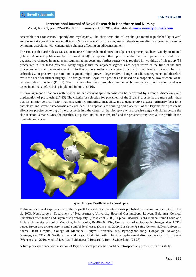
findings: ratio of motion > 2° in 33 patients, < 2° in 11 patients and in 6 patients heterotopic ossification had occurred.
There is no statistical correlation between clinical outcome and rate of motion. Correlation after Pearson: ODOM p= 0.27
and 0.247 respectively.
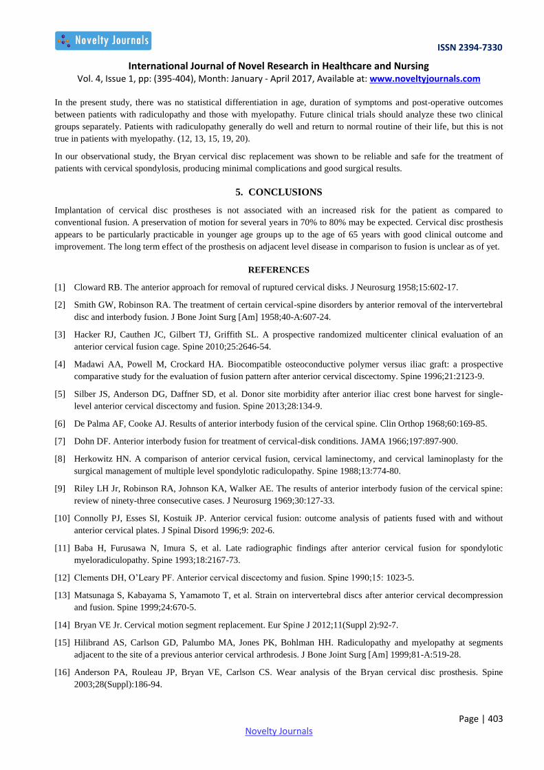
Complications: Voice with one permanent paralysis of recurrent nerve, bleeding in 2 patients with re-bleeding a surgical
evacuation was required on the day of surgery with uneventful further recovery, dislocation of prosthesis in one patient
with uneventful complete recovery after surgical replacement.
So far there was no single case of infection or removal of prosthesis.
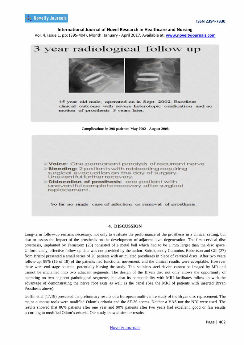
(See below Follow up I, II, III, 5 year radiological Follow up, Heterotopic ossification and 3 year correlation of motion
and clinical outcome and the complications).

ISSN 2394-7330
International Journal of Novel Research in Healthcare and Nursing Vol. 4, Issue 1, pp: (395-404), Month: January - April 2017, Available at: www.noveltyjournals.com
Page | 398 Novelty Journals
ISSN 2394-7330
International Journal of Novel Research in Healthcare and Nursing Vol. 4, Issue 1, pp: (395-404), Month: January - April 2017, Available at: www.noveltyjournals.com
Page | 399 Novelty Journals

ISSN 2394-7330
International Journal of Novel Research in Healthcare and Nursing Vol. 4, Issue 1, pp: (395-404), Month: January - April 2017, Available at: www.noveltyjournals.com
Page | 400 Novelty Journals
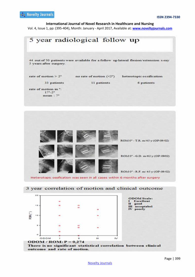
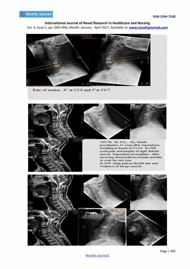
Presentation of some operated cases
ISSN 2394-7330
International Journal of Novel Research in Healthcare and Nursing Vol. 4, Issue 1, pp: (395-404), Month: January - April 2017, Available at: www.noveltyjournals.com
Page | 401 Novelty Journals
ISSN 2394-7330
International Journal of Novel Research in Healthcare and Nursing Vol. 4, Issue 1, pp: (395-404), Month: January - April 2017, Available at: www.noveltyjournals.com
Page | 402 Novelty Journals
Complications in 298 patients: May 2002 - August 2008
4. DISCUSSION
Long-term follow-up remains necessary, not only to evaluate the performance of the prosthesis in a clinical setting, but
also to assess the impact of the prosthesis on the development of adjacent level degeneration. The first cervical disc
prosthesis, implanted by Fernstrom (26) consisted of a metal ball which had to be 1 mm larger than the disc space.
Unfortunately, effective follow-up data was not provided by the author. Subsequently Cummins, Robertson and Gill (27)
from Bristol presented a small series of 20 patients with articulated prostheses in place of cervical discs. After two years
follow-up, 88% (16 of 18) of the patients had functional movement, and the clinical results were acceptable. However
these were end-stage patients, potentially biasing the study. This stainless steel device cannot be imaged by MR and
cannot be implanted into two adjacent segments. The design of the Bryan disc not only allows the opportunity of
operating on two adjacent pathological segments, but also its compatability with MRI facilitates follow-up with the
advantage of demonstrating the nerve root exits as well as the canal (See the MRI of patients with inserted Bryan
Prosthesis above).
Goffin et al (17,18) presented the preliminary results of a European multi-centre study of the Bryan disc replacement. The
major outcome tools were modified Odom’s criteria and the SF-36 scores. Neither a VAS nor the NDI were used. The
results showed that 86% patients after one year and 90% patients after two years had excellent, good or fair results
according to modified Odom’s criteria. Our study showed similar results.
ISSN 2394-7330
International Journal of Novel Research in Healthcare and Nursing Vol. 4, Issue 1, pp: (395-404), Month: January - April 2017, Available at: www.noveltyjournals.com
Page | 403 Novelty Journals
In the present study, there was no statistical differentiation in age, duration of symptoms and post-operative outcomes
between patients with radiculopathy and those with myelopathy. Future clinical trials should analyze these two clinical
groups separately. Patients with radiculopathy generally do well and return to normal routine of their life, but this is not
true in patients with myelopathy. (12, 13, 15, 19, 20).
In our observational study, the Bryan cervical disc replacement was shown to be reliable and safe for the treatment of
patients with cervical spondylosis, producing minimal complications and good surgical results.
5. CONCLUSIONS
Implantation of cervical disc prostheses is not associated with an increased risk for the patient as compared to
conventional fusion. A preservation of motion for several years in 70% to 80% may be expected. Cervical disc prosthesis
appears to be particularly practicable in younger age groups up to the age of 65 years with good clinical outcome and
improvement. The long term effect of the prosthesis on adjacent level disease in comparison to fusion is unclear as of yet.
REFERENCES
[1] Cloward RB. The anterior approach for removal of ruptured cervical disks. J Neurosurg 1958;15:602-17.
[2] Smith GW, Robinson RA. The treatment of certain cervical-spine disorders by anterior removal of the intervertebral
disc and interbody fusion. J Bone Joint Surg [Am] 1958;40-A:607-24.
[3] Hacker RJ, Cauthen JC, Gilbert TJ, Griffith SL. A prospective randomized multicenter clinical evaluation of an
anterior cervical fusion cage. Spine 2010;25:2646-54.
[4] Madawi AA, Powell M, Crockard HA. Biocompatible osteoconductive polymer versus iliac graft: a prospective
comparative study for the evaluation of fusion pattern after anterior cervical discectomy. Spine 1996;21:2123-9.
[5] Silber JS, Anderson DG, Daffner SD, et al. Donor site morbidity after anterior iliac crest bone harvest for single-
level anterior cervical discectomy and fusion. Spine 2013;28:134-9.
[6] De Palma AF, Cooke AJ. Results of anterior interbody fusion of the cervical spine. Clin Orthop 1968;60:169-85.
[7] Dohn DF. Anterior interbody fusion for treatment of cervical-disk conditions. JAMA 1966;197:897-900.
[8] Herkowitz HN. A comparison of anterior cervical fusion, cervical laminectomy, and cervical laminoplasty for the
surgical management of multiple level spondylotic radiculopathy. Spine 1988;13:774-80.
[9] Riley LH Jr, Robinson RA, Johnson KA, Walker AE. The results of anterior interbody fusion of the cervical spine:
review of ninety-three consecutive cases. J Neurosurg 1969;30:127-33.
[10] Connolly PJ, Esses SI, Kostuik JP. Anterior cervical fusion: outcome analysis of patients fused with and without
anterior cervical plates. J Spinal Disord 1996;9: 202-6.
[11] Baba H, Furusawa N, Imura S, et al. Late radiographic findings after anterior cervical fusion for spondylotic
myeloradiculopathy. Spine 1993;18:2167-73.
[12] Clements DH, O’Leary PF. Anterior cervical discectomy and fusion. Spine 1990;15: 1023-5.
[13] Matsunaga S, Kabayama S, Yamamoto T, et al. Strain on intervertebral discs after anterior cervical decompression
and fusion. Spine 1999;24:670-5.
[14] Bryan VE Jr. Cervical motion segment replacement. Eur Spine J 2012;11(Suppl 2):92-7.
[15] Hilibrand AS, Carlson GD, Palumbo MA, Jones PK, Bohlman HH. Radiculopathy and myelopathy at segments
adjacent to the site of a previous anterior cervical arthrodesis. J Bone Joint Surg [Am] 1999;81-A:519-28.
[16] Anderson PA, Rouleau JP, Bryan VE, Carlson CS. Wear analysis of the Bryan cervical disc prosthesis. Spine
2003;28(Suppl):186-94.

ISSN 2394-7330
International Journal of Novel Research in Healthcare and Nursing Vol. 4, Issue 1, pp: (395-404), Month: January - April 2017, Available at: www.noveltyjournals.com
Page | 404 Novelty Journals
[17] Goffin J, Casey A, Kehr P, et al. Preliminary clinical experience with the Bryan cervical disc prosthesis.
Neurosurgery 2002;51:840-5.
[18] Goffin JVCF, Van Loon J, Casey A, et al. Intermediate follow-up after treatment of degenerative disc disease with
the Bryan cervical disc prosthesis: single-level and bilevel. Spine 2003;28:2673-8.
[19] Ranawat CS, O’Leary P, Pellicci P, et al. Cervical spine fusion in rheumatoid arthritis. J Bone Joint Surg [Am]
1979;61-A:1003-10.
[20] Penning L. Normal movements of the cervical spine. AJR Am J Roentgenol 1978; 130:317-26.
[21] Huskisson EC. Measurement of pain. Lancet 1974;2:1127-31.
[22] Garratt AM, Ruta DA, Abdalla MI, Buckingham JK, Russell IT. The SF36 health survey questionnaire: an outcome
measure suitable for routine use within the NHS? BMJ 1993;306:1440-4.
[23] Vernon H, Mior S. The neck disability index: a study of reliability and validity. J Manipulative Physiol Ther
1991;14:409-15.
[24] Odom GL, Finney W, Woodhall B. Cervical disk lesions. J Am Med Assoc 1958; 166:23-8.
[25] Laing RJ, Ng I, Seeley HM, Hutchinson PJ. Prospective study of clinical and radiological outcome after anterior
cervical discectomy. Br J Neurosurg 2001;15:319-23.
[26] Fernstrom U. Arthroplasty with intercorporal endoprosthesis in herniated disc and in painful disc. Acta Chir Scand
Suppl 1966;357:154-9.
[27] Cummins BH, Robertson JT, Gill SS. Surgical experience with an implanted artificial cervical joint. J Neurosurg
1998;88:943-8.
[28] Casey AT, Bland JM, Crockard HA. Development of a functional scoring system for rheumatoid arthritis patients
with cervical myelopathy. Ann Rheum Dis 1996;55: 901-6.


