
LIVER II

A - picornavirusB - hepadnavirusC - flavivirusD - defective virus
E - calcivirus
Physically
Handicapped
Fellow
Died
Cycling

HEPATITIS Acute hepatitis(<6 months )
-It is based upon the following: incubation period preicteric phase- malaise , fever,
fatigue, nausea muscle and joint ache etc.
icteric phase- jaundice –conjugated type
convalescence

HEPATITIS Microscopically: (acute hepatitis)
-hepatocyte swelling ( ballooning degeneration)- presence of apoptotic hepatocytes are known as Councilman bodies -cholestasis –due to bile plug formation-cytolysis or apoptosis-lobular disarray- due to loss of architecture-regenerative changes- hepatocyte proliferation.
Severe lesions may present with confluent necrosis of hepatocytes-Bridging Necrosis

HEPATITIS There are many attributable causes of
hepatitis, which include: -viruses- CMV, EBV, herpesvirus, etc-autoimmune
The term viral hepatitis is often thought to be synonymous with diseases caused by the known hepatotropic viruses, including hepatitis viruses (HAV), B (HBV), C (HCV), D (HDV), and E (HEV)
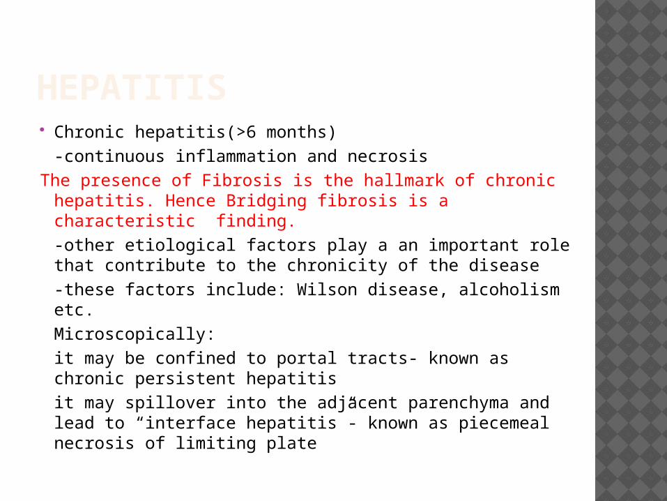
HEPATITIS Chronic hepatitis(>6 months)
-continuous inflammation and necrosisThe presence of Fibrosis is the hallmark of chronic
hepatitis. Hence Bridging fibrosis is a characteristic finding.-other etiological factors play a an important role that contribute to the chronicity of the disease-these factors include: Wilson disease, alcoholism etc. Microscopically:it may be confined to portal tracts- known as chronic persistent hepatitisit may spillover into the adjacent parenchyma and lead to “interface hepatitis”- known as piecemeal necrosis of limiting plate


ACUTE/CHRONIC HEPATITIS

HEPATITIS M/E findings con’t:
HBV type may produce a ground glass appearance- due to the accumulation of HBsAg

HEPATITIS CON’T Lab findings: increased ALT/AST
ALT/AST ratio at least 2:1
Diagnosis is done by serological markers

Acute viral hepatitis

CHRONIC VIRAL HEPATITIS

CHRONIC VIRAL HEPATITIS,GROUND GLASS APPEARANCE

HEPATITIS Fulminant Hepatitis: Hepatic failure that progresses within 2-3
weeks period to hepatic encephalopathy in the absence of chronic liver disease.
Viral hepatitis accounts for 12% of Fulminant hepatic failure.
Mainly Hep.B and HAV viruses. Massive necrosis of contigous
hepatocytes with or without accompanying inflammation.
Mortality is 80% without treatment, with renal transplant mortality drops to 35 %.
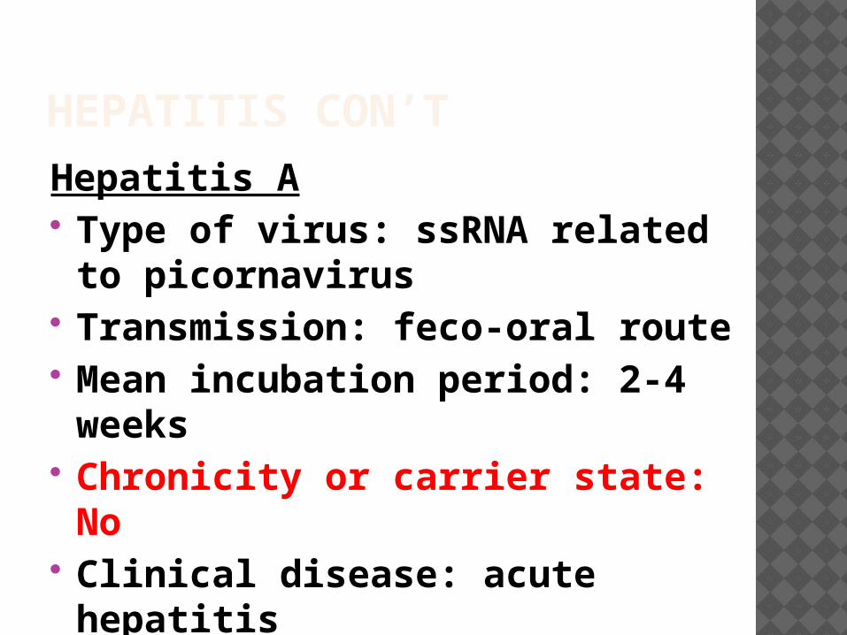
HEPATITIS CON’THepatitis A Type of virus: ssRNA related to picornavirus
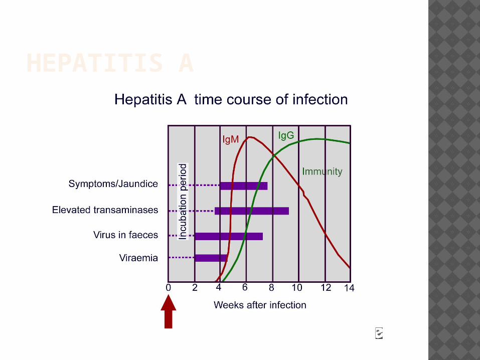
Transmission: feco-oral route Mean incubation period: 2-4 weeks
Chronicity or carrier state: No Clinical disease: acute hepatitis
Diagnosis: anti-HAV IgM Ab’s

HEPATITIS CON’THepatitis A This is a benign and self limited disease
Clinical disease is usually mild or asymptomatic
Factors predisposing humans to HAV include: overcrowding, poor sanitation and lack of a reliable clean water
Since viremia is transient, donated blood is not screened for
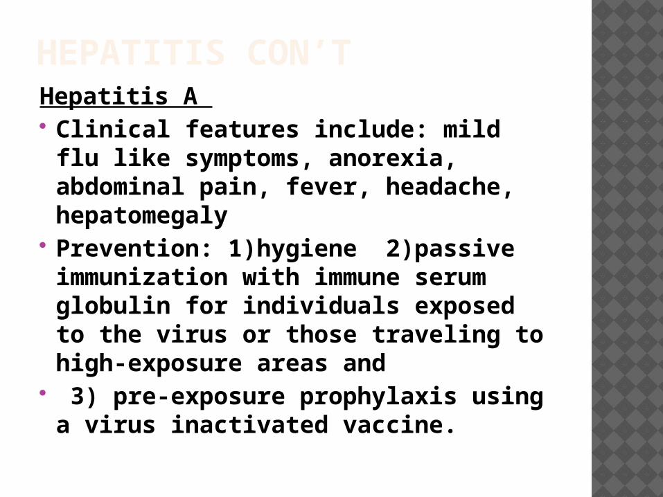
HEPATITIS CON’T Hepatitis A Clinical features include: mild flu
like symptoms, anorexia, abdominal pain, fever, headache, hepatomegaly
Prevention: 1)hygiene 2)passive immunization with immune serum globulin for individuals exposed to the virus or those traveling to high-exposure areas and
3) pre-exposure prophylaxis using a virus inactivated vaccine.
HEPATITIS A

HEPATITIS CON’T Hepatitis B Hepatitis B is a worldwide healthcare problem, especially in developing areas. An estimated one third of the global population has been infected with the hepatitis B
Prevention: vaccines and blood donor screening
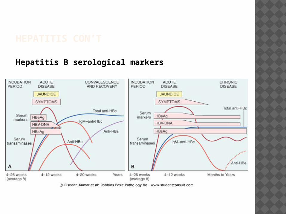
HEPATITIS CON’T
Hepatitis B serological markers
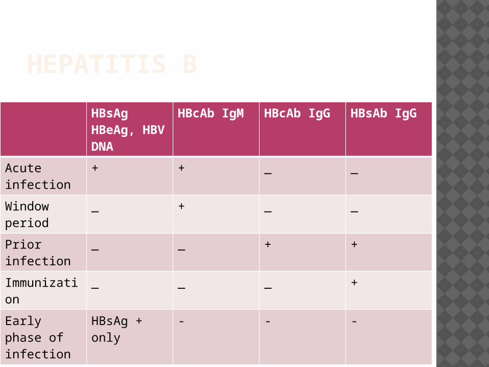
HEPATITIS B
HBsAgHBeAg, HBV DNA
HBcAb IgM HBcAb IgG HBsAb IgG
Acute infection
+ + _ _
Window period
_ + _ _
Prior infection
_ _ + +
Immunization
_ _ _ +
Early phase of infection
HBsAg + only
- - -
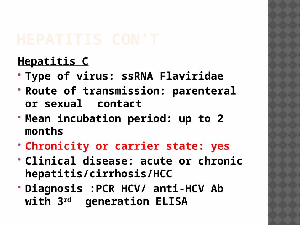
HEPATITIS CON’THepatitis C Type of virus: ssRNA Flaviridae Route of transmission: parenteral
or sexual contact Mean incubation period: up to 2
months Chronicity or carrier state: yes Clinical disease: acute or chronic
hepatitis/cirrhosis/HCC Diagnosis :PCR HCV/ anti-HCV Ab
with 3rd generation ELISA

HEPATITIS CON’T Hepatitis C Hepatitis C is the major cause of chronic hepatitis in the United States. HCV infections account for 20% of all cases of acute hepatitis
Patients may present asymptomatically or with subclinical disease.
Persistence of infection is key in HCV infections
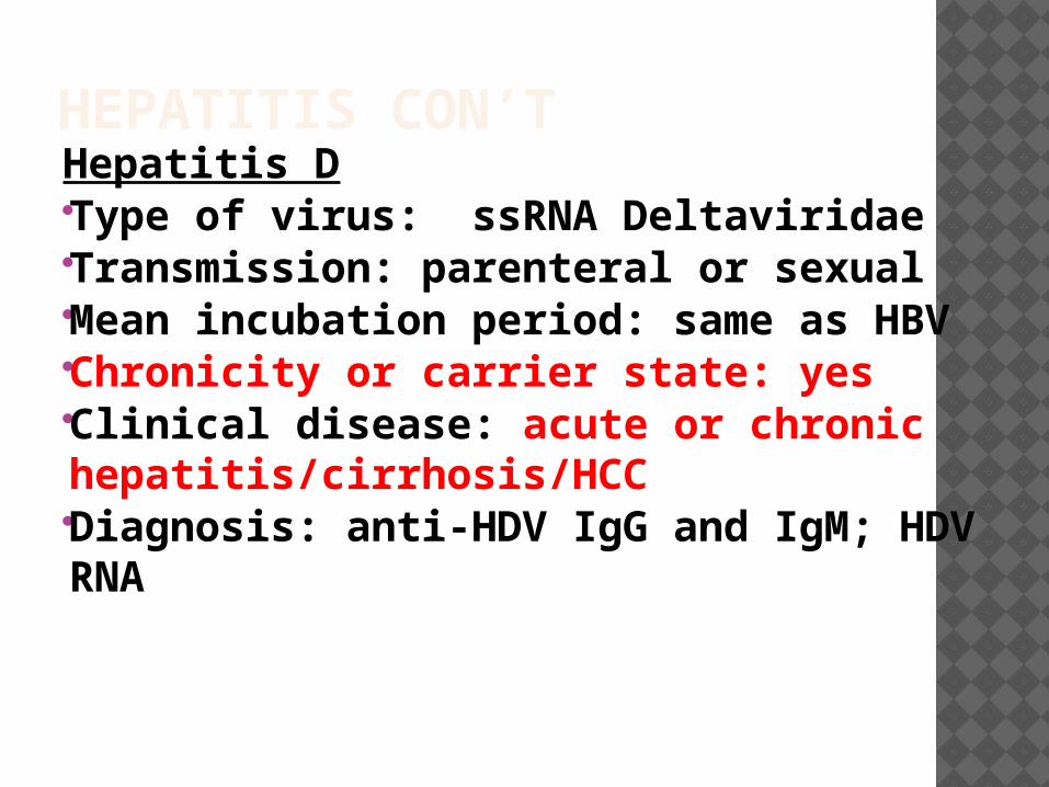
HEPATITIS CON’THepatitis D Type of virus: ssRNA Deltaviridae Transmission: parenteral or sexual
Mean incubation period: same as HBV
Chronicity or carrier state: yes Clinical disease: acute or chronic hepatitis/cirrhosis/HCC
Diagnosis: anti-HDV IgG and IgM; HDV RNA
HEPATITIS CON’T HDV causes a unique infection
HBV infection must be present, in order for the development of HDV virion
Co-infected individuals recover completely, whereas, individuals with super-infection progress to severe chronic hepatitis

HEPATITIS CON’THepatitis E Type of virus: ssRNA calcivirus Transmission:feco-oral Mean incubation period: 4-5 weeks Chronicity or carrier state: no Clinical disease: acute hepatitis Diagnosis: anti-HEV Ab for IgM and
IgG/PCR HEV RNA
Extremely fatal in pregnant women

ALCOHOLIC LIVER DISEASE Pathologic changes observed in
patients with alcohol-induced liver disease can be divided into the following 3 groups: alcoholic fatty liver (hepatic steatosis), alcoholic hepatitis, and cirrhosis.
All three forms of disease may present as a spectrum of disease, or they may occur independently of one another

ALCOHOLIC LIVER DISEASE CON’T
Mild and reversible changes, as seen in fatty liver, occur when an individual ingests as much as 80 g/day of alcohol
Chronic intake of alcohol of 50 -60 g/day may lead to severe forms of liver disease
Females are at an increased risk of liver disease, when compared to males
Genetics may play a role; however, no identifiable genetic markers are known
ALCOHOLIC LIVER DISEASE Alcohol metabolism: most of the alcohol is catabolized by
several pathways, which include: 1) ADH – alcohol dehydrogenase2) Cytochrome P-4503) MEOS- microsomal enzyme
oxidation system

ALCOHOLIC LIVER DISEASE CON’TFatty liver (hepatic steatosis) Fatty liver is the accumulation of triglycerides and other fats in the liver cells
In some patients, this may be accompanied by hepatic inflammation and liver cell death
Alcoholic fatty liver is an early and reversible consequence of excessive alcohol consumption
ALCOHOLIC AND DRUG RELATED LIVER DISEASE Fatty liver (hepatic steatosis)
There is a defect in fatty acid oxidation and lipoprotein synthesis. This leads to peripheral conversion of fat and subsequent hyperlipidemia
Grossly: large, yellow, greasy liver Microscopically : macrovesicular
globules (lipid accumulation within the hepatocyte displaces the nucleus to the periphery)
ALCOHOLIC LIVER DISEASE
Fatty liver (hepatic steatosis) con’t
Lab findings: increased bilirubin and ALP
Clinical findings: hepatomegaly
Treatment: cessation of alcohol

ALCOHOLIC LIVER DISEASE Gross: fatty liverEnlarged, yellow, greasy liver
ALCOHOLIC LIVER DISEASE
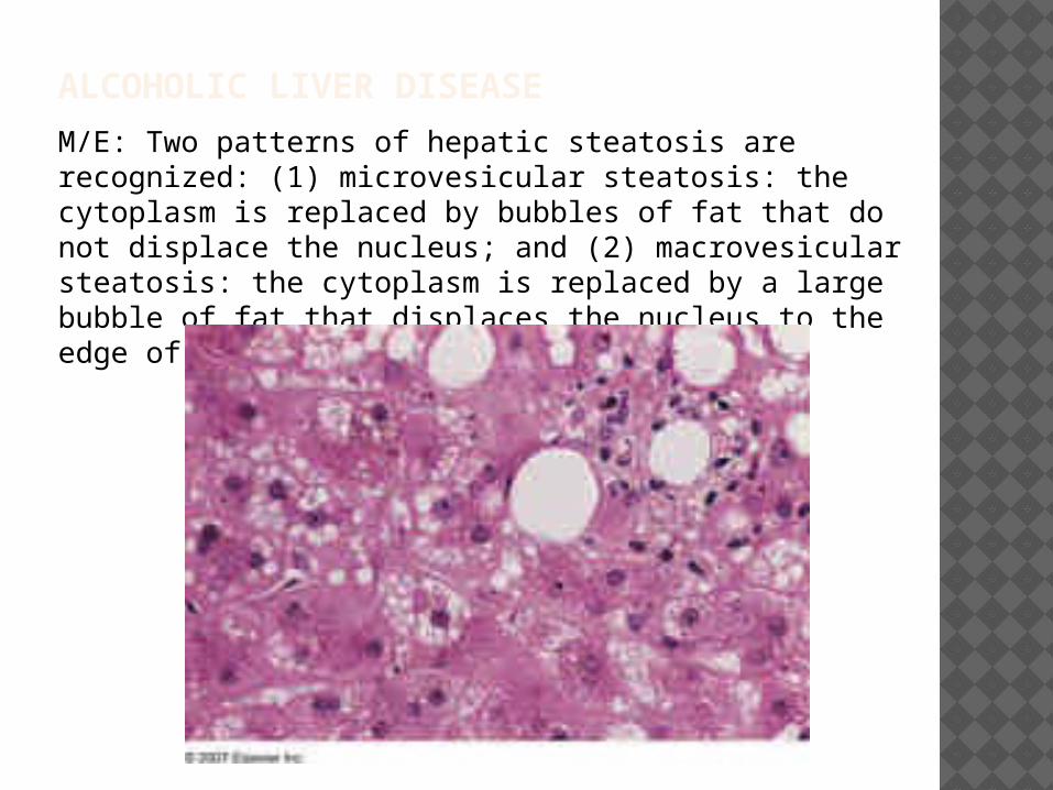
M/E: Two patterns of hepatic steatosis are recognized: (1) microvesicular steatosis: the cytoplasm is replaced by bubbles of fat that do not displace the nucleus; and (2) macrovesicular steatosis: the cytoplasm is replaced by a large bubble of fat that displaces the nucleus to the edge of the cell.

ALCOHOLIC LIVER DISEASE Alcoholic hepatitis aka steatohepatitis Alcoholic hepatitis is a syndrome of
progressive inflammatory liver injury associated with long-term heavy intake of ethanol- up to 2 decades
The MEOS system forms the reactive oxygen species (ROS) release cytokines i.e. TNF, IL-6, IL-8, IL-18
The combination of acetaldehyde and ROS leads to hepatic injury especially in the centrilobular region

ALCOHOLIC LIVER DISEASE Alcoholic hepatitis con’t Microscopically • Hepatic swelling (ballooning) and
necrosis• Mallory bodies- intermediate
filaments and proteins appear as eosinophilic cytoplasmic inclusions
• Neutrophil infiltration- these are present around degenerating hepatocytes
• Perivenular fibrosis
ALCOHOLIC LIVER DISEASE Alcoholic hepatitis Grossly: red and mottled liver Lab findings: increased ALP/ bilirubin / AST/ ALT
Clinical findings: malaise, anorexia, weight loss, hepatomegaly, abdominal tenderness.
Prognosis: complete cessation of alcohol may heal slowly; it may progress to cirrhosis

ALCOHOLIC LIVER DISEASE CON’T
M/E:Mallory body is shown within a ballooned hepatocyte.

ALCOHOLIC LIVER DISEASE Alcoholic cirrhosis This is the irreversible form of
alcoholic liver disease It has the same features as any
other cirrhosis- abnormal liver architecture, fibrosis, and vascular changes
Grossly : uniformly micronodular nodules < 0.3 cm in diameter. The liver initially is very large; it eventually transforms into a shrunken form

ALCOHOLIC LIVER DISEASE Alcoholic cirrhosisClinical findings: portal hypertensionhepatic encephalopathyjaundiceother findings thiamine and vitamin B12 deficiency
Lab findings: increased ALT/AST/bilirubin/ALP/ hypoproteinemia
AST/ALT ratio is increased to at least 2 to 1
This usually requires liver transplantation

ALCOHOLIC LIVER DISEASE Gross: There is diffuse nodularity of the liver- micronodular cirrhosis- <3mm in size
AST/ALT RATIO(DE RITIS RATIO)
http://www.ncbi.nlm.nih.gov/pmc/articles/PMC3866949/

METABOLIC LIVER DISEASE
NAFLD,Hemochromatosis,Wilson’s Disease

NON-ALCOHOLIC LIVER DISEASE
NAFLD Non-alcoholic Fatty Liver Disease Group of disorders with the common features of :Fatty liver and low (<20g/week)or absent alcohol consumption. Commonest cause of chronic liver disease in the U.S.
Includes: Simple hepatic steatosis, Steatosis with minor inflammation and NASH(non-alcoholic steato-hepatitis)
NON-ALCOHOLIC LIVER DISEASENAFLD/NASH Pathogenesis :unclear Two underlying events:1.Hepatic fat accumulation 2. Hepatic oxidative stress
Simple steatosis is usually asymptomatic but NASH presents with hepatocyte injury and may lead to cirrhosis(10-20%)
NON-ALCOHOLIC LIVER DISEASE
NAFLD/NASH
NASH strongly associated with other
components of the metabolic syndrome :Obesity, dyslipidemia, insulin resistance, hyperinsulinemia.
Lab: Elevated AST/ALT AST /ALT ratio less than 1.compared
with alcoholic hepatitis where the ratio is between 2-2.5.
HEMOCHROMATOSIS It is the abnormal accumulation of iron in parenchymal organs, leading to organ toxicity.
Males: females = 5:1(menstrual loss reduce progression in women)
The organs involved are the liver, heart, pancreas, pituitary, joints, gonads,skin ETC
Seen more commonly in people of northern European ancestry.

HEMOCHROMATOSIS Hereditary hemochromatosis (primary)Genetic mutations HFE gene mutation responsible for most
disease. This gene is located close to the HLA gene on chromosome 6
HFE regulate the levels of hepcidin.Mutation leads to low level of Hepcidin=>increased iron absorption and transport into plasma.
Disease becomes evident over the course of several years. Liver iron stores approaches 20g/l in symptomatic patients.

HEMOCHROMATOSIS Secondary hemochromatosis: this is the
acquired form of iron overload, which may be due to:
• blood transfusions, β-thalessemia, sideroblastic anemia
Hemosiderin gets deposited in various organs.Deposition of hemosiderin without clinical disease is referred to as hemosiderosis.
Grossly: micronodular cirrhosis
HEMACHROMATOSIS CON’T
Clinical findings: HepatomegalyAbdominal painHyperpigmentation of skin Pancreas –diabetes Heart: arrythmias, cardiomyopathyArthritis Ammenorrhea in females / impotence in malesLiver is affected in 100% of patients. The
pancreas and skin in 80% of patients.Bronze diabetes is diabetes mellitus plus skin
hyperpigmentation.
HEMOCHROMATOSIS Diagnosis: liver biopsy –hemosiderin granules stain with prussian blue
increased levels of iron and ferritin
Treatment: desferoxamine and phlebotomy
200 fold risk of developing HCC
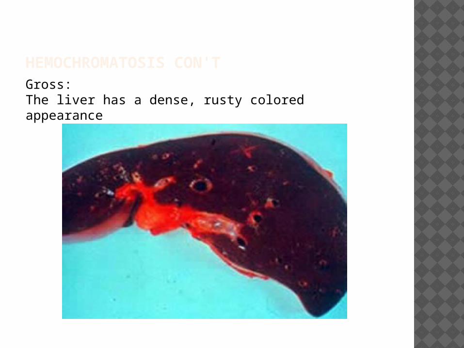
HEMOCHROMATOSIS CON'TGross: The liver has a dense, rusty colored appearance
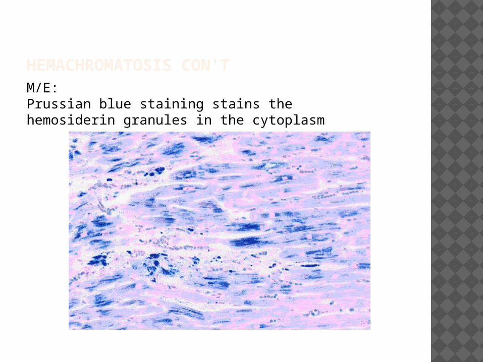
HEMACHROMATOSIS CON'T M/E: Prussian blue staining stains the hemosiderin granules in the cytoplasm
WILSON DISEASE Rare Autosomal recessive inherited disorder of copper metabolism.
Characterized by excessive deposition of copper in the liver, brain, and other tissues.
It mainly affects the liver, brain, and eye
It usually presents with Liver cirrhosis,behavioral changes, psychosis

WILSON DISEASE The genetic defect, localized to
chromosome arm 13q, has been shown to affect the copper-transporting ATPase gene (ATP7B) in the liver
Normally the process of copper metabolism is as follows:absorption of ingested copper copper- albumin complex goes to liver formation of ceruloplasmin gets released into the bloodstream senescent copper returns to the liver and gets degraded by lysosomes free copper gets secreted into bile eliminated from gut
WILSON DISEASE The gene (ATP7B) mutation prevents copper to be excreted into the bile
There is also inhibition of ceruloplasmin release into plasma
Thus, copper accumulates in the liver and causes free radical injury.
WILSON DISEASE CON’T There is increased urinary
excretion of copper
In the brain, affects basal ganglia, the putamen is mainly affected.
Grossly: it may present as fatty change of liver, acute or chronic hepatitis, micronodular cirrhosis

WILSON DISEASE CON’T Diagnosis: decreased ceruloplasmin levels
liver biopsy- increased copper
increased urinary copper
Measurement of serum copper level is not a reliable test to make diagnosis.
Treatment: D-pencillamine

WILSON DISEASE Kayser- Fleischer rings are due to deposition of copper in the limbus of the cornea. It is usually an orange brown discoloration

QUIZ Histologic examniation of liver biopsy
specimen from a 24 year old college student who presents with a chronic history of recurrent jaundice shows normal looking hepatocytes. Laboratory findings are shows unconjugated hyperbilirubinemia. Which of the following is most likely responsible?
A. Criggler Najar syndromeB. Gilbert syndromeC. Rotor syndromeD. Dubin-Johnson syndrome
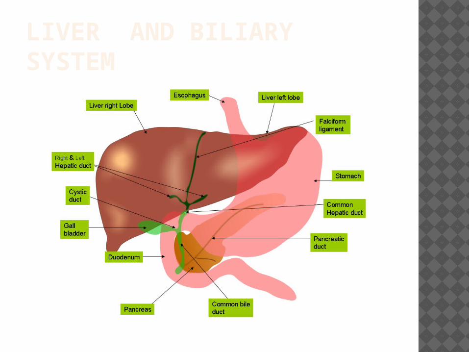
LIVER AND BILIARY SYSTEM
PRIMARY BILIARY CIRRHOSIS (PBC) Chronic and progressive cholestatic
disease of the liver. Considered to be an auto immune disorder. Inflammation and granulomatous
destruction of intrahepatic bile ducts Destruction of the small-to-medium bile
ducts, which leads to progressive cholestasis and often cirrhosis and end-stage liver disease
Anti-mitochondrial Ab’s in patients with PBC
F:M= 6 is to 1

PRIMARY BILIARY CIRRHOSIS (PBC) Other serological associations include: ANA and ANCA Ab’s
Associated with other auto immune disorders: Sjogren syndrome ETC
Subsequent to the loss of the intrahepatic bile ducts, a disruption of the normal bile flow occurs with retention and deposition of toxic substances, which are normally excreted into bile.

PRIMARY BILIARY CIRRHOSIS (PBC) The retention of toxic substances, such as bile acids, can cause further secondary destruction of the bile ducts and the hepatocytes
Clinical findings:pruritus/ fatigue/ xanthomas/ xanthelasma/ elevated serum cholesterol/ cirrhosis
Lab findings: Elevated AST/ALT,ALP bilirubinemia
Postive anti-mitochondrial antibody test

PRIMARY BILIARY CIRRHOSIS (PBC) CON'T End stage liver shows yellow green pigmentation
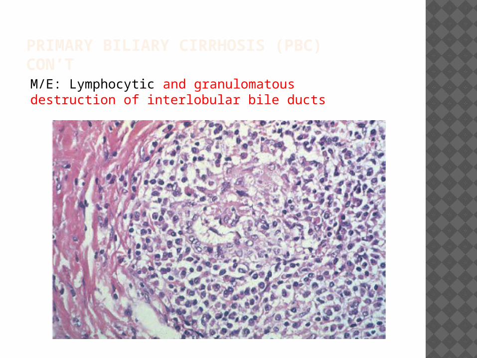
PRIMARY BILIARY CIRRHOSIS (PBC) CON’TM/E: Lymphocytic and granulomatous destruction of interlobular bile ducts
PRIMARY SCLEROSING CHOLANGITIS (PSC) Chronic liver disease characterized by cholestasis with inflammation and fibrosis of the intrahepatic and extrahepatic bile ducts.
May lead to cirrhosis of the liver with portal hypertension
PRIMARY SCLEROSING CHOLANGITIS (PSC) CON’T Associated with ANCA Ab’s. Note:
anti-mitochondrial Ab’s are present in minority
Associated with inflammmatory bowel disease i.e ulcerative colitis
Clinical findings: fatigue/pruritus/jaundice
Diagnosis ERCP- multiple strictures and dilations of the intrahepatic and extrahepatic biliary ducts

PRIMARY SCLEROSING CHOLANGITIS (PSC) Lab findings: ALP
There is increased chances of developing of cholangiocarcinoma
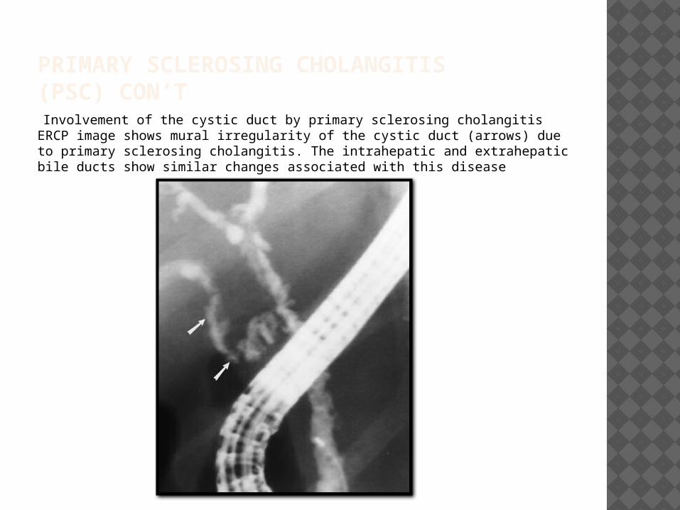
PRIMARY SCLEROSING CHOLANGITIS (PSC) CON’T Involvement of the cystic duct by primary sclerosing cholangitis ERCP image shows mural irregularity of the cystic duct (arrows) due to primary sclerosing cholangitis. The intrahepatic and extrahepatic bile ducts show similar changes associated with this disease

PRIMARY SCLEROSING CHOLANGITIS (PSC) CON’T M/E: there is periductal ‘onion skin’ fibrosis around the bile duct/ lymphocytic infiltration
HEPATIC ADENOMA Hepatocellular adenomas occur mostly in women of childbearing age and are strongly associated with the use of oral contraceptive pills (OCPs) and other estrogens
Hepatic adenomas consist of sheets of hepatocytes without bile ducts or portal areas.
HEPATIC ADENOMA
Hepatic adenomas are tan in color, smooth, well circumscribed, fleshy in appearance, and vary from 1 to 30 cm in size.
They have large blood vessels on the surface, and the lesions may outgrow their arterial blood supply, causing necrosis within the lesions.
A fibrous capsule may be present or absent; if absent, this may predispose to intrahepatic or extrahepatic hemorrhage
HEPATOCELLULAR CARCINOMA (HCC) Hepatocellular carcinoma is the most common primary malignancy of the liver.
It is also known as hepatoma Hepatocellular carcinoma occurs predominantly in patients with underlying chronic liver disease and cirrhosis Note: HCC may also occur in the absence of cirrhosis
HEPATOCELLULAR CARCINOMA (HCC) CON’T
Risk factors include:HBV or HCV infectionChronic alcoholismAflatoxin exposureHemochromatosisTyrosinemiaPBC,PSCAAT deficiencyNote: HCV is the most important risk factor (in
the western world)Hereditary Tyrosinemia (rare) is the condition
with the greatest association with PLCC(40% of cases would develop cancer)
HEPATOCELLULAR CARCINOMA (HCC) CON’T Pathogenesis- unclearit may develop from pre-existing nodules high grade dysplastic nodules i.e cirrhosis
DNA damage may be caused by cell death, inflammation, and hepatocyte replication
Hepatocyte replication may be due to point mutations or β-catenin overexpression or p53

HEPATOCELLULAR CARCINOMA CON’T Clinical findings:
HepatomegalyHistory of cirrhosis- blood in ascitic fluid
Diagnosis: α-FP (non specific).Useful for monitoring disease progression
Treatment: liver transplantation or
surgical resection

HEPATOCELLULAR CARCINOMA Gross: this is an example of a multifocal tumor, which is made up of nodules of varying sizesThere is also cirrhotic liver present below the cancerous liver

HEPATOCELLULAR CARCINOMA CON'T
M/E:This is malignant epithelial tumor consists of scant stroma and central necrosis because of the poor vascularization. In well differentiated forms, tumor cells resemble hepatocytes, form cords and nests, and may contain bile pigment in cytoplasm.

GALLSTONES/CHOLELITHIASIS Gallstone formation occurs because
certain substances in bile are present in concentrations that approach the limits of their solubility
When bile is concentrated in the gallbladder, it can become supersaturated with these substances, which then precipitate from solution as microscopic crystals

GALLSTONES/CHOLELITHIASIS There are mainly two types of
stones:1) Cholesterol stones –it is due to
supersaturation of bile with cholesterol/ calcium salt precipitation/stasis of bile/ mucus hypersecretion
2) Pigment stones- it is due to the presence of unconjugated bilirubin
GALLSTONES/CHOLELITHIASIS Cholesterol stone risk factors
Age- older individuals Gender- F>MHeredity-family historyEstrogen- OCP’s, pregnancyWeight lossHyperlipidemia Spinal cord injuryObesity Gallbladder stasis

GALLSTONES/CHOLELITHIASIS Cholesterol stones: These arise in the
gallbladder mainly Pure cholesterol
stones are pale yellow
Cholesterol stones are radiolucent – if calcium is present, they may appear radiopaque

GALLSTONES/CHOLELITHIASIS Pigment stones risk factors
Hemolytic anemiasBiliary tract infections (e.coli,ascariasis,liver fluke)Ileal defects- Crohn’s disease/ ileal resectionCystic fibrosis
Note: the pigment stones can appear brown or black. Black stones are usually exclusive to the gallbladder; brown stones are typically found in the bile ducts
GALLSTONES/CHOLELITHIASIS Pigment stones
Black stones are formed by combination of unconjugated bilirubin and calcium
Brown stones are formed when bacterial hydrolysis of lecithin leads to the release of fatty acids, which complex with calcium and precipitate from solution. The resulting concretions have a claylike consistency and are termed brown pigment stones

GALLSTONES/CHOLELITHIASIS Pigment stones These stones can
arise anywhere in the biliary tree
Black stones are mainly radiopaque because of calcium carbonate
Brown stones are radiolucent due to the presence of calcium soaps

GALLSTONES/CHOLELITHIASIS Clinical features:
it is asymptomatic mainly
if symptomatic- biliary colic- spasmodic pain
complications include: inflammation of the biliary tree/ empyema/ perforation/ fistula/ obstructive cholestasis/ pancreatitis

CHOLECYSTITIS It is defined as inflammation of the
gallbladder, which is due to obstruction of the cystic duct- most common cause: gallstone impaction
It manifests in several forms, which include: acute calculous cholecystitis, acute non-calculous cholecystitis, and chronic cholecystitis
Diagnosis: ultrasonography- thickened gallbladder wall

CHOLECYSTITIS Acute calculous cholecystitis
It is inflammation of the gallbladder that is caused by obstruction of the cystic duct by gallstonesObstruction of the ductlecithin converts to lysolecithin toxic to mucosal layer chemical type of irritation distension increases intraluminal pressure this leads to compromised blood flowThis condition may require emergent cholecystectomy or it may subside on its own

CHOLECYSTITIS CON’T Acute calculous cholecystitis
Clinical findings: fever, nausea,vomiting mid-epigastric pain to RUQ colicky pain- may radiate to the tip of the shoulder jaundice- obstruction may be presentleukocytosisgallstone ileus/ perforation/ gangrene

CHOLECYSTITIS CON’TAcute non-calculous cholecystitis
This is not associated with stones
It is associated with serious conditions i.e. trauma, burns, sepsis, major surgeries (post-operative)
Predisposing factors include: dehydration, gallbladder stasis, bacterial infection, vascular insufficiency
CHOLECYSTITIS CON’TChronic cholecystitis
Repeated attacks of acute cholecystitis, which is most commonly due to gallstones
There is consistent chronic inflammation and stone formation due to the presence of supersaturated bile
Infection is uncommon, if present, bile culture will reveal E.coli
Clinical findings: epigastric pain or RUQ pain
CHOLEDOCHOLITHIASIS/CHOLANGITIS Choledocholithiasis occurs as a result
of: 1)The primary formation of stones in
the common bile duct 2)the passage of gallstones from the
gallbladder through the cystic duct into the CBD
Cholangitis refers to the inflammation of the bile ducts. Bacterial infections are very common – due to E coli, Klebsiella, Bacteroides, Clostridium, Bacteroides etc.
CHOLEDOCHOLITHIASIS/CHOLANGITIS Cholangitis con’t Ascending cholangitis
It is an infection of the bile ducts that ascends up-to the liver and intrahepatic biliary ducts
Clinical findings:Fever, chills, and jaundiceLiver abscess

CARCINOMA GALL BLADDER It is the most common of all gall
bladder cancers. It is an adenocarcinoma
The most common risk factor for gallbladder cancer is gallstones, which are present in 75%-90% of gallbladder cancer cases
Thus, chronic inflammation and trauma predispose the individual to this type of cancer
CARCINOMA OF GALL BLADDER It may be associated with porcelain
gallbladder- there is dystrophic calcification within the chronically inflamed gallbladder wall. Gallstones are found in over 90% of cases.
Clinical findings include:it may remain asymptomatic enlarged gallbladder- abdominal pain/ jaundice/anorexia

CARCINOMA OF GALL BLADDER CON'T
Adenocarcinoma growing in diffuse fashion in the distal wall of the gallbladder, associated with extensive involvement of the liver. Most common type
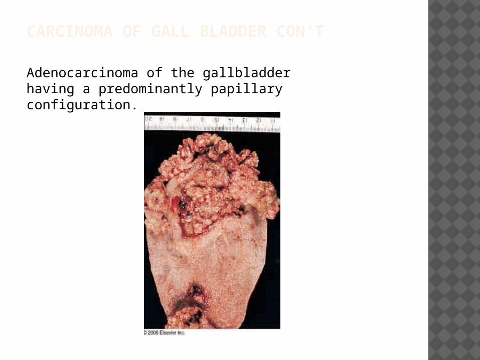
CARCINOMA OF GALL BLADDER CON'T
Adenocarcinoma of the gallbladder having a predominantly papillary configuration.
CHOLANGIOCARCINOMA Cholangiocarcinomas are
malignancies of the biliary duct system that may originate in the liver and extrahepatic bile ducts
The tumor may arise from the bifurcation of the right and left hepatic bile ducts- known as Klatskin tumor(60% of CCA)
They can arise more distally as well

CHOLANGIOCARCINOMA Risk factors include: primary
sclerosing cholangitis ,caroli disease,HCV infection or thorotrast dye exposure
O. sinensis and related liver flukes in South East asia.
Clinical findings: weight loss and anorexia-intrahepatic jaundice, clay colored stools
Lab findings: increased ALP/ AST/ALT


