Download - Ampullariidae: Pila globosa

Ruthenica, 2014, vol. 24, No. 1: 31-33.Published electronically May 21, 2014.
© Ruthenica, 2014http: www.ruthenica.com
ABSTRACT. The present investigation was undertak-en to study a shell disease of the freshwater snail, Pilaglobosa. Observations were made in June-July in fourconsecutive years. The disease first appears as blistersin the periostracum and then, once the periostracum islost from these lesions, dissolution of the underlyingcalcified layer. The numerically predominant bacterialgenera in the lesions included Aeromonas, Pseudomo-nas, Escherichia and Listeria. Communication de-scribes this previously unreported shell disease, whichmay be a health problem in apple snails.
The apple snail, Pila globosa (Swainson, 1822)is a vital component of biodiversity playing an im-portant role in the maintenance of aquatic ecosys-tems. They are widely distributed in Nepal andsouth western Asia. Apple snails are exceptionallywell adapted to tropical regions with periods ofdrought alternated with periods of excessive rainfall[Subba Rao, Dey, 1989]. Pila globosa occurs in alltypes of temporary and permanent water bodies likeponds, canals and ditches [Jahan et al., 2001].Though the species is widespread, in certain re-gions like Nepal, it is decreasing due to habitat loss,and fish poisoning [Subba Rao, Dey, 1989]. Mol-luscs are excellent sources of trace and minorelements that are essential for the growth and devel-opment of humans and they are also used as nutri-ent supplements for domestic animals and birds[Baby et al., 2010]. In Bangladesh and some partsof India, Pila globosa is used as a protein supple-ment for humans as well as in aquaculture, such asshrimp and catfish farming [Nath et al., 2008;Wilkins, Lee, 2002]. It has also been tested as abiocontrol agent for the aquatic weed Salvinia mo-lesta [Thomas, 1975]. The natural pathogens ofapple snails are not well known, though Godan[1983] reported the association of microorganismsespecially protozoa, both as parasites and as sym-bionts or commensals, but was cautious in assess-ing the role of parasites or pathogens in populationregulation. Occurrence of disease in Pila globosa
may pose a threat to some groups of people residingwithin its range.
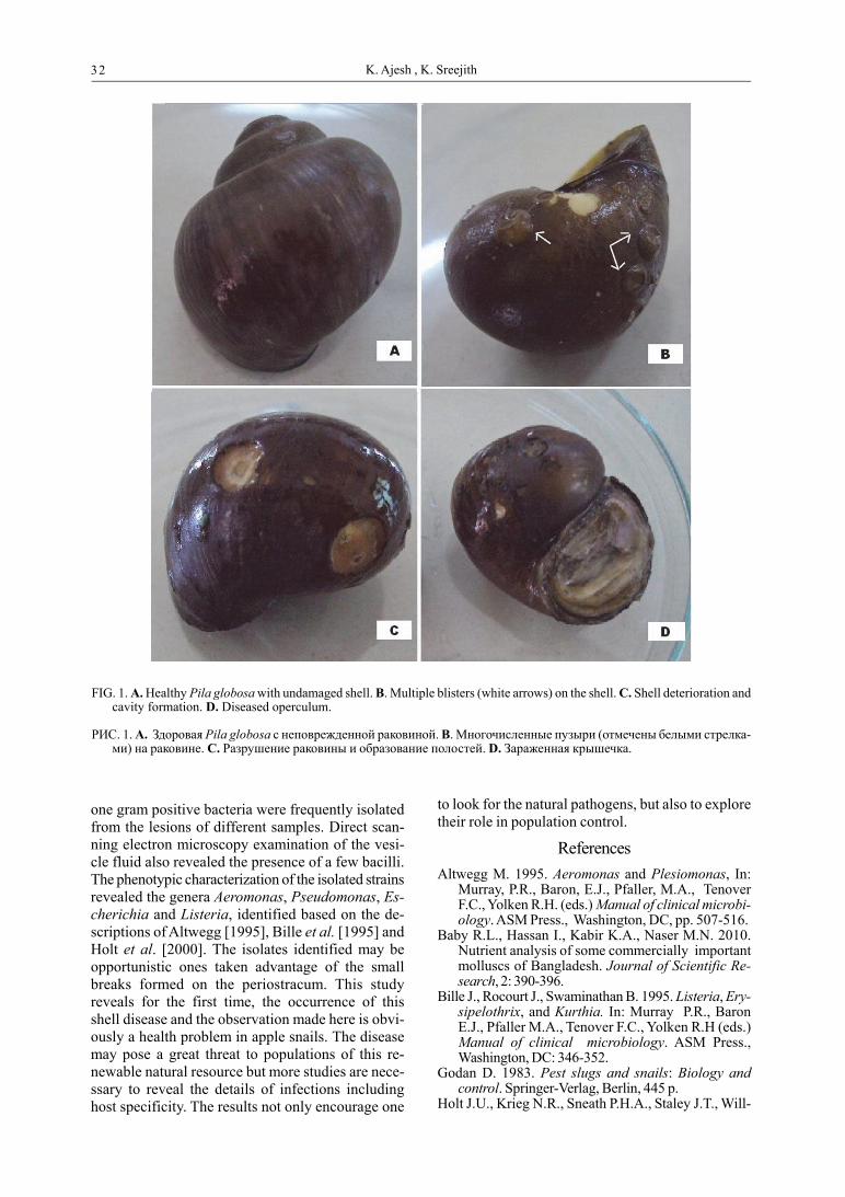
We report the occurrence of a shell disease inPila globosa. Initially, blister formation in the peri-ostracum is seen (Fig. 1B). As the disease progress-es, more blisters appear. Once the protein coat hasbeen lost, the calcified layer appears as white patch-es. This is followed by deterioration of the shell andcavity formation (Fig. 1C) when exposed to envi-ronment factors such as varying pH. Once the pHof the environment drops, the exposed calcium partstarts to dissolve. Problems may arise, when holesare formed in the cavity, exposing the soft tissuesbelow. The operculum, which helps to preventdrying out during aestivation [Meenakshi, 1964] isalso vulnerable to deterioration (Fig. 1D).
The infected specimens used in this study werecollected from paddy fields near brackish water inKannur district, Kerala, India during June – July of2010, 2011, 2012 and 2013 on the onset of themonsoon. The study was undertaken with the in-tention of isolating the causative agent from theinfected snails. Diseased snails were brought to thelaboratory and maintained under natural tempera-ture and moisture regimes. All experimental proce-dures were carried out under aseptic conditions.The shell was washed and then surface sterilizedwith 70% ethanol, carefully without rupturing theblisters. Then with the aid of a surgical blade, theblister was cut open and using a micropipette, 5 µLof the blister fluid was transferred to tubes contain-ing an enrichment broth (10 ml) prepared in snailinfusion (prepared by boiling healthy Pila globosain double distilled water) at pH 7, containing 1%tryptone, 0.5% yeast extract and 1% NaCl. Wehave found this medium to be preferable to otherbacteriological media used for studying the bacterialflora of snails. The inoculated tubes were incubatedat room temperature (30±2° C) for 3 days. Theturbid growth in the liquid medium was then streakedonto solidified snail infusion media and incubated atroom temperature for three days before the colo-nies were counted. Differently looking colonies wasisolated in pure cultures. Three gram negative and
Disease of the shells of Indian apple snails(Ampullariidae: Pila globosa)
AJESH K., SREEJITH K.*
1 Department of Biotechnology and Microbiology, Kannur University, Kerala–670 661 India* Corresponding author, E-mail: [email protected]
32 K. Ajesh , K. Sreejith
one gram positive bacteria were frequently isolatedfrom the lesions of different samples. Direct scan-ning electron microscopy examination of the vesi-cle fluid also revealed the presence of a few bacilli.The phenotypic characterization of the isolated strainsrevealed the genera Aeromonas, Pseudomonas, Es-cherichia and Listeria, identified based on the de-scriptions of Altwegg [1995], Bille et al. [1995] andHolt et al. [2000]. The isolates identified may beopportunistic ones taken advantage of the smallbreaks formed on the periostracum. This studyreveals for the first time, the occurrence of thisshell disease and the observation made here is obvi-ously a health problem in apple snails. The diseasemay pose a great threat to populations of this re-newable natural resource but more studies are nece-ssary to reveal the details of infections includinghost specificity. The results not only encourage one
to look for the natural pathogens, but also to exploretheir role in population control.
References
Altwegg M. 1995. Aeromonas and Plesiomonas, In:Murray, P.R., Baron, E.J., Pfaller, M.A., TenoverF.C., Yolken R.H. (eds.) Manual of clinical microbi-ology. ASM Press., Washington, DC, pp. 507-516.
Baby R.L., Hassan I., Kabir K.A., Naser M.N. 2010.Nutrient analysis of some commercially importantmolluscs of Bangladesh. Journal of Scientific Re-search, 2: 390-396.
Bille J., Rocourt J., Swaminathan B. 1995. Listeria, Ery-sipelothrix, and Kurthia. In: Murray P.R., BaronE.J., Pfaller M.A., Tenover F.C., Yolken R.H (eds.)Manual of clinical microbiology. ASM Press.,Washington, DC: 346-352.
Godan D. 1983. Pest slugs and snails: Biology andcontrol. Springer-Verlag, Berlin, 445 p.
Holt J.U., Krieg N.R., Sneath P.H.A., Staley J.T., Will-
FIG. 1. A. Healthy Pila globosa with undamaged shell. B. Multiple blisters (white arrows) on the shell. C. Shell deterioration andcavity formation. D. Diseased operculum.
РИС. 1. A. Здоровая Pila globosa с неповрежденной раковиной. B. Многочисленные пузыри (отмечены белыми стрелка-ми) на раковине. C. Разрушение раковины и образование полостей. D. Зараженная крышечка.

33Shell disease in Pila globosa
iams, S.T. 2000. Bergey’s manual of determinativebacteriology, Lippincott Williams & Wilkins, Phila-delphia, USA.
Jahan M. S., Akter M.S., Sarker M.M., Rahaman M.R.,Pramanik M.N. 2001. Growth ecology of Pila glo-bosa (Swainson) (Gastropoda: Pilidae) in stimulat-ed habitat. Pakistan Journal of Biological Scienc-es, 4: 581-584.
Meenakshi V.R. 1964. Aestivation in the Indian applesnail Pila. 1. Adaptation in natural and experimen-tal conditions. Comparative Biochemistry and Phys-iology, 11: 379-386.
Nath R.D., Rahi M.L., Hossain G.S., Huq K.A. 2008.Bangladesh status of fresh water snail in Khulnadistrict. Bangladesh Research Publication Jour-nal, 1: 337-347.
Subba Rao N.V., Dey A. 1989. Freshwater molluscs inaquaculture. In: Handbook of Freshwater Mol-luscs of India. Zoological Survey of India: 225-232.
Thomas K.J. 1975. Biological control of Salvinia by thesnail Pila globosa Swainson. Biological Journalof Linnean Society, 7: 243-247.
Wickins J.F., Lee, D.O’C. 2002. Crustacean farming:Ranching and culture. Blackwell Science, Oxford,480 p.
Заболевание раковины у индийской ампуллярии(Ampullariidae: Pila globosa)
АДЖЕШ К., СРИДЖИТ К.*
Department of Biotechnology and Microbiology, Kannur Uni-versity, Kerala–670 661 India; * автор для переписки[email protected]
РЕЗЮМЕ. Настоящая работа была проведена дляизучения заболевания раковины у пресноводногомоллюска Pila globosa. Наблюдения проводились виюне-июле в течение четырех последовательных лет.Заболевание проявляется вначале в появлении пу-зырей на периостракуме, а после того как периост-ракум исчезает в местах повреждений, в растворе-нии подлежащих кальцифицированных слоев. До-минирующими по численностями родами бактерийв местах повреждений являются Aeromonas,Pseudomonas, Escherichia и Listeria. Это первое опи-сание заболевания, которое моэжет вызывать про-блемы со здоровьем у ампуллярий.


