An Evidence-based approach to Upper GI Bleeding
Jonathan P. Terdiman, M.D.University of California, San
Francisco
Upper GI Bleeding
Consensus Recommendations on UGIB in Ann Intern Med
2003;139:843-857
Incidence
• Decreasing incidence over the past 10 years– 77/100, 000 in 1993– 53/100, 000 in 2003
• 250,000 hospitalizations per year in USA

Epidemiology
• Disease of the elderly– 20-30 fold increase from 3rd to 9th decade– median age > 70 years
• Men more common than women• Additional major comorbidity in 50%• NSAID use in > 50%
Outcomes• Mortality = 3.5%
– 15,000-30,000 deaths per year in USA– No change in rates over past decade
• Recurrent bleeding in 10-20%• Length of stay = 4-5 days• Need for surgical intervention as
decreased– 7% to 4% from 1993 to 2003
Outcomes: Population-based study in Canada
Am J Gastroenterol, 2004
• 1869 patients, 18 centers• EGD within 24 hours in 76%• Rebleeding in 14%, surgery in 7%,
death in 5%• High risk PUD: reduced mortality with
– PPI (OR, 0.18), EGD Rx (OR, 0.31)

Outcomes: importance ofcomorbidities
• After control of active PUD bleed with EGD Rx and PPI
• 7 day rebleed rate driven by other medical conditions– 2.5% in healthy patients– 25% with one major comorbidity– > 50% with > 2
Gastroesophageal Varices• 50 % of cirrhotics
– < 40 % Childs A; > 80 % Childs C
• Incidence of new varices: 5 % / year
• Incidence of bleeding : 10 –20 %/year
– 1/3 of deaths from cirrhosis
– Risk related to: Variceal size, “Red color signs”, Childs class, Active ETOH
Variceal Bleeding
Outcomes• Acute Bleeding Episodes: < 48 hours
– 50 % stop spontaneously– Least likely to stop: large varices, Childs C– Mortality: 10 %– Death: aspiration, sepsis, coma, renal failure >>
bleeding• High Risk Period: < 6 weeks
– Rebleeding: 40 – 50 %– Increased risk: large varices, age > 60, renal failure,
severe or active bleeding on presentation• > 6 Weeks to 1 year
– 70 % rebleeding– Survival similar to cirrhotics who have never bled
Variceal Bleeding
Initial Resuscitation
• Intravascular volume replacement
• Airway management
• Blood products
Volume Replacement
• Isotonic crystalloid solutions• Packed cells to keep Hct 25-30%
– anticipate nadir• FFP for PT > 1.5 x control, platelets
for count < 50,000
Airway Management
• Pulmonary complications a major source of morbidity and mortality– > new CXR infiltrates in ~ 15%– Significant cardiopulm comlications in 5%
• Endotracheal intubation for altered mental status or massive bleed
• Impact on outcome uncertain

Initial Risk Assessment• Important Factors
– Vital signs– Manifestation of bleed– Comorbid medical conditions– Age– Hematocrit– Coagulopathy
• Not important: ASA/NSAID, anticoagulant, steroid use, stable medical conditions
Nasogastric Lavage• Important for diagnosis and risk
assessment if location and severity of bleed is not clear
• False negatives in 10-15% of upper bleeds
• Does not induce bleed, even whenvarices present
• Large volume lavage in ED not useful, can be dangerous
Value of NGA Related to Pre-test Probability of High-risk Lesion
35%65%53%47%Bloody
80%20%77%23%Coffee grounds
87%13%79%21%Clear or bilious
LRLHRLLRLHRLNGA
Stable VSHemodynamic instability
Bloody NG aspirate: sensitivity 48%, specificity 75%, PPV 45 % for high risk lesions. Clear NG aspirate: PPV 85% and specificity 94% for low-risk lesions
Triage from Emergency Department
Home Endo Unit Floor ICU
Definition of Low Risk: < 5 % Rebleeding, < 1 % Mortality
BlatchfordSystolic BP > 110 Pulse < 100 Hgb > 12 (women), 13 (men)BUN < 6.5 mmol/LComorbities: none
Clinical RockallSystolic BP > 100Pulse < 100Age < 60 years
Comorbidities: none
Gralnek I, GIE 2004; 60:9.• Retrospective cohort (n = 175) hospitalized with
acute NVUGIB• Scoring of 0: 0 % rebleeding
Discharge w/o Endoscopy?
Limitations: 1. Identify only 8-12 % of patients with
NVUGIB as “low-risk”2. ER physicians reluctant to accept risk3. Concerns: generalizability and
validation

Who Requires ICU Admission
• Institution specific• In general:
– Ongoing hemodynamic instability despite IV fluids– Evidence of active bleeding: hematemesis, lg volume
bloody lavage, hematochezia– Significant comorbidities: ischemia,liver, CA– Advanced elderly
Early Endoscopy
• Diagnosis• Risk assessment• Therapy
Endoscopic Diagnoses at UCSF
30%
19%
11%
10%
7%6% 5% 12%
DUGUVaricesEsophagitis/ulcerMWTGastritis/duodentitsMassother
Other: Dieulafoy, vascular ectasia, AE fistula, diverticula, unknown
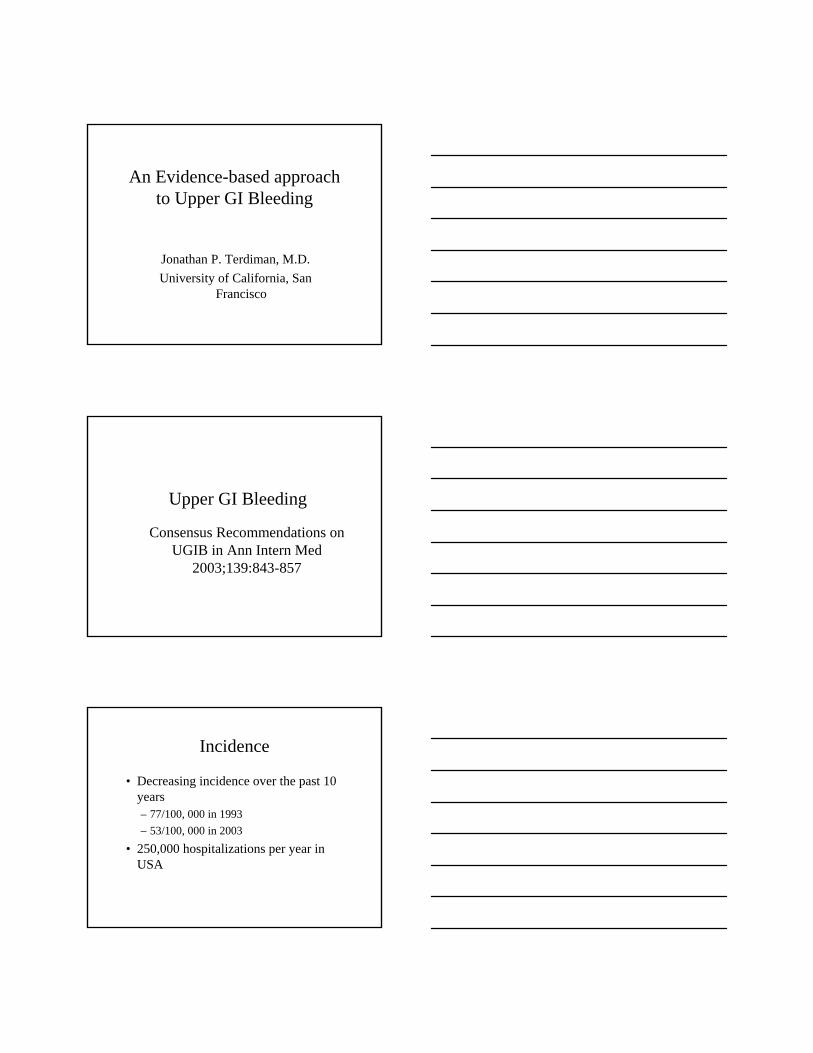
Ulcer with clean base
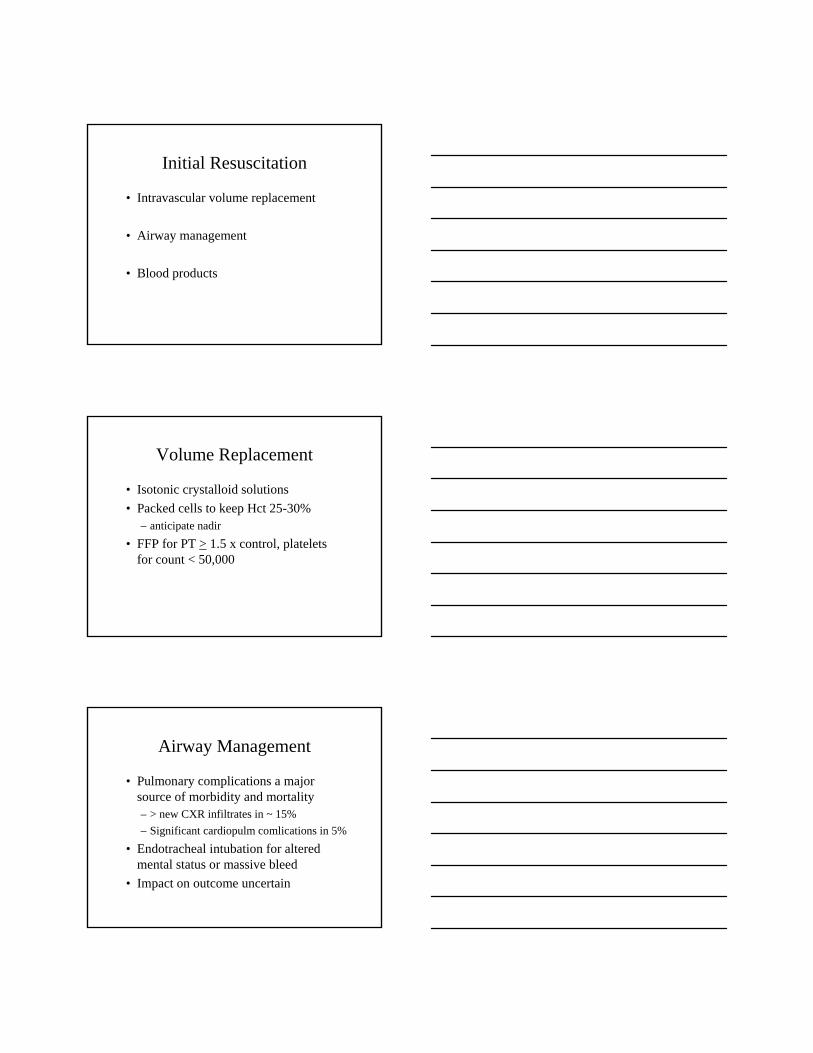
Ulcer with red/black spot
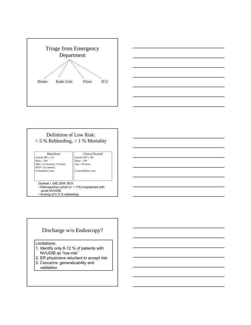
Ulcer with adherent clot
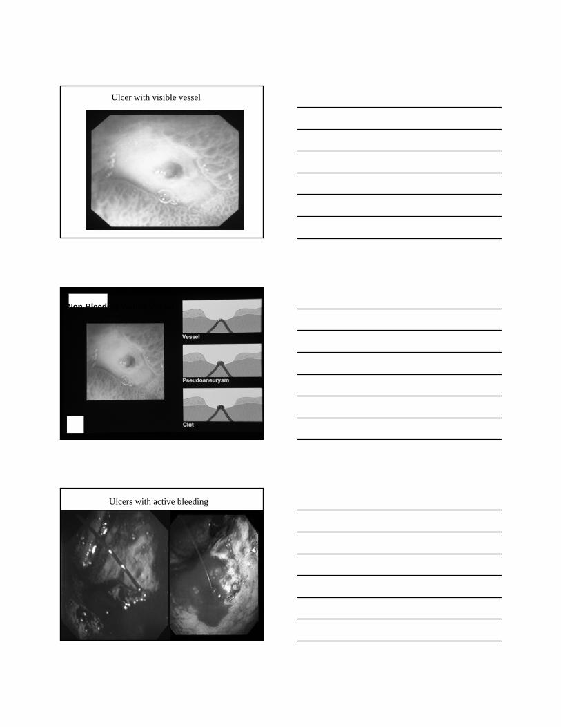
Ulcer with visible vessel
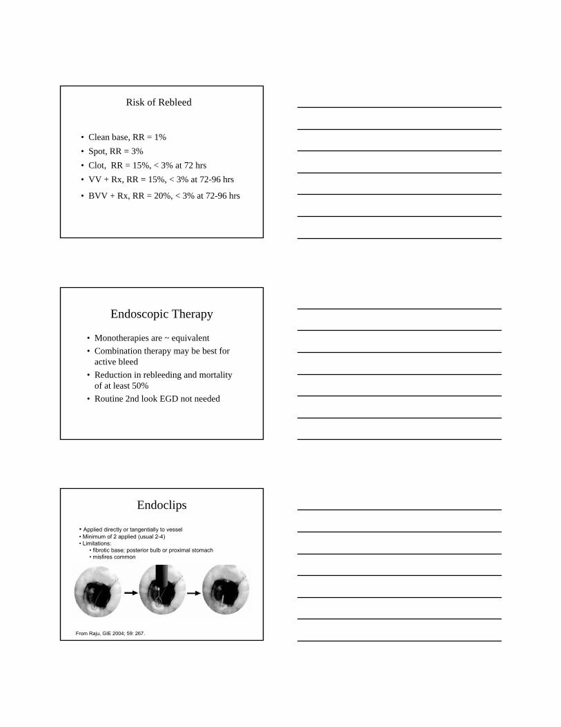
Non-Bleeding Visible Vessel
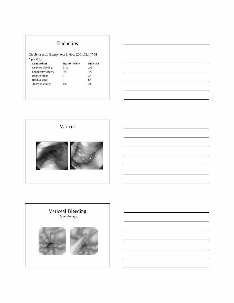
Ulcers with active bleeding

Endoscopic Stigmata of Bleeding Ulcers & Rebleed Risk
< 535Clean ulcer base
2510Adherent clot
5025Visible vessel
9010Arterial bleed
Rebleed (%)Prevalence (%)Stigmata
Rebleeding Rates in RCT’s of Treatment of Adherent Clots
35.0%34.3%
0.0%4.8%0%
10%
20%
30%
40%
50%
Mayo ClinicMulticenter Trial
UCLA CUREMulticenter Trial
Medical Therapy Endotherapy
P < 0.05
N = 56 N = 32
Risk of Rebleed
• Clean base, RR = 1%• Spot, RR = 3%• Clot, RR = 15%, < 3% at 72 hrs• VV + Rx, RR = 15%, < 3% at 72-96 hrs
• BVV + Rx, RR = 20%, < 3% at 72-96 hrs
Endoscopic Therapy
• Monotherapies are ~ equivalent• Combination therapy may be best for
active bleed• Reduction in rebleeding and mortality
of at least 50%• Routine 2nd look EGD not needed
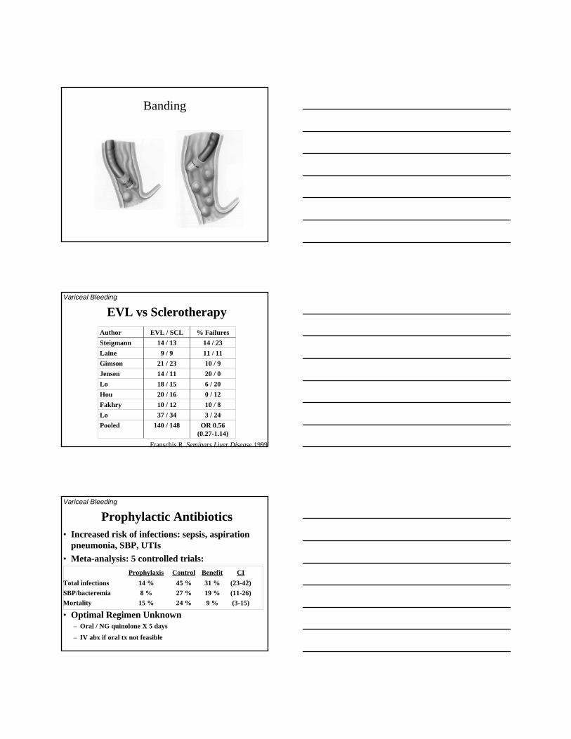
Endoclips
From Raju, GIE 2004; 59: 267.
• Applied directly or tangentially to vessel• Minimum of 2 applied (usual 2-4)• Limitations:
• fibrotic base; posterior bulb or proximal stomach• misfires common
Endoclips
Cipoletta et al. Gastrointest Endosc 2001;53:147-51* p < 0.05 Comparison Heater Probe Endoclip recurrent bleeding 21% 2%* Emergency surgery 7% 4% Units of blood 4 3* Hospital days 7 6* 30 day mortality 4% 4%
Varices
Variceal BleedingSclerotherapy
Banding
EVL vs SclerotherapyVariceal Bleeding
OR 0.56 (0.27-1.14)
140 / 148Pooled 3 / 2437 / 34Lo10 / 810 / 12Fakhry0 / 1220 / 16Hou6 / 2018 / 15Lo20 / 014 / 11Jensen10 / 921 / 23Gimson
11 / 119 / 9Laine14 / 2314 / 13Steigmann
% FailuresEVL / SCLAuthor
Franschis R, Seminars Liver Disease 1999
Prophylactic Antibiotics• Increased risk of infections: sepsis, aspiration
pneumonia, SBP, UTIs• Meta-analysis: 5 controlled trials:
Prophylaxis Control Benefit CITotal infections 14 % 45 % 31 % (23-42)SBP/bacteremia 8 % 27 % 19 % (11-26)Mortality 15 % 24 % 9 % (3-15)
• Optimal Regimen Unknown– Oral / NG quinolone X 5 days
– IV abx if oral tx not feasible
Variceal Bleeding
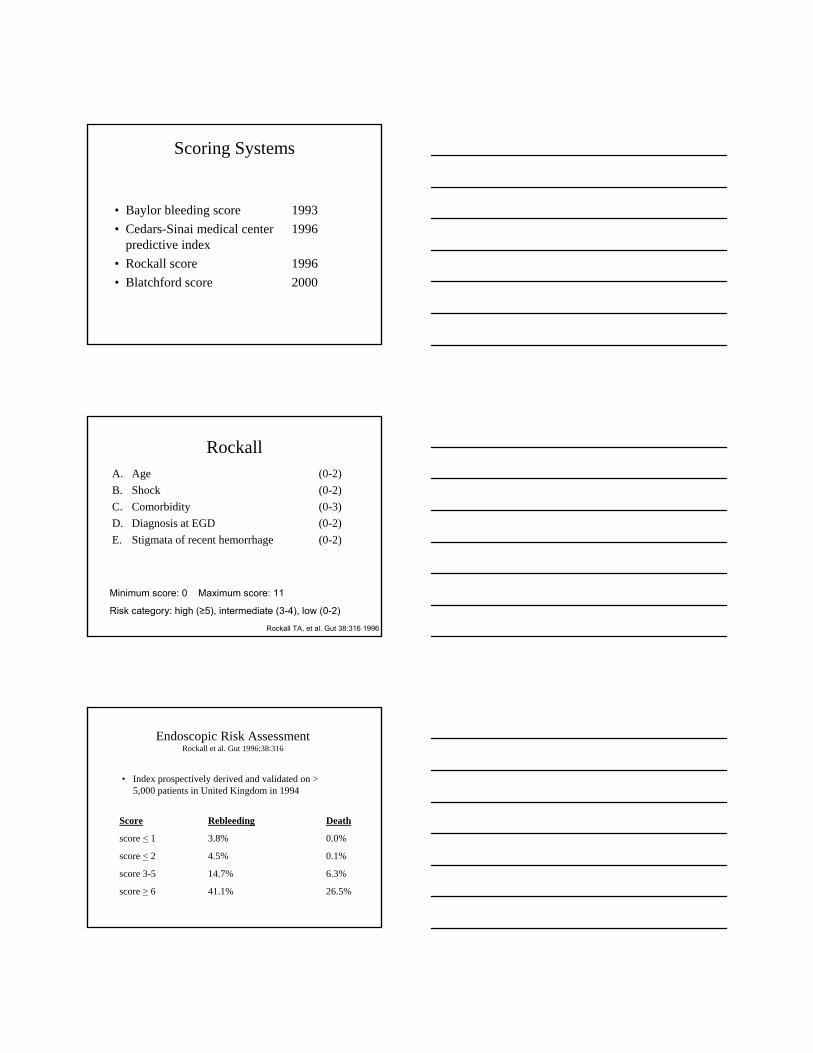
Scoring Systems
• Baylor bleeding score 1993• Cedars-Sinai medical center 1996
predictive index• Rockall score 1996• Blatchford score 2000
Rockall A. Age (0-2)B. Shock (0-2)C. Comorbidity (0-3)D. Diagnosis at EGD (0-2)E. Stigmata of recent hemorrhage (0-2)
Rockall TA, et al. Gut 38:316 1996
Minimum score: 0 Maximum score: 11
Risk category: high (≥5), intermediate (3-4), low (0-2)
Endoscopic Risk AssessmentRockall et al. Gut 1996;38:316
• Index prospectively derived and validated on > 5,000 patients in United Kingdom in 1994
Score Rebleeding Death
score < 1 3.8% 0.0%
score < 2 4.5% 0.1%
score 3-5 14.7% 6.3%
score > 6 41.1% 26.5%
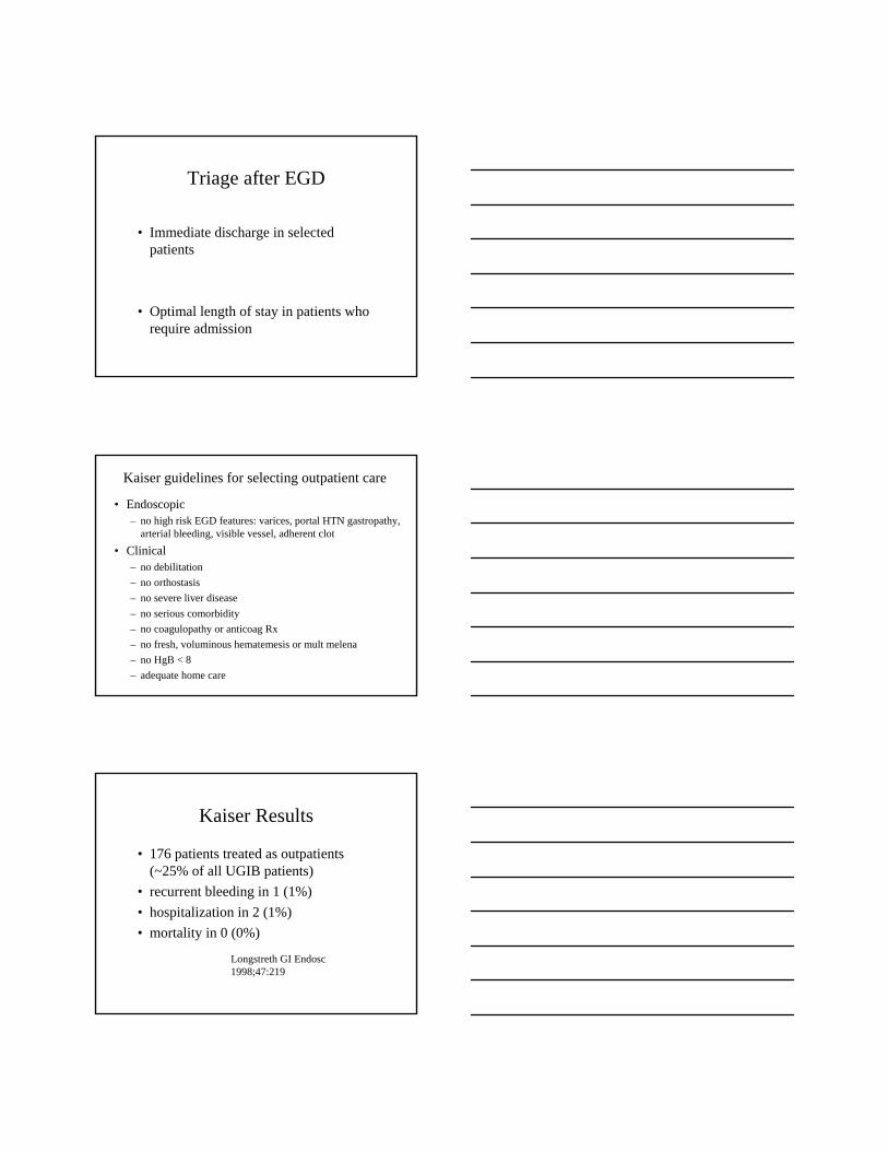
Triage after EGD
• Immediate discharge in selected patients
• Optimal length of stay in patients who require admission
Kaiser guidelines for selecting outpatient care
• Endoscopic– no high risk EGD features: varices, portal HTN gastropathy,
arterial bleeding, visible vessel, adherent clot
• Clinical– no debilitation– no orthostasis– no severe liver disease– no serious comorbidity– no coagulopathy or anticoag Rx– no fresh, voluminous hematemesis or mult melena– no HgB < 8– adequate home care
Kaiser Results
• 176 patients treated as outpatients (~25% of all UGIB patients)
• recurrent bleeding in 1 (1%)• hospitalization in 2 (1%)• mortality in 0 (0%)
Longstreth GI Endosc 1998;47:219

Early Endoscopy-based Triage:Three RCT’s
• Lee J, GIE 1999– N = 110: ER EGD vs routine admission– ER EGD: 46% immediate discharge; no rebleeds– LOS 1 vs 2 days; costs reduced 40%
• Cipolletta L, GIE 2002– N=464 pts underwent EGD w/in 12 hrs; 95 (20%) low risk.
Randomized to early discharge vs hospital care– Recurrent bleeding: 2 % both groups– Costs: 90% reduction
• Bjorkman D, GIE 2004– N=93: ER EGD vs routine admission– ER EGD: 40 % recommended for discharge; ER only discharged
10%
Optimal Length of Stay• Cedars-Sinai Guideline (Hay et al. JAMA
1997;278:2151)
• Clinical and EGD risk assessment• Daily reassessment• Reduction in LOS: 4.6 + 3.5 days to
2.9 + 1.3 days• No adverse events
Does Early EGD (< 24 hours) Improve Outcomes in the
Community?
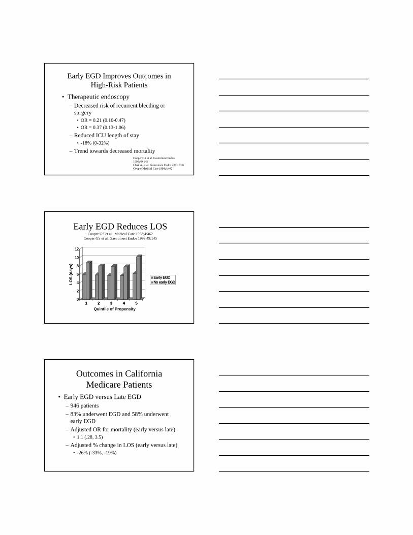
Early EGD Improves Outcomes in High-Risk Patients
• Therapeutic endoscopy– Decreased risk of recurrent bleeding or
surgery• OR = 0.21 (0.10-0.47)• OR = 0.37 (0.13-1.06)
– Reduced ICU length of stay• -18% (0-32%)
– Trend towards decreased mortalityCooper GS et al. Gastroinest Endos 1999;49:145Chak A, et al. Gastroinest Endos 2001;53:6Cooper Medical Care 1998;4:462
1 2 3 4 50
2
4
6
8
10
12
LOS
(day
s)
1 2 3 4 5Quintile of Propensity
Early EGDNo early EGD
Early EGD Reduces LOSCooper GS et al. Medical Care 1998;4:462
Cooper GS et al. Gastroinest Endos 1999;49:145
Outcomes in California Medicare Patients
• Early EGD versus Late EGD– 946 patients– 83% underwent EGD and 58% underwent
early EGD– Adjusted OR for mortality (early versus late)
• 1.1 (.28, 3.5)
– Adjusted % change in LOS (early versus late)• -26% (-33%, -19%)
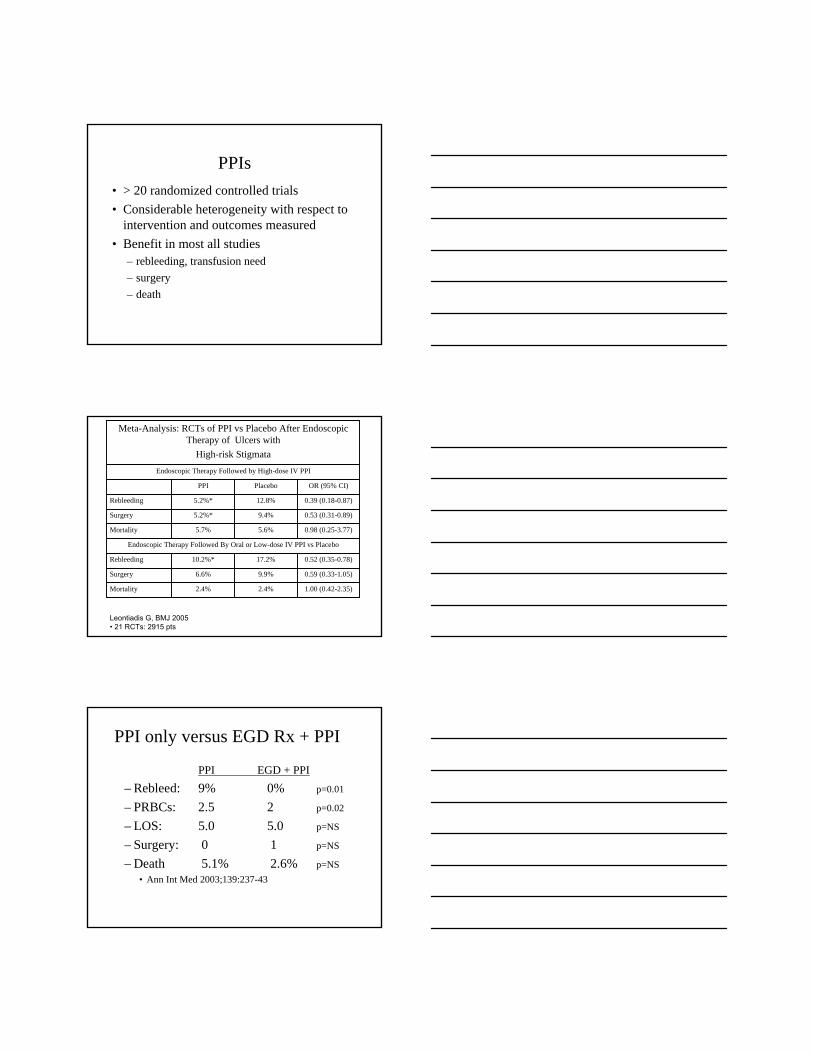
PPIs• > 20 randomized controlled trials• Considerable heterogeneity with respect to
intervention and outcomes measured• Benefit in most all studies
– rebleeding, transfusion need– surgery– death
Endoscopic Therapy Followed by High-dose IV PPI
OR (95% CI)PlaceboPPI
1.00 (0.42-2.35)2.4%2.4%Mortality
0.59 (0.33-1.05)9.9%6.6%Surgery
0.52 (0.35-0.78)17.2%10.2%*Rebleeding
Endoscopic Therapy Followed By Oral or Low-dose IV PPI vs Placebo
0.98 (0.25-3.77)5.6%5.7%Mortality
0.53 (0.31-0.89)9.4%5.2%*Surgery
0.39 (0.18-0.87)12.8%5.2%*Rebleeding
Meta-Analysis: RCTs of PPI vs Placebo After Endoscopic Therapy of Ulcers with
High-risk Stigmata
Leontiadis G, BMJ 2005• 21 RCTs: 2915 pts
PPI only versus EGD Rx + PPI
PPI EGD + PPI– Rebleed: 9% 0% p=0.01
– PRBCs: 2.5 2 p=0.02
– LOS: 5.0 5.0 p=NS
– Surgery: 0 1 p=NS
– Death 5.1% 2.6% p=NS• Ann Int Med 2003;139:237-43
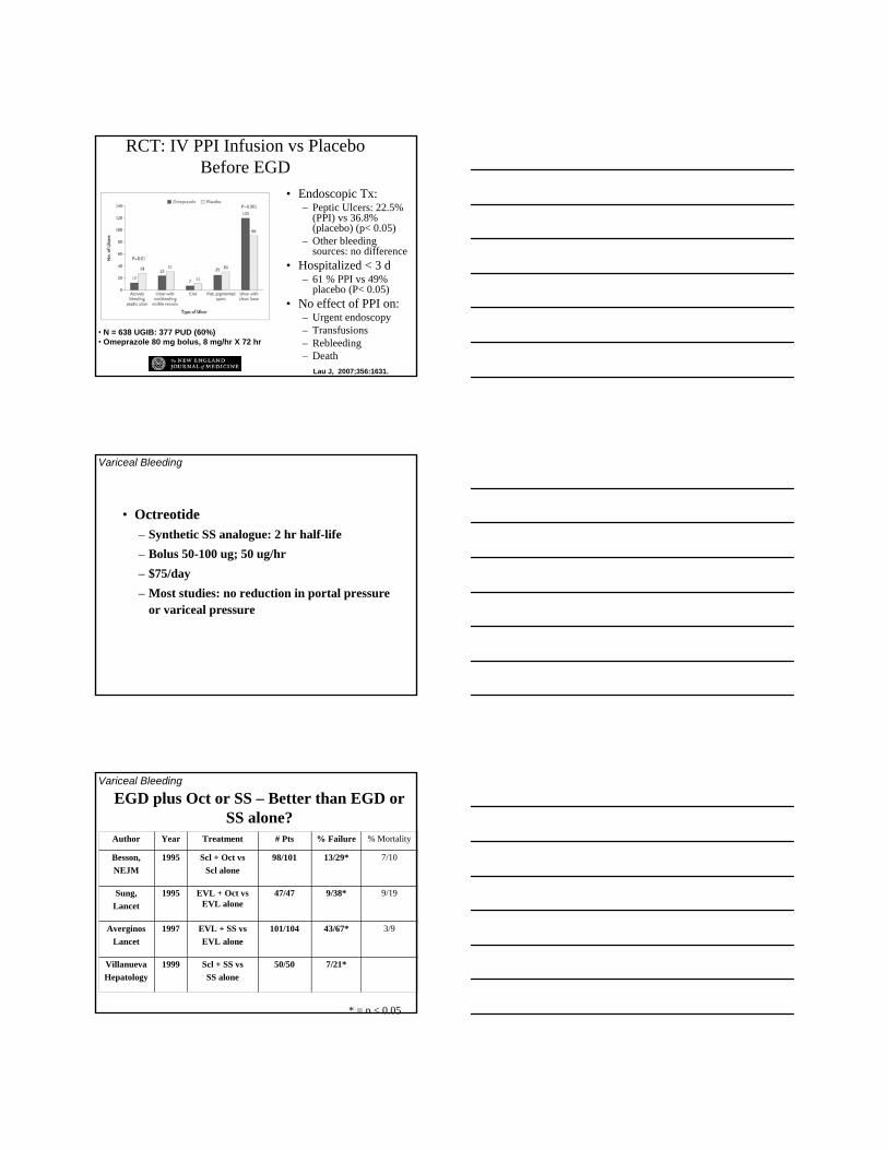
RCT: IV PPI Infusion vs Placebo Before EGD
• Endoscopic Tx:– Peptic Ulcers: 22.5%
(PPI) vs 36.8% (placebo) (p< 0.05)
– Other bleeding sources: no difference
• Hospitalized < 3 d– 61 % PPI vs 49%
placebo (P< 0.05)• No effect of PPI on:
– Urgent endoscopy– Transfusions– Rebleeding– Death
Lau J, 2007;356:1631.
• N = 638 UGIB: 377 PUD (60%)• Omeprazole 80 mg bolus, 8 mg/hr X 72 hr
• Octreotide– Synthetic SS analogue: 2 hr half-life– Bolus 50-100 ug; 50 ug/hr– $75/day– Most studies: no reduction in portal pressure
or variceal pressure
Variceal Bleeding
EGD plus Oct or SS – Better than EGD or SS alone?
Variceal Bleeding
3/943/67*101/104EVL + SS vs EVL alone
1997AverginosLancet
7/21*50/50Scl + SS vsSS alone
1999VillanuevaHepatology
9/199/38*47/47EVL + Oct vs EVL alone
1995Sung, Lancet
7/1013/29*98/101Scl + Oct vsScl alone
1995Besson,NEJM
% Mortality% Failure# PtsTreatmentYearAuthor
* = p < 0.05
Endoscopic Therapy Initial Control
Permanent Control
80-90%
Rebleeding
Endoscopic TherapyPermanent
Control75%
Rebleeding
Angiography
25%
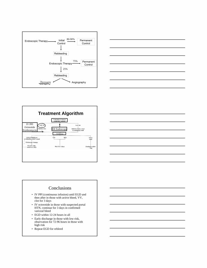
ER Endoscopy
IV PPIOctreotide
Erythromycin
Treatment Algorithm
Conclusions• IV PPI (continuous infusion) until EGD and
then after in those with active bleed, VV, clot for 3 days
• IV octreotide in those with suspected portal HTN, continue for 3 days in confirmedvariceal bleed
• EGD within 12-24 hours in all• Early discharge in those with low risk,
observation for 72-96 hours in those with high risk
• Repeat EGD for rebleed


