
Anamnese og klinisk undersøgelse

Clinical diagnosis of hip OA
American College of Rheumatology classification of hip OA Altman et al. 1991
Hip pain AND internal rotation <15˚ AND flexion <115˚ AND age >50 OR Hip pain AND (IR >15˚ AND pain) AND morning stiffness <60
minutes AND age >50
NICE guideline Age ≥45 AND Activity related joint pain No morning joint-related stiffness or morning stiffness <30 min.
Pain triad (smertetriade)
June 20092

Clinical diagnosis of hip OA cont.
In patients with hip symptoms Hip internal rotation <24˚ and flexion<114˚ were cutoffs Individual measurements of hip flexion seems to be of little
diagnostic value in diagnosing early symptomatic hip OA
June 20093

Risikofaktorer for udvikling af hofteartrose
Bierma-Zeinstra and Koes 2007
• Alder• Arvelighed, Skousgaard 2015
• Overvægt / fedme, Lohmander 2009
• Traume (ofte labrumskade)• Tungt fysisk arbejde, Sulsky et al. 2012
• Landmænd >10 år• Elitesportsudøvere, Molloy & Molloy 2011
• Dysplasi, Harris-Hayes & Royer 2011
• Cam, pincer, Harris-Hayes & Royer 2011, Agricola 2012
• Leg length inequality?

When patients present with activity related hip pain
June 20096
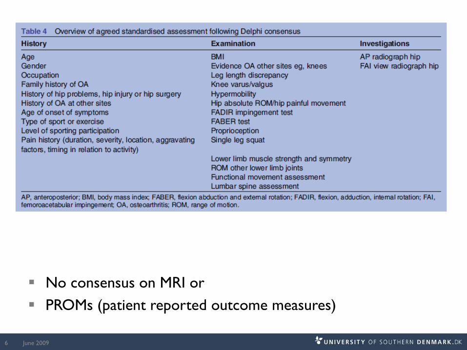
No consensus on MRI or PROMs (patient reported outcome measures)

Aktivitetsbegrænsning i dagligdagen
Upubliserede data
109 patienter med unilateral klinisk og radiologisk verificeret hofteartrose
Patient Specific Functional Scale Op til tre spørgsmål
Hvilke aktiviteter påvirker din dagligdag mest?

Aktivitetsbegrænsning i dagligdagen
Unpublished data
• Bending down to pick something up• Putting shoes and socks on• Cutting my toenails• Getting up from sitting• Walking on stairs – dominantly up the stairs

Aktivitetsbegrænsning i dagligdagen
Unpublished data
• Getting up on my bicycle• Getting in and out of my car• Gardening / kneeling down• Lying on the affected side• After walking for some time• Tage skridtet fuldt ud!

Hvor har patienterne med hofteartrose ondt?
June 200910

Patient characteristics
Valid pain drawings (n) 108 / 109Age in years, mean (SD) 65 (9)Females (%) 44Left hip (%) 40
Pain duration in monthsmean (range)
32 (4-300)
Pain intenstity (mean, SD) 5.4 (2.0)
Pain location by area- Greater trochanter- Groin- Anterior/lateral thigh- Buttock- Knee- Lower leg
77%53%42%38%17%15%

Percentageoverlap (# of drawings)
Colour
0-5% (1-3)
5-20% (4-12)
20-40% (13-24)
40-60% (25-35)
60-80% (36-47)
80-100% (48-59)
17% reported pain solely to the groin area No patient reported pain solely to the knee, lower leg or posterior thigh areas

Clinical examination

Inspection/observation during: Undressing, walking, active hip ROM
Manual examination Palpation of tissue texture Passive hip ROM evaluation incl. end-feel assessment Muscle strength assessment
Special tests Modified FABER, FAI/FADDIR, Trendelenburg, Ely’s, Thomas,
Obers, leg length equality, leg/log roll

Active hip range of motion
Patient walking during inspection Speed, guarding, compensation and symmetry
AROM can be performed with patient standing All six ranges are performed Used as a screening
15
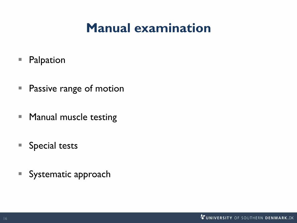
Manual examination
Palpation
Passive range of motion
Manual muscle testing
Special tests
Systematic approach
16

Hip range of motion and hip muscle strength areassociated with hip OA
Arokoski et al., 2004, Rasch et al., 2007, Pua et al., 2008; Pua et al., 2009, Rydevik et al., 2010

Passiv range of motion
Supine Flexion Abduction Adduction Internal rotation (90°
hip flexion) External rotation (90°
hip flexion)
Sitting IR ER
Prone Extension IR (0˚ hip flexion) ER (0˚ hip flexion)

Muscle strength evaluation Isometric vs. isotonic Isotonic excentric vs. concentric Literature: ‘make’ vs ‘break’ test Manual vs. HHD Position
Sitting, prone, supine, sidelying
FleksionIntern rotation
xxxxxxxAbduktion

21
Examine the intra- and inter-rater reproducibility of hip range of motion in patients with hip OA, when using a goniometer
Examine the intra- and inter-rater reproducibility of hip muscle strength in patients with hip OA, when using a dynamometer
Objectives

Clinical implications
If using hip range of motion and/or muscle strengthmeasurements in monitoring treatment progress, the measurements should be performed by the same clinician.

Clinical implications
Be careful when interpreting individualmeasurements of range of motion and musclestrength in the assessment of patients with hip osteoarthritis.

Orthopaedic tests
Trendelenburg Modified FABER Femoro-acetabulær impingement (FADDIR / FAI test) Leg/Log roll test Leg length
Obers Thomas Elys

Conclusion The Trendelenburg test is not useful in identifying subjects
in the early stages of hip joint OA
June 200925

FADDIR test / FAI test
Konlusion: kan bruges som en screening test pga. antal af falske positive
June 200926
BJSM 2015;49(12):811

Diagnostic test study 2 large cohorts: Framingham (n=946) and OAI (n=4366) Standardized questions about hip pain 16 and 9% of patients with hip pain demonstrated
radiographic hip OA 21 and 24% of patients with radiographic hip OA had hip pain
Diagnosticians may miss many older people with hip osteoarthritis if they rely on radiographic evidence for diagnosis
June 200927
BMJ 2015;351:h5983


