
AP Biology
Chapter 7.
The Cell: Cytoskeleton

AP Biology
Cytoskeleton Function
structural support maintains shape of cell provides anchorage for organelles
motility cell locomotion cilia, flagella, etc.
regulation organizes structures &
activities of cell

AP Biology 2005-2006
Cytoskeleton Structure
network of fibers extending throughout cytoplasm
3 main protein fibers microtubules microfilaments intermediate filaments
It’s a matter of size…

AP Biology
Evolutionary perspective Proteins that make up the fibers are
very similar in all living things from bacteria to humans
tubulin (all cells) actin (eukaryote cells)
Means that they are both ancient and essential for life

AP Biology 2005-2006
Microtubules Structure
thickest fibers hollow rods about 25nm
in diameter constructed of protein,
tubulin grow or shrink as more
tubulin molecules are added or removed
AP Biology 2005-2006
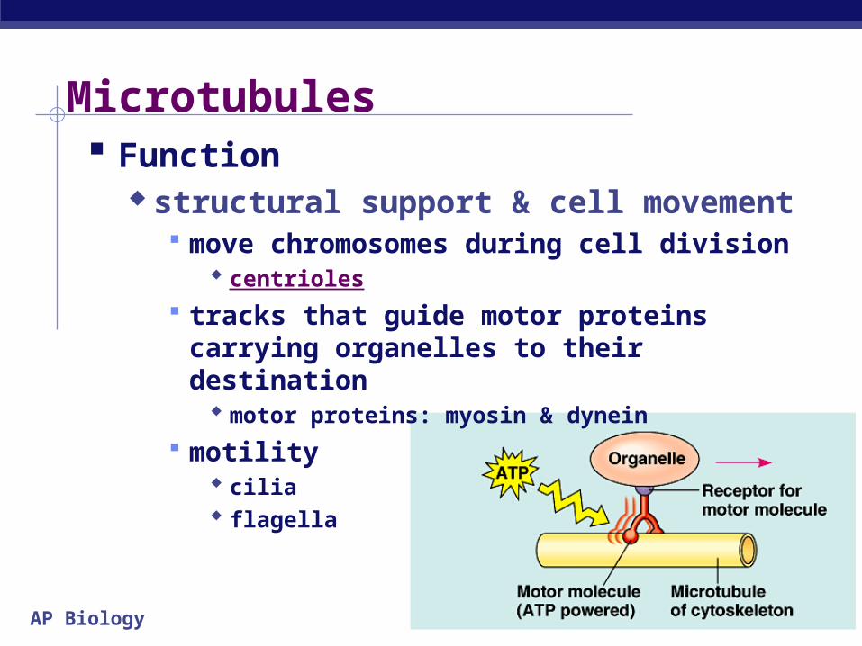
Microtubules Function
structural support & cell movement move chromosomes during cell division
centrioles
tracks that guide motor proteins carrying organelles to their destination
motor proteins: myosin & dynein
motility cilia flagella
AP Biology 2005-2006
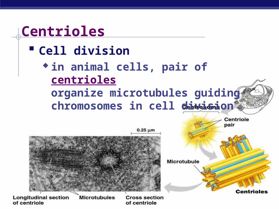
Centrioles Cell division
in animal cells, pair of centrioles organize microtubules guiding chromosomes in cell division

AP Biology
Cilia & flagella Extensions of eukaryotic cytoskeleton Cilia = numerous & short (hair-like) Flagella = 1-2/cell & longer (whip-like)
move unicellular & small multicellular organisms by propelling water past them
cilia sweep mucus & debris from lungs flagellum of sperm cells

AP Biology
Cilia Oar-like movement
alternating power & recovery strokes generate force perpendicular to cilia’s
axis

AP Biology
Flagella undulatory movement
force generated parallel to flagellum’s axis

AP Biology 2005-2006
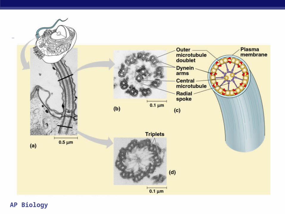
Cilia & Flagella Structure
remember 9+2! 9 pairs of
microtubules around 2 single microtubules in center
bending of cilia & flagella is driven by motor protein dynein
requires ATP
AP Biology

AP Biology
Microfilaments (actin filaments) Structure
thinnest class of fibers solid rods of protein, actin twisted double chain of actin
subunits about 7nm in diameter
Function 3-D network inside cell membrane in muscle cells, actin filaments
interact with myosin filaments to create muscle contraction
AP Biology 2005-2006
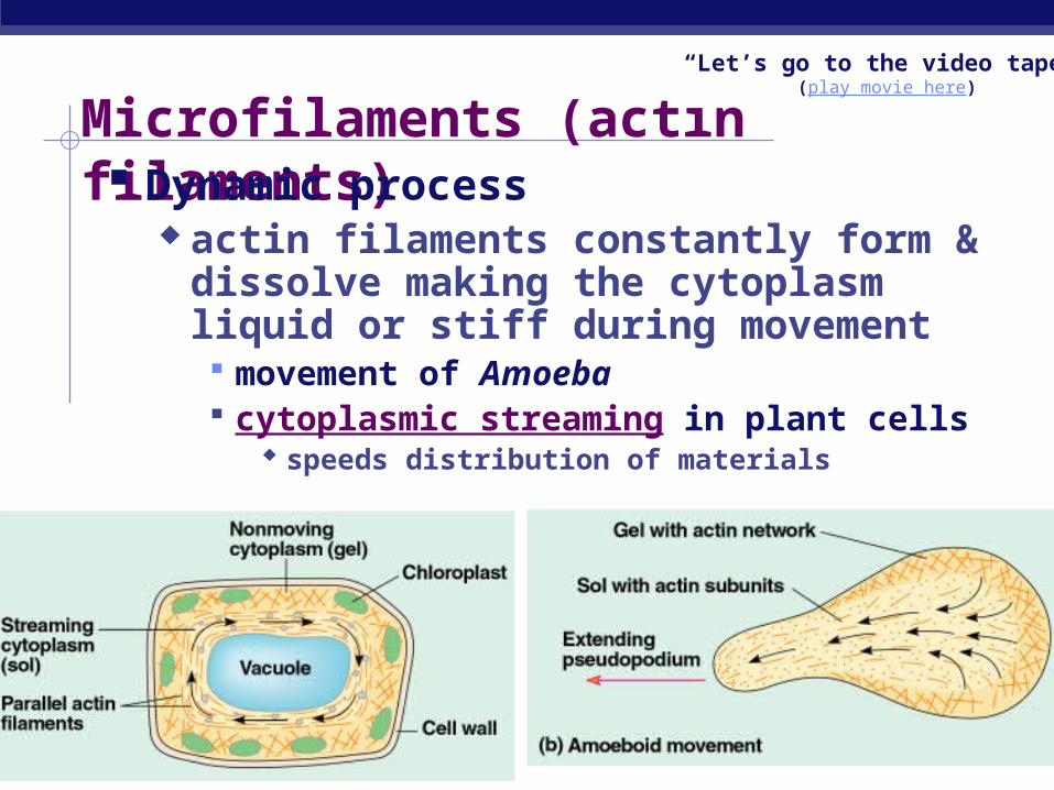
Microfilaments (actin filaments) Dynamic process
actin filaments constantly form & dissolve making the cytoplasm liquid or stiff during movement movement of Amoeba cytoplasmic streaming in plant cells
speeds distribution of materials
“Let’s go to the video tape!”(play movie here)

AP Biology
Intermediate filaments Structure
specialized for bearing tension built from keratin proteins
same protein as hair intermediate in size 8-12nm
Function hold “things” in place inside cell more permanent fixtures of
cytoskeleton reinforce cell shape & fix
organelle location nucleus is held in place by a
network of intermediate filaments

AP Biology 2005-2006
Summary Microtubules
thickest cell structure & cell motility tubulin
Microfilaments thinnest internal movements
within cell actin, myosin
Intermediate filaments intermediate more permanent fixtures keratin
• actin• microtubule• nuclei

AP Biology
Cell Junctions
Where cells touch each other…

AP Biology 2005-2006
Plant cell wall Structure
cellulose primary cell wall secondary cell wall middle lamella = sticky polysaccharides

AP Biology
Intercellular junctions Plant cells
plasmodesmata channels allowing cytosol
to pass between cells
plasmodesmata

AP Biology 2005-2006
Animal cell surface Extracellular matrix
collagen fibers in network of glycoproteins support adhesion movement regulation

AP Biology 2005-2006
Intercellular junctions in animals

AP Biology
Intercellular junctions Animal cells
tight junctions membranes of adjacent cells fused forming
barrier between cells forces material through cell membrane
gap junctions communicating junctions allow cytoplasmic movement between
adjacent cells desmosomes
anchoring junctions fasten cells together in strong sheets
AP Biology
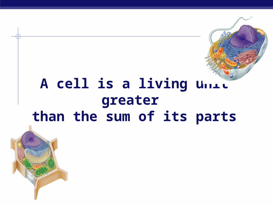
A cell is a living unit greater than the sum of its parts

AP Biology Thanks as always to Kim Foglia
And now for some…Stupid Penguin Tricks….
Any Questions??


