
B5 Summary

Inside or Outside
• Endo skeleton – internal skeleton made from cartilage (shark) or bone (human) e.g. humans, frogs, birds. These are living tissues.
• Exo skeleton – external skeleton made from chitin e.g. insects
• Some animals e.g. worms do not have a skeleton made of hard material

Skull
Rib
Pelvis
Femur
Patella
Tarsals
MetatarsalsPhalanges
Fibula
Tibia
Phalanges
Metacarpals
CarpalsRadiusUlnaVertebral Column
Humerus
Sternum
Scapula
Clavicle
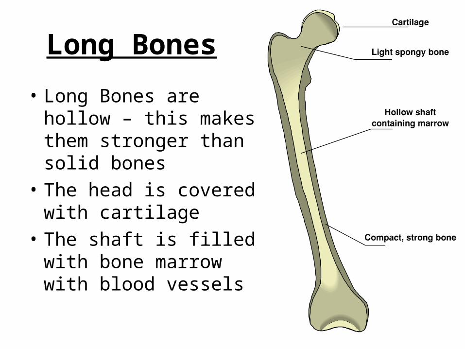
Long Bones
• Long Bones are hollow – this makes them stronger than solid bones
• The head is covered with cartilage
• The shaft is filled with bone marrow with blood vessels

Bones
• Cartilage and bone are susceptible to infection but can grow and repair themselves
• In humans, the skeleton starts off being cartilage which is slowly replaced by the addition of calcium and phosphorous (ossification)
• The more cartilage in a bone = the younger the person
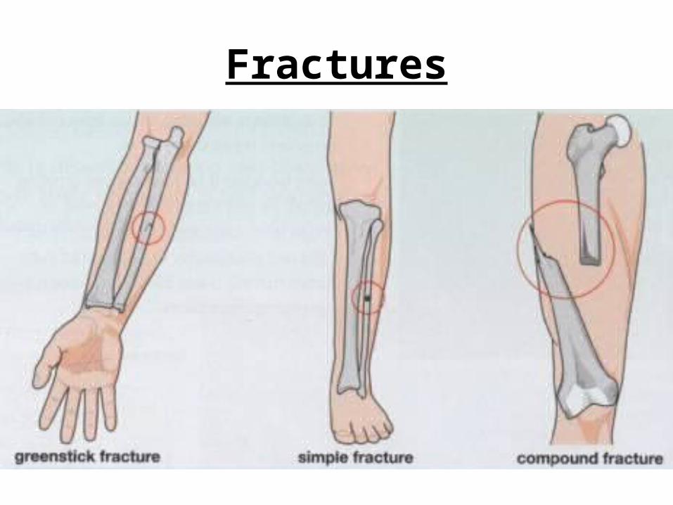
Fractures
Fractures
• Elderly people are more prone to fractures due to soft bones (osteoporosis)
• If you move someone with a fracture you can damage blood vessels and nerves.
• You should never move someone with a suspected spinal fracture without special first aid training.


Ball & Socket Joint and Hinge Joint

Joints
• Hip and knee joints can be replaced (danger of rejection and infection)
• Ligaments connect bone to bone (helps to prevent dislocations)
• Tendons connects muscle to bone• Cartilage absorb shock• Synovial fluid absorbs shock and acts
as a lubricant

rightatrium
leftatrium
semi-lunarvalve
tricuspid valve
rightventricle
leftventricle
bicuspid valve
aortapulmonaryartery
pulmonaryvein
vena cava from lungs
to headand bodyto lungs
from headand body
valve tendons
muscle
Arteries = high pressure
Veins = low pressure
Circulatory Systems
• No Circulation– Some animals are so small e.g. amoeba
that they do not have a blood circulatory system
• Open and Closed– Insects have an open CS, humans have
a closed CS e.g. blood flows in BV• Single Circulation
– One circuit from a 2 chambered heart• Double Circulation
– Two circuits from a 4 chambered heart

The History of Blood Circulation
• Galen, 200AD• First doctor to realise
the importance of the pulse in medicine
• He believed the liver made blood
• And the heart pumped blood around the body in a backwards and forwards motion
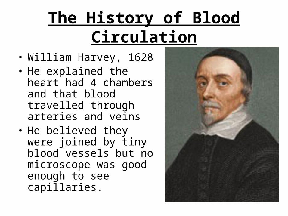
The History of Blood Circulation
• William Harvey, 1628• He explained the heart
had 4 chambers and that blood travelled through arteries and veins
• He believed they were joined by tiny blood vessels but no microscope was good enough to see capillaries.
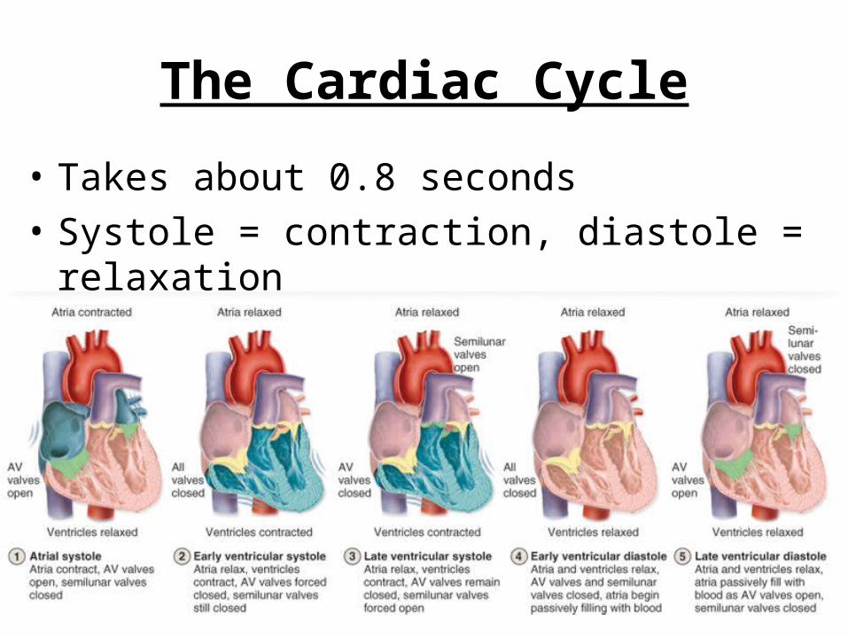
The Cardiac Cycle
• Takes about 0.8 seconds• Systole = contraction, diastole =
relaxation

The Cardiac Cycle
Heart relaxes and blood enters
both atria
Atria contract at the same time which
forces blood into both ventricles
Ventricles contract from the bottom
upwards which forces blood into the
pulmonary artery or aorta

Pacemaker Cells
• The Sino-Atrial Node (SAN) generates electrical impulses
• They spread across the atria causing them to contract

Pacemaker Cells
• The impulse reaches the Atrio-Ventricular Node (AVN)
• Impulses spread across the ventricles
• They contract from the bottom upwards
• Impulses from the vagus and sympathetic nerve can modify the heartbeat

Electrocardiogram
• An ECG shows the change in electrical impulses in the heart muscles
• The video of an echocardiogram shows if any parts of the heart, such as valves are not working properly.
P waves (impulses from SAN)
R waves (impulses
in ventricles)
T waves (as ventricles contract)

Heart-Assist Devices
• Doctors use ‘heart-assist’ devices to reduce the work done by heart muscles.
• They help to pump the blood.• This allows the heart muscles to
recover and then the device can be removed.
• Weak or damaged valves can also be replaced by artificial valves.

Heart Health
• Many factors can contribute to a poor circulatory system:– Fatty diets – can lead to cholesterol– Smoking – reduce amount of oxygen
available– Stress – high blood pressure– Inhaling solvents – can lead to heart attack– Injecting drugs – bacterial infections– Alcohol – lower BP, raise fat levels in blood

Blood Groups (Grade A)
• Before a blood transfusion is carried out, the new blood is checked to ensure it does not react with the patient’s blood
• Early attempts at blood transfusion were not successful
• Mixing blood from 2 people often caused blood clumping (agglutination)
• Karl Landsteiner (1901) solved this by discovering the four blood groups.

Blood Groups (Grade A)
• The groups depends on the presence or absence of agglutinins that consist of:– 2 proteins, antigen A or antigen B on the surface
of red blood cells– 2 antibodies, anit-A or anti-B in blood plasma
• Anti-A causes RBC containing antigen A to agglutinate.
• The same happens with anti-B and antigen B.

Rules for Donation
• You can give or receive from the same blood group.
• The person receiving blood does not have any antibodies against the donor’s antigens.

Blood Groups (Grade A)
AB
O
A, B, O
same group only


Blood Clotting
• Blood clots in order to seal wounds• Vitamin K is important for this to happen• Doctors use warfarin, heparin and aspirin to
prevent clotting• Haemophilia is an inherited condition where
the blood does not clot• It is a cascade process
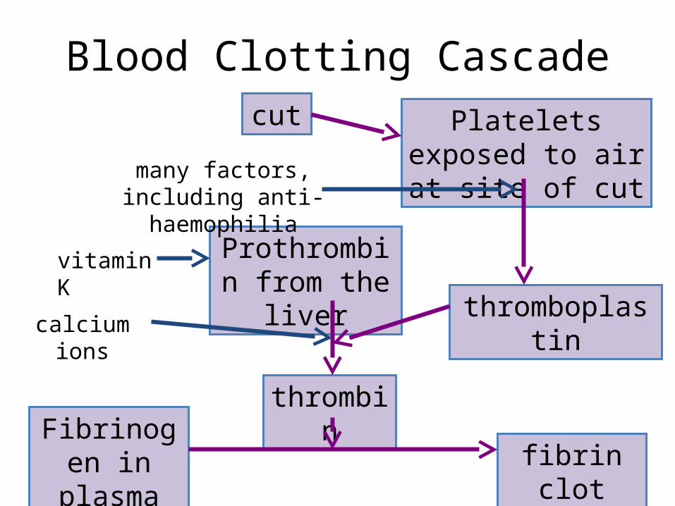
Blood Clotting Cascadecut Platelets exposed to
air at site of cut
thrombin
Prothrombin from the liver
Fibrinogen in plasma
thromboplastin
fibrin clot
many factors, including anti-haemophilia
vitamin K
calcium ions

Alveoli (air sac)
Bronchiole
Bronchus
Pleural membrane
Ribs
Intercostal Muscles
Lung
Trachea
Diaphragm

Respiration in Amoeba and WormsSmall, simple animals exchange gases like
oxygen through moist skin.
O2
O2
O2
O2
O2O2
O2

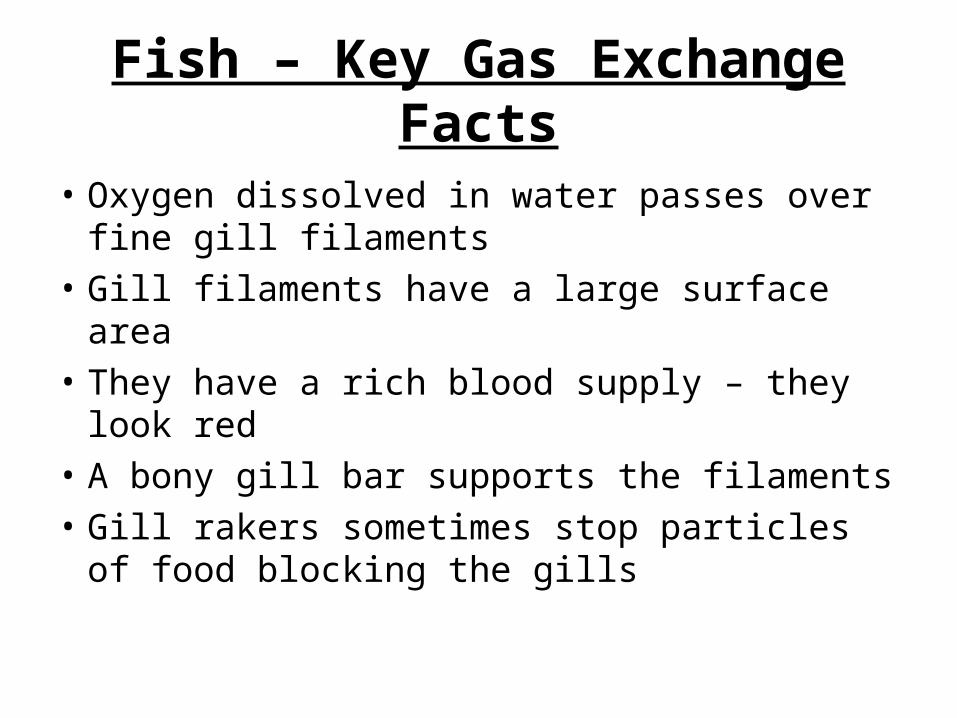
Fish – Key Gas Exchange Facts
• Oxygen dissolved in water passes over fine gill filaments
• Gill filaments have a large surface area• They have a rich blood supply – they look red• A bony gill bar supports the filaments• Gill rakers sometimes stop particles of food
blocking the gills
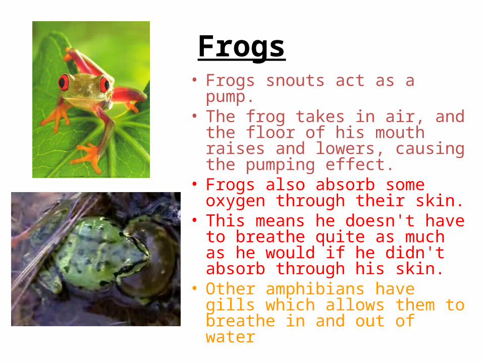
Frogs• Frogs snouts act as a pump. • The frog takes in air, and the floor
of his mouth raises and lowers, causing the pumping effect.
• Frogs also absorb some oxygen through their skin.
• This means he doesn't have to breathe quite as much as he would if he didn't absorb through his skin.
• Other amphibians have gills which allows them to breathe in and out of water
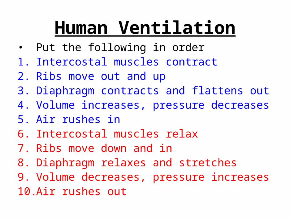
Human Ventilation• Put the following in order1. Intercostal muscles contract2. Ribs move out and up3. Diaphragm contracts and flattens out4. Volume increases, pressure decreases5. Air rushes in6. Intercostal muscles relax7. Ribs move down and in8. Diaphragm relaxes and stretches9. Volume decreases, pressure increases10. Air rushes out
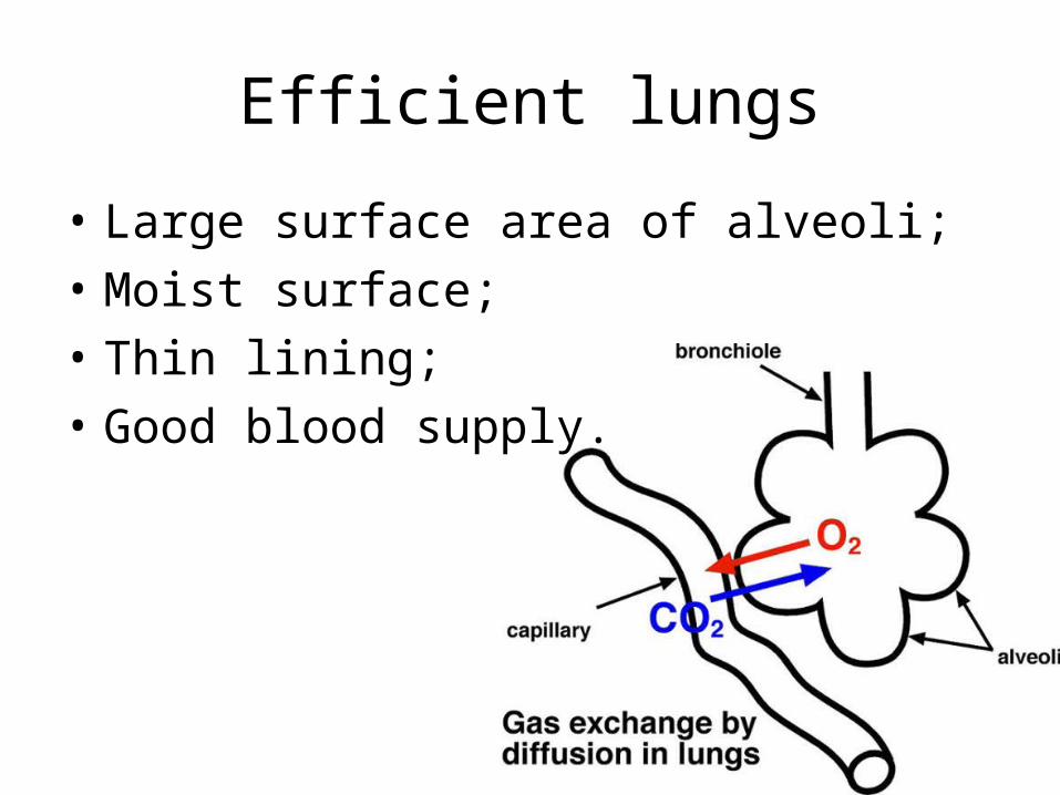
Efficient lungs
• Large surface area of alveoli;• Moist surface;• Thin lining;• Good blood supply.

Terminology• A spirometer is used to measure how much air is
breathed in and out• When you are resting and breathing normally, you
are exchanging tidal air.
• Vital capacity - The amount of air that can be forced out of the lungs
• Residual volume -The amount of air left in the lungs after a maximal exhalation. The amount of air that is always in the lungs and can never be expired

Larger volumes Smaller volumes
males females
taller people shorter people
non-smokers heavy smokers
professional athletes non-athletes
people living at high altitudes
people living at low altitudes

Symptoms of Asthma (Grade C)
• Shortness of breath• Wheezing• Coughing• Tightness in the chest
Treatment of Asthma (Grade C)
• Inhalers– Preventer– Reliever e.g. Ventolin (widens bronchioles)
• Primary care provided by doctors and nurses trained in asthma management.
• Weight reduction advice for obese patients with asthma to improve asthma control.
• Advice to smokers about the risks to themselves and their children with asthma.
• Vaccinations to reduce respiratory infection, such as flu.
Cause of Asthma (Grade A)
• Exact cause is not clear
• Combination of inherited, environmental, infectious and chemical factors

Cause of Asthma (Grade A)Sensitive airways Trigger
Immune response
Airways constrict More mucus produced
Mucus plugBody releases histamine
Airways inflamed
Less air available for gas exchange in the lungs

Starter - Waste Disposal
• Excretion – getting rid of waste products made by the body. – The lungs, liver, skin and kidneys all
excrete waste. – The kidneys excrete water, salt and
urea.
• Defecation – getting rid of solid waste through the anus that cannot be digested.

Function of the Kidney
• The kidneys filter the blood under high pressure.
• They remove all waste products and any excess water and salt ions.
• Any useful substances are returned (reabsorbed) to the blood eg glucose, some water, some salt.
• Urine is stored in the bladder.

Role of the Kidney - HIGHER
• The kidneys help to maintain the internal environment by:– First filtering the blood– Reabsorbing all the sugar– Reabsorbing the dissolved ions needed by
the body– Reabsorbing as much water as the body
needs– Releasing urea, excess ions and excess
water as urine
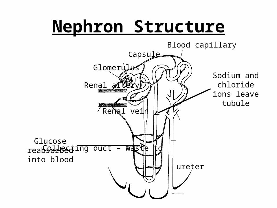
Nephron StructureBlood capillary
Capsule
Glomerulus
Renal artery
Renal vein
Collecting duct – waste to
ureter
Sodium and chloride ions leave tubule
Glucose reabsorbed into blood

Role of the Nephron (Grade A)
• A kidney contains about half a million tubules that filter the blood
• They are U-shaped• They contain a ‘filter unit’ – glomerulus
(collection of blood capillaries) surrounded by a capsule
• The loop is where the blood selectively reabsorbs useful substances such as glucose and some water
• The tubule also regulates water and salt levels

ADH – Anti Diuretic Hormone
• Urine concentration depends on how much water is reabsorbed by the kidney tubules
• This is controlled by ADH (made in the pituitary gland)
• It is a negative feedback system
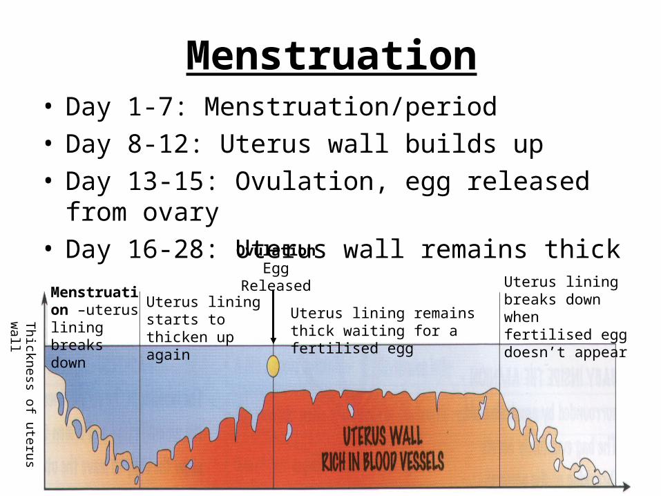
Menstruation• Day 1-7: Menstruation/period• Day 8-12: Uterus wall builds up• Day 13-15: Ovulation, egg released from
ovary• Day 16-28: Uterus wall remains thick
Thick
ness o
f ute
rus
wall
Menstruation –uterus lining breaks down
Uterus lining starts to thicken up again
OvulationEgg
Released
Uterus lining remains thick waiting for a fertilised egg
Uterus lining breaks down when fertilised egg doesn’t appear

Menstruation
• Hypothalamus in the brain triggers 2 hormones to be released from the pituitary gland
– FSH (Follicle Stimulating Hormone) – Stimulates a follicle in an ovary to start developing
– LH (Luteinising Hormone) – controls the release of an egg

Other Hormones
• As the follicle in the ovary develops it releases oestrogen and progesterone.
• These control the growth of the uterus cells and therefore the thickness of the uterus lining.
If the egg is fertilised…
• The levels of progesterone remain high
• No more eggs develop or are released
• No FSH is produced
• The uterus lining does not break down

Reasons for Infertility
• Not enough FSH produced• Inability to carry a foetus to term• Inability to fall pregnant naturally• Ovaries stop producing eggs at a
young age• Blockage in the oviducts• Partner with a low sperm count
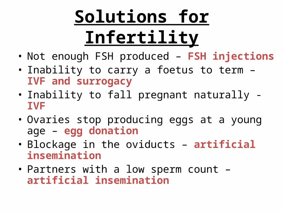
Solutions for Infertility
• Not enough FSH produced – FSH injections• Inability to carry a foetus to term – IVF and
surrogacy• Inability to fall pregnant naturally - IVF• Ovaries stop producing eggs at a young age
– egg donation• Blockage in the oviducts – artificial
insemination• Partners with a low sperm count – artificial
insemination

Mechanical Replacements
The lens in the eye is replaced
with a plastic one
Hip joints can wear out and are replaced with a metal ball and
socket
Knee joints can also wear out and are replaced
with a metal hinge joint
The heart is replaced with a
metal and plastic pump

Biological Replacements
The cornea of the eye can be
transplanted
One kidney can be transplanted
Bone marrow from inside bones can be transplanted
Blood is often transfused during operations
Both lungs, sometimes with the heart, can be
transplanted
The whole heart can be
transplanted

Disadvantages of Mechanical Replacements
• Size• Weight• Need a power supply• Materials must be inert, light and strong• Rejection must be controlled

Growth in Plants and Animals
• Animals normally only grow in the early stages of their life
• Plants grow during all their life• Animals grow all over their body• Plants only grow from their meristems (tips of
roots and shoots)• In animals or plants cells divide by mitosis to
make new cells to grow


