
Bacterial Cell Morphogenesi
Current Biology 24, 863–867, April 14, 2014 ª2014 The Authors http://dx.doi.org/10.1016/j.cub.2014.02.053
Reports Does Not
Require a Preexisting Template Structure
Yoshikazu Kawai,1,* Romain Mercier,1 and Jeff Errington1,*1Centre for Bacterial Cell Biology, Institute for Cell andMolecular Biosciences, Medical School, Newcastle University,Richardson Road, Newcastle upon Tyne NE2 4AX, UK
Summary
Morphogenesis, the development of shape or form in cells ororganisms, is a fundamental but poorly understood process
throughout biology. In the bacterial domain, cells have awide range of characteristic shapes, including rods, cocci,
and spirals. The cell wall, composed of a simple meshworkof long glycan strands crosslinked by short peptides (pepti-
doglycan, PG) and anionic cell wall polymers such as wallteichoic acids (WTAs), is the major determinant of cell
shape. It has long been debated whether the formation ofnew wall material or the transmission of shape from parent
to daughter cells requires existing wall material as a tem-plate [1–3]. However, rigorous testing of this hypothesis
has been problematical because the cell wall is normallyan essential structure. L-forms are wall-deficient variants
of common bacteria that have been classically identified asantibiotic-resistant variants in association with a wide range
of infectious diseases [4–6]. We recently determined thegenetic basis for the L-form transition in the rod-shaped bac-
terium Bacillus subtilis and thus how to generate L-formsreliably and reproducibly [7, 8]. Using the new L-form sys-
tem, we show here that we can delete essential genes for
cell wall synthesis and propagate cells in the long-termabsence of a cell wall template molecule. Following genetic
restoration of cell wall synthesis, we show that the abilityto generate a classical rod-shaped cell is restored, conclu-
sively rejecting template-directed models, at least for theestablishment of cell shape in B. subtilis.
Results and Discussion
It is well known that treatment of bacterial cells with cell wall-active antibiotics or enzymes such as lysozymes convertsthem to cell wall-deficient protoplasts, which can be main-tained in an osmoprotective medium (though with little netgrowth [8]). Such protoplasts can then be regenerated to pro-duce viable, walled cells with normal morphology [2], albeit atlow efficiency (reviewed by Hopwood [9]). In these experi-ments, it is difficult to exclude the presence of residual cellwall template fragments because synthesis and assembly ofthe wall can continue via newly synthesized precursors or cat-alytic enzymes (Figure 1A) [10–13]. However, bacterial variantscalled L-forms [5] are capable of prolonged growth in theabsence of cell wall synthesis and thus might be suited to adefinitive test of the need for a cell wall template. Becausethey are largely or completely lacking in cell wall, the basicshape of L-forms is spherical, but they are highly malleable
*Correspondence: [email protected] (Y.K.), [email protected] (J.E.)
This is an open access article under the CC BY license (http://
creativecommons.org/licenses/by/3.0/).
and take on an array of irregular shapes influenced by thesurrounding milieu. L-forms have often been classified as‘‘stable,’’ in which case the cells can be propagated in theL-form state indefinitely, or as ‘‘unstable’’ for strains capableof reverting to the normal walled state. In principle, theexistence of unstable L-forms suggests that a defined cellshape can be generated de novo. However, recent work onEscherichia coli L-forms suggests that unstable L-forms retainthe requirement for at least a low level of cell wall synthesis,because genes essential for cell wall synthesis or assemblyremain essential in the unstable L-forms [14, 15].We have been developingmethods for generating L-forms of
the Gram-positive model bacterium Bacillus subtilis [7, 8, 16].We found that at least two mutations are normally required forL-form growth. One mutation (e.g., ispA) has a poorly definedrole in maintaining cell integrity and is probably of littledirect functional significance. The key mutations enablingproliferation in the L-form state appear to work simply byincreasing the rate of membrane synthesis. L-forms proliferateby a strange mechanism of membrane blebbing, or tubulationand fission [7]; it seems that excessmembrane synthesis is suf-ficient to drive this mode of cell division [8]. Genetic screens re-vealed two classes of mutation that can generate the excessmembraneeffect.Oneclass (i.e., overexpressionof thegeneen-coding the catalytic subunit of acetyl-coenzyme A-carboxylase[AccDA] [8]) leads directly to upregulation of the fatty acid syn-thetic pathway and hence to increased membrane synthesis.The other class, inhibition of cell wall precursor synthesis (e.g.,by repression of the murE operon [7]), works indirectly by anas yet uncharacterized mechanism. Nevertheless, the fact thatrepression of peptidoglycan (PG) precursor synthesis can pro-mote the L-form transition provides a means, in principle, oftesting whether continued PG synthesis is needed to maintainthe ability to regenerate a rod-shaped walled cell (Figure 1B).In our previouswork,we identifiedan18kbpdeletion that en-
ables stable proliferation of L-forms [8]. This deletion removedthe murC gene, which encodes an essential enzyme in the PGprecursor pathway, together with 17 other coding regions ofmainly unknown function. (We assume that one or more ofthe other genes deleted confer a stabilizing effect similar tothat of the ispA mutation mentioned above, although we havenot yet fully characterized the effect.) We reconstructed the18 kbp deletion by replacement with a tetracycline resistancegene (D18::tet) (Figure 1C) and showed that the resultant strainhad the expected phenotype [8]. The D18::tet mutation wasintroduced into wild-type cells by a standard B. subtilis trans-formation method (see Experimental Procedures). Transform-ants were selected on our standard L-form plates (nutrientagar [NA]/magnesium-sucrose-maleic acid [MSM]) containingtetracycline. The plates contain an osmoprotectant (sucrose)and an inhibitor of cell division (benzamide [17]) that inhibitsthe growth of walled cells, but not of L-forms. After w3–4 days at 30�C, small tetracycline- and benzamide-resistantcolonies were visible (Figure 2A; the three large coloniesmarked by arrows contained rod-shaped walled cells andwere presumably spontaneous tetracycline-resistant mutantsor some kind of merodiploid recombinants). Phase-contrastmicroscopy of the small colonies revealed only L-form cells(Figure 2B). We confirmed the presence of the D18::tet

Figure 2. Inhibition of PG Precursor Synthesis by Deleting murC
(A) Transformation of wild-typewalled cells to L-forms by introduction of the
D18::tet mutation, which completely deletes the murC gene as well as 17
other genes. Colonies of D18::tet L-forms were selected on transformation
plates (NA/MSM) containing tetracycline and benzamide. The plate was
incubated for 4 days at 30�C after transformation. An enlarged image of
the typical colonies (dashed square) is shown to the right. Arrows point to
large dense colonies that were of spurious origin and contained walled
rod-shaped cells.
(B) Phase-contrast micrograph of D18::tet L-forms from a colony on the
transformation plate shown in (A). Scale bar represents 5 mm.
(C) Growth of wild-type protoplasts (WT, blue) and D18::tet L-forms
(D18::tet, red) in L-form-supporting medium (NB/MSM) with benzamide.
Cells were incubated at 30�C.(D) Induction of cell wall regeneration from L-forms of strain RM84 [8]
(accDA* ispA*) or D18::tet. Proliferating L-form cultures (OD600 = w0.2–
0.3) in L-form medium (NB/MSM) with penicillin G (PenG) and benzamide
were spotted onto L-form plates (NA/MSM) without PenG and benzamide.
The plates were incubated for 3 days at 30�C.(E) Phase-contrast micrograph of proliferating L-forms of strain RM84
(accDA* ispA*) in L-formmedium (NB/MSM)with penicillin G and benzamide
(left), and after induction of cell wall regeneration by cultivating L-forms (left)
on L-form plates (NA/MSM) in the absence of PenG and benzamide (right).
Phase-contrast micrograph of rod-shaped cells was taken from a colony
shown in (D) (accDA* ispA*). Scale bar represents 5 mm.
(F) Phase-contrast micrograph of proliferating L-forms of D18::tet in L-form
medium with penicillin G and benzamide (left) or from the L-form regenera-
tion plate (no penicillin G or benzamide) shown in (D) (D18::tet) (right). Scale
bar represents 5 mm.
Figure 1. Schematic View of Peptidoglycan Synthesis and the Models for
Cell Morphogenesis
(A) The peptidoglycan (PG) cell wall is built from long glycan strands
composed of N-acetylmuramic acid (MurNAc) and N-acetylglucosamine
(GlcNAc) crosslinked by peptide cross-bridges [10, 11]. The precursor for
PG is initially synthesized in the cytoplasm by the action of MurAA, MurB,
MurC, MurD, MurE, and MurF enzymes. MurNAc-pentapeptide is coupled
to a membrane carrier, undecaprenyl pyrophosphate, by MraY, and GlcNAc
is added by MurG to form lipid II, which is then transferred to the outside of
the cytoplasmic membrane. Newly synthesized PG is incorporated into the
existing PG meshwork by a combination of transglycosylation and trans-
peptidation reactions catalyzed by penicillin-binding proteins. Wall teichoic
acids (WTAs) are abundant PG-linked glycopolymers present inmost Gram-
positive organisms and are essential for maintaining rod shape in B. subtilis
[12]. WTA synthesis begins at the cytoplasmic side of membrane with the
coupling of GlcNAc to the same lipid carrier, undecaprenyl pyrophospate,
as is used for PG precursors. WTA polymer synthesis requires the action
of series of enzymes (TagA, TagB, TagD, TagE, and TgaF) in the cytosol.
The polymer is exported and coupled to the PG by the action of the TagTUV
enzymes [13].
(B) Models for bacterial cell morphogenesis. It has not been clear whether
the formation of new wall material and the transmission of rod shape
from parent to daughter cells require existing wall material as a template
(see text).
(C) Schematic representation of the chromosomal region deleted in D18::tet
L-forms [8].
Current Biology Vol 24 No 8864
mutation and deletion of the murC gene by PCR (see below).Consistent with our previous work [8], the newly selectedL-forms were able to grow in liquid L-form medium (nutrientbroth [NB]/MSM) in contrast to wild-type protoplasts notbearing the D18::tet mutation (Figure 2C). Certain types of L-forms are known to be able to regenerate cell wall and shapein the absence of selection pressure such as b-lactam antibi-otics [5]. Proliferating L-forms induced by AccDA overproduc-tion (accDA* ispA*), and thus with an intact PG syntheticpathway, were indeed able to revert to the walled state, whenspotted onto L-form plates without penicillin G. The left-handspot in Figure 2D shows the emergence of dense colonies,which contained classical rod-shaped walled cells (Figure 2E,right panel). However, complete deletion of murC, e.g., by theD18::tet mutation, irreversibly blocks the PG precursor syn-thetic pathway and thus prevents regeneration of the cell wall(Figures 2D, right-hand spot with no dense growth, and 2F).
Having established a stable L-form strain incapable of cellwall synthesis, we wished to test whether the resumption of
PG precursor synthesis would enable the regeneration of aPG cell wall and the restoration of rod-shaped cells. Weattempted to introduce an isopropyl b-D-thiogalactoside(IPTG)-inducible ectopic copy of murC on a plasmid (pLOSS-Pspac-murC lacZ ermR [8]) into the D18::tet L-form strain bymodifying an established polyethylene glycol (PEG)-mediatedprotoplast transformation method [18] (see details in Experi-mental Procedures). Transformantswere selectedonNA/MSM
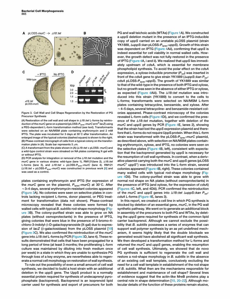
Figure 3. Cell Wall and Cell Shape Regeneration by the Restoration of PG
Precursor Synthesis
(A) Restoration of the cell wall and cell shape in D18::tet L-forms by reintro-
duction of themurC gene on aplasmid (pLOSS-Pspac-murC ermR lacZ) using
a PEG-dependent L-form transformation method (see text). Transformants
were selected on an NA/MSM plate containing erythromycin and 2 mM
IPTG. The plate was incubated for 3 days at 30�C after transformation. An
enlarged image of the typical colonies (dashed square) is shown to the right.
(B) Phase-contrast micrograph of cells from a typical colony on the transfor-
mation plate in (A). Scale bar represents 5 mm.
(C) A transformant from the plate shown in (A) (D18::tet + pLOSS-murC) and
a wild-type control strain were streaked on NA plates containing X-gal with
or without IPTG.
(D) PCR analysis for integration or removal of the D18::tet mutation and the
murC gene in various strains: wild-type (lane 1), RM121(lane 2), D18::tet
L-forms (lane 3), and D18::tet + pLOSS-Pspac-murC (lane 4). RM121
(D18::tet + pLOSS-Pspac-murC) was constructed in previous work [8] and
was used as a control.
Bacterial Cell Morphogenesis865
plates containing erythromycin and IPTG (for expression ofthe murC gene on the plasmid, Pspac-murC) at 30�C. Afterw3–4 days, several erythromycin-resistant colonies appeared(Figure 3A). No colonies were seen on selective plates in con-trols lacking recipient L-forms, donor plasmid, or PEG treat-ment for transformation (data not shown). Phase-contrastmicroscopy revealed that these colonies were formed bywalled cells with typicalB. subtilis rod-shapemorphology (Fig-ure 3B). The colony-purified strain was able to grow on NAplates (without osmoprotectants) in the presence of IPTG,giving colonies that were blue in the presence of 5-bromo-4-chloro-3-indolyl-b-D-galactopyranoside (X-gal) due to expres-sion of lacZ (b-galactosidase) from the pLOSS plasmid [19](Figure 3C). We also confirmed the reintroduction of the murCgene intoD18::tet L-formsbyPCR (Figure 3D, lane 4). These re-sults demonstrated that cells that have been propagated for along period of time (at least 3 months; the proliferating L-formculture was maintained by diluting into fresh medium onceper week) in a state in which they are unable to synthesize PGthrough loss of a key enzyme, are nevertheless able to regen-erate a normal cellmorphology on restoration ofwall synthesis.
To rule out the possibility of even a small amount of cell wallsynthesis, we decided to build a host strain with an additionaldeletion in the uppS gene. The UppS product is a normallyessential protein required for synthesis of undecaprenyl pyro-phosphate (bactoprenol). Bactoprenol is an isoprenoid lipidcarrier used for synthesis and export of precursors for both
PG andwall teichoic acids (WTAs) (Figure 1A). We constructeda uppS deletion mutant in the presence of an IPTG-induciblecopy of uppS carried on an unstable pLOSS plasmid (strainYK1888,DuppS::kan pLOSS-Pspac-uppS). Growth of this strainwas dependent on IPTG (Figure 4Aii), confirming that uppS isindeed essential for cell viability in normal walled cells. How-ever, the growth defect was not fully restored in the presenceof IPTG (Figure 4A, i and ii). We realized that uppS lies immedi-ately upstream of cdsA, which is essential for membranephospholipid synthesis. To avoid the polar effect on the cdsAexpression, a xylose-inducible promoter (Pxyl) was inserted infront of the cdsA gene to give strain YK1889 (DuppS::kan Pxyl-cdsA pLOSS-Pspac-uppS). The growth of YK1889 was similarto that of thewild-type in thepresenceof both IPTGandxylose,but no growthwas seen in the absence of either IPTGor xylose,as expected (Figure 4Aiii). The D18::tet mutation was intro-duced into this strain (YK1889) to convert to the cells toL-forms; transformants were selected on NA/MSM L-formplates containing tetracycline, benzamide, and xylose. Afterw4–5 days, several tetracycline- and benzamide-resistant col-onies appeared. Phase-contrast microscopy of the coloniesrevealed L-form cells (Figure 4Di), and we confirmed the pres-ence of the D18::tet mutation, together with deletion of themurC and uppS genes by PCR (Figure 4E, lanes 2), showingthat the strain had lost the uppS expression plasmid and there-fore that L-formsdonot requireUppSprotein.When this L-formstrain was transformed with the pLOSS-Pspac-murC plasmid,as described above,with selection onNA/MSMplates contain-ing erythromycin, xylose, and IPTG, no colonies were seen onthe selective plates (Figure 4B, left), consistent with expecta-tion that the bactoprenol generated by uppS is necessary forthe resumption of cell wall synthesis. In contrast, when a deriv-ative plasmid carrying both themurC and uppS genes (pLOSSmurC+ uppS+) was introduced into the L-form strain, severalcolonies appeared (Figure 4B, right). These colonies containedmany walled cells with typical rod-shape morphology (Fig-ure 4Dii). The colony-purified strain was able to grow withnormal rod shape on NA plates (without osmoprotectants) inthe presence of IPTG (and xylose, for the expression of cdsA)(Figures 4C, left, and 4Diii). PCR confirmed the reintroductionof the murC and uppS genes into D18::tet DuppS::kan Pxyl-cdsA L-forms (Figure 4E, lanes 3).In this report, we created a cell line in which PG synthesis is
blocked by deletion of an essential gene,murC, in the PG wallsynthetic pathway.Wewent on to generate an additional blockin assembly of the precursors to both PG and WTAs, by delet-ing the uppS gene required for synthesis of the common lipidcarrier bactoprenol. Although we cannot exclude the possi-bility that B. subtilis possesses a series of enzymes that cansupport wall polymer synthesis by an as yet undefined mech-anism, it seems highly likely that the double blockade wegenerated would have abolished all significant wall synthesis.We then developed a transformation method for L-forms andreturned the murC and uppS genes, enabling the resumptionof cell wall synthesis. Ultimately, we showed that de novoPG synthesis is sufficient to regenerate the cell wall andrestore a rod-shape morphology in B. subtilis in the absenceof an existing cell wall template, conclusively excluding theneed for a cell wall template in establishment of the rod shapeof B. subtilis. What then are the mechanisms responsible forestablishment and maintenance of cell shape? Several linesof evidence suggest that the actin-like MreB proteins play acentral role in shape determination [10, 20–22]. Although mo-lecular details of the function of these proteins remain elusive,

Figure 4. Regeneration of Rod-Shape
Morphology by De Novo PG Synthesis in the
Absence of an Existing Cell Wall Template
(A) Effect of repression of uppS and cdsA on
growth in the walled state. The following strains
were cultured on NA plates with 1% xylose and
1 mM IPTG (left), 1 mM IPTG (middle), or without
(right) at 30�C: wild-type (strain 168, i), DuppS::
kan pLOSS-Pspac-uppS (YK1888, ii), and DuppS::
kan Pxyl-cdsA pLOSS-Pspac-uppS (YK1889, iii).
(B) Regeneration of the cell wall in D18::tet
DuppS::kan Pxyl-cdsA L-forms by reintroduction
of the murC gene (pLOSS-Pspac-murC ermR
lacZ, left) or murC and uppS genes (pLOSS-
Pspac-murC PuppS-uppS ermR lacZ, right) using a
PEG-dependent L-form transformation method
(see text). Transformants were selected on an
NA/MSM plate containing erythromycin, 1%
xylose, and 2 mM IPTG. Plates were incubated
for w5–6 days at 30�C after transformation.
(C) One of the transformants shown in (B) (right,
D18::tet DuppS::kan Pxyl-cdsA + pLOSS-Pspac-
murC PuppS-uppS ermR lacZ) was streaked on
NA plates containing 1% xylose with (left) or
without (right) 2 mM IPTG.
(D) Phase-contrast micrograph of D18::tet
DuppS::kan Pxyl-cdsA L-forms on NA/MSM con-
taining 1% xylose (i) and of cells (D18::tet
DuppS::kan Pxyl-cdsA + pLOSS-Pspac-murC
PuppS-uppS ermR lacZ) from a typical colony on
the transformation plate (NA/MSM with 1%
xylose and 2 mM IPTG) as shown in (B) (ii) or on
NA plate containing xylose and IPTG as shown
at left in (C) (iii). Scale bar represents 5 mm.
(E) PCR analysis for integration or removal of the
D18::tet mutation, the murC gene, and the uppS
gene in various strains: YK1889 (DuppS::kan
Pxyl-cdsA pLOSS-Pspac-uppS, lanes 1), YK1913
(D18::tet DuppS::kan Pxyl-cdsA, lanes 2), and
YK1925 (D18::tet DuppS::kan Pxyl-cdsA +
pLOSS-Pspac-murC PuppS-uppS ermR lacZ, lanes
3). The ftsZ gene was also checked as a control.
Current Biology Vol 24 No 8866
the evidence that they regulate the synthesis of several keywall polymers during growth of the lateral wall is strong. More-over, their ability to form extended linear filaments provides ameans, at least in principle, of exerting long-range interactionson the cell wall synthetic machinery, leading to the control ofgross cell geometry. It will be interesting to investigate the pro-cess whereby cell shape is reestablished, although the low fre-quency of this event precludes detailed analysis at present.Nevertheless, extension of the methods that we have devel-oped for studying de novo cell wall synthesis promises to pro-vide a powerful new means of studying the establishment ofbacterial cell morphology.
Experimental Procedures
Bacterial Strains, Plasmids, Primers, and Growth Conditions
The bacterial strains, plasmid constructs, and primers for PCR analysis in
this study are shown in Tables S1 and S2 available online. DNA manipula-
tions were carried out using standard methods. Protoplasts were prepared
as described previously [8]. Normal B. subtilis cells were grown on NA
(Oxoid) and in Luria-Bertani broth. B. subtilis L-forms and protoplasts
were grown in osmoprotective medium composed of 23 MSM (pH 7)
(40 mM MgCl2, 1 M sucrose, and 40 mM maleic acid) mixed 1:1 with 23
NB (Oxoid) or 23 NA. Details of supplements, antibiotics, and microscopic
imaging used for this study can be found in the Supplemental Information.
Selection of D18::tet L-Forms
For selection of the D18::tet L-forms, we transformed chromosomal DNA of
the strain RM121 (D18::tet pLOSS-Pspac-murC lacZ ermR [8]) into wild-type
B. subtilis using standard methods [23]. Transformants were selected on
L-form plates (NA/MSM) containing 30 mg/ml tetracycline and 1 mg/ml
benzamide.
Transformation Method for L-Forms
L-form transformation was carried out by modifying a PEG-dependent pro-
toplast transformation method [18]. Proliferating L-form cultures (D18::tet)
were diluted at 1023 into fresh NB/MSM medium (10 ml) and incubated at
30�C until OD600 = w0.2 (2 days). The culture was centrifuged at
8,000 rpm for 10 min, and the L-forms were resuspended in 300 ml of NB/
MSM medium and then mixed with 2 mg of murC expression plasmid. For
L-form transformation, 150 ml of the L-form and plasmid mixture was trans-
ferred into 450 ml of MSM containing 40% PEG6000 (Sigma-Aldrich) and
gently mixed. After 2 min, 1 ml of NB/MSM was added and mixed, and the
cells were then centrifuged at 8,000 rpm for 10 min. The cell pellet was re-
suspended in 300 ml of NB/MSM and incubated for 120 min at 30�C. Finally,a 150 ml sample of the cell suspension was plated on NA/MSM plates
containing erythromycin and IPTG. The plates were incubated at 30�C.
Supplemental Information
Supplemental Information includes two tables and Supplemental Experi-
mental Procedures and can be found with this article online at http://dx.
doi.org/10.1016/j.cub.2014.02.053.
Acknowledgments
We thank Waldemar Vollmer for critical reading of the manuscript and Ling
Juan Wu for helpful discussions. We thank Patricia Domınguez-Cuevas
for the uppS mutant strain, Heath Murray for the pPxyl-cdsA plasmid, and
Elvira Olmedo-Verd for preliminary work on this problem. This work was

Bacterial Cell Morphogenesis867
funded by European Research Council grant 250363 to J.E. R.M. was sup-
ported by a Marie Curie Intra-European Fellowship.
Received: January 29, 2014
Revised: February 24, 2014
Accepted: February 25, 2014
Published: April 3, 2014
References
1. Holtje, J.V. (1998). Growth of the stress-bearing and shape-maintaining
murein sacculus of Escherichia coli. Microbiol. Mol. Biol. Rev. 62,
181–203.
2. Ranjit, D.K., and Young, K.D. (2013). The Rcs stress response and
accessory envelope proteins are required for de novo generation of
cell shape in Escherichia coli. J. Bacteriol. 195, 2452–2462.
3. Harold, F.M. (2007). Bacterial morphogenesis: learning how cells make
cells. Curr. Opin. Microbiol. 10, 591–595.
4. Klieneberger, E. (1935). The natural occurrence of pleuropneumonia-like
organisms in apparent symbiosis with Streptobacillus moniliformis and
other bacteria. J. Pathol. Bacteriol. 40, 93–105.
5. Allan, E.J., Hoischen, C., andGumpert, J. (2009). Bacterial L-forms. Adv.
Appl. Microbiol. 68, 1–39.
6. Domingue, G.J., Sr., and Woody, H.B. (1997). Bacterial persistence and
expression of disease. Clin. Microbiol. Rev. 10, 320–344.
7. Leaver, M., Domınguez-Cuevas, P., Coxhead, J.M., Daniel, R.A., and
Errington, J. (2009). Life without a wall or division machine in Bacillus
subtilis. Nature 457, 849–853.
8. Mercier, R., Kawai, Y., and Errington, J. (2013). Excess membrane syn-
thesis drives a primitive mode of cell proliferation. Cell 152, 997–1007.
9. Hopwood, D.A. (1981). Genetic studies with bacterial protoplasts. Annu.
Rev. Microbiol. 35, 237–272.
10. Typas, A., Banzhaf, M., Gross, C.A., and Vollmer, W. (2012). From
the regulation of peptidoglycan synthesis to bacterial growth and
morphology. Nat. Rev. Microbiol. 10, 123–136.
11. Bhavsar, A.P., and Brown, E.D. (2006). Cell wall assembly in Bacillus
subtilis: how spirals and spaces challenge paradigms. Mol. Microbiol.
60, 1077–1090.
12. Brown, S., Santa Maria, J.P., Jr., and Walker, S. (2013). Wall teichoic
acids of gram-positive bacteria. Annu. Rev. Microbiol. 67, 313–336.
13. Kawai, Y., Marles-Wright, J., Cleverley, R.M., Emmins, R., Ishikawa, S.,
Kuwano, M., Heinz, N., Bui, N.K., Hoyland, C.N., Ogasawara, N., et al.
(2011). A widespread family of bacterial cell wall assembly proteins.
EMBO J. 30, 4931–4941.
14. Joseleau-Petit, D., Liebart, J.C., Ayala, J.A., and D’Ari, R. (2007).
Unstable Escherichia coli L forms revisited: growth requires peptido-
glycan synthesis. J. Bacteriol. 189, 6512–6520.
15. Young, K.D. (2007). Reforming L forms: they need part of a wall after all?
J. Bacteriol. 189, 6509–6511.
16. Domınguez-Cuevas, P., Mercier, R., Leaver, M., Kawai, Y., and
Errington, J. (2012). The rod to L-form transition of Bacillus subtilis
is limited by a requirement for the protoplast to escape from the cell
wall sacculus. Mol. Microbiol. 83, 52–66.
17. Adams, D.W., Wu, L.J., Czaplewski, L.G., and Errington, J. (2011).
Multiple effects of benzamide antibiotics on FtsZ function. Mol.
Microbiol. 80, 68–84.
18. Chang, S., and Cohen, S.N. (1979). High frequency transformation of
Bacillus subtilis protoplasts by plasmid DNA. Mol. Gen. Genet. 168,
111–115.
19. Claessen, D., Emmins, R., Hamoen, L.W., Daniel, R.A., Errington, J., and
Edwards,D.H. (2008).Controlof thecell elongation-divisioncyclebyshut-
tling of PBP1 protein in Bacillus subtilis. Mol. Microbiol. 68, 1029–1046.
20. Kawai, Y., Asai, K., and Errington, J. (2009). Partial functional redun-
dancy of MreB isoforms, MreB, Mbl and MreBH, in cell morphogenesis
of Bacillus subtilis. Mol. Microbiol. 73, 719–731.
21. Jones, L.J., Carballido-Lopez, R., and Errington, J. (2001). Control of cell
shape in bacteria: helical, actin-like filaments in Bacillus subtilis. Cell
104, 913–922.
22. White, C.L., and Gober, J.W. (2012). MreB: pilot or passenger of cell wall
synthesis? Trends Microbiol. 20, 74–79.
23. Hamoen, L.W., Smits, W.K., de Jong, A., Holsappel, S., and Kuipers,
O.P. (2002). Improving the predictive value of the competence transcrip-
tion factor (ComK) binding site in Bacillus subtilis using a genomic
approach. Nucleic Acids Res. 30, 5517–5528.


