1
Biosynthesis and Chemical Applications of Thioamides
Nilkamal Mahanta,a,b,‡ D. Miklos Szantai-Kis,c,‡ E. James Petersson,c,d,* and Douglas A. Mitchella,b*
aDepartment of Chemistry, bCarl R. Woese Institute for Genomic Biology, University of Illinois,
600 S Mathews Avenue, Urbana, IL, 61801, USA cDepartment of Biochemistry and Molecular Biophysics, Perelman School of Medicine, University
of Pennsylvania, 3700 Hamilton Walk, Philadelphia, PA, 19104, USA dDepartment of Chemistry, University of Pennsylvania, 231 S 34th Street, Philadelphia, PA, 19104,
USA
‡ These authors contributed equally.
Corresponding authors:
[email protected], phone: 1-215-746-2221, fax: 1-215-573-2112
[email protected], phone: 1-217-333-1345, fax: 1-217-333-0508
2
Abstract
Thioamidation as a posttranslational modification is exceptionally rare, with only a few reported
natural products and exactly one known protein example (methyl-coenzyme M reductase from
methane-metabolizing archaea). Recently, there has been significant progress in elucidating the
biosynthesis and function of several thioamide-containing natural compounds. Separate
developments in the chemical installation of thioamides into peptides and proteins have enabled
cell biology and biophysical studies that advance the current understanding of natural thioamides.
This review highlights the various strategies used by Nature to install thioamides in peptidic
scaffolds and the potential functions of this rare but important modification. We also discuss
synthetic methods used for the site-selective incorporation of thioamides into polypeptides with a
brief discussion of the physicochemical implications. This account will serve as a foundation for
further study of thioamides in natural products and their various applications.
3
Keywords
Thioamide = an analog of the amide bond in which the carbonyl oxygen has been replaced by a sulfur atom Thionucleoside = a nucleic acid analog in which a carbonyl oxygen has been replaced by a sulfur atom Closthioamide = a thioamide-containing non-ribosomally synthesized peptide exhibiting antibiotic activity RiPP = ribosomally synthesized and posttranslationally modified peptide Thioviridamide = a representative of a class of thioamide-containing RiPPs that also includes thioholgamide A/B and thioalbamide Methanobactin = a thioamide-containing RiPP that functions in copper acquisition by methanotrophs Methyl Coenzyme M Reductase (MCR) = the only currently known thioamide-containing natural protein, catalyzes the reversible conversion of methyl-coenzyme M (CoM) and coenzyme B (CoB) to methane and a CoB-CoM heterodisulfide Thiopeptide = a class of RiPPs containing thiazole, thiazoline, and/or thioamide modifications; also sometimes used to refer to any peptide with a thioamide modification of the peptide backbone
4
Introduction
Prior to their discovery in natural molecules, thioamides were considered as a synthetic isostere
for amides in peptide backbones1 and have been employed by medicinal chemists for improving
thermal/proteolytic stability and the pharmacokinetic properties of amide-containing
compounds.2,3 In some cases, the thioamide-modified peptides exhibited improved bioactivity as
well.4,5,6,7 However, while there are pharmaceutical compounds containing thiocarbonyl groups
(e.g. thyroid medications methyl- and propylthiouracil, anti-tuberculosis drug thioacetazone,
androgen receptor agonist enzalutamide, and antimetabolite tioguanine), to date, no marketed
therapeutics contain a peptidic thioamide.8 Biophysical chemists have used thioamides for
perturbing protein folding or introducing spectroscopic probes.9 The discovery of thioamides in
several natural products and in a single protein provides new perspective and prompts
investigations of the role of the thioamide as well as a search for additional natural thioamide-
containing molecules.
In this review, we have summarized the current state of knowledge of Nature’s biosynthetic
strategies for generating thioamide-containing polypeptides. The overall understanding of the
biosynthesis pathways and the function of this posttranslational modification (PTM) is still rather
limited, but rapidly growing. In addition, we have summarized the synthetic methods developed
for site-selective incorporation of thioamides into peptides and proteins. These methods can be
used to prepare substrates to investigate biosynthetic mechanisms or analogs to investigate natural
product bioactivity, as well as thioamide variants of peptides and proteins which may have
improved properties relative to the all-amide congeners. Recent progress in the elucidation of
5
thioamide biosynthetic pathways and methods for their chemical installation has positioned the
field for new breakthroughs.
Thioamide Properties
The potential benefits of thioamide incorporation in both natural and synthetic compounds result
from the at once subtle and dramatic changes to amide interactions that can come from this single
atom substitution. Thioamides are more reactive with both nucleophiles and electrophiles than
amides,10,11 with a weaker carbonyl bond (130 vs. 170 kcal/mol),12 and therefore have been used
as chemical synthesis intermediates. Thioamides also have greater affinity for certain metals over
amides. For instance, the natural product methanobactin (Figure 2) exhibits extremely high
affinity copper binding,13 and selective, silver-catalyzed transformations have been used to convert
thioamides into other carbonyl derivatives.14 The differences in amide and thioamide geometry
govern many of the non-covalent interactions exhibited by thioamide-containing peptides. The
thioamide C=S bond is 1/3 longer than the amide C=O bond (1.71 vs. 1.23 Å)15 and sulfur has a
1/3 larger van der Waals radius compared to oxygen (1.85 vs 1.40 Å).16 Peptide conformational
changes can result from the elongated C=S bond and the higher rotational barrier for the C-N bond
(~5 kcal/mol),17 which reduces conformational flexibility. Additional altered physiochemical
properties include: (i) thioamide N-H groups are more acidic (DpKa = -6) than the corresponding
amide,18 (ii) thioamide N-H groups are better hydrogen bond donors,19 and (iii) the sulfur lone pairs
of thioamides are weaker hydrogen bond acceptors relative to oxygen lone pairs in amides.20
Therefore, thioamides are suitable to evaluate the contribution of single hydrogen bonds to protein
folding/stability. Substitution of an amide with a thioamide also imparts significant spectroscopic
and electrochemical changes. The thioamide C=S bond has an UV absorption maximum at 265
6
(±5) nm and an IR stretch at 1120 (±20) cm-1, compared to 220 (±5) nm and 1660 (±20) cm-1,
respectively, for the amide C=O bond.21 Moreover, the 13C NMR chemical shift of the thioamide
carbonyl is found 30 ppm downfield (200 – 210 ppm) of the corresponding amide resonance.22 The
oxidation potential of a model thioamide (1.21 eV) is significantly lower than that of the amide
(3.29 eV),23 which has prompted speculation about a potential role in electron-transfer in biological
settings.
The origin of the higher C-N rotational barrier in thioamides has been examined by NMR17 and ab
initio calculations.24 It was found that the amino group of thioformamide is more conformationally
rigid than in formamide. Additionally, the change in charge density at sulfur upon rotation of the
amino group in thioformamide is greater than that at oxygen in formamide due to a predominant
bipolar amide resonance form. The small difference in electronegativity between carbon and sulfur
(Pauling electronegativities for C=2.55 and for S=2.58), along with the larger size of sulfur, are
the predominant factors that allow charge transfer from nitrogen to sulfur in thioamides. A special
feature of the thioamide bond arises from the red-shifted absorption bands and the higher barrier
for cis-trans isomerization. This property allows access to either the cis or trans isomer by
irradiation with the appropriate UV wavelength. The photoinduced isomerization is efficient
(30%) and fast (<600 ps), whereas thermal relaxation is comparatively slow (>10 min).21 Hence,
thioamide-containing peptides are good candidates for fast photo-switches in proteins, either to
regulate activity, or to initiate conformational transitions for time-resolved studies.25 The above
properties, summarized in Table 1, allow thioamide modification to affect biomolecule function
in valuable ways which have led to the evolution of several distinct mechanisms for thioamidation
in natural products.
7
Thioamides in Nature: Biosynthesis, Structure, and Function
Thioamides are exceptionally rare in biology. Most reported natural thioamides are of bacterial
origin, except for the plant-derived cycasthioamide (Figure 1). Among these are the ribosomally
synthesized and posttranslationally modified peptide (RiPP) natural products thioviridamide and
its analogs, methanobactin, and thioamidated thiopeptides as well as closthioamide, which is a
non-ribosomal natural product (Figures 1 and 2).26,27,28,29 Known thioamide-containing nucleotides
include thiouridine, thiocytidine, and thioguanine (Figure 1).30,31 Recent identification of the
proteins responsible for thioglycine formation in the active site of methyl-coenzyme M reductase
(MCR), the only known thioamidated protein, further adds impetus to this area.32,33 On the other
hand, a distinct biosynthetic mechanism has been identified for closthioamide,34 which shares
features of thionated nucleoside biosynthesis,30 as well as for methanobactin.35 Collectively, these
revelations suggest that natural thioamidation pathways arose through multiple different
independent routes.32,33,34
Non-ribosomal natural products
Cycasthioamide
Cyscasthioamide is a thioamide-containing, non-proteinaceous amino acid obtained from the plant
Cycas revoluta Thunb with unknown biological function (Figure 1).36 In addition, another
thioamidated compound is reported from the tunicate Polycarpa aurata; however, it is believed to
be an artifact of product isolation or degradation of the major alkaloid compound polycarpine.37
Closthioamide
8
Closthioamide, a polythioamide compound, produced by Ruminiclostridium cellulolyticum
(previously Clostridium cellulolyticum), a strictly anaerobic, Gram-positive, soil-dwelling
bacterium (Figure 1).27 Until recently, no R. cellulolyticum natural products were isolated from
standard laboratory cultivation. Hertweck and co-workers mimicked the natural environment by
adding soil extracts to the culture which led to the production of closthioamide.27 Closthioamide
is a symmetrical hexathioamide and displays a central diaminopropyl group with four β-alanyl
extender units and two terminal p-hydroxybenzoyl groups. Closthioamide is growth-suppressive
towards several human pathogens, including Staphylococcus aureus, Enterococcus faecalis, and
Neisseria gonorrhoeae.27,38,39 Replacement of the thioamides with amides abolished the
antibacterial activity of closthioamide implicating the importance of thioamide moieties in its
biological activity.27
Studies using whole cell-based assays showed that closthioamide inhibits the ATPase activity of
DNA gyrase and topoisomerase IV, thus effectively blocking DNA replication.40 Closthioamide
also inhibits the relaxation activity of DNA gyrase, which does not require ATP hydrolysis and
thus may allosterically, rather than directly, interfere with the ATPase activity of gyrase. Notably,
this mode of action differs from that of the other DNA gyrase inhibitors such the fluoroquinolones
and aminocoumarins.40 The discovery of closthioamide from an underexplored anaerobe with a
new mode of action holds promise that additional, novel antibiotics might be discovered by parallel
methods.
Manipulation of the regulatory elements involved in the global activation of secondary metabolism
in R. cellulolyticum sustained the production of closthioamide and allowed isolation of seven

9
congeners which showed varied levels of bioactivity.41 Evaluation of the antibacterial activity of
the congeners demonstrated the importance of all six thioamide moieties, terminal aromatic
residues, the modular arrangement of the β-thioalanyl units, and the distinct length of spacer units
for antibacterial activity.41,39 Moreover, it was also found that closthioamide is a selective Cu(I)
chelator akin to methanobactin and forms a compact symmetrical dinuclear copper complex.13,42
Through synthesis and application of deuterium- and fluorine-labeled probes (see below), initial
insights into closthioamide biosynthesis were obtained, which predicted the involvement of an
unusual non-ribosomal peptide synthetase (NRPS).22,41 A very recent study reported the
closthioamide biosynthetic gene cluster (BGC) in R. cellulolyticum and demonstrated that
closthioamide biosynthesis involves a novel thiotemplated peptide assembly line that differs from
known NRPSs.34,43 Extensive genome editing and isolation of intermediates from the knock out
mutants revealed that the closthioamide BGC consisted of genes ctaA-M along (Figure 3A) and a
preliminary biosynthetic pathway has been proposed (Figure 3B).34 First, p-hydroxybenzoic acid
(PHBA) is synthesized from chorismate by chorismate lyase CtaA (NCBI accession number:
WP_015926607.1) and loaded onto a peptidyl carrier protein (PCP), either CtaE
(WP_015926603.1) or CtaH (WP_015926600.1). 44 This step is probably catalyzed by either CtaD
(WP_015926604.1) or CtaI (WP_015926599.1), as both enzymes are members of PCP-loading
protein families (ATP-grasp and AMP-dependent ligase, respectively).45 The second PCP would
be loaded with two molecules of β-alanine by the iterative action of CtaD/I, which itself could be
biosynthesized from L-aspartate by CtaF (decarboxylase, WP_015926602.1). Then, the PHBA
thioester is proposed to be loaded onto this PCP-bound β-alanyl dipeptide. CtaG
(WP_015926601.1), whose N-terminus shows homology to an amide synthase, BtrH from
10
butirosin biosynthesis, is the likely candidate.46 The resultant PCP-bound product would be
converted to the corresponding polythioamide intermediate by CtaC (WP_015926605.1), which
shows homology to the alpha-adenine nucleotide hydrolase (AANH) superfamily.28 Finally, it
would be coupled to diaminopropane (DAP) by CtaJ (amide synthase, WP_015926598.1) and
thionated by CtaC to yield mature closthioamide.34 The biosynthetic intermediate, DAP, could be
biosynthesized from L-aspartate by the sequential action of CtaK (WP_015926597.1), CtaB
(WP_015926606.1), and CtaF.47 Currently, the source of sulfur is not known.34 This is the first
example of a thioamidated compound that employs a novel NRPS biosynthetic assembly line.34
Thioamide-containing nucleosides
6-Thioguanine (6TG)
6-Thioguanine (6TG) is a highly potent cytotoxin developed in the 1950s by Elion and Hitchings,
who received the Nobel Prize for discovering antimetabolite drugs (Figure 1).48 6TG exerts its
cytotoxicity by in situ transformation to 6-thioguanine nucleosides and subsequent incorporation
into DNA.49 As a result, a mismatch repair pathway triggers cell-cycle arrest and apoptosis.50 6TG
is used for the treatment of several ailments such as leukemia, psoriasis, and inflammatory bowel
disease; however, it is known to exhibit some deleterious side effects.51,52,53
Naturally produced 6TG plays a critical role in the pathogenesis of Erwinia amylovora, the
causative agent for fire blight, a devastating disease that affects apple and pear trees, causing
immense ecological and economic damage.54,31,55 Functional gene analyses revealed that the
biosynthesis of 6TG is encoded by a small gene locus (ycfABCD), as shown by successful
heterologous expression of the ycf gene cluster in E. coli (Figure 4A).31 YcfA (WP_004156383.1)
11
is homologous to proteins of the diverse AANH-like superfamily, which display a conserved ATP-
binding αβα fold and includes enzymes responsible for sulfur incorporation on tRNA
nucleobases.56 YcfB (WP_004156386.1) is a homolog of NUDIX hydrolase while YcfD
(WP_004156390.1) is a transporter, and thus, neither were implicated directly in thioamidation
chemistry. YcfC (WP_004156388.1) is distantly related to pyridoxal phosphate (PLP)-dependent
transferases. It was also demonstrated that an E. coli strain expressing only ycfA and ycfC produces
6TG equivalent to the one that has ycfABC,56 indicating that YcfA and YcfC are necessary and
sufficient for heterologous 6TG biosynthesis. Besides these, YcfR (WP_004156381.1) has been
identified as the major transcriptional regulator of 6TG biosynthesis and related pathogenicity.55
A recent in vitro study using purified enzymes revealed that 6TG thioamidation is achieved by a
bipartite enzyme system consisting of YcfA and YcfC with cysteine as the sulfur source.31 Guanine
nucleotides were shown to be the substrates, indicating that thioamide formation occurs prior to
cleavage of the glycosidic bond. Detection of AMP and pyrophosphate as reaction byproducts and
mutational analysis of YcfA provided evidence for an adenylation mechanism.56 According to the
current mechanism (Figure 4B), YcfA activates guanosine nucleotides by adenylating the
carbonyl oxygen. Then, YcfC provides the sulfur nucleophile from cysteine that is transferred onto
YcfA, most likely to Cys113 in the form of a persulfide, which in turn, would attack the activated
carbonyl group.31 AMP would be released after sulfur insertion yielding 6TG. This study highlights
the distinct mechanism that Nature employs for thioamidation of nucleosides.
4-Thiouridine (s4U)
4-Thiouridine (s4U) is a modified nucleobase in tRNA of some prokaryotes and is proposed to
serve primarily as a photosensor (Figure 1).57,58 When the organism is exposed to UV light, s4U
12
(position 8 in E. coli tRNA) undergoes photoinduced crosslinking with nearby cytidine 13. This
stalls protein synthesis and triggers a controlled growth arrest, allowing the organism time to repair
the UV-damaged DNA with growth resuming after the UV exposure ends.30
Biosynthesis of s4U in E. coli tRNA is accomplished by two enzymes, IscS (named for its role in
iron sulfur cluster assembly, WP_001295373.1)59 and ThiI60 (an enzyme involved in thiamin
biosynthesis, NP_414957.1) with the sulfur donor being L-cysteine.61,62 Like YcfC in 6TG
biosynthesis, IscS is a PLP-dependent cysteine desulfurase which transfers the sulfur atom of
cysteine to generate a persulfide (R-SSH) group on an active site cysteine residue.63 The terminal
sulfur of the persulfide is then transferred to ThiI (s4U synthetase) where it also resides on an active
site cysteine in what is referred to as a rhodanese homology domain (RHD, Cys456 in E. coli
ThiI).64,65 The N-terminal portion of ThiI, which is composed of three domains (ferredoxin-like,
THUMP, and pyrophosphatase), is responsible for tRNA-binding and activating uridine-8 using
ATP while the C-terminal portion delivers sulfur to the adenylated uridine in tRNA.66,67 Two
mechanisms were proposed for the formation of s4U (Figure 5).30,65,68,69 In one, the persulfide
attacks the activated uridine to release AMP and generate a disulfide bond that links ThiI to the
tRNA. A second active site cysteine (Cys344 in E. coli ThiI) then attacks the ThiI-tRNA disulfide
bond to liberate s4U in tRNA and make a disulfide bond (Cys344-Cys456), reduction of which
regenerates ThiI for another catalytic cycle (Figure 5A). In the second proposed mechanism,
Cys344 attacks the persulfide on Cys456 to form the same disulfide but instead generate bisulfide
nucleophile (-SH) that displaces AMP from the adenylated uridine (Figure 5B). Both the Cys456
persulfide and the Cys344-Cys456 disulfide bond have been observed.69 However, it remains
unclear whether persulfide or bisulfide acts as a nucleophile.30
13
The mechanism of s4U biosynthesis shows striking differences based on the organism. It was
demonstrated that ThiI from Bacillus subtilis, which lacks a RHD (WP_003229326.1), strictly
depends on a sulfurtransferase, NifZ (WP_072589459.1), which transfers the sulfhydryl group
onto a yet-unidentified cysteine.70 RHD-deficient ThiI from Methanococcus maripaludis
(WP_011171298.1) showed as well that s4U biosynthesis involves a conserved CXXC motif
within the N-terminal pyrophosphatase domain that forms the persulfide and disulfide using the
active site cysteines (Cys265 and Cys268) with bisulfide as a potential sulfur source.71 This is
supported by the fact that Methanococci are well adapted to live in sulfide-rich environments and
also use free sulfide, not cysteine, for Fe-S cluster biosynthesis.72 In addition, recent structural
studies of ThiI from Bacillus anthracis (WP_011053248.1) and Thermotoga maritima
(WP_010865383.1) further define substrate specificity during s4U biosynthesis.67,73
2-Thiouridine (s2U)
Several derivatives of 2-thiouridine (s2U) are found in the anticodon loop of tRNAs (at wobble
position 34) which are specific for glutamate, glutamine, and lysine (Figure 1).74 This modification
stabilizes anticodon structure, confers ribosome binding ability to tRNA, and improves reading
frame maintenance.74 In E. coli, seven proteins have been identified which are responsible for
generation of 5-methylaminomethyl-2-thiouridine (mnm5s2U): IscS, a modification enzyme
(MnmA) and three persulfide carriers (TusA, TusBCD complex, and TusE).75,76 Using this sulfur
relay system (Figure 6), the sulfur atom of cysteine is first activated by IscS to form an enzyme-
bound persulfide. The group is then transferred to the small sulfur-carrier protein TusA
(NP_417927.1), which, in turn, passes it on to TusD in the α2β2γ2 TusB (NP_417802.1)/TusC
14
(NP_417803.1)/TusD (NP_417804.1) hexamer for delivery to TusE (NP_415489.4), and then
finally to tRNA bound to MnmA (NP_415651.4).78 Biochemical studies revealed that the
conserved cysteines in TusA, TusD, and TusE are essential for sulfur relay.76 MnmA is a N-type
ATP-pyrophosphatase with two catalytic cysteines (Cys102 and Cys199).77 Mechanistic insights
were obtained from a MnmA-tRNA complex which revealed that MnmA binds the anticodon arm
and D-stem regions of tRNA and activates the C2-position of the uracil ring at position 34 as an
acyl-adenylated intermediate. Thioamidation is then achieved through nucleophilic attack by the
MnmA persulfide (Figure 6).79 Bioinformatics revealed that IscS, TusA, and MnmA are mostly
conserved among bacteria, while TusBCD and TusE homologs are relatively rare, suggesting there
may be alternative s2U biosynthetic pathways.80,81,82
2-Thiocytidine (s2C)
2-Thiocytidine, s2C (position 32 of E. coli tRNA), is another modified tRNA base in the anticodon
loop and plays a critical role in maintaining translational fidelity and efficiency (Figure 1).75 There
are two pathways for thioamidation of the four nucleosides in E. coli tRNA.58 First, an Fe-S cluster-
independent pathway leads to the formation of s4U (position 8) and mnm5s2U (position 34) which
uses the persulfide (IscS-SSH) intermediate.30 The second pathway is Fe-S cluster-dependent and
carries out the biosynthesis of s2C (position 32) and N-6-isopentenyl-2-methylthioadenosine,
ms2i6A (position 37).58 In addition to IscS, a major scaffold protein involved in the biosynthesis of
Fe-S clusters, IscU (WP_000331707.1), is required for the second pathway. Biosynthesis of ms2i6A
requires a radical S-adenosylmethionine enzyme, MiaB (WP_000162740.1), that contains two
[4Fe-4S] clusters which participate during methylthiolation of N-6-isopentenyl adenosine, i6A.83,84
15
TtcA (WP_001157406.1), responsible for s2C biosynthesis, is intriguing as it contains the
characteristic nucleotide-binding motif and ATPase activity as in ThiI and MnmA (Fe-S cluster-
independent pathway), but also TtcA contains a MiaB-like motif that comprises a [4Fe-4S]
cluster.85,86 TtcA is the first example of an Fe-S cluster- and ATP-dependent enzyme that
thioamidates tRNA via a non-radical mechanism.86 Based on several lines of evidence, it was
proposed that the [4Fe-4S] cluster participates in sulfur transfer. The bisulfide group is transferred
from the IscS-SSH (obtained from Cys/IscS) to an Fe of the cluster, forming an Fe-SH
intermediate.84 Subsequently, a nucleophilic attack on the activated cytidine is proposed that would
release AMP and form s2C.82,86 These studies on nucleoside thioamidation indicate that analogous
mechanisms of sulfur transfer might be operating in the thioamidation of some peptidic scaffolds
as well.
YcaO-independent RiPP pathways:
Methanobactin
Methanotrophic bacteria, obligate anaerobes that oxidize methane, produce a small copper-
chelating molecule called methanobactin (Mbn).13 Mbn is a RiPP natural product (Figure 2), first
discovered in Methylosinus (Ms.) trichosporium OB3b which binds Cu(I) with high affinity (or
Cu(II) via a reductive process)87,26 Methanotrophs use the Cu-dependent particulate methane
monooxygenase or, when starved for copper, the Fe-dependent soluble methane monooxygenase
for oxidation of methane. Mbns play a critical role in their metabolism by participating in Cu
uptake, transport, and homeostasis.88,89 The high affinity of Mbns for Cu(I) is conferred by a pair
of bidentate ligands, each comprising of a nitrogen heterocycle (primarily an oxazolone ring) and
an adjacent thioamide or enethiol, which chelate Cu(I) in a distorted tetrahedral geometry.88
16
The Mbn BGC was first identified in Ms. trichosporium OB3b and is present in a range of
methanotrophic bacteria.88,90 Mbn operons consist of biosynthesis genes mbnABC along with other
genes involved in secondary modification, regulation, and transport (Figure 7A). The precursor
peptide, MbnA (WP_003614758.1), consists of an N-terminal leader peptide, which is cleaved in
the mature Mbn (Figure 7B), and a C-terminal core peptide (LCGSCYPCSCM), in which
thioamide modifications are installed at two cysteines (Cys21 and Cys27 in Ms. trichosporium
MbnA).88,90,91 For the structurally characterized Mbns, there are two heterocycle-thioamide
moieties in the final compound, with the second heterocycle uniformly being an oxazolone.
Formation of each oxazolone-thioamide group requires a net four-electron oxidation which
suggested that the modification enzymes contained redox cofactors.35 88
MbnB (WP_065083569.1) belongs to an uncharacterized protein family (DUF692) and is
predicted to be an Fe-containing enzyme based on distantly related enzymes of the triose phosphate
isomerase (TIM) barrel family 15.92 The crystal structure from a related homolog from
Haemophilus somnus 129Pt revealed a diiron cluster at the center of the TIM barrel fold, based on
which a model for the MbnB active site was generated.35 MbnC (WP_003614727.1) is a protein
of unknown function, sometimes found separately from mbnB as in Pseudomonas sp.35 MbnB/C
does not contain a RiPP precursor peptide recognition element (RRE), a ~90 amino acid
structurally conserved domain which is present in many leader peptide-dependent modifying
enzymes from several RiPP families.26,93
17
Rosenzweig and co-workers recently showed that purified MbnA and MbnBC from Ms.
trichosporium OB3b form a heterotrimeric complex35 and spectroscopic studies revealed that all
the Fe(II) was associated with MbnB. MbnBC reacts with MbnA in the presence of O2, to yield a
MbnA-related product that is 4 Da lighter and absorbs strongly at 335 nm. The mass loss was
localized to the N-terminal Cys21, which is the position of the N-terminal oxazolone ring in the
final Mbn. MbnBC did not modify the core peptide alone and truncation of the leader peptide
resulted in diminished activity. This indicates that RRE-independent MbnBCs possess an alternate
way of engaging the MbnA leader peptide.35 In the mature Mbn, the leader peptide is cleaved, and
the N-terminal primary amine is converted to a carbonyl group by the PLP-dependent
aminotransferase MbnN (WP_051418802.1),35,94 an enzyme previously purported to be
responsible for N-terminal oxazolone formation.95 This modification extends the conjugation of
the N-terminal oxazolone ring, accounting for the bathochromic shift of the mature Mbn to 392
nm. On the other hand, C-terminal oxazolone moieties in all Mbns lack such extended conjugation
and their absorption fall within 325-345 nm (Figure 7B).35
The formation of the oxazolone thioamide moiety is dependent on the presence of the predicted
multinuclear Fe (di/tri) cofactor.35 Substitution of the proposed iron ligands in Ms. trichosporium
OB3b MbnB model structure diminished or abolished activity. The absolute requirements for
Fe(II) and O2 suggest that MbnBC is an oxidase that, possibly via the Fe center, activates O2 for
the cleavage of the three aliphatic C-H bonds on Cα and Cβ of Cys21 of MbnA to catalyze the
four-electron oxidation to form the oxazolone-thioamide group.35 MbnA processing could be
initiated by abstraction of a hydrogen atom from the Cβ of the Cys21 by a superoxo (di/tri) Fe(III)
intermediate.96 However, how MbnBC proceeds to the second C-terminal site on MbnA (Cys27),
18
remains unclear. Leader peptide removal also remains enigmatic. Nonetheless, the formation of
the oxazolone thioamide moiety in Mbn using a metalloenzyme-mediated radical mechanism
presents a novel way for carrying out this chemistry.35,89
YcaO-TfuA dependent RiPP pathways:
Thioviridamide and related compounds
Thioviridamide (and its derivative JBIR-140) is a RiPP natural product obtained from
Streptomyces olivoviridis NA05001 which exhibits activity in several cancer cell lines as well as
antibiotic properties.97,98 Thioviridamide features an N-terminal pyruvyl group, a β-hydroxy-N1,
N3-dimethylhistidinium (hdmHis) residue, and a S-(2-aminovinyl)-cysteine (AviCys) residue that
forms part of a macrocycle (Figure 2).99,100,101 Thioviridamide also has five thioamide groups in
place of backbone amide groups. The thioviridamide (tva) BGC was identified from S. olivoviridis
and confirmed by heterologous production of thioviridamide in Streptomyces lividans TK23.102
This demonstrated the ribosomal origin of this molecule, which derives from a 12 amino acid core
peptide at the C-terminus of the TvaA precursor peptide (BAN83916.1). An additional 11 proteins
encoded by this gene cluster (TvaB-TvaL) are predicted to be involved in the maturation of the
precursor peptide into thioviridamide, although virtually nothing has been reported regarding the
individual steps (Figure 8 A,B).102 Two proteins encoded by the thioviridamide BGC have
plausible roles in thioamide synthesis. TvaH is a member of the YcaO superfamily
(BAN83923.1),103 while the second, TvaI, is annotated as a “TfuA-like” protein (BAN83924.1).102
Biochemical characterization of YcaO-family proteins illustrates that they catalyze ATP-
dependent cyclodehydration of cysteine, serine, and threonine residues to the corresponding
thiazoline, oxazoline, and methyloxazoline (Figure 9) in the biosynthesis of various RiPP natural
19
products.26,103 Characterized YcaOs often require a partner protein for efficient
cyclodehydration.104,105 Two types of such partner proteins are reported so far, with one type
resembling E1-ubiquitin activating like enzymes while the other is referred to as an “ocin-ThiF”
protein.104,106 These partner proteins harbor N-terminal RREs93 which bind mostly the N-terminal
leader peptide region. Once the substrate peptide is bound, the YcaO performs modifications in
the C-terminal core region.103 In contrast, two recently characterized YcaOs from bottromycin
biosynthesis can catalyze the formation of thiazoline and macrolactamidine moieties
independently of a partner protein.103,107,108
Based on sequence similarity, the TfuA protein encoded adjacent to the YcaO in the thioviridamide
BGC and in numerous other genomic contexts,32 is predicted to assist in the thioamidation reaction,
although this proposal awaits biochemical validation in thioviridamide biosynthesis. However, an
exogenous source of a sulfide equivalent will be required for thioamide formation (Figure 9).
Recently, genetic deletion studies in Methanosarcina acetivorans corroborated with in vitro
studies on thioglycine formation, a universal PTM in MCR, provide the first evidence for this
proposal.32,33 Potential roles for TfuA include allosteric activation of the YcaO and/or delivery of
sulfur equivalents, possibly in collaboration with sulfurtransferases.
Recent genome-mining efforts led to the discovery of several thioviridamide-like compounds with
improved bioactivities (Figure 2). Truman and co-workers reported three such analogs,
thioalbamide (from Amycolatopsis alba DSM44262), thiostreptamide S4 (from Streptomyces sp.
NRRL S-4), and thiostreptamide S87 (from Streptomyces sp. NRRL S-87).100 Of these,
thioalbamide possesses nanomolar antiproliferative activity with about 6-fold selectivity for cancer
20
cells.100 Around the same time, the Müller and Koehnke groups reported the discovery of
thioholgamides A/B from Streptomyces malaysiense (Figure 2).109 Thioholgamide A showed
markedly increased activity compared to thioviridamide with submicromolar activity against
several cancer cell lines (30 nM against HCT-116 cells) and 10-fold higher potency than
thioholgamide B.109 The minimal set of responsible biosynthetic genes have been identified,
however, biochemical evaluation of the pathways has yet to be achieved.109
Thioamidated thiopeptides
Thiazolylpeptides (or thiopeptides) are RiPP natural products with many displaying nanomolar
growth suppression activity towards Gram-positive human pathogens.110,111 Thiostrepton is used as
a topical agent in veterinary medicine, while LFF571, a semisynthetic analog of the thiopeptide
GE2270A, displayed promising efficacy in human clinical trials against C. difficile infections.111,112
Nosiheptide has been widely used in animal feed as a growth promoter. Thiopeptides exert their
antibiotic activity by inhibiting protein translation, with the exception of cyclothiazomycins
(reported as RNA polymerase inhibitors) and lactazoles (no reported activity).111,113,114
A few thiopeptides have been reported that bear a thioamide group, but until recently, the origin
of this group was unknown (Figure 2).115,116 A recent discovery of a thiopeptide from
Amycolatopsis saalfeldensis NRRL B-24474 also displays a thioamide moiety.29 A bioinformatic
survey detected a thiostrepton-like BGC which contained a ycaO-tfuA gene pair (YcaO:
SEO90172.1; TfuA: SEO90188.1) in addition to other thiopeptide biosynthetic genes. Upon
isolation and structure elucidation, it was confirmed that the new thiopeptide “saalfelduracin” is a
thioamidated thiopeptide as evident from extensive NMR studies (Figure 10).29 The antibiotic
21
activity of saalfelduracin is approximately equivalent to thiostrepton.29, 111 This study also provided
insight into thioamide biosynthesis for the previously reported thioamidated thiopeptides, which
include thiopeptin (from Micromonospora arborensis NRRL 8041) and Sch 18640 (Streptomyces
tateyamensis ATCC 21389).115,116 The genomes for the thiopeptide- and Sch 18640-producing
organisms were sequenced, which upon analysis found ycaO-tfuA genes present in a BGC along
with other anticipated thiopeptide biosynthetic genes.29 The function of the YcaO-TfuA proteins
was corroborated by functional expression in S. laurentii (producer of thiostrepton). Mass
spectrometry analysis identified a derivative 16 Da heavier, consistent with thioamidation (formal
replacement of oxygen by sulfur, +15.9772 Da, error ~0.5 ppm) as opposed to oxidation or
hydroxylation (formal addition of oxygen, +15.9949 Da, error >12 ppm). Further in vitro
experiments are required to decipher the biosynthetic timing and role of this PTM in thiopeptides.
Thioamide-containing protein: Methyl-coenzyme M reductase (MCR)
MCR is found strictly in methanogenic (methane-producing) and methanotrophic (methane-
consuming) archaea and carries out the reversible conversion of methyl-coenzyme M (CoM, 2-
methylmercaptoethanesulfonate) and coenzyme B (CoB, 7-thioheptanoylthreoninephosphate) to
methane and a CoB-CoM heterodisulfide.117,118 MCR plays an important role in the global carbon
cycle by maintaining steady-state levels of atmospheric methane, a potent greenhouse gas.119 The
300 kDa enzyme is a heterodimer of three subunits in an α2β2γ2 arrangement and uses a tightly
bound, Ni-containing coenzyme F430.117,120 The Ni(I) oxidation state of this porphinoid cofactor is
crucial for catalysis.121
22
Notably, the α subunit of MCR (MCRα, WP_011024419.1) has several PTMs including 3-
methylhistidine, S-methylcysteine, 5-methylarginine, 2-methylglutamine, didehydroaspartate, and
thioglycine with varying degrees of occurrence.122,123,124 Thioglycine is present in all the
methanogens analyzed thus far and MCR is the only protein known to biology to bear a thioamide
PTM.123 Although there have been proposals for thioglycine in the MCR catalytic
mechanism,125,126,127 recent work has shown that it might instead play a structural role in properly
organizing the active site.32
Thauer originally proposed that thioglycine formation in MCR could be similar to thioviridamide
biosynthesis.97, 123 Upon elucidation of the thioviridamide BGC, it was then proposed that ycaO-
tfuA would be involved in MCR thioamidation. Bioinformatics analysis of all available
methanogen genomes revealed universal occurrence of ycaO with ~90% also encoding a tfuA
gene.32 To evaluate any role in thioglycine formation, ycaO (WP_011020223.1) and tfuA
(WP_011020222.1) genes were deleted (individually and together) in Methanosarcina
acetivorans.32 MCRα peptide fragments from the wild type and deletion strains were analyzed by
mass spectrometry and it was found that each variant (i.e. ∆ycaO, ∆tfuA, and ∆ycaO-tfuA) lacked
thioglycine (at Gly465 in MCRα), implicating their importance in thioamide installation.32 While
each variant was viable, they were incapable of growth at higher temperatures and displayed
marked growth defects compared to wild type M. acetivorans.32 This could be due to the increased
flexibility of the unmodified glycine in the ∆ycaO-tfuA strain, which would render the contorted
conformation of the peptide backbone, otherwise stabilized by several interactions involving the
thioglycine, in the Gly462-Leu469 region of MCRα considerably less stable (Figure 11).32
23
This genetic deletion study was further supported by in vitro thioamidation using purified YcaO
TfuA, ATP, and substrate peptides from M. acetivorans MCRα. The requisite bisulfide
nucleophile was supplied chemically as sodium sulfide or produced enzymatically from L-cysteine
and M. acetivorans IscS (AAM06097.1).33 The residues encompassing Gly465 that are directly
engaged by YcaO were evaluated using biochemical and biophysical methods. Structural insights
were obtained using thermophilic YcaO homologs from Methanopyrus kandleri (AAM01332.1,
PDB codes: 6CIB and 6CI7) and Methanocaldococcus jannaschii (WP_010870606.1) which
confirmed the ATP-binding pocket. Sequence- and structure-guided alanine scanning was
performed to elucidate the role of residues involved in ATP/Mg2+ and possible peptide binding.33
According to a mechanistic proposal supported by spectroscopic and isotopic labeling experiments
(Figure 9), upon substrate binding, an external source of sulfide will attack the target amide bond
(in this case, Gly465) generating a tetrahedral intermediate. The amide oxyanion will then attack
the γ-phosphate of ATP, releasing ADP and a phosphorylated thiolate intermediate. This
thermodynamically favorable step in which ATP cleavage is coupled with C-S bond formation
could be concerted or step wise. Finally, the tetrahedral intermediate will collapse by releasing
phosphate and the thioamidated peptide.33 Further studies are needed to elucidate the substrate
orientation in the active site as well as the role of TfuA, which is currently proposed to act as a
partner to YcaO that may regulate ATP usage or participate in sulfur delivery in collaboration with
sulfurtransferases.33
Potential for other thioamide natural products
The discoveries of thioamide biosynthesis pathways and the assignments of thioamide-specific
functions in polypeptides reveal the potential that this backbone modification might be widely
24
used in nature to confer evolutionarily valuable properties that presumably cannot be attained
through sidechain modification. However, one is then left to wonder why natural thioamides are
not more prevalent. Studies on MCR, thioviridamide-like molecules, and thioamidated
thiopeptides have identified biosynthetic signatures of such compounds, which surveys predict to
be considerably more numerous than currently appreciated.29,32,33 Indeed, given that the mass for a
thioamide-containing peptide is ~16 Da heavier than the corresponding amide, the mass change
can easily be misinterpreted as oxidation. This may explain why some thioamide-containing
natural products have gone undetected, although thioamidation may still represent a relatively rare
PTM. Bioinformatic-driven searches based on the presence of ycaO and tfuA genes are already
aiding in the discovery of thioamide-containing molecules and will undoubtedly help to resolve
such mis-assignments. As more thioamide-containing compounds are discovered, the ability to
synthetically introduce thioamides will be important to define the functional role of the thioamide.
This knowledge can be applied by exogenously introducing thioamides to confer new functions
on peptides and proteins.
Synthetic Thioamides in Peptides and Proteins
Studies of the roles of natural thioamides are enhanced by the ability to site-specifically
incorporate thioamides in order to generate biosynthetic precursors, probe molecules, or natural
product analogs. This is well-illustrated in the above-noted studies of closthioamide by Hertweck
and coworkers.42 40 In addition to testing the activity of synthetic closthioamide,128 closthioamide
variants were prepared with some or all of the thioamides replaced by amides, variants which had
been observed in R. cellulolyticum extracts.42 Using these compounds, amide variants were not
converted to thioamide variants, but that thioamide variants were hydrolyzed to form the amide
25
variants. Thus, it appeared that thioamide incorporation occurred during closthioamide backbone
synthesis, not through downstream modification of amides. In the second study, the motifs that
crucial to the antibiotic properties of closthioamide were investigated by modulating, amongst
other things, the number and position of thioamides.40 Removal of even one thioamide caused a
greater than 100-fold decrease in potency toward methicillin-resistant Staphylococcus aureus and
vancomycin-resistant Enterococcus faecalis. As these studies make clear, the ability to insert
thioamides at specific locations allows one to interrogate both the biosynthesis and mechanism of
action of thioamide-containing natural products.
Chemical synthesis of thioamide-containing peptides
Site-specific installation of a thioamide into a peptide of interest is readily accomplished by
incorporating a thioamide precursor during peptide synthesis rather than attempting a selective
transformation of a specific amide bond. Various routes have been investigated, including
activation of thioacids with phosphonium-based reagents like benzotriazol-1-yl-
oxytripyrrolidinophosphonium hexafluorophosphate (PyBOP)40 or the synthesis of thioacyl-
benzimidazolinones.129 However, these reactions suffered from low yields and racemization of the
thioamide residue. An improved route was devised based on thioacyl-benzotriazoles, which are
sufficiently reactive that they can thioacylate amines in the presence of base with no other
activating reagent.130 While tert-butyloxycarbonyl (Boc)-protected precursors were synthesized in
the initial report, the methods have been shown to be compatible with fluorenylmethyloxycarbonyl
(Fmoc)-protected amino acids,131 which are more commonly used. These precursors have become
widely used; the route to their synthesis and incorporation is illustrated in Figure 12. The synthesis
starts with Fmoc-protected amino acids, which are commercially available in most cases, with
26
typical yields of 40% over the three steps. So far, thioamide versions of 15 of the 20 canonical
amino acids (Ala,132 Arg,133 Asp,134 Cys,133 Glu,135 Gly,133 Ile,136 Leu,137 Lys,133 Phe,138 Pro,138 Ser,133
Trp,133 Tyr,136 and Val139), as well as hydroxyproline,136 have been reported.
While the aforementioned benzotriazole synthesis can be used for Boc- or Fmoc- based precursors,
thioamides are incompatible with the harsh HF cleavage used in Boc-based solid phase peptide
synthesis (SPPS). However, thioamides are compatible with the conditions and reagents used in
Fmoc-based SPPS, including various activating reagents (for the other amino acids), bases,
cleavage additives, capping solutions, and strong acids like trifluoroacetic acid (TFA) that are used
in cleavage reactions. Fmoc-based SPPS using thioacyl-benzotriazole building blocks has been
used to routinely prepare thioamide-containing peptides of 20-40 amino acids and on the multi-
mg scale.136 4
There are several side-reactions that must be considered during SPPS and cleavage. First,
prolonged exposure (>30 minutes) to strongly acidic conditions (95% trifluoroacetic acid, TFA)
typically used during peptide cleavage can lead to an Edman degradation-type reaction that results
in backbone cleavage at the n+1 site.140 Therefore, one must limit TFA concentrations and
deprotection times to achieve a delicate balance between full removal of all acid-labile side-chain
protecting groups while also maintaining the integrity of the thioamide. Second, thioamide
precursors can react with residual amounts of water during coupling reactions, resulting in an S-
to-O exchange reaction to yield the corresponding amide. Fortunately, the use of anhydrous
methylene chloride during thioamide coupling can greatly reduce this side reaction.133 Last,
thioamides can undergo epimerization during peptide synthesis. While there have been few studies
27
in peptides, studies of model compounds consistently show that the pKa of the thioamide Cα proton
is about 5 pH units lower than that of the corresponding amide, equating to a pKa of 12-13 in
peptides. A more detailed analysis can be found elsewhere.141 Therefore, prolonged exposure to
base during Fmoc-deprotection steps can lead to peptide epimerization. Although there have been
various approaches to this problem,135 141 the use of a 2% (v/v) solution of 1,8-
diazabicyclo[5.4.0]undec-7-ene (DBU) seems to give the best combination of high deprotection
efficiency and minimal epimerization. This is particularly important for the synthesis of peptides
containing multiple adjacent thioamides,137 142 motifs found in the thioviridamide family of
compounds. While alternate methods such as recently developed ynamide-based couplings may
lead to further improvements,143 with these considerations, the thioacyl-benzotriazoles can be used
to access many thioamide-containing peptides.
Uses of synthetic thioamide peptides
The prospects of using the nearly isosteric thioamide to intentionally modify the physical
properties of the peptide backbone have long intrigued biochemists. As noted above, these
properties can be roughly categorized into four categories: increased reactivity with nucleophiles,
electrophiles, and soft metals; altered conformational properties; altered hydrogen-bonding
propensity; and altered photophysical or electrochemical properties. All of these properties have
been exploited for various purposes.
The first biochemical study of a synthetic thioamide containing peptide was published by Nobel
laureate Vincent du Vigneaud in 1974.144 This study focused on the C-terminal amide of the cyclic
9-mer peptide hormone oxytocin. It was known that the C-terminal amide was crucial for
28
biological activity, but none of the previously studied analogues altered the carbonyl moiety.
Thioamide substitution at this site reduced the biological activity to ≤6% of oxytocin in all assays
tested. This study demonstrated that although it is nearly isosteric with an amide and retains
functional groups capable of donating and accepting hydrogen bonds, a thioamide modification
can have significant effects on biological signaling. Since this pioneering work, thioamides have
been used in biophysical studies of protein folding, as spectroscopic labels, as probes of hydrogen
bonding in protein interactions, and as synthetic intermediates in the synthesis of other carbonyl
modifications. For example, Boger’s laboratory used thioamides both as synthetic tools and as
probes of hydrogen bonding interactions with a backbone carbonyl in vancomycin.14 Through the
introduction of thioamides as useful intermediates, synthetic chemists have been able to synthesize
analogues that have different hydrogen bonding patterns and test the derivatives with model target
substrates to confirm the assumed mechanism of action,14 as well as to develop more potent
variants of vancomycin.145 146 The use of thioamides as spectroscopic labels, particularly as circular
dichroism probes or fluorescence quenchers, has limited utility in complex biological
environments and has been summarized elsewhere.147 9 Thus, we will focus here on studies
applicable to cell-based or in vivo experiments.
One photophysical property that has a demonstrated utility in biological systems is the ability to
use thioamides as photoswitches. Excitation at 270 nm or 340 nm (πàπ* and nàπ* transitions,
respectively),21 allows for selective photoswitching of the thioamide between the trans and cis
isomers. Additionally, the metastable photo-induced cis state has a slow thermal relaxation rate (k
< 1 x 10⁻³ s-1)148 and can populate up to 50% of the photostationary state.149 Two studies illustrate
the use of thioamide photoswitching in biological contexts. Kiefhaber and Fischer investigated the
29
enzymatic activity of ribonuclease S (NP_937878.1).150 This cleaved enzyme requires the S-
protein and complimentary S-peptide for activity. Incorporation of a thioamide moiety into the
helical S-peptide at central positions had no significant of enzyme activity as measured by
hydrolysis of cytidine 2’,3’-cyclic monophosphate (cCMP). However, UV irradiation induced
30% isomerization of the peptide to the cis state, correlating with a 30% decrease in enzyme
activity without dissociation of the protein/peptide complex. This indicated that switching the
thioamide into the cis state lead to a conformational change that abolished enzyme activity. In
another study, the conformation-activity relationship of a C-terminal pentapeptide activator of
insect kinin was investigated.149 NMR and molecular modeling studies were inconclusive as to
whether a 1-4 or 2-5 β-turn was the active confirmation of the peptide.151 In a cockroach hindgut
myotropic activity assay, the photo-switched thioamide peptide in the cis confirmation had a 4-
fold lower EC50 compared to the native peptide or trans thioamide peptide, confirming that a 1-4
turn was necessary for bioactivity (Figure 13).149 These examples show that thioamide
photoswitching can regulate peptide activity in cell lysates and similar systems, providing a
backbone switch with much less structural perturbation than an azobenzene replacement.152
The distinct properties of thioamides have also been exploited in studies involving proteases. Most
investigations have centered on the effects of thioamides in short peptide substrates of various
proteases, with the intention of developing inhibitors and investigating the protease mechanism
(e.g., favored interactions with soft metals substituted in the active sites of metalloproteases).153
For example, Fischer and coworkers investigated thioalanine-proline-p-nitroaniline (AlaS-Pro-
pNA) as a substrate for dipeptidyl peptidase-4 (DPP-4, NP_001926.2), where the enzyme
hydrolyses the bond between Pro and pNA.154 They found a 1,100-fold decrease of kcat/Km
30
compared to the amide, which they attributed to the decrease of kcat, caused by the increased
rotational barrier of the thioamide. Recently, these results were extended by introducing thioamide
substitutions in glucagon-like peptide 1 (GLP-1),4 a natural substrate of DPP-4 that stimulates the
release of insulin while suppressing glucagon release.155 However, GLP-1 is rapidly degraded (t1/2
= 2 min) in vivo by DPP-4, which cleaves the two N-terminal amino acids and renders GLP-1
inactive.156 Injection of DPP-4 resistant GLP-1 variants has become a common treatment for type
II diabetes.156 The single-atom thioamide substitution at either of the two terminal positions
increased the peptide half-life in an in vitro proteolysis assay from 2 min to up to 12 h (Figure
14). Competition experiments with an alternate DPP-4 substrate revealed that thioamide GLP-1
was not a competitive inhibitor, seemingly in conflict with the finding of a primary kcat effect. This
may be due to the fact that the 36 residue GLP-1 peptide cannot be repositioned in the active site
to accommodate the thioamide, whereas the AlaS-Pro-pNA can, but in a way that is not optimal
for catalysis. Examination of the crystal structure of DPP-4 with a peptide substrate157 reveals
bifurcated hydrogen bonds with the carbonyls of the two N-terminal amino acids (Figure 14B).
Thioamide incorporation at either site leads to DPP-4 resistance, a finding which can be explained
based on weakened hydrogen bond acceptance by the thioamide (although the rotational barrier
may still play a role).
Rats injected with the thioamide containing version of GLP-1 had a significantly reduced blood
glucose spike in an oral glucose tolerance test compared to those injected with GLP-1 or the vehicle
control, showing that DPP-4 resistance was retained in vivo. In cell-based receptor activation
assays, thioamide GLP-1 had comparable potency for cyclic AMP activation (which controls
insulin release), but substantially lower potency for activation of β-arrestin 1 and 2 (which has
31
been correlated with GLP-1 effects on appetite).158 159 While a detailed analysis of this bias effect
was not possible at the time of publication of the thioamide GLP-1 study, subsequent disclosure
of a cryo-EM structure of the GLP-1 receptor (PDB code: 6B3J)could be used as a basis for
modeling to explain both the signal bias as well as thioamide positional effects.160 These studies
illustrate how thioamide substitution has the potential to influence interactions of peptides with
other proteins, preventing proteolytic cleavage and eliciting subtle changes in receptor activation.
While thioamide substitution has been used to confer protease resistance in several other in vivo
studies,6 7 we highlight the DPP-4 work because of the level of mechanistic understanding
available.
A recent publication by Chatterjee and coworkers nicely illustrates how the altered conformational
properties of thioamides can be exploited in medicinal chemistry applications for macrocyclic
peptides.5 Due to their high surface area per molecular weight, easy variability of functional
groups, and improved bioavailability compared to linear congeners, peptide macrocycles are
primed to be used as protein-protein interaction inhibitors.161 However, in spite of their cyclic
constraint, macrocycles can exhibit significant conformational flexibility, often making them low
affinity or non-specific binders in vivo. NMR experiments, including 13C spin-lattice relaxation
time and temperature coefficient measurements, were used to show that introduction of a
thioamide into a peptide macrocycle narrowed the breadth of the conformational ensemble,
limiting the macrocycle to a single observable conformation. After initial investigation of model
peptide macrocycles, the authors turned to bioactive RGD peptides. RGD peptides are known
antagonists of pro-angiogenic integrins and represent attractive drug targets161 for cancer and other
indications. Cilengitide, an N-methylated cyclic peptide that reached Phase III clinical trials
32
against glioblastoma, was used as a reference for in vitro and in cellulo binding assays.162 Some of
the thioamide-containing RGD peptides bound more tightly to integrins than cilengitide, and
computational docking of their solution NMR structures aligned well to a bound cilengitide
molecule in an integrin receptor cocrystal structure (Figure 15).163 These docking studies allowed
them to identify the basis for increased affinity in the optimal thioamide RGD macrocycle as
arising from stabilization of a certain ring conformation. Interestingly, ex vivo metabolic stability
assays in human serum over 72 h revealed that all tested thioamide RGD macrocycles were more
stable than cilengitide, independent of the position of the thioamide. Like the thioamide GLP-1
molecules, these RGD analogs show strong prospects as injectable therapeutics or vehicles for
imaging probes.
Together, these studies show that thioamides are able to profoundly modulate the biological
activity of peptides. Their unique combination of easy installation and amide mimicry with distinct
physical properties allows targeted manipulation of various systems to tune protein interactions.
These provide valuable tools for basic science or translational research and help to shed light on
the roles of thioamides in natural peptides.
Thioprotein semisynthesis
SPPS is largely sufficient to synthesize thioamide analogs of peptide hormones or natural product
derivatives. However, to investigate the role of thioamides in proteins like MCR or to use them as
probes in full-sized proteins, other methods are necessary. To the best of our knowledge, the
longest directly synthesized thioamide peptide/small protein was a thioleucine-modified version
of the 56 amino acid containing B1 domain from protein G (GB1, UniProtKB-P06654).136
33
However, the isolated yield of this GB1 variant was very low (1%), showing the size limitations
of SPPS for generating thioamide proteins. This problem has been rectified using native chemical
ligation (NCL). NCL allows the ligation of two peptides or protein fragments together at low mM
concentrations under neutral pH, aqueous conditions, resulting in a native amide bond.164 The main
requirement/limitation of this method is that proteins must be able to refold into their native
structure after ligation in a denatured state. For this reaction to occur, the C-terminal fragment
requires an N-terminal Cys or a β-, γ- or δ-thiol containing amino acid derivative, and the N-
terminal fragment requires a C-terminal thioester. While the generation of C-terminal peptide
fragments containing thioamides is straightforward, the generation of N-terminal fragments is
complicated by the need to generate a C-terminal thioester. Thioamides have been shown to be
compatible with various peptide activation methods to generate C-terminal thioesters, which have
been reviewed elsewhere.165 Many of these older methods suffer from low yields, slow reactions,
or racemization. Currently, the fastest and most effective way of generating C-terminal thioesters
in thioamide containing peptides makes use of acyl hydrazide methods, for which detailed
procedures are available.166 The acyl hydrazide modification is stable to most purification
conditions and latent until activation using sodium nitrite at pH 4.0, which converts it to an acyl
azide. Readjustment of the pH to 7.0 and addition of an aromatic thiol generates a thioester that
can be immediately used in NCL reactions. Thioamide containing peptides have been shown to be
compatible with this activation reaction with no observed side-reactions and have been used to
generate proteins with thioamides near the N-terminus.167
Another concern in the field of NCL is that the requirement for a Cys residue at the ligation site
forces one to ligate long fragment sequences or results in semisynthetic proteins with non-native
34
Cys residues included. There are two general approaches to circumventing this issue and both have
been demonstrated to be compatible with thioamides (Figure 16). The first strategy involves
masking the cysteine or analogue thereof, resulting in a mimic of another amino acid. For example,
homocysteine can be used as ligation handle and treated with methyl iodide after ligation to convert
it to methionine.134 Alternatively, cysteine can be treated with alkyl halide reagents like
iodoacetamide or 2-bromoethylamine which convert it to thioether mimics of (homo)glutamine or
lysine, respectively. The second strategy, radical desulfurization, converts cysteine to alanine and
converts β-, γ- or δ-thiol analogs of various amino acids to their respective canonical amino
acids.168 Desulfurization of thioamide containing peptides using classical Raney-Nickel procedures
results in undesired backbone cleavage at the thioamide position.167 The more recently developed
VA-044, a water-soluble diazo compound that acts as a radical initiator, has been shown to be
compatible with backbone thioamides, provided that thioacetamide is used as a scavenger to
suppress an S-to-O exchange side-reaction.167 With this combination of methods, thioamides can
be incorporated in essentially any protein that is amenable to synthesis by NCL. At the mM
concentrations typically used in NCL, reactions proceed in a few hours for ligations of short
peptides and in 1-2 days for ligations with at least one large protein fragment (>60 residues). The
limiting reagent in this process is the amount purified thioamide-containing peptide, which usually
is obtained in a 20-50% yield relative to SPPS of the corresponding all-amide peptide.
Thioamide effects in full length proteins
With the possibility of incorporating thioamides as spectroscopic probes into full-length
proteins138, the question of their influence on protein stability has emerged. Due to their increased
size and altered hydrogen bonding properties, they can stabilize or destabilize protein folds. While
35
several older studies had focused on thioamide effects on the stability of peptide models of α-
helical and β-sheet secondary structure,132 169 two recent publications have investigated their effects
in full-sized proteins.136 170 First, Raines and coworkers investigated the influence of thioamides on
the thermal stability of collagen model peptides and identified thioamides as the first backbone
modification that does not compromise the thermal stability of these peptides. A separate team of
investigators conducted a more comprehensive investigation, examining thioamide substitutions
in three different protein model systems each representing a different class of secondary
structure.136 Taken together, the two studies indicate that thioamides can have a slight stabilizing
effect in α-helices and polyproline type II (PPII) helices, if the additional steric bulk of the
thiocarbonyl is accommodated and the thioamide acts primarily as a hydrogen bond donor.
However, in a sterically crowded environment, thioamides can be destabilizing. All tested
(internal) β-sheet positions, as well as some positions in a PPII helix, had a significant negative
impact on protein stability. Destabilizing effects in α-helices however, were comparatively mild,
and some of the destabilizing effects in α-helices can be ‘rescued’ with the incorporation of a
second thioamide.142 While Raines and others have shown that thioamides can exert stabilizing
effects in model peptide systems through increasing the strength of nàπ* interactions between i
and i+1 carbonyls, these have not obviously contributed to the stabilizing effects observed to date
in protein systems.171,137,172 However, many of the observed effects cannot fully be rationalized
using crystal structures of the native proteins. Determining high-resolution structures of the
thioamide proteins themselves will benefit the field substantially. Combining structural
information with biophysical studies should help to provide guidelines for thioamide placement
beyond simple rules of avoiding steric clashes of the longer thiocarbionyl and taking advantage of
stronger thioamide hydrogen bond donation.
36
Conclusions: Thioamidation is an intriguing modification of the peptide unit that can have both subtle and
profound effects, depending on the interactions of the thioamide site. Thioamides appear to be
exceptionally rare PTMs, with only one protein example known (MCR) and a small number of
peptide natural products. In these cases, the thioamide moiety has been shown to be important to
biological activity ranging from copper acquisition by methanobactin to DNA gyrase inhibition by
closthioamide, to imparting structural stability near the active site of MCR. Bioinformatic studies
enabled by the identification of the YcaO//TfuA thioamide installation mechanisms in MCR and
the thioviridamide family point to an unappreciated prevalence of thioamides in peptides and
possibly in additional proteins. As the biosynthetic mechanisms of methanobactin and
closthioamide are elucidated, similar genome-scanning approaches can be employed. We expect
that the ability to anticipate the presence of thioamides based on bioinformatic approaches will
lead to a rapid expansion of the number of identified thioamide-containing peptides and proteins,
even in classes of previously-studied natural products, as illustrated by the identification of
thioamide thiopeptides like saalfelduracin. Supportive evidence comes from a pre-print
publication on the thiovarsolins from Truman and co-workers that appeared while this manuscript
was under revision.173 Beyond the discovery of additional thioamidated compounds, key areas for
future investigation include determining the sulfur transfer mechanism during their biosynthesis
and assigning biological function(s) for this rare but important PTM in peptides and proteins.
The ability to synthetically install thioamides provides opportunities to better understand natural
thioamides and to apply them as tools in biophysical and medicinal chemistry. Chemical synthesis
of intermediates can be used to study biosynthetic pathways, and analogs of the natural products
37
can be used to determine their mechanism of action. As new biological functions are uncovered
for natural thioamides, these can inform the use of thioamides in a designed fashion, leading to
applications beyond the current directions of improving the stability and activity of peptide drugs.
Of course, the study of thioamide-containing peptides and proteins in biophysical and medicinal
chemistry experiments also generates data providing insight into cryptic functions of natural
products. The current genetic and synthetic tools constitute an excellent platform for further
investigation of thioamide chemistry in living organisms and we hope that this review will inspire
new interest in this under-studied peptide modification.
Additional information:
Funding Sources
This work was supported by funding from the National Institutes of Health (GM097142 to D.A.M.)
and the National Science Foundation (CHE-1708759 to E.J.P.).
38
References: (1) Patani, G. A.; LaVoie, E. J. Bioisosterism: A Rational Approach in Drug Design. Chem. Rev. 1996, 96, 3147–3176.
(2) Lee, H.-J.; Choi, Y.-S.; Lee, K.-B.; Park, J.; Yoon, C.-J. Hydrogen Bonding Abilities of Thioamide. J. Phys. Chem. A 2002, 106, 7010–7017.
(3) Reiner, A.; Wildemann, D.; Fischer, G.; Kiefhaber, T. Effect of Thioxopeptide Bonds on α-Helix Structure and Stability. J. Am. Chem. Soc. 2008, 130, 8079–8084.
(4) Chen, X.; Mietlicki-Baase, E. G.; Barrett, T. M.; McGrath, L. E.; Koch-Laskowski, K.; Ferrie, J. J.; Hayes, M. R.; Petersson, E. J. Thioamide Substitution Selectively Modulates Proteolysis and Receptor Activity of Therapeutic Peptide Hormones. J. Am. Chem. Soc. 2017, 139, 16688–16695.
(5) Verma, H.; Khatri, B.; Chakraborti, S.; Chatterjee, J. Increasing the Bioactive Space of Peptide Macrocycles by Thioamide Substitution. Chem. Sci. 2018, 9, 2443–2451.
(6) Zacharie, B.; Lagraoui, M.; Dimarco, M.; Penney, C. L.; Gagnon, L. Thioamides: Synthesis, Stability, and Immunological Activities of Thioanalogues of Imreg. Preparation of New Thioacylating Agents Using Fluorobenzimidazolone Derivatives. J. Med. Chem. 1999, 42, 2046–2052.
(7) Zhang, W.; Li, J.; Liu, L.-W.; Wang, K.-R.; Song, J.-J.; Yan, J.-X.; Li, Z.-Y.; Zhang, B.-Z.; Wang, R. A Novel Analog of Antimicrobial Peptide Polybia-MPI, with Thioamide Bond Substitution, Exhibits Increased Therapeutic Efficacy against Cancer and Diminished Toxicity in Mice. Peptides 2010, 31, 1832–1838.
(8) Scott, K. A.; Njardarson, J. T. Analysis of US FDA-Approved Drugs Containing Sulfur Atoms. Top. Curr. Chem. 2018, 376, 5.
(9) Walters, C. R.; Ferrie, J. J.; Petersson, E. J. Thioamide Labeling of Proteins through a Combination of Semisynthetic Methods. In Chemical Ligation; Wiley-Blackwell, 2017; 355–390.
(10) Jagodziński, T. S. Thioamides as Useful Synthons in the Synthesis of Heterocycles. Chem. Rev. 2003, 103, 197–228.
(11) Cour, T. F. M.; Hansen, H. A. S.; Clausen, K.; Lawesson, S.-O. The Geometry of the Thiopeptide Unit. Int. J. Pept. Protein Res. 22, 509–512.
(12) Sifferlen, T.; Rueping, M.; Gademann, K.; Jaun, B.; Seebach, D. β-Thiopeptides: Synthesis, NMR Solution Structure, CD Spectra, and Photochemistry. Helv. Chim. Acta 1999, 82, 2067–2093.
(13) Kim, H. J.; Graham, D. W.; DiSpirito, A. A.; Alterman, M. A.; Galeva, N.; Larive, C. K.; Asunskis, D.; Sherwood, P. M. A. Methanobactin, a Copper-Acquisition Compound from Methane-Oxidizing Bacteria. Science 2004, 305, 1612-1615.
(14) Okano, A.; James, R. C.; Pierce, J. G.; Xie, J.; Boger, D. L. Silver(I)-Promoted Conversion of Thioamides to Amidines: Divergent Synthesis of a Key Series of Vancomycin Aglycon Residue 4 Amidines That Clarify Binding Behavior to Model Ligands. J. Am. Chem. Soc. 2012, 134, 8790–8793.
(15) Truter, M. R. 207. An Accurate Determination of the Crystal Structure of Thioacetamide. J. Chem. Soc. 1960, No. 0, 997–1007.
39
(16) Bondi, A. Van Der Waals Volumes and Radii. J. Phys. Chem. 1964, 68, 441–451.
(17) Wiberg, K. B.; Rush, D. J. Solvent Effects on the Thioamide Rotational Barrier: An Experimental and Theoretical Study. J. Am. Chem. Soc. 2001, 123, 2038–2046.
(18) Bordwell, F. G. Equilibrium Acidities in Dimethyl Sulfoxide Solution. Acc. Chem. Res. 1988, 21, 456–463.
(19) Dudek, E. P.; Dudek, G. O. Proton Magnetic Resonance Spectra of Thiocarboxamides. J. Org. Chem. 1967, 32, 823–824.
(20) Hollósi, M.; Zewdu, M.; Kollát, E.; Majer, Z.; Kajtár, M.; Batta, G.; Kövér, K.; Sándor, P. Intramolecular H-Bonds and Thioamide Rotational Isomerism in Thiopeptides. Int. J. Pept. Protein Res. 1990, 36, 173–181.
(21) Helbing, J.; Bregy, H.; Bredenbeck, J.; Pfister, R.; Hamm, P.; Huber, R.; Wachtveitl, J.; De Vico, L.; Olivucci, M. A Fast Photoswitch for Minimally Perturbed Peptides: Investigation of the Trans → Cis Photoisomerization of N-Methylthioacetamide. J. Am. Chem. Soc. 2004, 126, 8823–8834.
(22) Banala, S.; Süssmuth, R. D. Thioamides in Nature: In Search of Secondary Metabolites in Anaerobic Microorganisms. ChemBioChem 2010, 11, 1335–1337.
(23) Bordwell, F. G.; Algrim, D. J.; Harrelson, J. A. The Relative Ease of Removing a Proton, a Hydrogen Atom, or an Electron from Carboxamides versus Thiocarboxamides. J. Am. Chem. Soc. 1988, 110, 5903–5904.
(24) Wiberg, K. B.; Rablen, P. R. Why Does Thioformamide Have a Larger Rotational Barrier Than Formamide? J. Am. Chem. Soc. 1995, 117, 2201–2209.
(25) Reiner, A.; Wildemann, D.; Fischer, G.; Kiefhaber, T. Effect of Thioxopeptide Bonds on α-Helix Structure and Stability. J. Am. Chem. Soc. 2008, 130, 8079–8084.
(26) Arnison, P. G.; Bibb, M. J.; Bierbaum, G.; Bowers, A. A.; Bugni, T. S.; Bulaj, G.; Camarero, J. A.; Campopiano, D. J.; Challis, G. L.; Clardy, J.; Cotter P. D.; Craik D. J.; Dawson M.; Dittmann E.; Donadio S.; Dorrestein P. C.; Entian K. D.; Fischbach M. A.; Garavelli J. S.; Göransson U.; Gruber C. W.; Haft D. H.; Hemscheidt T. K.; Hertweck C.; Hill C.; Horswill A. R.; Jaspars M.; Kelly W. L.; Klinman J. P.; Kuipers O. P.; Link A. J.; Liu W.; Marahiel M. A.; Mitchell D. A.; Moll G. N.; Moore B. S.; Müller R.; Nair S. K.; Nes I. F.; Norris G. E.; Olivera B. M.; Onaka H.; Patchett M. L.; Piel J.; Reaney M. J.; Rebuffat S.; Ross R. P.; Sahl H. G.; Schmidt E. W.; Selsted M. E.; Severinov K.; Shen B.; Sivonen K.; Smith L.; Stein T.; Süssmuth R. D.; Tagg J. R.; Tang G. L.; Truman A. W.; Vederas J. C.; Walsh C. T.; Walton J. D.; Wenzel S. C.; Willey J. M.; van der Donk W. A. Ribosomally Synthesized and Post-Translationally Modified Peptide Natural Products: Overview and Recommendations for a Universal Nomenclature. Nat. Prod. Rep. 2013, 30, 108–160.
(27) Lincke, T.; Behnken, S.; Ishida, K.; Roth, M.; Hertweck, C. Closthioamide: An Unprecedented Polythioamide Antibiotic from the Strictly Anaerobic Bacterium Clostridium Cellulolyticum. Angew. Chem. Int. Ed.. 2010, 49, 2011–2013.
(28) Dunbar, K. L.; Scharf, D. H.; Litomska, A.; Hertweck, C. Enzymatic Carbon–Sulfur Bond Formation in Natural Product Biosynthesis. Chem. Rev. 2017, 117, 5521–5577.
40
(29) Schwalen, C. J.; Hudson, G. A.; Kille, B.; Mitchell, D. A. Bioinformatic Expansion and Discovery of Thiopeptide Antibiotics. J. Am. Chem. Soc. 2018, 140, 9494–9501.
(30) Mueller, E. G. Trafficking in Persulfides: Delivering Sulfur in Biosynthetic Pathways. Nat. Chem. Biol. 2006, 2, 185-194.
(31) Coyne, S.; Chizzali, C.; Khalil, M. N. A.; Litomska, A.; Richter, K.; Beerhues, L.; Hertweck, C. Biosynthesis of the Antimetabolite 6-Thioguanine in Erwinia Amylovora Plays a Key Role in Fire Blight Pathogenesis. Angew. Chem. Int. Ed. 2013, 52, 10564–10568.
(32) Nayak, D. D.; Mahanta, N.; Mitchell, D. A.; Metcalf, W. W. Post-Translational Thioamidation of Methyl-Coenzyme M Reductase, a Key Enzyme in Methanogenic and Methanotrophic Archaea. eLife 2017, 6, e29218. https://doi.org/10.7554/eLife.29218.
(33) Mahanta, N.; Liu, A.; Dong, S.; Nair, S. K.; Mitchell, D. A. Enzymatic Reconstitution of Ribosomal Peptide Backbone Thioamidation. Proc. Natl. Acad. Sci. U. S. A. 2018, 115, 3030-3035.
(34) Dunbar, K.; Büttner, H.; Molloy, E.; Dell, M.; Kumpfmueller, J.; Hertweck, C. Genome Editing Reveals Novel Thiotemplated Assembly of Polythioamide Antibiotics in Anaerobic Bacteria. Angew. Chem. Int. Ed. 2018, 57, 14080-14084.
(35) Kenney, G. E.; Dassama, L. M. K.; Pandelia, M.-E.; Gizzi, A. S.; Martinie, R. J.; Gao, P.; DeHart, C. J.; Schachner, L. F.; Skinner O. S.; Ro S. Y.; Zhu X.; Sadek M.; Thomas P. M.; Almo S. C.; Bollinger J. M. Jr.; Krebs C.; Kelleher N. L.; Rosenzweig A. C. The Biosynthesis of Methanobactin. Science 2018, 359, 1411-1416.
(36) Pan, M.; Mabry, T. J.; Beale, J. M.; Mamiya, B. M. Nonprotein Amino Acids from Cycas Revoluta. Phytochemistry 1997, 45, 517–519.
(37) Abas, S. A.; Hossain, M. B.; van der Helm, D.; Schmitz, F. J.; Laney, M.; Cabuslay, R.; Schatzman, R. C. Alkaloids from the Tunicate Polycarpa Aurata from Chuuk Atoll. J. Org. Chem. 1996, 61, 2709–2712.
(38) Miari, V. F.; Solanki, P.; Hleba, Y.; Stabler, R. A.; Heap, J. T. In Vitro Susceptibility to Closthioamide among Clinical and Reference Strains of Neisseria Gonorrhoeae. Antimicrob. Agents Chemother. 2017, 61, e00929-17. https://doi .org/10.1128/AAC.00929-17.
(39) Lincke, T.; Behnken, S.; Ishida, K.; Roth, M.; Hertweck, C. Closthioamide: An Unprecedented Polythioamide Antibiotic from the Strictly Anaerobic Bacterium Clostridium Cellulolyticum. Angew Chem. Int. Ed. Engl. 2010, 49, 2011–2013.
(40) Kloss, F.; Chiriac, A. I.; Hertweck, C. Mapping of the Modular Closthioamide Architecture Reveals Crucial Motifs of Polythioamide Antibiotics. Chem. Eur. J. 2014, 20, 15451–15458.
(41) Chiriac, A. I.; Kloss, F.; Krämer, J.; Vuong, C.; Hertweck, C.; Sahl, H.-G. Mode of Action of Closthioamide: The First Member of the Polythioamide Class of Bacterial DNA Gyrase Inhibitors. J. Antimicrob. Chemother. 2015, 70, 2576–2588.
(42) Behnken, S.; Lincke, T.; Kloss, F.; Ishida, K.; Hertweck, C. Antiterminator-Mediated Unveiling of Cryptic Polythioamides in an Anaerobic Bacterium. Angew. Chem. Int. Ed. 2012, 51, 2425–2428.
41
(43) Kloss, F.; Pidot, S.; Goerls, H.; Friedrich, T.; Hertweck, C. Formation of a Dinuclear Copper(I) Complex from the Clostridium-Derived Antibiotic Closthioamide. Angew. Chem. Int. Ed. 2013, 52, 10745–10748.
(44) Süssmuth, R. D.; Mainz, A. Nonribosomal Peptide Synthesis—Principles and Prospects. Angew. Chem. Int. Ed. 2017, 56, 3770–3821.
(45) Beld, J.; Sonnenschein, E. C.; Vickery, C. R.; Noel, J. P.; Burkart, M. D. The Phosphopantetheinyl Transferases: Catalysis of a Post-Translational Modification Crucial for Life. Nat. Prod. Rep. 2014, 31, 61–108.
(46) Finking, R.; Marahiel, M. A. Biosynthesis of Nonribosomal Peptides. Annu. Rev. Microbiol. 2004, 58, 453–488.
(47) Llewellyn, N. M.; Li, Y.; Spencer, J. B. Biosynthesis of Butirosin: Transfer and Deprotection of the Unique Amino Acid Side Chain. Chem. Biol. 2007, 14, 379–386.
(48) Michael, A. J. Polyamines in Eukaryotes, Bacteria, and Archaea. J. Biol. Chem. 2016, 291, 14896–14903.
(49) Elion, G. The Purine Path to Chemotherapy. Science 1989, 244, 41-46. https://doi.org/10.1126/science.2649979.
(50) Cara. C. J.; Pena, A. S.; Sans, M.; Rodrigo, L.; Guerrero-Esteo, M.; Hinojosa, J.; García-Paredes, J.; Guijarro. L. G. Reviewing the Mechanism of Action of Thiopurine Drugs: Towards a New Paradigm in Clinical Practice. Med. Sci. Monit. 2004, 10:RA247–254.
(51) Swann, P. F.; Waters, T. R.; Moulton, D. C.; Xu, Y.-Z.; Zheng, Q.; Edwards, M.; Mace, R. Role of Postreplicative DNA Mismatch Repair in the Cytotoxic Action of Thioguanine. Science 1996, 273, 1109-1111.
(52) Stanulla, M.; Schünemann, H. J. Thioguanine versus Mercaptopurine in Childhood ALL. Lancet 2006, 368, 1304–1306.
(53) Elion, G. B. The Purine Path to Chemotherapy. Biosci. Rep. 1989, 9, 509–529.
(54) Stork, L. C.; Matloub, Y.; Broxson, E.; La, M.; Yanofsky, R.; Sather, H.; Hutchinson, R.; Heerema, N. A.; Sorrell, A. D.; Masterson, M.; Bleyer A.; Gaynon P. S. Oral 6-Mercaptopurine versus Oral 6-Thioguanine and Veno-Occlusive Disease in Children with Standard-Risk Acute Lymphoblastic Leukemia: Report of the Children’s Oncology Group CCG-1952 Clinical Trial. Blood 2010, 115, 2740-2748.
(55) Malnoy, M.; Martens, S.; Norelli, J. L.; Barny, M.-A.; Sundin, G. W.; Smits, T. H. M.; Duffy, B. Fire Blight: Applied Genomic Insights of the Pathogen and Host. Annu. Rev. Phytopathol. 2011, 50, 475–494.
(56) Coyne, S.; Litomska, A.; Chizzali, C.; Khalil, M. N. A.; Richter, K.; Beerhues, L.; Hertweck, C. Control of Plant Defense Mechanisms and Fire Blight Pathogenesis through the Regulation of 6-Thioguanine Biosynthesis in Erwinia Amylovora. ChemBioChem 2014, 15, 373–376.
(57) Litomska, A.; Ishida, K.; Dunbar, K. L.; Boettger, M.; Coyne, S.; Hertweck, C. Enzymatic Thioamide Formation in a Bacterial Antimetabolite Pathway. Angew. Chem. Int. Ed. 2018, 57, 11574–11578.
42
(58) Leipuviene, R.; Qian, Q.; Björk, G. R. Formation of Thiolated Nucleosides Present in tRNA from Salmonella Enterica serovar Typhimurium Occurs in Two Principally Distinct Pathways. J. Bacteriol. 2004, 186, 758-766.
(59) Favre, A.; Michelson, A. M.; Yaniv, M. Photochemistry of 4-Thiouridine in Escherichia Coli Transfer RNA1Val. J. Mol. Biol. 1971, 58, 367–379.
(60) Kambampati, R.; Lauhon, C. T. IscS Is a Sulfurtransferase for the in Vitro Biosynthesis of 4-Thiouridine in Escherichia Coli tRNA. Biochemistry 1999, 38, 16561–16568.
(61) Mueller, E. G.; Buck, C. J.; Palenchar, P. M.; Barnhart, L. E.; Paulson, J. L. Identification of a Gene Involved in the Generation of 4-Thiouridine in tRNA. Nucleic Acids Res. 1998, 26, 2606–2610.
(62) Abrell, J. W.; Kaufman, E. E.; Lipsett, M. N. The Biosynthesis of 4-Thiouridylate: Separation and Purification of Two Enzymes in the Transfer Ribonucleic Acid-Sulfurtransferase System J. Biol. Chem. 1971, 246, 294–301.
(63) Lipsett, M. N. Biosynthesis of 4-Thiouridylate: Participation of a Sulfurtransferase Containing Pyridoxal 5’-Phosphate J. Biol. Chem. 1972, 247, 1458–1461.
(64) Lauhon, C. T.; Kambampati, R. The IscS Gene in Escherichia Coli Is Required for the Biosynthesis of 4-Thiouridine, Thiamin, and NAD. J. Biol. Chem. 2000, 275, 20096–20103.
(65) Palenchar, P. M.; Buck, C. J.; Cheng, H.; Larson, T. J.; Mueller, E. G. Evidence That ThiI, an Enzyme Shared between Thiamin and 4-Thiouridine Biosynthesis, May Be a Sulfurtransferase That Proceeds through a Persulfide Intermediate. J. Biol. Chem. 2000, 275, 8283–8286.
(66) Wright, C. M.; Palenchar, P. M.; Mueller, E. G. A Paradigm for Biological Sulfur Transfers via Persulfide Groups: A Persulfide–Disulfide–Thiol Cycle in 4-Thiouridine Biosynthesis. Chem. Commun. 2002, 0, 2708–2709.
(67) Mueller, E. G.; Palenchar, P. M. Using Genomic Information to Investigate the Function of ThiI, an Enzyme Shared between Thiamin and 4-Thiouridine Biosynthesis. Protein Sci. 1999, 8, 2424–2427.
(68) Waterman, D. G.; Ortiz-Lombardía, M.; Fogg, M. J.; Koonin, E. V.; Antson, A. A. Crystal Structure of Bacillus Anthracis ThiI, a tRNA-Modifying Enzyme Containing the Predicted RNA-Binding THUMP Domain. J. Mol. Biol. 2006, 356, 97–110.
(69) Mueller, E. G.; Palenchar, P. M.; Buck, C. J. The Role of the Cysteine Residues of ThiI in the Generation of 4-Thiouridine in tRNA. J. Biol. Chem. 2001, 276, 33588–33595.
(70) Wright, C. M.; Christman, G. D.; Snellinger, A. M.; Johnston, M. V.; Mueller, E. G. Direct Evidence for Enzyme Persulfide and Disulfide Intermediates during 4-Thiouridine Biosynthesis. Chem. Commun. 2006, 0, 3104–3106.
(71) Rajakovich, L. J.; Tomlinson, J.; Dos Santos, P. C. Functional Analysis of Bacillus Subtilis Genes Involved in the Biosynthesis of 4-Thiouridine in tRNA. J. Bacteriol. 2012, 194, 4933-4940.
(72) Liu, Y.; Zhu, X.; Nakamura, A.; Orlando, R.; Söll, D.; Whitman, W. B. Biosynthesis of 4-Thiouridine in tRNA in the Methanogenic Archaeon Methanococcus Maripaludis. J. Biol. Chem. 2012, 287, 36683–36692.
43
(73) Liu, Y.; Sieprawska-Lupa, M.; Whitman, W. B.; White, R. H. Cysteine Is Not the Sulfur Source for Iron-Sulfur Cluster and Methionine Biosynthesis in the Methanogenic Archaeon Methanococcus Maripaludis. J. Biol. Chem. 2010, 285, 31923–31929.
(74) Neumann, P.; Lakomek, K.; Naumann, P.-T.; Erwin, W. M.; Lauhon, C. T.; Ficner, R. Crystal Structure of a 4-Thiouridine Synthetase–RNA Complex Reveals Specificity of tRNA U8 Modification. Nucleic Acids Res. 2014, 42, 6673–6685.
(75) Shigi, N. Biosynthesis and Functions of Sulfur Modifications in tRNA. Frontiers in Genetics 2014, 5, 67. https://doi.org/10.3389/fgene.2014.00067.
(76) Ikeuchi, Y.; Shigi, N.; Kato, J.; Nishimura, A.; Suzuki, T. Mechanistic Insights into Sulfur Relay by Multiple Sulfur Mediators Involved in Thiouridine Biosynthesis at tRNA Wobble Positions. Mol. Cell 2006, 21, 97–108.
(77) Kambampati, R.; Lauhon, C. T. MnmA and IscS Are Required for in Vitro 2-Thiouridine Biosynthesis in Escherichia Coli. Biochemistry 2003, 42, 1109–1117.
(78) Numata, T.; Fukai, S.; Ikeuchi, Y.; Suzuki, T.; Nureki, O. Structural Basis for Sulfur Relay to RNA Mediated by Heterohexameric TusBCD Complex. Structure 2006, 14, 357–366.
(79) Numata, T.; Ikeuchi, Y.; Fukai, S.; Suzuki, T.; Nureki, O. Snapshots of tRNA Sulphuration via an Adenylated Intermediate. Nature 2006, 442, 419-424.
(80) Kotera, M.; Bayashi, T.; Hattori, M.; Tokimatsu, T.; Goto, S.; Mihara, H.; Kanehisa, M. Comprehensive Genomic Analysis of Sulfur-Relay Pathway. In Genome Informatics 2010; Imperial College Press, 2010; pp 104–115.
(81) Umeda, N.; Suzuki, T.; Yukawa, M.; Ohya, Y.; Shindo, H.; Watanabe, K.; Suzuki, T. Mitochondria-Specific RNA-Modifying Enzymes Responsible for the Biosynthesis of the Wobble Base in Mitochondrial tRNAs: Implications for the Molecular Pathogenesis of Human Mitochondrial Diseases. J. Biol. Chem. 2005, 280, 1613–1624.
(82) Shigi, N. Recent Advances in Our Understanding of the Biosynthesis of Sulfur Modifications in tRNAs. Frontiers in Microbiology 2018, 9, 2679. https://doi.org/10.3389/fmicb.2018.02679.
(83) Pierrel, F.; Björk, G. R.; Fontecave, M.; Atta, M. Enzymatic Modification of tRNAs: MiaB is an Iron-Sulfur Protein. J. Biol. Chem. 2002, 277, 13367–13370.
(84) Forouhar, F.; Arragain, S.; Atta, M.; Gambarelli, S.; Mouesca, J.-M.; Hussain, M.; Xiao, R.; Kieffer-Jaquinod, S.; Seetharaman, J.; Acton, T. B.; Montelione, G. T.; Mulliez, E.; Hunt, J. F.; Fontecave, M. Two Fe-S Clusters Catalyze Sulfur Insertion by Radical-SAM Methylthiotransferases. Nat. Chem. Biol. 2013, 9, 333-338.
(85) Jäger, G.; Leipuviene, R.; Pollard, M. G.; Qian, Q.; Björk, G. R. The Conserved Cys-X(1)-X(2)-Cys Motif Present in the TtcA Protein Is Required for the Thiolation of Cytidine in Position 32 of tRNA from Salmonella Enterica Serovar Typhimurium. J. Bacteriol. 2004, 186, 750–757.
(86) Bouvier, D.; Labessan, N.; Clémancey, M.; Latour, J.-M.; Ravanat, J.-L.; Fontecave, M.; Atta, M. TtcA a New tRNA-Thioltransferase with an Fe-S Cluster. Nucleic Acids Res. 2014, 42, 7960–7970.
(87) Kenney, G. E.; Rosenzweig, A. C. Chemistry and Biology of the Copper Chelator Methanobactin. ACS Chem. Biol. 2012, 7, 260–268.
44
(88) Dassama, L. M. K.; Kenney, G. E.; Rosenzweig, A. C. Methanobactins: From Genome to Function. Metallomics 2017, 9, 7–20.
(89) Kenney, G. E.; Rosenzweig, A. C. Methanobactins: Maintaining Copper Homeostasis in Methanotrophs and Beyond. J. Biol. Chem. 2018, 293, 4606–4615.
(90) Kenney, G. E.; Rosenzweig, A. C. Genome Mining for Methanobactins. BMC Biology 2013, 11:17. https://doi.org/10.1186/1741-7007-11-17.
(91) Krentz, B. D.; Mulheron, H. J.; Semrau, J. D.; DiSpirito, A. A.; Bandow, N. L.; Haft, D. H.; Vuilleumier, S.; Murrell, J. C.; McEllistrem, M. T.; Hartsel, S. C.; Gallagher, W. H. A Comparison of Methanobactins from Methylosinus Trichosporium OB3b and Methylocystis Strain SB2 Predicts Methanobactins Are Synthesized from Diverse Peptide Precursors Modified To Create a Common Core for Binding and Reducing Copper Ions. Biochemistry 2010, 49, 10117–10130.
(92) Nagano, N.; Orengo, C. A.; Thornton, J. M. One Fold with Many Functions: The Evolutionary Relationships between TIM Barrel Families Based on Their Sequences, Structures and Functions. J. Mol. Biol. 2002, 321, 741–765.
(93) Burkhart, B. J.; Hudson, G. A.; Dunbar, K. L.; Mitchell, D. A. A Prevalent Peptide-Binding Domain Guides Ribosomal Natural Product Biosynthesis. Nat. Chem. Biol. 2015, 11, 564-570.
(94) Park, Y. J.; Kenney, G. E.; Schachner, L. F.; Kelleher, N. L.; Rosenzweig, A. C. Repurposed HisC Aminotransferases Complete the Biosynthesis of Some Methanobactins. Biochemistry 2018, 57, 3515–3523.
(95) Gu, W.; Baral, B. S.; DiSpirito, A. A.; Semrau, J. D. An Aminotransferase Is Responsible for the Deamination of the N-Terminal Leucine and Required for Formation of Oxazolone Ring A in Methanobactin of Methylosinus Trichosporium OB3b. Appl. Environ. Microbiol. 2017, 83, e02619-19. https://doi.org/10.1128/AEM.02619-16.
(96) Trehoux, A.; Mahy, J.-P.; Avenier, F. A Growing Family of O2 Activating Dinuclear Iron Enzymes with Key Catalytic Diiron(III)-Peroxo Intermediates: Biological Systems and Chemical Models. Coord. Chem. Rev. 2016, 322, 142–158.
(97) Hayakawa, Y.; Sasaki, K.; Adachi, H.; Furihata, K.; Nagai, K.; Shin-ya, K. Thioviridamide, a Novel Apoptosis Inducer in Transformed Cells from Streptomyces Olivoviridis. J. Antibiot 2006, 59, 1-5.
(98) Izumikawa, M.; Kozone, I.; Hashimoto, J.; Kagaya, N.; Takagi, M.; Koiwai, H.; Komatsu, M.; Fujie, M.; Satoh, N.; Ikeda, H.; Shin-ya K. Novel Thioviridamide Derivative—JBIR-140: Heterologous Expression of the Gene Cluster for Thioviridamide Biosynthesis. J. Antibiot 2015, 68, 533-536.
(99) Hayakawa, Y.; Sasaki, K.; Nagai, K.; Shin-ya, K.; Furihata, K. Structure of Thioviridamide, a Novel Apoptosis Inducer from Streptomyces Olivoviridis. J. Antibiot 2006, 59, 6-10.
(100) Frattaruolo, L.; Lacret, R.; Cappello, A. R.; Truman, A. W. A Genomics-Based Approach Identifies a Thioviridamide-Like Compound with Selective Anticancer Activity. ACS Chem. Biol. 2017, 12, 2815–2822.
(101) Sit, C. S.; Yoganathan, S.; Vederas, J. C. Biosynthesis of Aminovinyl-Cysteine-Containing Peptides and Its Application in the Production of Potential Drug Candidates. Acc. Chem. Res. 2011, 44, 261–268.
45
(102) Izawa, M.; Kawasaki, T.; Hayakawa, Y. Cloning and Heterologous Expression of the Thioviridamide Biosynthesis Gene Cluster from Streptomyces Olivoviridis. Appl. Environ. Microbiol. 2013, 79, 7110-7113.
(103) Burkhart, B. J.; Schwalen, C. J.; Mann, G.; Naismith, J. H.; Mitchell, D. A. YcaO-Dependent Posttranslational Amide Activation: Biosynthesis, Structure, and Function. Chem. Rev. 2017, 117, 5389–5456.
(104) Dunbar, K. L.; Melby, J. O.; Mitchell, D. A. YcaO Domains Use ATP to Activate Amide Backbones during Peptide Cyclodehydrations. Nat. Chem. Biol. 2012, 8, 569-575.
(105) Dunbar, K. L.; Chekan, J. R.; Cox, C. L.; Burkhart, B. J.; Nair, S. K.; Mitchell, D. A. Discovery of a New ATP-Binding Motif Involved in Peptidic Azoline Biosynthesis. Nat. Chem. Biol. 2014, 10, 823-829.
(106) Dunbar, K. L.; Tietz, J. I.; Cox, C. L.; Burkhart, B. J.; Mitchell, D. A. Identification of an Auxiliary Leader Peptide-Binding Protein Required for Azoline Formation in Ribosomal Natural Products. J. Am. Chem. Soc. 2015, 137, 7672–7677.
(107) Schwalen, C. J.; Hudson, G. A.; Kosol, S.; Mahanta, N.; Challis, G. L.; Mitchell, D. A. In Vitro Biosynthetic Studies of Bottromycin Expand the Enzymatic Capabilities of the YcaO Superfamily. J. Am. Chem. Soc. 2017, 139, 18154–18157.
(108) Franz, L.; Adam, S.; Santos-Aberturas, J.; Truman, A. W.; Koehnke, J. Macroamidine Formation in Bottromycins Is Catalyzed by a Divergent YcaO Enzyme. J. Am. Chem. Soc. 2017, 139, 18158–18161.
(109) Kjaerulff, L.; Sikandar, A.; Zaburannyi, N.; Adam, S.; Herrmann, J.; Koehnke, J.; Müller, R. Thioholgamides: Thioamide-Containing Cytotoxic RiPP Natural Products. ACS Chem. Biol. 2017, 12, 2837–2841.
(110) Bagley, M. C.; Dale, J. W.; Merritt, E. A.; Xiong, X. Thiopeptide Antibiotics. Chem. Rev. 2005, 105, 685–714.
(111) Just-Baringo, X.; Albericio, F.; Álvarez, M. Thiopeptide Antibiotics: Retrospective and Recent Advances. Mar. Drugs 2014, 12, 317–351.
(112) Mullane, K.; Lee, C.; Bressler, A.; Buitrago, M.; Weiss, K.; Dabovic, K.; Praestgaard, J.; Leeds, J. A.; Blais, J.; Pertel, P. Multicenter, Randomized Clinical Trial To Compare the Safety and Efficacy of LFF571 and Vancomycin for Clostridium Difficile Infections. Antimicrob. Agents Chemother. 2015, 59, 1435.
(113) Hashimoto, M.; Murakami, T.; Funahashi, K.; Tokunaga, T.; Nihei, K.; Okuno, T.; Kimura, T.; Naoki, H.; Himeno, H. An RNA Polymerase Inhibitor, Cyclothiazomycin B1, and Its Isomer. Bioorg. Med. Chem. 2006, 14, 8259–8270.
(114) Hayashi, S.; Ozaki, T.; Asamizu, S.; Ikeda, H.; Ōmura, S.; Oku, N.; Igarashi, Y.; Tomoda, H.; Onaka, H. Genome Mining Reveals a Minimum Gene Set for the Biosynthesis of 32-Membered Macrocyclic Thiopeptides Lactazoles. Chem. Biol. 2014, 21, 679–688.
(115) Hensens, O. D.; Albers-Schönberg, G. Total Structure of the Peptide Antibiotic Components of Thiopeptin by 1H and 13C NMR Spectroscopy. Tetrahedron Lett. 1978, 19, 3649–3652.
46
(116) Puar, M. S.; Ganguly, A. K.; Afonso, A.; Brambilla, R.; Mangiaracina, P.; Sarre, O.; MacFarlane, R. D. Sch 18640. A New Thiostrepton-Type Antibiotic. J. Am. Chem. Soc. 1981, 103, 5231–5233.
(117) Ermler, U.; Grabarse, W.; Shima, S.; Goubeaud, M.; Thauer, R. K. Crystal Structure of Methyl-Coenzyme M Reductase: The Key Enzyme of Biological Methane Formation. Science 1997, 278, 1457-1462.
(118) Scheller, S.; Goenrich, M.; Boecher, R.; Thauer, R. K.; Jaun, B. The Key Nickel Enzyme of Methanogenesis Catalyses the Anaerobic Oxidation of Methane. Nature 2010, 465, 606-608.
(119) Thauer, R. K.; Kaster, A.-K.; Seedorf, H.; Buckel, W.; Hedderich, R. Methanogenic Archaea: Ecologically Relevant Differences in Energy Conservation. Nat. Rev. Microbiol. 2008, 6, 579.
(120) Moore, S. J.; Sowa, S. T.; Schuchardt, C.; Deery, E.; Lawrence, A. D.; Ramos, J. V.; Billig, S.; Birkemeyer, C.; Chivers, P. T.; Howard, M. J.; Rigby, S. E.; Layer, G.; Warren, M. J. Elucidation of the Biosynthesis of the Methane Catalyst Coenzyme F430. Nature 2017, 543, 78-82.
(121) Wongnate, T.; Sliwa, D.; Ginovska, B.; Smith, D.; Wolf, M. W.; Lehnert, N.; Raugei, S.; Ragsdale, S. W. The Radical Mechanism of Biological Methane Synthesis by Methyl-Coenzyme M Reductase. Science 2016, 352, 953-958.
(122) Selmer, T.; Kahnt, J.; Goubeaud, M.; Shima, S.; Grabarse, W.; Ermler, U.; Thauer, R. K. The Biosynthesis of Methylated Amino Acids in the Active Site Region of Methyl-Coenzyme M Reductase. J. Biol. Chem. 2000, 275, 3755–3760.
(123) Kahnt, J.; Buchenau, B.; Mahlert, F.; Krüger, M.; Shima, S.; Thauer, R. K. Post-Translational Modifications in the Active Site Region of Methyl-Coenzyme M Reductase from Methanogenic and Methanotrophic Archaea. FEBS J. 2007, 274, 4913–4921.
(124) Wagner, T.; Kahnt, J.; Ermler, U.; Shima, S. Didehydroaspartate Modification in Methyl-CoenzymeM Reductase Catalyzing Methane Formation. Angew. Chem. Int. Ed. 2016, 55, 10630–10633.
(125) Horng, Y.-C.; Becker, D. F.; Ragsdale, S. W. Mechanistic Studies of Methane Biogenesis by Methyl-Coenzyme M Reductase: Evidence That Coenzyme B Participates in Cleaving the C−S Bond of Methyl-Coenzyme M. Biochemistry 2001, 40, 12875–12885.
(126) Grabarse, W.; Mahlert, F.; Duin, E. C.; Goubeaud, M.; Shima, S.; Thauer, R. K.; Lamzin, V.; Ermler, U. On the Mechanism of Biological Methane Formation: Structural Evidence for Conformational Changes in Methyl-Coenzyme M Reductase upon Substrate Binding. J. Mol. Biol. 2001, 309, 315–330.
(127) Goenrich, M.; Duin, E. C.; Mahlert, F.; Thauer, R. K. Temperature Dependence of Methyl-Coenzyme M Reductase Activity and of the Formation of the Methyl-Coenzyme M Reductase Red2 State Induced by Coenzyme B. JBIC, J. Biol. Inorg. Chem. 2005, 10, 333–342.
(128) Kloss, F.; Lincke, T.; Hertweck, C. Highly Efficient Total Synthesis of the Clostridium-Derived Anti-MRSA Antibiotic Closthioamide. Eur. J. Org. Chem. 2011, 2011, 1429–1431. https://doi.org/10.1002/ejoc.201001695.
(129) Zacharie, B.; Sauvé, G.; Penney, C. Thioacylating Agents. Use of Thiobenzimidazolone Derivatives for the Preparation of Thiotuftsin Analogs. Tetrahedron 1993, 49, 10489–10500.
(130) Shalaby, M. A.; Grote, C. W.; Rapoport, H. Thiopeptide Synthesis. α-Amino Thionoacid Derivatives of Nitrobenzotriazole as Thioacylating Agents. J. Org. Chem. 1996, 61, 9045–9048.
47
(131) Miwa, J. H.; Patel, A. K.; Vivatrat, N.; Popek, S. M.; Meyer, A. M. Compatibility of the Thioamide Functional Group with β-Sheet Secondary Structure: Incorporation of a Thioamide Linkage into a β-Hairpin Peptide. Org. Lett. 2001, 3, 3373–3375.
(132) Culik, R. M.; Jo, H.; DeGrado, W. F.; Gai, F. Using Thioamides To Site-Specifically Interrogate the Dynamics of Hydrogen Bond Formation in β-Sheet Folding. J. Am. Chem. Soc. 2012, 134, 8026–8029.
(133) Mukherjee, S.; Verma, H.; Chatterjee, J. Efficient Site-Specific Incorporation of Thioamides into Peptides on a Solid Support. Org. Lett. 2015, 17, 3150–3153.
(134) Wissner, R. F.; Wagner, A. M.; Warner, J. B.; Petersson, E. J. Efficient, Traceless Semi-Synthesis of α-Synuclein Labeled with a Fluorophore/Thioamide FRET Pair. Synlett 2013, 24, 2454–2458.
(135) Mukherjee, S.; Chatterjee, J. Suppressing the Epimerization of Endothioamide Peptides during Fmoc/t-Bu-Based Solid Phase Peptide Synthesis. J. Pept. Sci. 2016, 22, 664–672.
(136) Walters, C. R.; Szantai-Kis, D. M.; Zhang, Y.; Reinert, Z. E.; Horne, W. S.; Chenoweth, D. M.; Petersson, E. J. The Effects of Thioamide Backbone Substitution on Protein Stability: A Study in α-Helical, β-Sheet, and Polyproline II Helical Contexts. Chem. Sci. 2017, 8, 2868–2877.
(137) Huang, Y.; Ferrie, J. J.; Chen, X.; Zhang, Y.; Szantai-Kis, D. M.; Chenoweth, D. M.; Petersson, E. J. Electronic Interactions of i, i + 1 Dithioamides: Increased Fluorescence Quenching and Evidence for n-to-Π* Interactions. Chem. Commun. 2016, 52, 7798–7801.
(138) Wissner, R. F.; Batjargal, S.; Fadzen, C. M.; Petersson, E. J. Labeling Proteins with Fluorophore/Thioamide Förster Resonant Energy Transfer Pairs by Combining Unnatural Amino Acid Mutagenesis and Native Chemical Ligation. J. Am. Chem. Soc. 2013, 135, 6529–6540.
(139) Batjargal, S.; Huang, Y.; Wang, Y. J.; Petersson, E. J. Synthesis of Thioester Peptides for the Incorporation of Thioamides into Proteins by Native Chemical Ligation. J. Pept. Sci. 2014, 20, 87–91.
(140) Stolowitz, M. L.; Paape, B. A.; Dixit, V. M. Thioacetylation Method of Protein Sequencing: Derivatization of 2-Methyl-5(4H)-Thiazolones for High-Performance Liquid Chromatographic Detection. Anal. Biochem. 1989, 181, 113–119.
(141) Szantai-Kis, D. M.; Walters, C. R.; Barrett, T. M.; Hoang, E. M.; Petersson, E. J. Improved Fmoc Deprotection Methods for the Synthesis of Thioamide-Containing Peptides and Proteins. Synlett 2017, 28, 1789–1794.
(142) Walters, C. R.; Ferrie, J. J.; Petersson, E. J. Dithioamide Substitutions in Proteins: Effects on Thermostability, Peptide Binding, and Fluorescence Quenching in Calmodulin. Chem. Commun. 2018, 54, 1766–1769.
(143) Zhao, J.; Yang, J.; Wang, C.; Xu, S. Ynamide-Mediated Thiopeptide Synthesis. Angew. Chem., Int. Ed. 2019, 58, 1-6.
(144) Jones, W. C.; Nestor, J. J.; Du Vigneaud, V. Synthesis and Some Pharmacological Properties of [1-Deamino-9-Thioglycine]Oxytocin. J. Am. Chem. Soc. 1973, 95, 5677–5679.
(145) Okano, A.; Nakayama, A.; Wu, K.; Lindsey, E. A.; Schammel, A. W.; Feng, Y.; Collins, K. C.; Boger, D. L. Total Syntheses and Initial Evaluation of [Ψ[C(═S)NH]Tpg4]Vancomycin,
48
[Ψ[C(═NH)NH]Tpg4]Vancomycin, [Ψ[CH2NH]Tpg4]Vancomycin, and Their (4-Chlorobiphenyl)Methyl Derivatives: Synergistic Binding Pocket and Peripheral Modifications for the Glycopeptide Antibiotics. J. Am. Chem. Soc. 2015, 137, 3693–3704.
(146) Okano, A.; Isley, N. A.; Boger, D. L. Peripheral Modifications of [Ψ[CH2NH]Tpg4]Vancomycin with Added Synergistic Mechanisms of Action Provide Durable and Potent Antibiotics. Proc. Natl. Acad. Sci. U. S. A. 2017, 114, E5052–E5061.
(147) Petersson, E. J.; Goldberg, J. M.; Wissner, R. F. On the Use of Thioamides as Fluorescence Quenching Probes for Tracking Protein Folding and Stability. Phys. Chem. Chem. Phys. 2014, 16, 6827–6837.
(148) Frank, R.; Jakob, M.; Thunecke, F.; Fischer, G.; Schutkowski, M. Thioxylation as One-Atom-Substitution Generates a Photoswitchable Element within the Peptide Backbone. Angew. Chem. Int. Ed. 2000, 39, 1120–1122.
(149) Huang, Y.; Cong, Z.; Yang, L.; Dong, S. A Photoswitchable Thioxopeptide Bond Facilitates the Conformation-Activity Correlation Study of Insect Kinin. J. Pept. Sci. 2008, 14, 1062–1068.
(150) Wildemann, D.; Schiene-Fischer, C.; Aumüller, T.; Bachmann, A.; Kiefhaber, T.; Lücke, C.; Fischer, G. A Nearly Isosteric Photosensitive Amide-Backbone Substitution Allows Enzyme Activity Switching in Ribonuclease S. J. Am. Chem. Soc. 2007, 129, 4910–4918.
(151) Cong, Z.; Huang, Y.; Yang, L.; Ye, D.; Dong, S. Solution Structure of a Photo-Switchable Insect Kinin Thioxo-Analog. Protein Pept. Lett. 2010, 17, 343–346.
(152) Broichhagen, J.; Podewin, T.; Meyer-Berg, H.; vonOhlen, Y.; Johnston, N. R.; Jones, B. J.; Bloom, S. R.; Rutter, G. A.; Hoffmann-Röder, A.; Hodson, D. J.; Trauner, D. Optical Control of Insulin Secretion Using an Incretin Switch. Angew. Chem. Int. Ed. 2015, 54, 15565–15569.
(153) Bond, M. D.; Holmquist, B.; Vallee, B. L. Thioamide Substrate Probes of Metal-Substrate Interactions in Carboxypeptidase a Catalysis. J. Inorg. Biochem. 1986, 28, 97–105.
(154) Schutkowski, M.; Neubert, K.; Fischer, G. Influence on Proline-Specific Enzymes of a Substrate Containing the Thioxoaminoacyl–Prolyl Peptide Bond. Eur. J. Biochem. 1994, 221, 455–461.
(155) Komatsu, R.; Matsuyama, T.; Namba, M.; Watanabe, N.; Itoh, H.; Kono, N.; Tarui, S. Glucagonostatic and Insulinotropic Action of Glucagonlike Peptide I-(7–36)-Amide. Diabetes 1989, 38, 902-905.
(156) Kim, W.; Egan, J. M. The Role of Incretins in Glucose Homeostasis and Diabetes Treatment. Pharmacol. Rev. 2008, 60, 470-512.
(157) Aertgeerts, K.; Ye, S.; Tennant, M. G.; Kraus, M. L.; Rogers, J.; Sang, B.-C.; Skene, R. J.; Webb, D. R.; Prasad, G. S. Crystal Structure of Human Dipeptidyl Peptidase IV in Complex with a Decapeptide Reveals Details on Substrate Specificity and Tetrahedral Intermediate Formation. Protein Sci. 2009, 13, 412–421.
(158) Whalen, E. J.; Rajagopal, S.; Lefkowitz, R. J. Therapeutic Potential of β-Arrestin- and G Protein-Biased Agonists. Trends Mol. Med. 2011, 17, 126–139.
(159) Zhang, H.; Sturchler, E.; Zhu, J.; Nieto, A.; Cistrone, P. A.; Xie, J.; He, L.; Yea, K.; Jones, T.; Turn, R.; Di Stefano, P. S.; Griffin, P. R.; Dawson, P. E.; McDonald, P. H.; Lerner, R. A. Autocrine
49
Selection of a GLP-1R G-Protein Biased Agonist with Potent Antidiabetic Effects. Nat. Commun. 2015, 6, 8918. https://doi.org/ 10.1038/ncomms9918.
(160) Liang, Y.-L.; Khoshouei, M.; Glukhova, A.; Furness, S. G. B.; Zhao, P.; Clydesdale, L.; Koole, C.; Truong, T. T.; Thal, D. M.; Lei, S.; Radjainia M.; Danev R.; Baumeister W.; Wang M. W.; Miller L. J.; Christopoulos A.; Sexton P. M.; Wootten D.. Phase-Plate Cryo-EM Structure of a Biased Agonist-Bound Human GLP-1 Receptor–Gs Complex. Nature 2018, 555, 121-125.
(161) Bock, J. E.; Gavenonis, J.; Kritzer, J. A. Getting in Shape: Controlling Peptide Bioactivity and Bioavailability Using Conformational Constraints. ACS Chem. Biol. 2013, 8, 488–499.
(162) Mas-Moruno, C.; Rechenmacher, F.; Kessler, H. Cilengitide: The First Anti-Angiogenic Small Molecule Drug Candidate. Design, Synthesis and Clinical Evaluation. Anti-Cancer Agents Med. Chem. 2010, 10, 753–768.
(163) Xiong, J.-P.; Stehle, T.; Zhang, R.; Joachimiak, A.; Frech, M.; Goodman, S. L.; Arnaout, M. A. Crystal Structure of the Extracellular Segment of Integrin ΑVβ3 in Complex with an Arg-Gly-Asp Ligand. Science 2002, 296, 151-155.
(164) Dawson, P. E. Synthesis of Chemokines by Native Chemical Ligation. In Methods in Enzymology; Academic Press, 1997; Vol. 287, pp 34–45.
(165) Wang, Y. J.; Szantai-Kis, D. M.; Petersson, E. J. Semi-Synthesis of Thioamide Containing Proteins. Org. Biomol. Chem. 2015, 13, 5074–5081.
(166) Zheng, J.-S.; Tang, S.; Qi, Y.-K.; Wang, Z.-P.; Liu, L. Chemical Synthesis of Proteins Using Peptide Hydrazides as Thioester Surrogates. Nat. Protoc. 2013, 8, 2483-2495.
(167) Wang, Y. J.; Szantai-Kis, D. M.; Petersson, E. J. Chemoselective Modifications for the Traceless Ligation of Thioamide-Containing Peptides and Proteins. Org. Biomol. Chem. 2016, 14, 6262–6269.
(168) Malins, L. R.; Payne, R. J. Recent Extensions to Native Chemical Ligation for the Chemical Synthesis of Peptides and Proteins. Curr. Opin. Chem. Biol. 2014, 22, 70–78.
(169) Bachmann, A.; Wildemann, D.; Praetorius, F.; Fischer, G.; Kiefhaber, T. Mapping Backbone and Side-Chain Interactions in the Transition State of a Coupled Protein Folding and Binding Reaction. Proc. Natl. Acad. Sci. U. S. A. 2011, 108, 3952-3957.
(170) Newberry, R. W.; VanVeller, B.; Raines, R. T. Thioamides in the Collagen Triple Helix. Chem. Commun. 2015, 51, 9624–9627.
(171) Newberry, R. W.; VanVeller, B.; Guzei, I. A.; Raines, R. T. N→π* Interactions of Amides and Thioamides: Implications for Protein Stability. J. Am. Chem. Soc. 2013, 135, 7843–7846.
(172) Engel-Andreasen, J.; Wich, K.; Laursen, J. S.; Harris, P.; Olsen, C. A. Effects of Thionation and Fluorination on Cis–Trans Isomerization in Tertiary Amides: An Investigation of N-Alkylglycine (Peptoid) Rotamers. J. Org. Chem. 2015, 80, 5415–5427.
(173) Santos-Aberturas, J.; Chandra, G.; Frattaruolo, L.; Lacret, R.; Pham, T. H.; Vior, N. M.; Eyles, T. H.; Truman, A. W. Uncovering the Unexplored Diversity of Thioamidated Ribosomal Peptides in Actinobacteria Using the RiPPER Genome Mining Tool. bioRxiv 2018, 494286. https://doi.org/10.1101/494286.
50
(174) Grabarse, W.; Mahlert, F.; Shima, S.; Thauer, R. K.; Ermler, U. Comparison of Three Methyl-Coenzyme M Reductases from Phylogenetically Distant Organisms: Unusual Amino Acid Modification, Conservation and Adaptation. J. Mol. Biol. 2000, 303, 329–344.
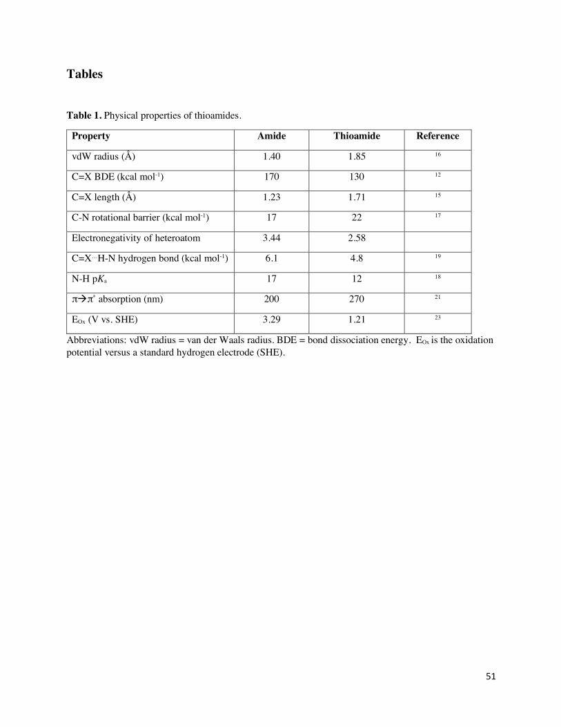
51
Tables
Table 1. Physical properties of thioamides.
Property Amide Thioamide Reference
vdW radius (Å) 1.40 1.85 16
C=X BDE (kcal mol-1) 170 130 12
C=X length (Å) 1.23 1.71 15
C-N rotational barrier (kcal mol-1) 17 22 17
Electronegativity of heteroatom 3.44 2.58
C=X…H-N hydrogen bond (kcal mol-1) 6.1 4.8 19
N-H pKa 17 12 18
πàπ* absorption (nm) 200 270 21
EOx (V vs. SHE) 3.29 1.21 23
Abbreviations: vdW radius = van der Waals radius. BDE = bond dissociation energy. EOx is the oxidation potential versus a standard hydrogen electrode (SHE).
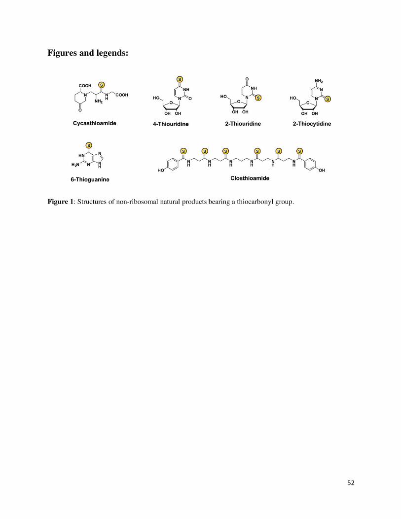
52
Figures and legends:
Figure 1: Structures of non-ribosomal natural products bearing a thiocarbonyl group.
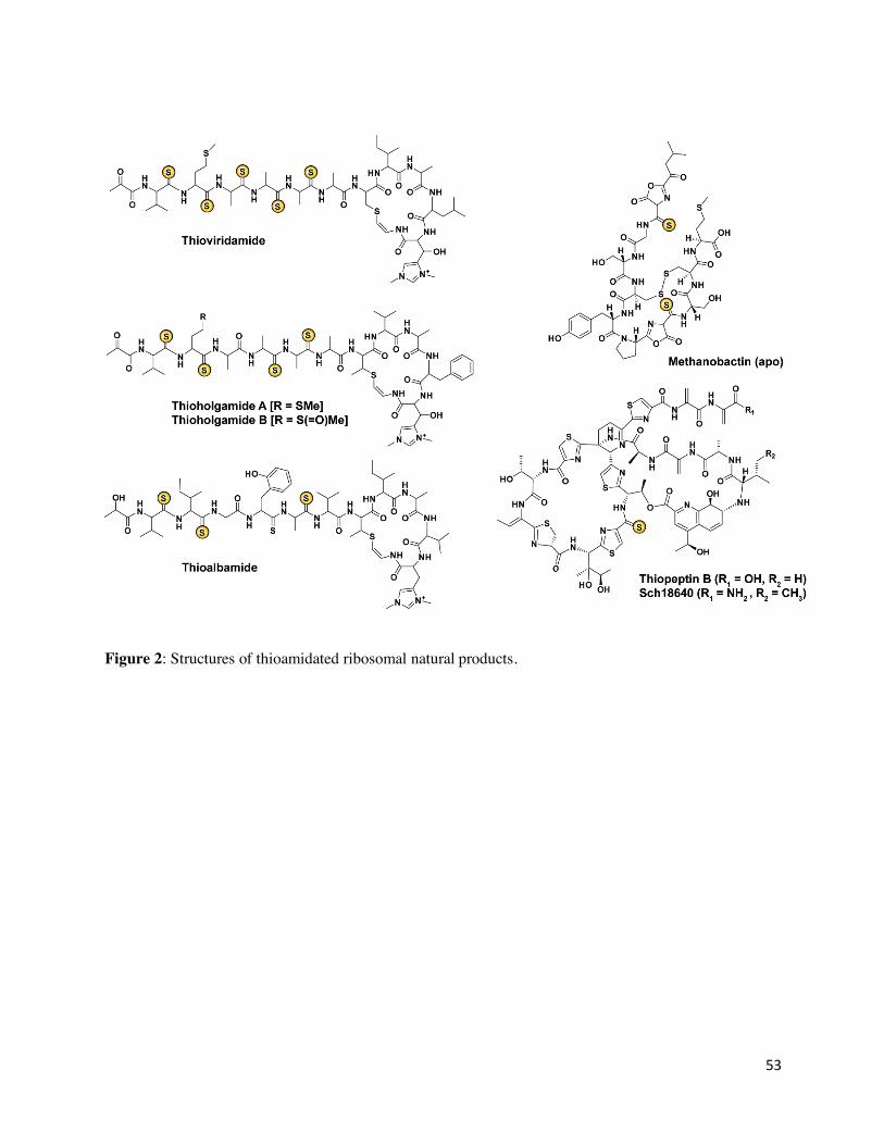
53
Figure 2: Structures of thioamidated ribosomal natural products.
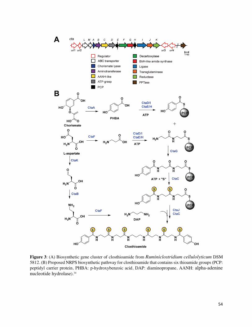
54
Figure 3: (A) Biosynthetic gene cluster of closthioamide from Ruminiclostridium cellulolyticum DSM 5812. (B) Proposed NRPS biosynthetic pathway for closthioamide that contains six thioamide groups (PCP: peptidyl carrier protein, PHBA: p-hydroxybenzoic acid, DAP: diaminopropane, AANH: alpha-adenine nucleotide hydrolase).34
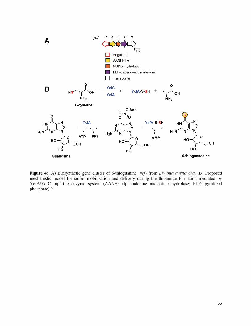
55
Figure 4: (A) Biosynthetic gene cluster of 6-thioguanine (ycf) from Erwinia amylovora. (B) Proposed mechanistic model for sulfur mobilization and delivery during the thioamide formation mediated by YcfA/YcfC bipartite enzyme system (AANH: alpha-adenine nucleotide hydrolase; PLP: pyridoxal phosphate).57
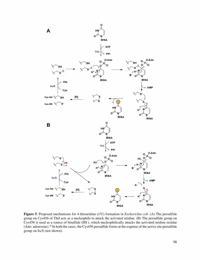
56
Figure 5: Proposed mechanisms for 4-thiouridine (s4U) formation in Escherichia coli. (A) The persulfide group on Cys456 of ThiI acts as a nucleophile to attack the activated uridine. (B) The persulfide group on Cys456 is used as a source of bisulfide (HS-), which nucleophilically attacks the activated uridine residue (Ado: adenosine).30 In both the cases, the Cys456 persulfide forms at the expense of the active site persulfide group on IscS (not shown).
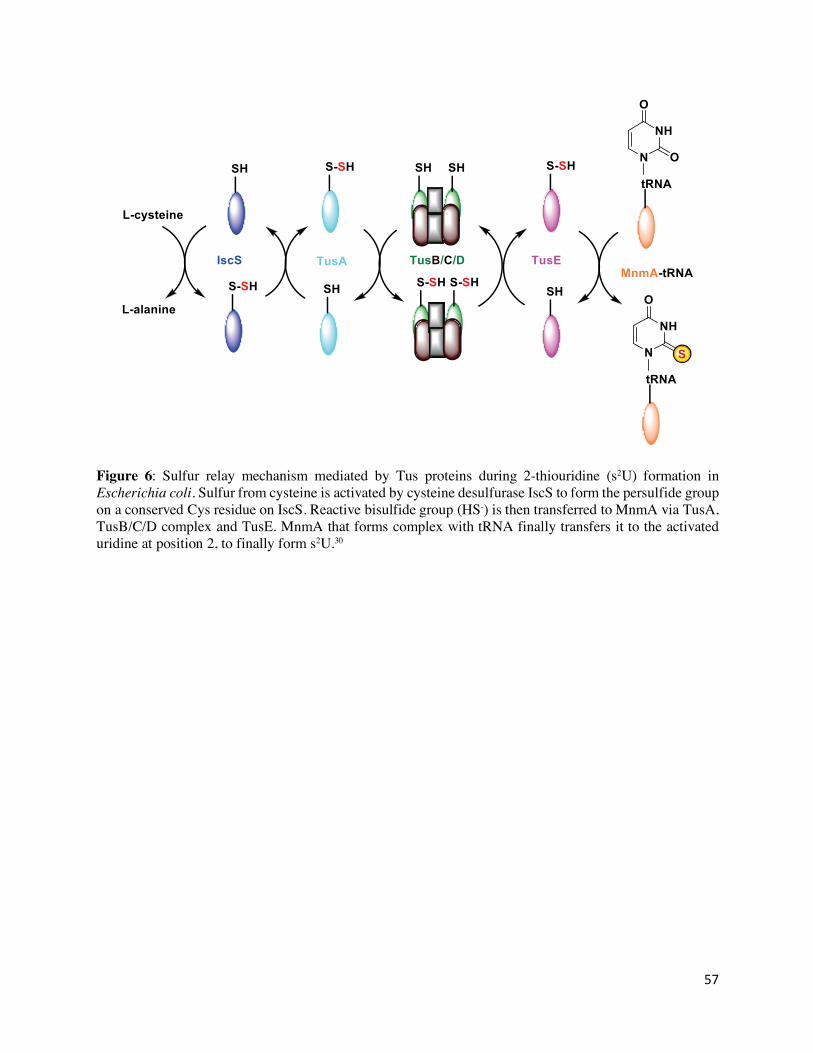
57
Figure 6: Sulfur relay mechanism mediated by Tus proteins during 2-thiouridine (s2U) formation in Escherichia coli. Sulfur from cysteine is activated by cysteine desulfurase IscS to form the persulfide group on a conserved Cys residue on IscS. Reactive bisulfide group (HS-) is then transferred to MnmA via TusA, TusB/C/D complex and TusE. MnmA that forms complex with tRNA finally transfers it to the activated uridine at position 2, to finally form s2U.30
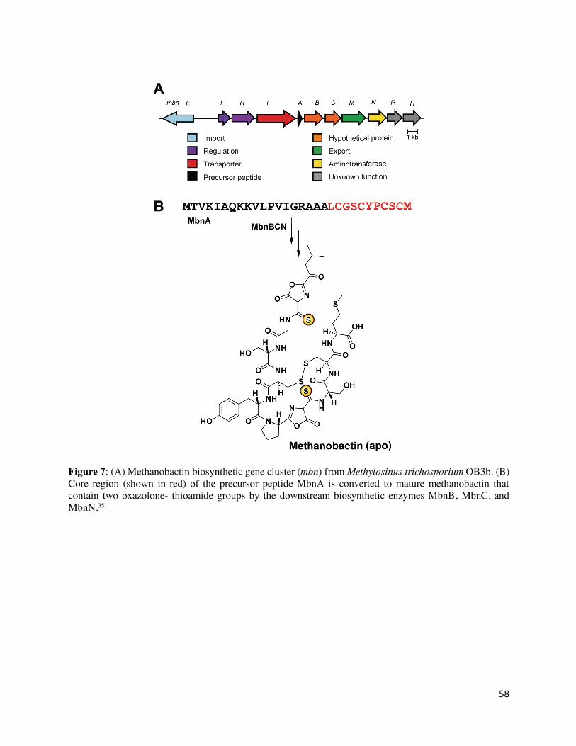
58
Figure 7: (A) Methanobactin biosynthetic gene cluster (mbn) from Methylosinus trichosporium OB3b. (B) Core region (shown in red) of the precursor peptide MbnA is converted to mature methanobactin that contain two oxazolone- thioamide groups by the downstream biosynthetic enzymes MbnB, MbnC, and MbnN.35
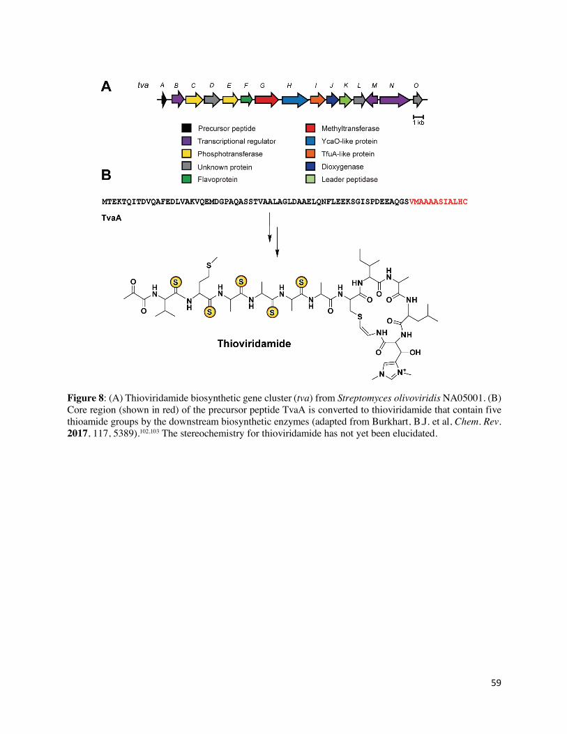
59
Figure 8: (A) Thioviridamide biosynthetic gene cluster (tva) from Streptomyces olivoviridis NA05001. (B) Core region (shown in red) of the precursor peptide TvaA is converted to thioviridamide that contain five thioamide groups by the downstream biosynthetic enzymes (adapted from Burkhart, B.J. et al, Chem. Rev. 2017, 117, 5389).102,103 The stereochemistry for thioviridamide has not yet been elucidated.
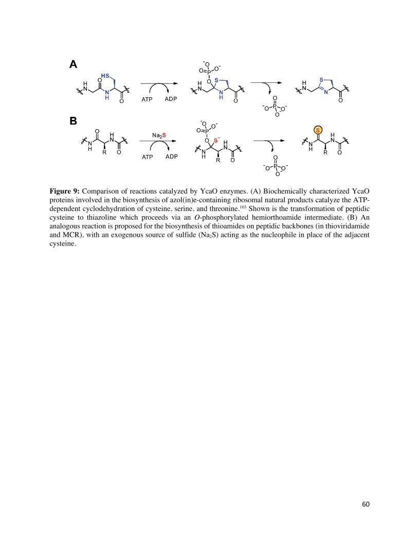
60
Figure 9: Comparison of reactions catalyzed by YcaO enzymes. (A) Biochemically characterized YcaO proteins involved in the biosynthesis of azol(in)e-containing ribosomal natural products catalyze the ATP-dependent cyclodehydration of cysteine, serine, and threonine.103 Shown is the transformation of peptidic cysteine to thiazoline which proceeds via an O-phosphorylated hemiorthoamide intermediate. (B) An analogous reaction is proposed for the biosynthesis of thioamides on peptidic backbones (in thioviridamide and MCR), with an exogenous source of sulfide (Na2S) acting as the nucleophile in place of the adjacent cysteine.
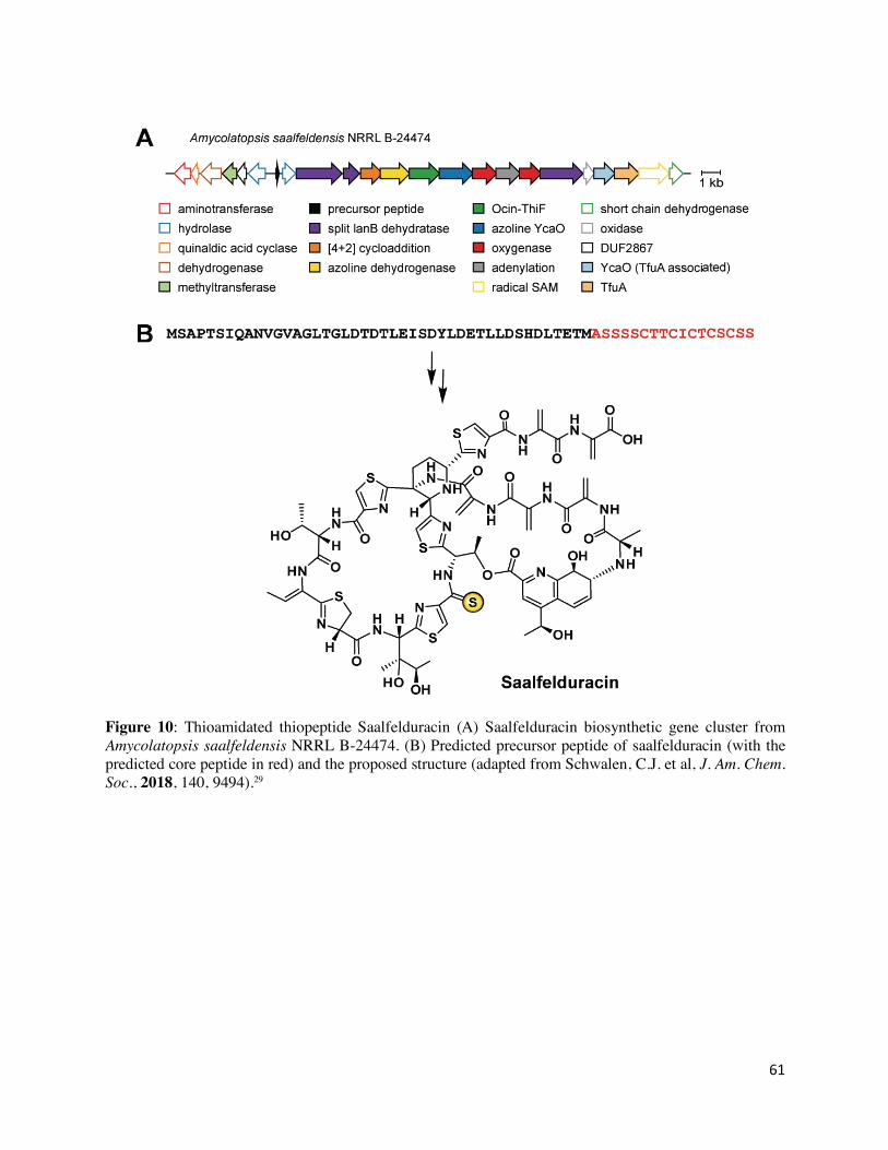
61
Figure 10: Thioamidated thiopeptide Saalfelduracin (A) Saalfelduracin biosynthetic gene cluster from Amycolatopsis saalfeldensis NRRL B-24474. (B) Predicted precursor peptide of saalfelduracin (with the predicted core peptide in red) and the proposed structure (adapted from Schwalen, C.J. et al, J. Am. Chem. Soc., 2018, 140, 9494).29
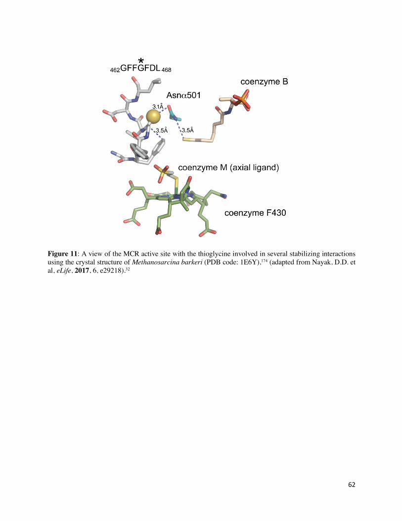
62
Figure 11: A view of the MCR active site with the thioglycine involved in several stabilizing interactions using the crystal structure of Methanosarcina barkeri (PDB code: 1E6Y),174 (adapted from Nayak, D.D. et al, eLife, 2017, 6, e29218).32
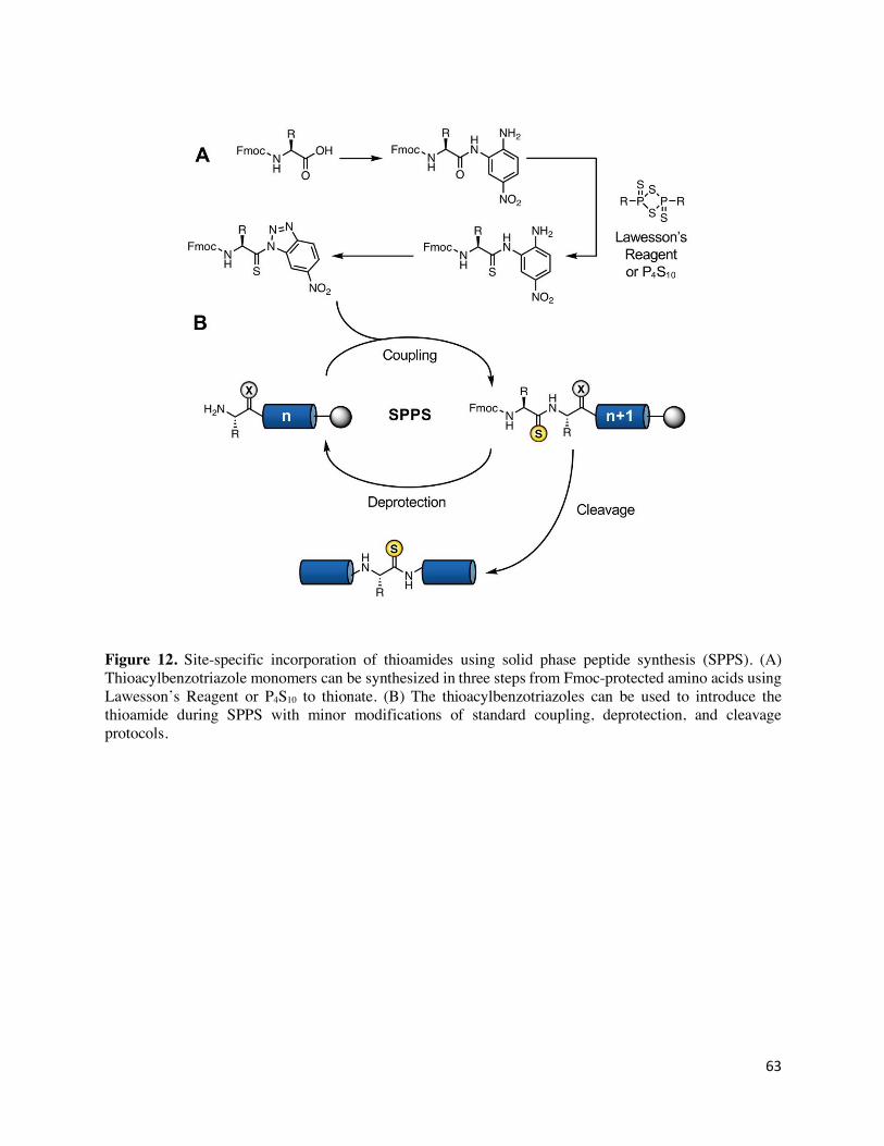
63
Figure 12. Site-specific incorporation of thioamides using solid phase peptide synthesis (SPPS). (A) Thioacylbenzotriazole monomers can be synthesized in three steps from Fmoc-protected amino acids using Lawesson’s Reagent or P4S10 to thionate. (B) The thioacylbenzotriazoles can be used to introduce the thioamide during SPPS with minor modifications of standard coupling, deprotection, and cleavage protocols.
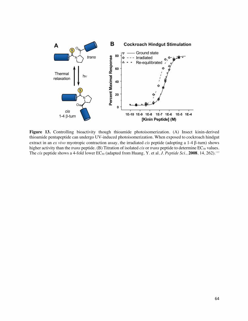
64
Figure 13. Controlling bioactivity though thioamide photoisomerization. (A) Insect kinin-derived thioamide pentapeptide can undergo UV-induced photoisomerization. When exposed to cockroach hindgut extract in an ex vivo myotropic contraction assay, the irradiated cis peptide (adopting a 1-4 b-turn) shows higher activity than the trans peptide. (B) Titration of isolated cis or trans peptide to determine EC50 values. The cis peptide shows a 4-fold lower EC50 (adapted from Huang, Y. et al, J. Peptide Sci., 2008, 14, 262).149
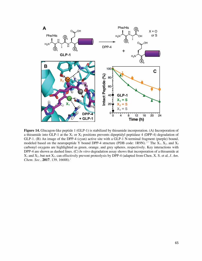
65
Figure 14. Glucagon-like peptide 1 (GLP-1) is stabilized by thioamide incorporation. (A) Incorporation of a thioamide into GLP-1 at the X1 or X2 positions prevents dipeptidyl peptidase 4 (DPP-4) degradation of GLP-1. (B) An image of the DPP-4 (cyan) active site with a GLP-1 N-terminal fragment (purple) bound, modeled based on the neuropeptide Y bound DPP-4 structure (PDB code: 1R9N).157 The X1, X2, and X3 carbonyl oxygens are highlighted as green, orange, and grey spheres, respectively. Key interactions with DPP-4 are shown as dashed lines. (C) In vitro degradation assay shows that incorporation of a thioamide at X1 and X2, but not X3, can effectively prevent proteolysis by DPP-4 (adapted from Chen, X. S. et al, J. Am. Chem. Soc., 2017, 139, 16688).4
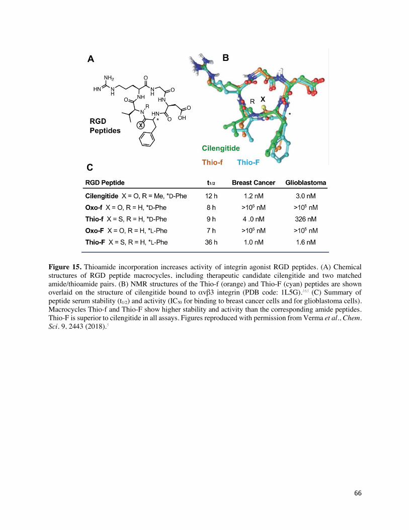
66
Figure 15. Thioamide incorporation increases activity of integrin agonist RGD peptides. (A) Chemical structures of RGD peptide macrocycles, including therapeutic candidate cilengitide and two matched amide/thioamide pairs. (B) NMR structures of the Thio-f (orange) and Thio-F (cyan) peptides are shown overlaid on the structure of cilengitide bound to αvβ3 integrin (PDB code: 1L5G).163 (C) Summary of peptide serum stability (t1/2) and activity (IC50 for binding to breast cancer cells and for glioblastoma cells). Macrocycles Thio-f and Thio-F show higher stability and activity than the corresponding amide peptides. Thio-F is superior to cilengitide in all assays. Figures reproduced with permission from Verma et al., Chem. Sci. 9, 2443 (2018).5
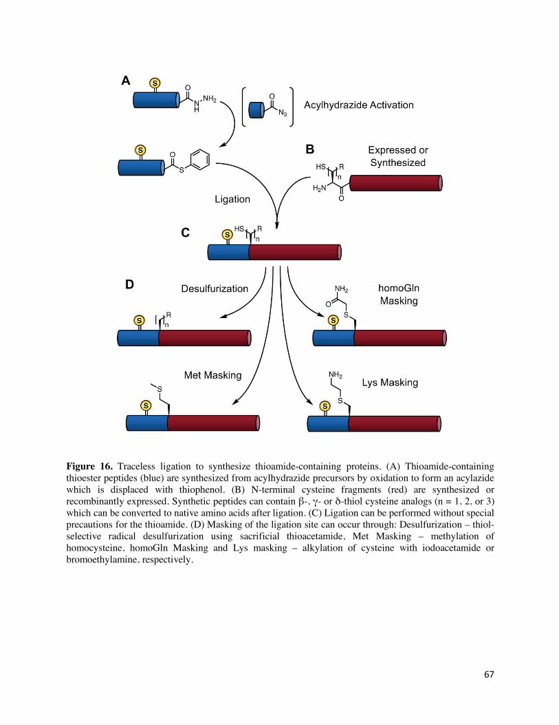
67
Figure 16. Traceless ligation to synthesize thioamide-containing proteins. (A) Thioamide-containing thioester peptides (blue) are synthesized from acylhydrazide precursors by oxidation to form an acylazide which is displaced with thiophenol. (B) N-terminal cysteine fragments (red) are synthesized or recombinantly expressed. Synthetic peptides can contain β-, γ- or δ-thiol cysteine analogs (n = 1, 2, or 3) which can be converted to native amino acids after ligation. (C) Ligation can be performed without special precautions for the thioamide. (D) Masking of the ligation site can occur through: Desulfurization – thiol-selective radical desulfurization using sacrificial thioacetamide, Met Masking – methylation of homocysteine, homoGln Masking and Lys masking – alkylation of cysteine with iodoacetamide or bromoethylamine, respectively.


