Download - BRAIN - med.upenn.edu

BRAINA JOURNAL OF NEUROLOGY
Hereditary spastic paraplegia is a novel phenotypefor GJA12/GJC2 mutationsJennifer L. Orthmann-Murphy,1 Ettore Salsano,2 Charles K. Abrams,3,4 Alberto Bizzi,5
Graziella Uziel,6 Mona M. Freidin,3 Eleonora Lamantea,7 Massimo Zeviani,7 Steven S. Scherer1
and Davide Pareyson2
1 Department of Neurology, University of Pennsylvania School of Medicine, Room 464 Stemmler Hall, 3450 Hamilton Walk, Philadelphia,
PA 19104-6077, USA
2 Biochemistry and Genetics Unit, IRCCS Foundation, C. Besta Neurological Institute, Via Celoria 11, 20133 Milan, Italy
3 Department of Neurology, SUNY Downstate Medical Center, Box 1213, 450 Clarkson Avenue, Brooklyn, NY 11203, USA
4 Department of Physiology and Pharmacology, SUNY Downstate Medical Center, Box 1213, 450 Clarkson Avenue, Brooklyn, NY 11203, USA
5 Neuroradiology Unit, IRCCS Foundation, C. Besta Neurological Institute, Via Celoria 11, 20133 Milan, Italy
6 Child Neurology Unit, IRCCS Foundation, C. Besta Neurological Institute, Via Celoria 11, 20133 Milan, Italy
7 Molecular Neurogenetics Unit, IRCCS Foundation, C. Besta Neurological Institute, Via Celoria 11, 20133 Milan, Italy
Correspondence to: Davide Pareyson, MD,
Biochemistry and Genetics Unit, IRCCS Foundation,
C. Besta Neurological Institute, Via Celoria 11,
20133 Milan, Italy
E-mail: [email protected]
Recessive mutations in GJA12/GJC2, the gene that encodes the gap junction protein connexin47 (Cx47), cause Pelizaeus-
Merzbacher-like disease (PMLD), an early onset dysmyelinating disorder of the CNS, characterized by nystagmus, psychomotor
delay, progressive spasticity and cerebellar signs. Here we describe three patients from one family with a novel recessively
inherited mutation, 99C4G (predicted to cause an Ile4Met amino acid substitution; I33M) that causes a milder phenotype. All
three had a late-onset, slowly progressive, complicated spastic paraplegia, with normal or near-normal psychomotor devel-
opment, preserved walking capability through adulthood, and no nystagmus. MRI and MR spectroscopy imaging were con-
sistent with a hypomyelinating leukoencephalopathy. The mutant protein forms gap junction plaques at cell borders similar to
wild-type (WT) Cx47 in transfected cells, but fails to form functional homotypic channels in scrape-loading and dual whole-
cell patch clamp assays. I33M forms overlapping gap junction plaques and functional channels with Cx43, however, I33M/
Cx43 channels open only when a large voltage difference is applied to paired cells. These channels probably do not function
under physiological conditions, suggesting that Cx47/Cx43 channels between astrocytes and oligodendrocytes are disrupted,
similar to the loss-of-function endoplasmic reticulum-retained Cx47 mutants that cause PMLD. Thus, GJA12/GJC2 mutations
can result in a milder phenotype than previously appreciated, but whether I33M retains a function of Cx47 not directly related
to forming functional gap junction channels is not known.
Keywords: spastic paraplegias; Pelizaeus-Merzbacher-like disease; gap junction; connexin; oligodendrocyte
Abbreviations: Cx47 = connexin47; DTRs = deep tendon reflexes; ER = endoplasmic reticulum; HSP = hereditary spastic paraplegia;PMD = Pelizaeus-Merzbacher disease; PMLD = Pelizaeus-Merzbacher-like disease
doi:10.1093/brain/awn328 Brain 2009: 132; 426–438 | 426
Received September 16, 2008. Revised October 27, 2008. Accepted November 4, 2008. Advance Access publication December 4, 2008
� The Author (2008). Published by Oxford University Press on behalf of the Guarantors of Brain. All rights reserved.
For Permissions, please email: [email protected]

IntroductionPelizaeus-Merzbacher disease (PMD) is an X-linked disorder
caused by mutations in PLP1, the gene encoding proteolipid pro-
tein, the main protein in CNS myelin. Classic PMD affects boys
and is characterized by nystagmus and impaired psychomotor
development within the first year of life, followed by progressive
spasticity, ataxia, choreoathetosis and diffuse white matter
changes on MRI (Nave and Boespflug-Tanguy, 1996; Hudson
et al., 2004; Inoue, 2005). PLP1 mutations may also cause a
more severe ‘connatal’ PMD phenotype, which is characterized
by dramatic psychomotor impairment from birth, with hypotonia,
quadriplegia, nystagmus, seizures and early death. Yet other PLP1
mutations cause a much milder disease, a ‘pure’ spastic paraplegia
type 2 (SPG2), or a more ‘complicated’ form, with onset in child-
hood or adolescence, and dysarthria, mental retardation and
ataxia (Garbern et al., 1999; Hudson et al., 2004; Garbern, 2007).
Pelizaeus-Merzbacher-like disease (PMLD) is clinically and neu-
roradiologically similar to classic PMD, but is not associated with
PLP1 mutations. Recessive mutations in GJA12 are one cause of
PMLD (Uhlenberg et al., 2004; Bugiani et al., 2006; Salviati et al.,
2007; Wolf et al., 2007; Henneke et al., 2008). GJA12/GJC2
(GJA12 was recently renamed GJC2; http://www.genename-
s.org/genefamily/gj.php) encodes connexin47 (Cx47), which is a
member of the connexin family, highly conserved integral mem-
brane proteins usually named according to their predicted mole-
cular mass (Willecke et al., 2002). Connexins form gap junctions,
which are intercellular channels that form between apposed cell
membranes to permit the diffusion of ions and small molecules
typically less than 1000 Da (Bruzzone et al., 1996). Six connexins
oligomerize into a hemichannel (or connexon), and two apposing
hemichannels form the gap junction; aggregates of tens to
thousands of intercellular channels form a gap junction plaque.
The potential diversity of gap junction composition is immense,
as over 20 mammalian connexins have been described.
Hemichannels may be homomeric, containing one type of con-
nexin, or heteromeric, containing more than one type. Gap
junctions are termed homotypic if the apposed hemichannels
contain the same connexin, and heterotypic if they contain differ-
ent connexins (Kumar and Gilula, 1996).
Anatomical and functional studies of the mammalian CNS have
demonstrated that astrocytes and oligodendrocytes are coupled by
gap junctions, forming a ‘glial syncytium’ (Mugnaini, 1986; Rash
et al., 2001; Orthmann-Murphy et al., 2008). There are abundant
gap junctions between astrocytes (A/A), fewer between oligoden-
drocytes and astrocytes (O/A), and few or none between
oligodendrocytes themselves. Oligodendrocytes express Cx32 and
Cx47 (Dermietzel et al., 1989; Micevych and Abelson, 1991;
Scherer et al., 1995; Li et al., 1997; Menichella et al., 2003; Nagy
et al., 2003a; Odermatt et al., 2003; Kamasawa et al., 2005),
as well as Cx29, which does not appear to form gap junctions
(Altevogt et al., 2002; Li et al., 2002; Altevogt and Paul, 2004;
Kleopa et al., 2004); cf. (Nagy et al., 2003a). Astrocytes express
Cx30 and Cx43 (Dermietzel et al., 1989; Yamamoto et al., 1990;
Micevych and Abelson, 1991; Nagy et al., 1997; Kunzelmann et al.,
1999; Nagy et al., 1999, 2001, 2003b; Rash et al., 2001). A/A
coupling appears to be limited to homotypic channels [Cx43/Cx43
and Cx30/Cx30; (Swenson et al., 1989; Werner et al., 1989; Dahl
et al., 1996)], but does not include Cx30/Cx43 heterotypic channels
(Orthmann-Murphy et al., 2007b). O/A coupling is most likely
mediated by Cx47/Cx43 and Cx32/Cx30 heterotypic channels
(Orthmann-Murphy et al., 2007b).
In humans, Cx47/Cx43 channels appear to be essential for
the proper maintenance of myelin. PMLD-associated mutations
are recessive and result in the loss-of-function of Cx47, including
the ability to form functional channels with Cx43, suggesting
that the loss of O/A coupling mediated by Cx47/Cx43 channels
causes PMLD (Orthmann-Murphy et al., 2007a, b). Here, we
describe a novel mutation (I33M) in GJA12/GJC2 in three
members of one family who have complicated hereditary spastic
paraplegia (HSP). In a cell model system, the mutant protein
forms gap junction plaques with Cx43, but I33M/Cx43 channels
have such severely altered voltage dependent gating that
they would not be predicted to function under physiological
conditions.
Methods
MRI studiesMRI was performed with a 1.5 Tesla MR unit (Siemens Magnetom
Avanto, Erlangen, Germany). The imaging protocol included sagittal
T1-weighted spin-echo, axial T1-weighted inversion-recovery turbo
spin-echo, axial proton-spin density and T2-weighted turbo spin-echo
and coronal FLAIR. Single-section multivoxel 2D 1H MR spectroscopic
imaging (1H MRSI) was acquired in all patients with a PRESS technique
(repetition time (TR)/echo time (TE) = 1200/144 ms), with a nominal
planar resolution of 1.04 mm3 (matrix = 24�24; field of view =
200�200�15 mm3). The volume of interest was positioned at the
level of the centrum semiovale. Scan acquisition time was 14 min. 1H
MRSI data were reconstructed and processed by using ‘csx2’ software
(Soher et al., 1996). Imaging of the cervical spine with sagittal T1- and
T2-weighted and axial T2-weighted MR images was acquired for
patient III-8.
Mutation analysisOligonucleotide primers (Uhlenberg et al., 2004) were used to PCR-
amplify the single coding exon of GJA12/GJC2 from genomic DNA in
three overlapping fragments, which were sequenced as previously
described (Bugiani et al., 2006).
Expression analysis of the I33M mutantWe generated the 99C4G (I33M) GJA12/GJC2 mutation from a
human Cx47 cDNA sequence (GenBank accession number
AF014643) using the QuikChange kit for PCR site-directed mutagen-
esis (Stratagene, La Jolla, CA, USA), with the following oligonucleotide
primers (the underlined codon encodes the altered amino acid):
50-ggtggtcttccgcatggtgctgacggctg-30; 50-cagccgtcagcaccatgcggaagac-
cacc-30, as previously described (Orthmann-Murphy et al., 2007a).
The resulting DNA [in pIRES2-EGFP and subcloned into pIRESpuro3
(Clontech, Mountain View, CA, USA)] and the mutation was con-
firmed by sequencing at the Sequencing Core of the University of
Pennsylvania.
GJA12 mutation causes spastic paraplegia Brain 2009: 132; 426–438 | 427
Neuro2A cells (N2A, from American Type Culture Collection,
Manassas, VA, USA) and communication-incompetent HeLa cells
(gift of Dr Klaus Willecke, University of Bonn, Bonn, Germany) were
maintained and transfected to transiently or stably express mutant
or WT Cx47 as previously described (Orthmann-Murphy et al.,
2007a, b). Heterotypic mix experiments were performed on HeLa
cells stably expressing WT Cx43, WT Cx47, I33M or P87S as described
previously (Orthmann-Murphy et al., 2007b).
Cells were immunostained as previously described (Orthmann-
Murphy et al., 2007a, b) using rabbit antisera raised against the
C-terminus of human Cx47 [diluted 1:1500; (Orthmann-Murphy
et al., 2007a)] or mouse monoclonal antibodies against mouse Cx43
(diluted 1:1000; Millipore Corporation, Billerica, MA, USA) or
pan-cadherin (Novus Biologicals, Littleton, CO, USA; diluted 1:200)
and FITC-, TRITC-, and Cy5-conjugated secondary antibodies.
Images were acquired using a Leica fluorescence microscope and
Openlab 3.1.7 or a FluoView FV1000 Olympus laser scanning confocal
microscope (60�, oil immersion objective). Confocal images were
merged and pseudocoloured using Image J 1.37v (National Institutes
of Health, USA) then imported into Adobe Photoshop (San Jose, CA,
USA) for post-processing. Transfected HeLa and N2A cells were col-
lected and processed for immunoblotting as previously described
(Orthmann-Murphy et al., 2007a).
Functional tests of the I33M mutantConfluent HeLa cell monolayers were scrape loaded as previously
described (Orthmann-Murphy et al., 2007a). We used dual whole-
cell patch clamping to assess formation of both homotypic channels,
on pairs of N2A cells transiently transfected with WT Cx43, WT Cx47
or I33M subcloned into pIRES2-EGFP, and heterotypic channels, on
pairs of cells each expressing a pIRES2-EGFP construct or pIRES2-
DsRed construct, as described previously (Orthmann-Murphy et al.,
2007b). Recording solutions used were as follows (in mM): pipette
solution, 145 CsCl2, 5 EGTA, 0.5 CaCl2 and 10.0 HEPES, pH 7.2;
bath solution, 150 NaCl, 4 KCl, 1 MgCl2, 2 CaCl2, 5 dextrose, 2
pyruvate and 10 HEPES, pH 7.4. Heterotypic pairings between two
cells are shown as ‘connexin expressed in cell 2/connexin expressed in
cell 1’; junctional conductance (Gj)—junctional voltage (Vj) relations
were determined from isolated pairs by measuring instantaneous junc-
tional current (Ij) responses in cell 2 following 12.5-s Vj pulses (from
�100 to 100 mV in 20 mV increments) applied to cell 1 and applying
Ohm’s law. Baseline Gj was similarly determined by measuring instan-
taneous Ij responses to �40 mV Vj pulses.
Statistical analysesFor heterotypic mix experiments, epifluorescence images were
imported into Adobe Photoshop and analysed as previously described
(Orthmann-Murphy et al., 2007b). At least 11 DsRed + cells from two
cover slips were analysed for each heterotypic mixture; this was
repeated in three independent experiments. For each DsRed + cell,
we determined the mean and 95% CI of the number of overlapping
cell surface puncta. The number of DsRed + cells with or without one
or more overlapping puncta was compared using Fisher’s exact test
with Bonferroni’s correction for multiple comparisons. All statistical
tests were performed in GraphPad Prism (San Diego, CA, USA). For
dual whole-cell patch clamp assays, values are presented as mean
Gj� SEM, and were compared using Fisher’s exact test or Mann–
Whitney test in GraphPad Prism.
Results
Clinical findingsThe family pedigree is shown in Fig. 1. There were three affected
members—the proband (III-8, arrowhead), his brother (III-11) and
their female cousin (III-5). At age 39, the proband presented with
an 8-year history of slowly progressive walking difficulties, leg
stiffness and slurred speech. He felt ‘awkward’ since his school
years, had minimal writing difficulties, but was fit for military ser-
vice at age 18. At age 21, he underwent surgery for concomitant
strabismus. Currently, he is able to walk without assistance, and
works as a priest. His legs and gait were spastic, and bilateral
Babinski signs were found. The jaw jerk and deep tendon reflexes
(DTRs) were brisk throughout, with non-sustained ankle clonus.
Mild pes cavus was noted. He had no nystagmus, but pursuit eye
movements were slightly saccadic. The following findings were
mild: pseudobulbar dysarthria, loss of finger dexterity, dysmetria
and intention tremor on finger-to-nose and heel-to-knee testing.
The proband’s 36-year-old brother (III-11) had minimal motor
difficulties since infancy and mild learning impairment at school.
He reported mild intention tremor during late-childhood, and a
few tonic-clonic seizures, mainly febrile, between childhood and
his early teens. These problems had long been attributed to a
dystocic delivery with cyanosis at birth. After age 20, he began
to show a slow worsening of walking difficulties and slight dysar-
thria, followed by decreased dexterity with his hands. Presently, he
walks with a cane, but he can still walk a few steps without aid.
On neurological examination he had a shuffling, scissor-like gait,
marked spasticity with mild weakness of the lower limbs, an
increased jaw jerk and increased DTRs in all limbs, with sustained
clonus at the ankles and bilateral Babinski signs. He had slight
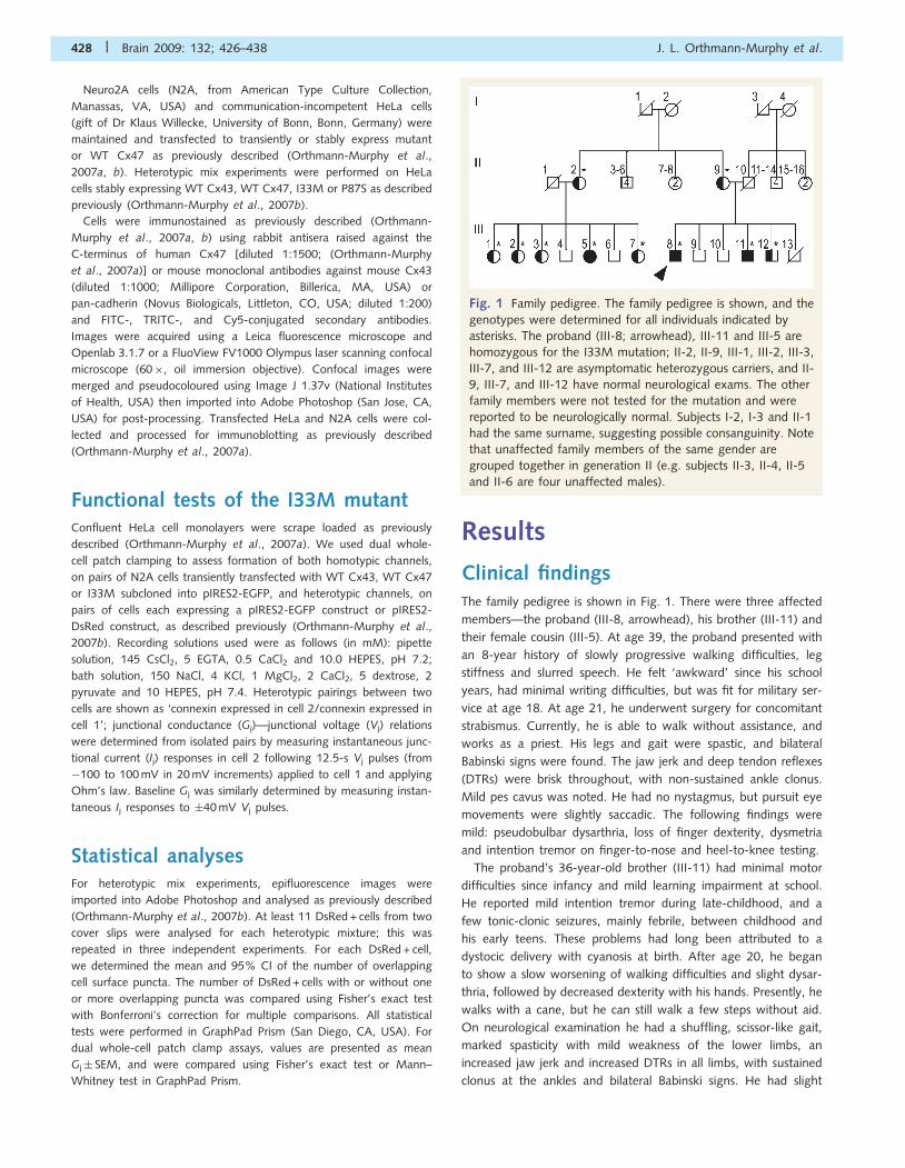
Fig. 1 Family pedigree. The family pedigree is shown, and the
genotypes were determined for all individuals indicated by
asterisks. The proband (III-8; arrowhead), III-11 and III-5 are
homozygous for the I33M mutation; II-2, II-9, III-1, III-2, III-3,
III-7, and III-12 are asymptomatic heterozygous carriers, and II-
9, III-7, and III-12 have normal neurological exams. The other
family members were not tested for the mutation and were
reported to be neurologically normal. Subjects I-2, I-3 and II-1
had the same surname, suggesting possible consanguinity. Note
that unaffected family members of the same gender are
grouped together in generation II (e.g. subjects II-3, II-4, II-5
and II-6 are four unaffected males).
428 | Brain 2009: 132; 426–438 J. L. Orthmann-Murphy et al.

dysarthria, mild loss of finger dexterity, dysmetria, intention
tremor on finger-to-nose testing and moderate hypodiadochoki-
nesia. He had pes cavus with hammer toes, lumbar hyperlordosis
and concomitant strabismus but no nystagmus.
The proband’s 53-year-old cousin (III-5) had always been con-
sidered somewhat dull, but completed primary school without
help. She walked at 20 months, and developed progressive spastic
paraplegia and dysarthric speech beginning in her teens, becoming
wheelchair bound at age 30. She later developed urinary incon-
tinence followed by retention, and required a permanent catheter
at age 46. More recently, she has developed episodic painful
spasms in lower limbs, and marked constipation with rare fecal
incontinence. On exam, she was unable to stand or walk without
bilateral support. Her arms were mildly spastic, atrophied and
weak; her legs were markedly spastic, wasted and completely
paralysed. Her jaw jerk and all DTRs were exaggerated, with sus-
tained right ankle clonus and bilateral Babinski signs. She had
slightly saccadic pursuits without nystagmus, and moderate dysar-
thria with nasal, scanning and spastic qualities. She had moderate
dysmetria and intention tremor on finger-to-nose testing, with
marked hand hypodiadochokinesia. Light touch and pain sensa-
tions were reduced in the left upper limb and both legs. Position
and vibration sense were severely impaired in the legs. She also
had severe dorsal scoliosis, bilateral pes cavus and ankle contrac-
tures (Table 1).
Clinical investigationsThe following blood tests were normal for all affected patients:
creatine kinase, uric acid, ammonia, lactate, pyruvate, alanine and
other amino acids, very-long-chain fatty acids and phytanic acid.
Urinary levels of organic acids, amino acids and sulfatides were
also normal. Lysosomal enzyme assessment in blood leukocytes
ruled out metachromatic leukodystrophy, Krabbe disease and
GM1 and GM2 gangliosidoses. Neuro-ophthalmologic examina-
tion revealed only left eye amblyopia in patients III-8 and III-11,
attributed to congenital strabismus. Audiometry revealed mild sen-
sorineural hearing loss in Patient III-5 and left ear conduction
hypoacusia due to previous trauma in Patient III-8.
Electromyography and motor and sensory nerve conduction stu-
dies were normal in all three patients. Motor evoked potential
(MEP) studies showed prolongation of central motor conduction
time (CMCT) in upper limbs, and no response in lower limbs in
Patients III-8 and III-11; in Patient III-5, MEPs could be recorded
only from the left upper limb, with central motor conduction time
prolongation, whereas no response was obtained from the other
limbs. Their electroretinogram was normal, whereas visual evoked
potentials showed delayed P100 wave latencies. Brainstem audi-
tory evoked potentials disclosed absence or severe distortion and
delay of waves III and V; wave I was normal in Patients III-11 and
III-5, but almost unrecognizable in Patient III-8, particularly on the
left side. Upper and lower limb somatosensory evoked potential
studies demonstrated delayed latencies of central components in
all patients; no response could be evoked in lower limbs in Patient
III-5. In all patients, EEG showed a posterior background activity
of about 8 Hz with a slightly irregular morphology. The cardiovas-
cular reflexes were normal in Patient III-8. Neuropsychological
assessment revealed IQ scores of 94 in Patient III-8, 83 in
Patient III-11 and 77 in Patient III-5 (normal value470).
Table 1 Summary of clinical and instrumental data for patients with the I33M GJA12/GJC2 mutation
Patient III-8 Patient III-11 Patient III-5
Sex/Age at examination (years) M/39 M/36 F/53
Age at disease onset 2nd decade 1st decade 1st decade
Age of disease progression (years) 430 420 410
Initial motor development Normal Normal Walking at 2 yrs
Onset symptom Difficulty in walking Hand tremor Difficulty in walking
Walking ability at exam Without aid With unilateral aid Chairbound since age 30
Lower limb spasticity ++ +++ ++++
Upper limb motor involvement � + ++
Dysarthria + ++ ++
Nystagmus – – –
Cerebellar ataxia � + ++
Mental impairment/IQ score �/94 �/83 +/77
Epilepsy – + –
Sphincter dysfunction – – ++
Sensory loss – – ++
Sensorineural hearing loss – – +
Other signs Pes cavus, strabismus Pes cavus, strabismus Pes cavus, scoliosis
MRI Hypomyelination Hypomyelination Hypomyelination and atrophy1H-MRSI #Cho/NAA, Cho/Cr ratios #Cho/NAA, Cho/Cr ratios #Cho/NAA, Cho/Cr ratios
EEG Near-normal Near-normal Near-normal
EMG and nerve conduction studies Normal Normal Normal
VEPs, BAEPs, MEPs and SEPs Abnormal Abnormal Abnormal
Autonomic tests Normal ND ND
–absent;�= minimal; + = mild; ++ = moderate; +++ = marked; ++++ = severe; ND = not done. BAEPs = brainstem auditory evoked potentials; EMG = electromyography;MEPs = motor evoked potentials; SEPs = somatosensory evoked potentials; VEPs = visual evoked potentials.
GJA12 mutation causes spastic paraplegia Brain 2009: 132; 426–438 | 429

Brain MR imaging and spectroscopic findings were homoge-
neous in the three patients and highly suggestive of a hypomye-
linating leukoencephalopathy. White matter regions showed
diffuse high signal intensity on T2-weighted and low signal on
T1-weighted images (Fig. 2, Supplementary Fig. 1). T2-weighted
signal hyperintensity was evident at level of the pons, in the
region of corticospinal and spinothalamic tracts (Fig. 2B and F).
The corpus callosum was thin in all patients. The cervical spine of
Patient III-8 appeared normal (data not shown). Ventricular and
posterior fossa dilation, likely secondary to white matter volume
loss, was mild in Patients III-8 and III-11, but advanced in Patient
III-5. Choline (Cho), N-acetyl-aspartate (NAA) and creatine (Cr)
were measured in the white matter of the centrum semiovale by1H MR spectroscopic imaging. The NAA/Cr ratios were within
normal values, consistent with what has been reported for PMD
and PMLD (Lee et al., 2004; Bizzi et al., 2008). Average Cho/
NAA and Cho/Cr ratios, however, were reduced—0.49 and 0.84
in Patient III-8; 0.47 and 0.80 in Patient III-11; 0.44 and 0.75 in
Patient III-5—compared with 0.60 and 1.0, respectively, in normal
adults.
Affected patients have an I33Mmutation in GJA12/GJC2The above data indicated that the three patients from
this family had a recessively inherited hypomyelinating
leukoencephalopathy with an unusual clinical phenotype of
a late onset complicated HSP. Although the parents of our
patients were reported to be unrelated, they all originated from
a small village in northern Italy, and Subjects I-2, I-3 and II-1
(Fig. 1) had the same family name. Hence, we sequenced ampli-
fied genomic DNA of the GJA12/GJC2 gene, and found a novel
missense mutation (99C4G), predicted to cause an Ile4Met
amino acid substitution (I33M) in Cx47. This mutation was
homozygous in the three affected individuals, heterozygous in
obligate healthy carriers and some clinically normal relatives
(asterisks in Fig. 1 denote DNA analysis), and absent in 210
control alleles. Heterozygous relatives (Fig. 1) were asymptomatic
and had a normal neurological exam (II-9, III-7 and III-12);
one of these relatives (III-7) had a normal brain MRI and1H MRSI.
As shown in Fig. 3, I33 is located in the first transmembrane
domain, which is a highly conserved region of connexins (Yeager
and Nicholson, 1996). This amino acid residue is identical in Cx47
orthologues of other vertebrates (cow, mouse, frog and zebrafish;
Supplementary Fig. 2), but Val (Cx30, Cx30.3, Cx31, Cx31.1), Leu
(Cx30.2) or even Met (Cx40, Cx59, Cx62) are found at the corre-
sponding position in other connexins (Supplementary Fig. 2).
Although mutations in the genes encoding Cx26, Cx30, Cx30.3,
Cx31, Cx32, Cx40, Cx43, Cx46 and Cx50 have been described,
mutations in the residues corresponding to I33 have not been
identified previously.
Fig. 2 Imaging studies show white matter abnormalities. These are MRI images from patients III-8 (A–D) and III-5 (E–H). Sagittal
T1-weighted (T1W) images [spin-echo (SE): repetition time (TR)/echo time (TE) 556/13 ms; 6-mm thickness] shows thinning of the
corpus callosum, posteriorly (A, arrowhead) or diffusely (E). Axial T2W images (SE: TR/TE = 3200/90 ms; 5-mm thickness) show
symmetric hyperintensity in the region of the corticospinal/corticobulbar tracts at the level of the pons B and F, arrowhead) and the
posterior limb of internal capsule (C and G, arrowhead). In addition, there is diffuse hyperintensity in the subcortical, lobar and
periventricular white matter (C, G and H), and enlarged ventricles, especially in Patient III-5 (G). Axial T1W image [inversion recovery
turbo spin-echo (IRTSE): TR/TE = 5600/70 ms; flip angle 150�; 4-mm thickness] shows diffuse hypointensity in the white matter,
including the posterior limb of the internal capsule (D, arrowhead).
430 | Brain 2009: 132; 426–438 J. L. Orthmann-Murphy et al.

I33M forms gap junction plaques inHeLa cellsTo investigate the underlying molecular defects of the I33M mutant,
we expressed it in communication-incompetent HeLa and N2A cells.
For comparison, we also expressed WT Cx47 and P87S, a missense
mutant that causes PMLD (Uhlenberg et al., 2004), and results in
loss-of-function (Orthmann-Murphy et al., 2007a, b). We con-
firmed expression by immunoblotting lysates from bulk-selected
and transiently transfected cells (Supplementary Fig. 3). As expected
(Orthmann-Murphy et al., 2007a, b), P87S was localized the endo-
plasmic reticulum (ER) in both transiently (Supplementary Figs. 4
and 5) and permanently transfected (Fig. 4) cells, whereas I33M
(and WT Cx47) formed gap junction plaques at apposed cell bor-
ders. We confirmed the cell surface localization of WT Cx47 and
I33M by double labelling with a monoclonal antibody that recog-
nizes cadherins. Neither parental HeLa cells, nor bulk-selected HeLa
cells that had been transfected to express vector alone, expressed
Cx47 (data not shown). We obtained similar results in four separate
transient transfection experiments and two different bulk-selected
cell lines expressing I33M.
I33M does not form functionalhomotypic channelsBecause patients expressing I33M have a milder phenotype than
do patients with PMLD, and I33M can form gap junction plaques,
Fig. 4 The I33M mutant forms gap junction plaques. These are
confocal images of bulk-selected HeLa cells that express WT
Cx47 or the indicated mutants, immunostained with a rabbit
antiserum against human Cx47 (red) and a mouse monoclonal
antibody against pan-cadherin (green), and counterstained with
DAPI. The pan-cadherin staining at cell borders interdigitates with
the cell surface staining of Cx47 in cells that express WT Cx47
(arrowheads) or I33M (arrowheads), but surrounds the staining
of cells expressing the mutant P87S, which is localized in the
endoplasmic reticulum. Scale bar: 10 mm.
Fig. 3 GJA12/GJC2 mutations associated with CNS diseases.
This is a schematic drawing of human Cx47, illustrating the
position and nature of mutations associated with CNS diseases.
I33M (grey circle) is located in the first transmembrane
domain. All of the other mutations depicted cause PMLD
(black circles), either as homozygous mutations or as com-
pound heterozygote mutations (missense mutations: P87S,
G146S, G233R, G233S, T262A, Y269D, M283T and T395I;
frameshift mutations: L25fs, P128fs, E204fs, L278fs, P302fs,
C315fs, A322fs and P327fs; non-sense mutations: Y229stop
and R237stop; complex mutations: A95G____V96insertT)
(Uhlenberg et al., 2004; Bugiani et al., 2006; Salviati et al.,
2007; Wolf et al., 2007; Henneke et al., 2008). The positions
of the transmembrane domains are based on the work of
Yeager and Nicholson (Yeager and Nicholson, 1996).
GJA12 mutation causes spastic paraplegia Brain 2009: 132; 426–438 | 431

we hypothesized that I33M might form gap junction channels
with altered functional properties. To determine whether I33M
could transfer small molecules, we scrape loaded cells (el-Fouly
et al., 1987; Trosko et al., 2000), using gap junction tracers of
different sizes, shapes and charge. A confluent monolayer of cells
was injured with a scalpel blade in media that contained 2%
neurobiotin (NB; MW 287, + 1) or 0.1% Lucifer Yellow (LY;
MW 443, �2). As in parental cells or cells expressing vector
alone (data not shown), no transfer was seen past the scrape
line in bulk-selected cells expressing I33M or P87S (Fig. 5B–C
and E–F), whereas NB and LY transferred to cells beyond the
scrape line for cells expressing WT Cx47 (Fig. 5A and D). We
also confirmed that the cells along the scrape line expressed the
appropriate connexin (Supplementary Fig. 6). This experiment was
repeated at least twice in two different bulk-selected cells lines
expressing I33M with similar results.
We used a more sensitive assay to determine whether Cx47
mutants can form functional homotypic gap junctions—dual
whole-cell patch clamping on transiently transfected N2A cells.
In this experiment, each cell was transiently transfected to express
a single connexin as well as EGFP. For negative control pairs, cells
expressing a single connexin were paired with cells expressing
EGFP alone (Cx/EGFP). Voltage ramps or steps were applied
to one cell of an EGFP-expressing pair, and current responses
were measured in the second cell; the junctional voltage (Vj)
corresponds to the difference in voltage between the two cells.
As shown in Table 2, I33M/I33M homotypic pairings failed to
form functional channels (0/6 pairs), whereas cell pairs expressing
Fig. 5 I33M is impermeable to low molecular weight tracers. These are representative images of bulk-selected HeLa cells that stably
express WT Cx47 or the indicated mutants, scrape-loaded with neurobiotin (A—C) or Lucifer Yellow (D—F). Cells scrape-loaded
with neurobiotin were fixed with 4% paraformaldehyde, then visualized using FITC-conjugated extravidin (green) and DAPI
counterstain (blue). Cells scrape-loaded with Lucifer Yellow were imaged by epifluorescence (top row) and phase contrast (bottom row)
5 min after scrape loading. Only HeLa cells expressing WT Cx47 showed transfer of neurobiotin (A) or Lucifer Yellow (D) to
neighbouring cells. The images for each type of scrape loading experiment were acquired on the same day at the same exposure.
(A—C) scale bar: 20 mm; (D—F) scale bar: 20 mm.
432 | Brain 2009: 132; 426–438 J. L. Orthmann-Murphy et al.
WT Cx47 were coupled. The difference between I33M/I33M and
WT Cx47/Cx47 pairings was statistically significant (I33M/I33M
versus Cx47/Cx47, P = 0.0022, Fisher’s exact test). Thus, I33M
does not appear to form functional homotypic gap junctions by
two different assays.
I33M forms functional heterotypicchannels with Cx43 in a cell modelsystemTo determine whether I33M can form heterotypic channels with
Cx43, we applied morphological and functional assays we used
previously to show that Cx47 mutants associated with PMLD do
not form functional heterotypic gap junctions with WT Cx43
(Orthmann-Murphy et al., 2007b). In the morphological assay,
cell lines in which at least 90% of cells stably expressed Cx47
(I33M, P87S or WT Cx47) or Cx43 were transiently transfected
to express DsRed (DsRed + cells). The DsRed + cells (expressing
Cx47 or Cx43) were mixed with DsRed- cells (expressing Cx43
or Cx47, respectively) at a ratio of 1:20, and, 24 h after
plating, immunostained for Cx47 and Cx43. For each combina-
tion, we determined whether the connexin puncta at the periphery
of the DsRed + cell overlapped with connexin puncta expressed by
the surrounding DsRed- cells. As shown in Fig. 6, P87S was loca-
lized to the ER, and did not appear to form overlapping puncta
with Cx43 (Orthmann-Murphy et al., 2007b). In contrast, cells
expressing I33M or WT Cx47 formed overlapping puncta with
Cx43, suggesting that I33M/Cx43 may form functional channels.
We quantified these results by counting the number of puncta at
the cell membrane of each central DsRed + cell, and determining
whether these puncta overlapped with the connexin signal in the
surrounding DsRed- cells (Fig. 6D). In this analysis, the mixtures
are designated as ‘CxA�/CxB,’ where the asterisk denotes the
connexin expressed by the DsRed + cell. Mixtures containing
I33M/Cx43 (I33M�/Cx43 and Cx43�/I33M) produced a signifi-
cantly larger proportion of DsRed + cells with at least one
overlapping punctum than did P87S/Cx43 mixtures (I33M�/
Cx43 versus P87S�/Cx43 or Cx43�/P87S, Cx43�/I33M versus
P87S�/Cx43, P50.0001; Cx43�/I33M versus Cx43�/P87S,
P = 0.0013, Fisher’s test), but were not significantly different
than mixtures containing Cx47/Cx43 (Cx47�/Cx43 and Cx43�/
Cx47).
In the electrophysiogical assay, we used dual whole-cell patch
clamping on pairs of N2A cells. In these experiments, each cell was
transiently transfected to express a single connexin as well as EGFP
or monomeric DsRed, so that each member of a cell pair could be
unambiguously identified. To confirm that the cells expressing
EGFP or DsRed also expressed the expected connexin, we immu-
nostained transiently transfected cells (Supplementary Fig. 5). In
this way, we previously showed that three loss-of-function Cx47
mutants that cause PMLD (P87S, Y269D and M283T) do not form
functional heterotypic channels with Cx43 (Orthmann-Murphy
et al., 2007b). In contrast, we detected Ij activation for six of
13 (46%) I33M/Cx43 pairs tested, somewhat less than we pre-
viously found for all of the WT Cx47/Cx43 heterotypic pairs we
have tested (40 of 55; 73%). As shown in Table 2, the mean Gj
measured at Vj = 0 for I33M/Cx43 channels is significantly smaller
than that of WT Cx47/Cx43 channels (I33M/Cx43 versus WT
Cx47/Cx43, P50.05, Mann–Whitney test). Negative control
pairs, in which cells expressing a single connexin were paired
with cells expressing EGFP or DsRed alone, showed low levels of
coupling probably due to formation of heterotypic channels with
an endogenous connexin expressed by parental N2A cells
(Table 2; Orthmann-Murphy et al., 2007b).
To better define the functional differences between I33M/Cx43
and Cx47/Cx43 channels, we examined macroscopic current
responses and Gj–Vj relations. By convention, pairing designation
is ‘connexin expressed by cell 2/connexin expressed by cell 1’.
Both cells in the pair were voltage clamped to 0 mV; cell 1 was
stepped between �100 and 100 mV in 20 mV increments, and
current was recorded from cell 2. For I33M/Cx43 heterotypic
pairings, negative pulses (4�40 mV) applied to the cell expressing
Cx43 activate junctional currents (Ij) (Fig. 7A). Assuming that Gj is
a linear function of Vj between �20 mV and +20 mV, the normal-
ized Gj versus junctional voltage (Vj) relation reveals that I33M/
Cx43 channels are about fifty times more likely to be open after a
12.5-s pulse to Vj =�100 mV than at Vj = 0 mV (solid line, Fig. 7B).
This is strikingly different than WT Cx47/Cx43 channels (dashed
line), which are more likely to be open at Vj = 0 mV than at either
�100 mV or + 100 mV (Fig. 7B; Orthmann-Murphy et al., 2007b).
The left shift of the Gj–Vj relation for I33M/Cx43 channels is likely
explained by an extremely low open probability of the I33M hemi-
channel when Vj5�40 mV are applied to the cell expressing
Cx43; this can account for the significantly reduced Gj values
detected at Vj = 0 mV for both I33M/Cx43 and I33M/I33M chan-
nels (Table 2). Although the WT Cx43 hemichannel has negative
Vj gating polarity (Bukauskas et al., 2001) and should be closed
under the conditions that open I33M hemichannels, Cx43 rarely
closes fully (Bukauskas et al., 2000). It is possible, then, that I33M
is forming a channel with a substate of Cx43. Properties of I33M/
WT Cx47 channels (in 5/15 pairs) are similar to those described
for I33M/Cx43 (data not shown).
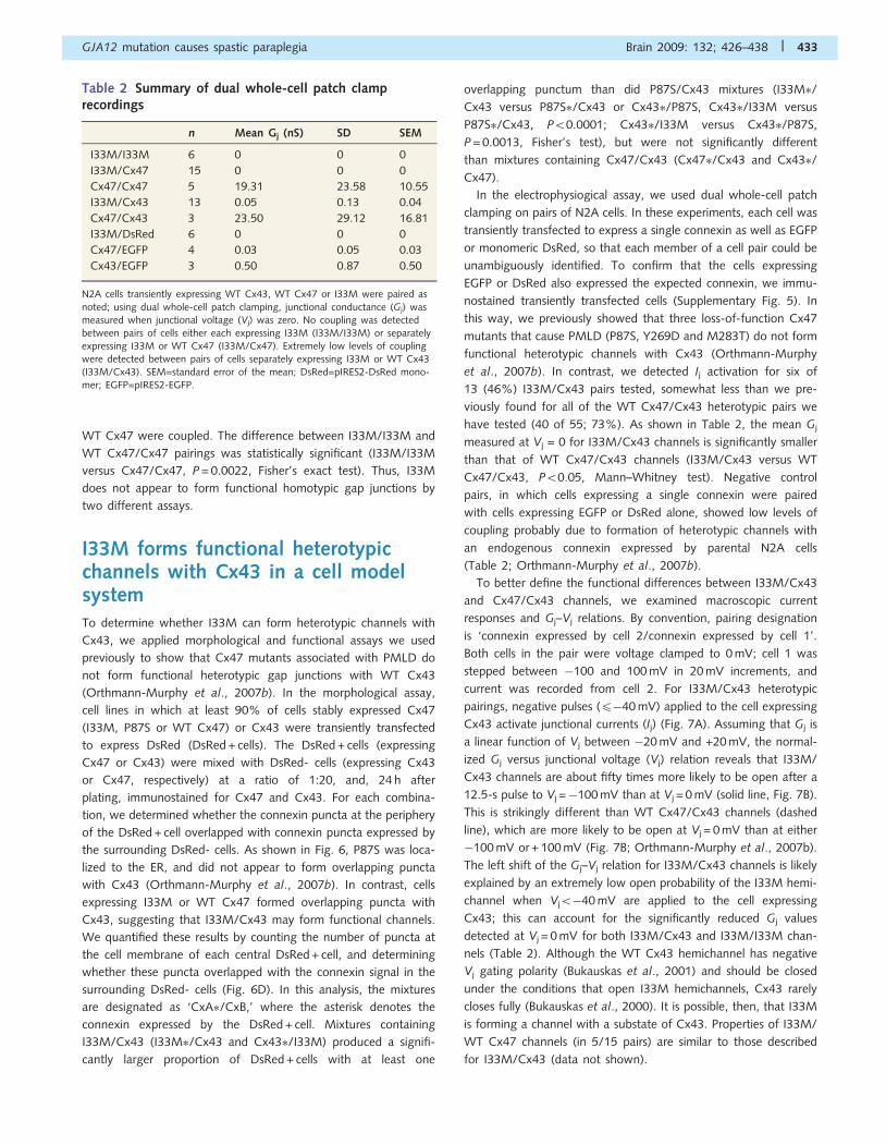
Table 2 Summary of dual whole-cell patch clamprecordings
n Mean Gj (nS) SD SEM
I33M/I33M 6 0 0 0
I33M/Cx47 15 0 0 0
Cx47/Cx47 5 19.31 23.58 10.55
I33M/Cx43 13 0.05 0.13 0.04
Cx47/Cx43 3 23.50 29.12 16.81
I33M/DsRed 6 0 0 0
Cx47/EGFP 4 0.03 0.05 0.03
Cx43/EGFP 3 0.50 0.87 0.50
N2A cells transiently expressing WT Cx43, WT Cx47 or I33M were paired as
noted; using dual whole-cell patch clamping, junctional conductance (Gj) wasmeasured when junctional voltage (Vj) was zero. No coupling was detectedbetween pairs of cells either each expressing I33M (I33M/I33M) or separatelyexpressing I33M or WT Cx47 (I33M/Cx47). Extremely low levels of couplingwere detected between pairs of cells separately expressing I33M or WT Cx43(I33M/Cx43). SEM=standard error of the mean; DsRed=pIRES2-DsRed mono-mer; EGFP=pIRES2-EGFP.
GJA12 mutation causes spastic paraplegia Brain 2009: 132; 426–438 | 433

Discussion
The I33M Cx47 mutant causeshereditary spastic paraplegiaThe three patients with a homozygous 99C4G mutation in
GJA12/GJC2 all developed HSP, characterized by a progressive
gait disorder during the first to fourth decades of life. The proband
exhibited an almost pure spastic paraplegia with mild ataxia and
dysarthria, and walked unaided at age 39. His brother had mod-
erately complicated spastic paraplegia, with mild ataxia and dysar-
thria, a few seizures and walked with unilateral support at age 36.
Their cousin had a more severe and complex phenotype, with
earlier onset and became wheelchair dependent at age 30. The
late onset, preserved ability to walk unaided until adulthood, and
Fig. 6 I33M/Cx43 pairings form gap junction plaques. (A–C) HeLa cells stably expressing Cx47 WT or one of the mutants (I33M or
P87S) or Cx43 were transiently transfected to express DsRed (DsRed + ) and mixed with cells expressing one of the connexins, but not
transfected with DsRed (DsRed�), in a ratio of 1:20, respectively. After 24 h, cells were immunostained as indicated and counterstained
for DAPI. One of the two possible pairings for each combination is illustrated. The DsRed + cell is pseudocoloured blue in the first and
third columns, and indicated by an asterisk in the fourth column. Note that I33M/Cx43 (B) pairings have overlapping puncta at the
border of the DsRed signal (arrowhead) similar to Cx47/Cx43 pairings (A), whereas P87S/Cx43 pairings (C) do not. (D) Quantitative
summary of three independent experiments such as illustrated in (A–C). The asterisk denotes the DsRed + cell. Each dot represents the
number of overlapping puncta determined for 1 DsRed + cell. In each column, the horizontal bar denotes the mean, the vertical bar
represents the 95% confidence interval, and the total number of DsRed + cells is shown in parentheses. Both pairings of I33M/Cx43
(I33M�/43 and 43�/I33M) have overlapping puncta similar to pairings of Cx47/Cx43 (47�/43 and 43�/47). Results for Cx47/Cx43
and P87S/Cx43 pairings are similar to those previously reported (Orthmann-Murphy et al., 2007b).
434 | Brain 2009: 132; 426–438 J. L. Orthmann-Murphy et al.

absence of nystagmus clearly distinguish these patients from the
more severe PMLD phenotype caused by other recessive muta-
tions in GJA12/GJC2 (Fig. 3). These PMLD patients typically pre-
sent by 12 months of age with nystagmus and impaired
psychomotor development, and rarely walk unaided in childhood
(Uhlenberg et al., 2004; Bugiani et al., 2006; Salviati et al., 2007;
Wolf et al., 2007; Henneke et al., 2008).
The diffuse white matter MR signal abnormalities and mildly
altered 1H MRSI metabolic profiles in the three patients homozy-
gous for the I33M mutation suggested a hypomyelinating leu-
koencephalopathy, which is consistent with oligodendrocyte
expression of Cx47. In our adults patients, the T2-signal abnorm-
alities were more subtle than those shown for previously reported
PMLD patients (caused by GJA12/GJC2 mutations), who ranged
in age from 5 months to 12 years (Uhlenberg et al., 2004; Bugiani
et al., 2006; Salviati et al., 2007; Wolf et al., 2007). Notably,
T2-signal hyperintensity for patients with the I33M mutation
was discrete and prominent in the corticospinal and spinothalamic
tracts (especially at the level of the pons). A similar involvement of
these tracts is evident in the MR images of spastic PMLD patients
reported by Wolf and colleagues (2007), whereas the corticospinal
tracts appeared to be relatively spared in PMLD patients with
other GJA12/GJC2 mutations (Bugiani et al., 2006). In our
patients, the involvement of the long white matter tracts may
correlate with the severity of spastic paraplegia. The mild decrease
in metabolite ratios (Cho/NAA and Cho/Cr) measured in our
patients, and previously reported for one of two PMLD patients
with GJA12/GJC2 mutations (Bugiani et al., 2006), are in agree-
ment with a recent study suggesting that hypomyelinating disor-
ders (including PMLD due to GJA12/GJC2 mutations) are
associated with normal or near-normal metabolite ratios, com-
pared to a large increase in choline or decrease in NAA typically
seen in actively demyelinating leukoencephalopathies (Bizzi et al.,
2008). Finally, like PMLD patients with GJA12/GJC2 mutations,
our patients exhibited central conduction slowing without electro-
physiological evidence for peripheral neuropathy.
Hereditary spastic paraplegias are a heterogeneous group of
genetic disorders characterized by progressive lower limb spas-
ticity (pure HSP) that may be associated with other neuro-
logical abnormalities (complicated HSP; Fink, 2006). We propose
that autosomal recessive mutations in GJA12/GJC2 that cause
HSP should be termed SPG44 (http://www.genenames.org).
The patients described here have a slowly progressive disease
consisting of a spastic paraplegia; the inclusion of minimal to
moderate ataxia and dysarthria, and seizures suggest that they
have a complicated HSP. Although some of the clinical findings
indicate that the pathophysiology is not limited to the longest CNS
axons, other forms of ‘complicated’ HSP have comparable, or
even more severe, findings (http://www.ncbi.nlm.nih.gov/sites/
entrez?db = OMIM). Similarly, MRI abnormalities in the white
matter, comparable to the ones we report, have been found in
one patient with SPG2 (Lee et al., 2004). Thus, GJA12/GJC2 is
only the second example (PLP1 is the other) of mutations that
cause HSP in a gene that is primarily expressed by oligodendro-
cytes; the other HSP-causing loci are thought to primarily affect
neurons/axons (Fink, 2006). Screening for GJA12/GJC2 muta-
tions, therefore, should be considered for patients and/or families
that present with a complicated HSP phenotype and hypomyeli-
nating leukoencephalopathic findings on MRI.
Like PLP1, mutations in GJA12/GJC2 appear to cause a spec-
trum of CNS white matter disease, including HSP and PMD/
PMLD. Based on the similar phenotypes caused by PLP1 and
GJA12/GJC2 mutations, one suspects that other causes of the
PMLD (and HSP with primarily white matter pathology) will turn
Fig. 7 Functional properties of I33M/Cx43 channels. N2A cells
were transiently transfected with a pIRES2-EGFP or a pIRES2-
DsRed bicistronic expression vector that also contained WT
Cx43 or I33M. After 24 h, red and green cell pairs were gen-
erated by mixing the transfected cells at a 1 to 1 ratio, and
assessed by dual whole-cell patch clamping 24–48 h later. (A)
These are representative current traces recorded from an I33M/
Cx43 pairing; both cells were voltage clamped to 0 mV and the
cell expressing WT Cx43 was stepped in 20 mV increments
from Vj =�100 to Vj = 100 mV, and junctional current (Ij) was
recorded from the cell expressing I33M. Note that the polarity
of Ij is opposite that of Vj. Current responses were only acti-
vated when a pulse less than or equal to �40 mV was applied
to the Cx43-expressing cell. Traces were filtered at 200 Hz. (B)
Average normalized Gj–Vj relations for heterotypic I33M/Cx43
channels (solid line) and WT Cx47/Cx43 channels (dashed line).
For I33M/Cx43 channels, the average Gj� SEM at each Vj
(filled triangles) was calculated from current traces such as
those shown in (A) and normalized to the value at –100 mV.
The WT Cx47/Cx43 channel trace is from Orthmann-Murphy
et al., 2007b. Note that I33M/Cx43 channels are only open
when Vj is less than �40 mV, and are closed when WT Cx47/
Cx43 channels are open (Vj = 0 mV). Each point in the Gj–Vj
plot is the average of data from three independent
experiments.
GJA12 mutation causes spastic paraplegia Brain 2009: 132; 426–438 | 435
out to be caused by mutations in genes that primarily affect oli-
godendrocytes. One example of this may be mutations of HSPD1
(the gene encoding the mitochondrial chaperonin, Hsp60), which
cause either uncomplicated autosomal dominant SPG13 (Hansen
et al., 2002) or an autosomal recessive lethal disease similar to
connatal PMD (Magen et al., 2008).
The molecular pathogenesis of HSPand PMLD due to mutations inGJA12/GJC2Because the patients with homozygous I33M mutations have a
milder phenotype than do patients with PMLD, we predicted
that I33M would have a partial loss-of-function, as compared to
complete loss-of-function we found for Cx47 mutants that cause
PMLD (Orthmann-Murphy et al., 2007a, b). Furthermore,
because oligodendrocytes form gap junctions with adjacent astro-
cytes, we predicted that I33M would still form functional, but
likely altered, channels with Cx43. The finding that I33M
formed gap junction plaques with itself and with Cx43 is consis-
tent with this hypothesis, and demonstrates that I33M oligo-
merizes into hemichannels that traffic to the cell surface (Kumar
and Gilula, 1996), compared with the ER-retained Cx47 mutants
which cause PMLD. The failure of I33M to transfer small mole-
cules such as Lucifer Yellow or neurobiotin (in scrape loading
assays), or to form detectable homotypic channels (by dual
whole-cell patch clamping), argues against this hypothesis. Both
the failure to detect I33M/I33M dye coupling or currents and the
alterations in the I33M/Cx43 Gj–Vj relation are likely due to a shift
of the open probability of the I33M hemichannel as a function of
voltage, such that its open probability increases only when a large
voltage difference (Vj4�40 mV) is applied with the respect to the
cell expressing Cx43. Some disease-associated Cx32 mutants show
similar alterations in voltage dependence and are also thought to
lead to loss-of-function (Oh et al., 1997; Abrams et al., 2001).
Because large voltage gradients that open I33M/Cx43 channels
probably do not occur across O/A junctions, the I33M mutant
should cause loss-of-function of these channels. Large O/A gra-
dients would be particularly improbable if these cells are well
coupled by Cx30/Cx32 channels (Orthmann-Murphy et al.,
2007b, 2008). Consequently, similar to patients with GJA12/
GJC2 mutations that cause PMLD, it is unlikely that Cx47/Cx43
gap junctions contribute to O/A coupling in patients with homo-
zygous I33M mutations.
If the I33M mutation completely disrupts gap junction coupling
via Cx47/Cx43 channels like the Cx47 mutants that cause PMLD,
then another mechanism must account for the milder phenotype
in our three patients. One possibility is that Cx47 has functions in
oligodendrocytes beyond forming functional O/A channels.
Because Cx47 has a PDZ binding domain [and at least one binding
partner, ZO-1; (Li et al., 2004)], it is possible that I33M, in parti-
cular, retains some important function (such as binding ZO-1) that
requires proper localization at the cell surface. Along these lines, it
is possible that like some other connexins (Prochnow and
Dermietzel, 2008), Cx47 is required for cell adhesion, as shown
for Cx26 and Cx43 in neuronal migration in developing cortex
(Elias et al., 2007), and Cx32 and Cx43 in an in vitro aggregation
assay (Cotrina et al., 2008). Another possibility is that the reces-
sive Cx47 mutants that cause PMLD (P87S, Y269D, M283T) actu-
ally have dominant effects (that the I33M mutant does not have)
that contribute to the more severe phenotype of PMLD. Because
the P87S, Y269D and M283T mutants are mostly found in the ER,
it seems unlikely that they would form an abnormal hemichannel
on the cell membrane (Richardson et al., 2004; Dobrowolski et al.,
2007). Alternatively, because they accumulate in the ER, these
mutants could induce an unfolded protein response with deleter-
ious effects, but we found no evidence for this possibility in a cell
model system (Orthmann-Murphy et al., 2007a).
Finally, it is possible that Cx47 mutants associated with more
severe phenotypes result in more axonal degeneration, indepen-
dent of their effects on myelin. In the peripheral nervous system,
mutations in genes expressed by Schwann cells (such as GJB1,
PMP22 and MPZ) cause demyelinating neuropathy; although the
demyelination is Schwann cell autonomous, clinical disability cor-
relates with the degree of axonal damage (Nave et al., 2007;
Scherer and Wrabetz, 2008). The enlarged ventricles in Patient
III-5 (who was the most severely affected) are likely a result of
axonal loss; such secondary axonal degeneration probably contri-
butes to progressive neurological deterioration in PMD (Inoue,
2005; Garbern, 2007), and multiple sclerosis (Bjartmar et al.,
1999). In spite of its importance, it is unclear how axons are
damaged in any of these demyelinating disorders; in the case of
Cx47 mutants, it is possible that disrupted O/A coupling impedes
K+ buffering mediated by glial gap junctions (Menichella et al.,
2006).
Supplementary materialSupplementary material is available at Brain online.
FundingNational Institutes of Health (NS050345 and NS050705 to C.K.A.)
(NS55284 to S.S.S.); the National Multiple Sclerosis Society (to
S.S.S.); Mariani Foundation (to G.U. and M.Z.).
ReferencesAbrams CK, Freidin MM, Verselis VK, Bennett MVL, Bargiello TA.
Functional alterations in gap junction channels formed by mutant
forms of connexin 32: evidence for loss of function as a pathogenic
mechanism in the X-linked form of Charcot-Marie-Tooth disease. Brain
Res 2001; 900: 9–25.
Altevogt BM, Kleopa KA, Postma FR, Scherer SS, Paul DL. Cx29 is
uniquely distributed within myelinating glial cells of the central and
peripheral nervous systems. J Neurosci 2002; 22: 6458–70.Altevogt BM, Paul DL. Four classes of intercellular channels between glial
cells in the CNS. J Neurosci 2004; 24: 4313–23.Bizzi A, Castelli G, Bugiani M, Barker PB, Herskovits EH, Danesi U, et al.
Classification of childhood white matter disorders using proton MR
spectroscopic imaging. Am J Neuroradiol 2008; 29: 1270–5.
Bjartmar C, Yin XH, Trapp BD. Axonal pathology in myelin disorders. J
Neurocytol 1999; 28: 383–95.
436 | Brain 2009: 132; 426–438 J. L. Orthmann-Murphy et al.

Bruzzone R, White TW, Paul DL. Connections with connexins: the mole-
cular basis of direct intercellular signaling. Eur J Biochem 1996; 238:
1–27.
Bugiani M, Al Shahwan S, Lamantea E, Bizzi A, Bakhsh E, Moroni I, et al.
GJA12 mutations in children with recessive hypomyelinating leukoen-
cephalopathy. Neurology 2006; 67: 273–9.
Bukauskas FF, Bukauskiene A, Bennett MVL, Verselis VK. Gating proper-
ties of gap junction channels assembled from connexin43 and con-
nexin43 fused with green fluorescent protein. Biophys J 2001; 81:
137–52.
Bukauskas FF, Jordan K, Bukauskiene A, Bennett MVL, Lampe PD,
Laird DW, et al. Clustering of connexin 43-enhanced green fluorescent
protein gap junction channels and functional coupling in living cells.
Proc Natl Acad Sci USA 2000; 97: 2556–61.
Cotrina ML, Lin JHC, Nedergaard M. Adhesive properties of connexin
hemichannels. Glia 2008; 56: 1791–8.
Dahl E, Manthey D, Chen Y, Schwarz HJ, Chang YS, Lalley PA, et al.
Molecular cloning and functional expression of mouse connexin-30,
a gap junction gene highly expressed in adult brain and skin. J Biol
Chem 1996; 271: 17903–10.
Dermietzel R, Traub O, Hwang TK, Beyer E, Bennett MVL, Spray DC,
et al. Differential expression of three gap junction proteins in
developing and mature brain tissues. Proc Natl Acad Sci USA 1989;
86: 10148–52.
Dobrowolski R, Sommershof A, Willecke K. Some oculodentodigital
dysplasia-associated Cx43 mutations cause increased hemichannel
activity in addition to deficient gap junction channels. J Membrane
Biol 2007; 219: 9–17.
el-Fouly MH, Trosko JE, Chang C. Scrape-loading and dye transfer. A
rapid and simple technique to study gap junctional intercellular com-
munication. Exp Cell Res 1987; 168: 422–30.
Elias LAB, Wang DD, Kriegstein AR. Gap junction adhesion is
necessary for radial migration in the neocortex. Nature 2007; 448:
901–7.
Fink JK. Hereditary spastic paraplegia. Curr Neurol Neurosci Rep 2006; 6:
65–76.
Garbern J, Cambi F, Shy M, Kamholz J. The molecular pathogenesis of
Pelizaeus-Merzbacher disease. Arch Neurol 1999; 56: 1210–4.
Garbern JY. Pelizaeus-Merzbacher disease: genetic and cellular patho-
genesis. Cell Mol Life Sci 2007; 64: 50–65.
Hansen JJ, Durr A, Cournu-Rebeix I, Georgopoulos C, Ang D,
Nielsen MN, et al. Hereditary spastic paraplegia SPG13 is associated
with a mutation in the gene encoding the mitochondrial chaperonin
Hsp60. Am J Hum Genet 2002; 70: 1328–32.
Henneke M, Combes P, Diekmann S, Bertini E, Brockmann K, Burlina AP,
et al. GJA12 mutations are a rare cause of Pelizaeus-Merzbacher-like
disease. Neurology 2008; 70: 748–54.
Hudson LD, Garbern JY, Kamholz JA. Pelizaeus-Merzbacher disease. In:
Lazzarini RA, editor. Myelin biology and disorders. Vol 2. San Diego:
Elsevier; 2004. p. 867–85.
Inoue K. PLP1-related inherited dysmyelinating disorders: Pelizaeus-
Merzbacher disease and spastic paraplegia type 2. Neurogenetics
2005; 6: 1–16.
Kamasawa N, Sik A, Morita M, Yasumura T, Davidson KGV, Nagy JI,
et al. Connexin-47 and connexin-32 in gap junctions of oligodendro-
cyte somata, myelin sheaths, paranodal loops and Schmidt-Lanterman
incisures: Implications for ionic homeostasis and potassium siphoning.
Neuroscience 2005; 136: 65–86.
Kleopa KA, Orthmann JL, Enriquez A, Paul DL, Scherer SS. Unique dis-
tributions of the gap junction proteins connexin29, connexin32, and
connexin47 in oligodendrocytes. Glia 2004; 47: 346–57.
Kumar NM, Gilula NB. The gap junction communication channel. Cell
1996; 84: 381–9.
Kunzelmann P, Schroder W, Traub O, Steinhauser C, Dermietzel R,
Willecke K. Late onset and increasing expression of the gap junction
protein connexin30 in adult murine brain and long-term cultured
astrocytes. Glia 1999; 25: 111–9.
Lee ES, Moon HK, Park YH, Garbern J, Hobson GM. A case of compli-
cated spastic paraplegia 2 due to a point mutation in the proteolipid
protein 1 gene. J Neurol Sci 2004; 224: 83–7.
Li J, Hertzberg EL, Nagy JI. Connexin32 in oligodendrocytes and associa-
tion with myelinated fibers in mouse and rat brain. J Comp Neurol
1997; 379: 571–91.
Li X, Ionescu AV, Lynn BD, Lu S, Kamasawa N, Morita M, et al.
Connexin47, connexin29 and connexin32 co-expression in oligoden-
drocytes and Cx47 association with zonula occludens-1 (ZO-1) in
mouse brain. Neuroscience 2004; 126: 611–30.
Li X, Lynn BD, Olson C, Meier C, Davidson KGV, Yasumura T, et al.
Connexin29 expression, immunocytochemistry and freeze-fracture
replica immunogold labelling (FRIL) in sciatic nerve. Eur J Neurosci
2002; 16: 795–806.
Magen D, Georgopoulos C, Bross P, Ang D, Segev Y, Goldsher D, et al.
Mitochondrial Hsp60 chaperonopathy causes an autosomal-recessive
neurodegenerative disorder linked to brain hypomyelination and leu-
kodystrophy. Am J Hum Genet 2008; 83: 30–42.
Menichella DM, Goodenough DA, Sirkowski E, Scherer SS, Paul DL.
Connexins are critical for normal myelination in the central nervous
system. J Neurosci 2003; 23: 5963–73.
Menichella DM, Majdan M, Awatramani R, Goodenough DA,
Sirkowski E, Scherer SS, et al. Genetic and physiological evidence
that oligodendrocyte gap junctions contribute to spatial buffering of
potassium released during neuronal activity. J Neurosci 2006; 26:
10984–91.
Micevych PE, Abelson L. Distribution of mRNAs coding for liver and
heart gap junction proteins in the rat central nervous system. J
Comp Neurol 1991; 305: 96–118.
Mugnaini E. Cell junctions of astrocytes, ependyma, and related cells in
the mammalian central nervous system, with emphasis on the hypoth-
esis of a generalized functional syncytium of supporting cells. In:
Federoff S, Vernadakis A, editors. Astrocytes. Vol. 1. Orlando:
Academic Press (Elsevier); 1986. p. 329–71.Nagy JI, Ionescu AV, Lynn BD, Rash JE. Connexin29 and connexin32 at
oligodendrocyte and astrocyte gap junctions and in myelin of the
mouse central nervous system. J Comp Neurol 2003a; 464: 356–70.Nagy JI, Ionescu AV, Lynn BD, Rash JE. Coupling of astrocyte connexins
Cx26, Cx30, Cx43 to oligodendrocyte Cx29, Cx32, Cx47: implications
from normal and connexin32 knockout mice. Glia 2003b; 44: 205–18.Nagy JI, Li XB, Rempel J, Stelmack G, Patel D, Staines WA, et al.
Connexin26 in adult rodent central nervous system: demonstration
at astrocytic gap junctions and colocalization with connexin30 and
connexin43. J Comp Neurol 2001; 441: 302–23.Nagy JI, Ochalski PAY, Li J, Hertzberg EL. Evidence for the co-localiza-
tion of another connexin with connexin-43 at astrocytic gap junctions
in rat brain. Neuroscience 1997; 78: 533–48.Nagy JI, Patel D, Ochalski PAY, Stelmack GL. Connexin30 in rodent, cat
and human brain: selective expression in gray matter astrocytes, co-
localization with connexin43 at gap junctions and late developmental
appearance. Neuroscience 1999; 88: 447–68.
Nave K-A, Boespflug-Tanguy O. X-linked developmental defects of
myelin formation: from mouse mutants to human genetic diseases.
Neuroscientist 1996; 2: 33–43.
Nave KA, Sereda MW, Ehrenreich H. Mechanisms of disease: inherited
demyelinating neuropathies - from basic to clinical research. Nat Clin
Pract Neurol 2007; 3: 453–64.
Odermatt B, Wellershaus K, Wallraff A, Seifert G, Degen G, Euwens C,
et al. Connexin 47 (Cx47)-deficient mice with enhanced green fluor-
escent protein reporter gene reveal predominant oligodendrocytic
expression of Cx47 and display vacuolized myelin in the CNS. J
Neurosci 2003; 23: 4549–59.
Oh S, Ri Y, Bennett MVL, Trexler EB, Verselis VK, Bargiello TA. Changes
in permeability caused by connexin 32 mutations underlie X-linked
Charcot-Marie-Tooth disease. Neuron 1997; 19: 927–38.
Orthmann-Murphy JL, Abrams CK, Scherer SS. Gap junctions couple
astrocytes and oligodendrocytes. J Mol Neurosci 2008; 35: 101–16.
GJA12 mutation causes spastic paraplegia Brain 2009: 132; 426–438 | 437

Orthmann-Murphy JL, Enriquez AD, Abrams CK, Scherer SS. Loss-of-function GJA12/Connexin47 mutations cause Pelizaeus-Merzbacher-
like disease. Mol Cell Neurosci 2007a; 34: 629–41.
Orthmann-Murphy JL, Freidin M, Fischer E, Scherer SS, Abrams CK. Two
distinct heterotypic channels mediate gap junction coupling betweenastrocyte and oligodendrocyte connexins. J Neurosci 2007b; 27:
13949–57.
Prochnow N, Dermietzel R. Connexons and cell adhesion: a romantic
phase. Histochemistry Cell Biol 2008; 130: 71–7.Rash JE, Yasumura T, Dudek FE, Nagy JI. Cell-specific expression of
connexins and evidence of restricted gap junctional coupling between
glial cells and between neurons. J Neurosci 2001; 21: 1983–2000.Richardson RR, Donnai D, Meire F, Dixon MJ. Expression of Gja1 corre-
lates with the phenotype observed in oculodentodigital syndrome/type
III syndactyly. J Med Genet 2004; 41: 60–7.
Salviati L, Trevisson E, Baldoin MC, Toldo I, Sartori S, Calderone M, et al.A novel deletion in the GJA12 gene causes Pelizaeus-Merzbacher-like
disease. Neurogenetics 2007; 8: 57–60.
Scherer SS, Deschenes SM, Xu Y-T, Grinspan JB, Fischbeck KH, Paul DL.
Connexin32 is a myelin-related protein in the PNS and CNS. J Neurosci1995; 15: 8281–94.
Scherer SS, Wrabetz L. Molecular mechanisms of inherited demyelinating
neuropathies. Glia 2008; 56: 1578–89.
Soher B, van Zijl P, Duyn J, Barker PB. Quantitative proton MR spectro-scopic imaging of the human brain. Magn Reson Med 1996; 35: 356–63.
Swenson KI, Jordan JR, Beyer EC, Paul DL. Formation of gap junctions
by expression of connexins in Xenopus oocyte pairs. Cell 1989; 57:
145–55.
Trosko JE, Change CC, Wilson MR, Upham B, Hayashi T, Wade M.
Gap junctions and the regulation of cellular functions of stem cells
during development and differentiation. Methods 2000; 20: 245–64.
Uhlenberg B, Schuelke M, Ruschendorf F, Ruf N, Kaindl AM,
Henneke M, et al. Mutations in the gene encoding gap junction
protein alpha 12 (connexin 46.6) cause Pelizaeus-Merzbacher-like
disease. Amer J Hum Genet 2004; 75: 251–60.
Werner R, Levine E, Rabadan-Diehl C, Dahl G. Formation of hybrid cell-
cell channels. Proc Natl Acad Sci USA 1989; 86: 5380–4.
Willecke K, Eiberger J, Degen J, Eckardt D, Romualdi A, Guldenagel M,
et al. Structural and functional diversity of connexin genes in the
mouse and human genome. Biol Chem 2002; 383: 725–37.
Wolf NI, Cundall M, Rutland P, Rosser E, Surtees R, Benton S, et al.
Frameshift mutation in GJA12 leading to nystagmus, spastic ataxia and
CNS dys-/demyelination. Neurogenetics 2007; 8: 39–44.
Yamamoto T, Ochalski A, Hertzberg EL, Nagy JI. LM and EM immuno-
localization of the gap junctional protein connexin 43 in rat brain.
Brain Res 1990; 508: 313–9.
Yeager M, Nicholson BJ. Structure of gap junction intercellular channels.
Curr Opin Struct Biol 1996; 6: 183–92.
438 | Brain 2009: 132; 426–438 J. L. Orthmann-Murphy et al.


