1
BRIGHTFIELD MICROSCOPY FOR THE DIAGNOSIS OF STDs
By Sally S. Cherry, MT(ASCP)Laboratory Consultant & Instructor
Cherry Consulting Network
BRIGHTFIELD MICROSCOPY FOR THE DIAGNOSIS OF STDs
MICROSCOPIC DIAGNOSIS OF TRICHOMONIASIS, CANDIDIASIS & BACTERIAL VAGINOSIS
Please note that written permission from the author must be obtained before using, or copying this PowerPoint laboratory presentation.
Objective:The STD laboratory presentation is designed to provide the knowledge and skills to properly perform saline and potassium hydroxide (KOH) wet preparations used in the identification of:
Clue Cells (Bacterial vaginosis)Trichomonas vaginalis
Candida species

2
Recommended EquipmentBrightfield Microscope
• Eyepieces (Oculars)
• Body Tube
• Arm• Objectives (10X-40x-100x)
• Condenser, Brightfield
• Iris Diaphragm Lever
• Stage
• Fine Focus Adjustment Knob
• Coarse Focus Adjustment Knob
• Light (Spare Light Bulb)
• Base
Recommended SuppliesExamination Room
Specimen Collection
• Gloves
• Swabs, Cotton, or Dacron
• pH Paper
• Gauze (Squares)
• Microscope Slides
• Cover Slips, Glass
• Saline Solution
• Potassium Hydroxide (10%)
Saline Wet PreparationsSupplies Needed
• Latex Gloves
• Sterile Pipette or dropper
• Small Test Tube - 12 x 75 mm
• Normal Saline (0.5 - 1.0 ml)
• Tube label, or ID marker
3
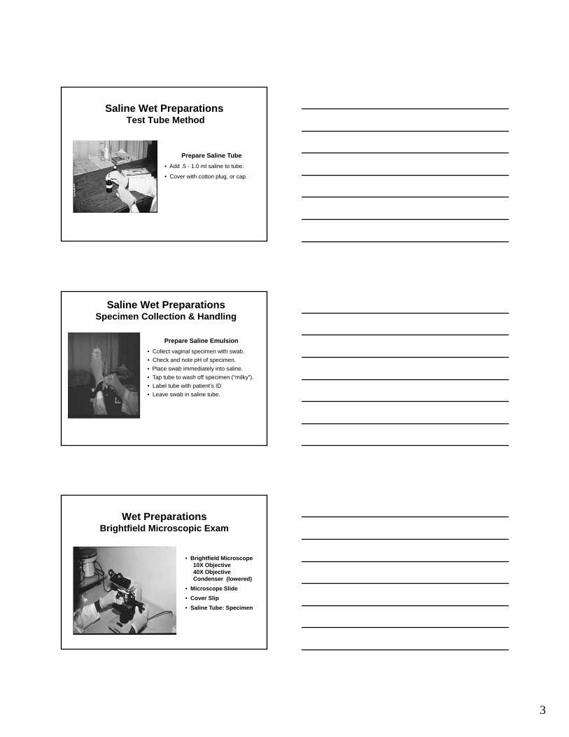
Saline Wet PreparationsTest Tube Method
Prepare Saline Tube
• Add .5 - 1.0 ml saline to tube.
• Cover with cotton plug, or cap.
Saline Wet PreparationsSpecimen Collection & Handling
Prepare Saline Emulsion
• Collect vaginal specimen with swab.
• Check and note pH of specimen.
• Place swab immediately into saline.
• Tap tube to wash off specimen (“milky”).
• Label tube with patient’s ID
• Leave swab in saline tube.
Wet PreparationsBrightfield Microscopic Exam
• Brightfield Microscope10X Objective40X Objective Condenser (lowered)
• Microscope Slide
• Cover Slip
• Saline Tube: Specimen
4
Saline Wet PreparationsExamine Specimen for:
Trichomonas vaginalis
Clue Cells (Bacterial vaginosis)
Yeast Cells and Hyphae
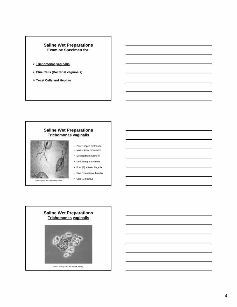
Saline Wet PreparationsTrichomonas vaginalis
• Pear-shaped protozoan
• Motile; jerky movement
• Directional movement
• Undulating membrane
• Four (4) anterior flagella
• One (1) posterior flagella
• One (1) nucleus(Illustration of Trichomonas vaginalis)
Saline Wet PreparationsTrichomonas vaginalis
(Note: Motility can not shown here.)
5
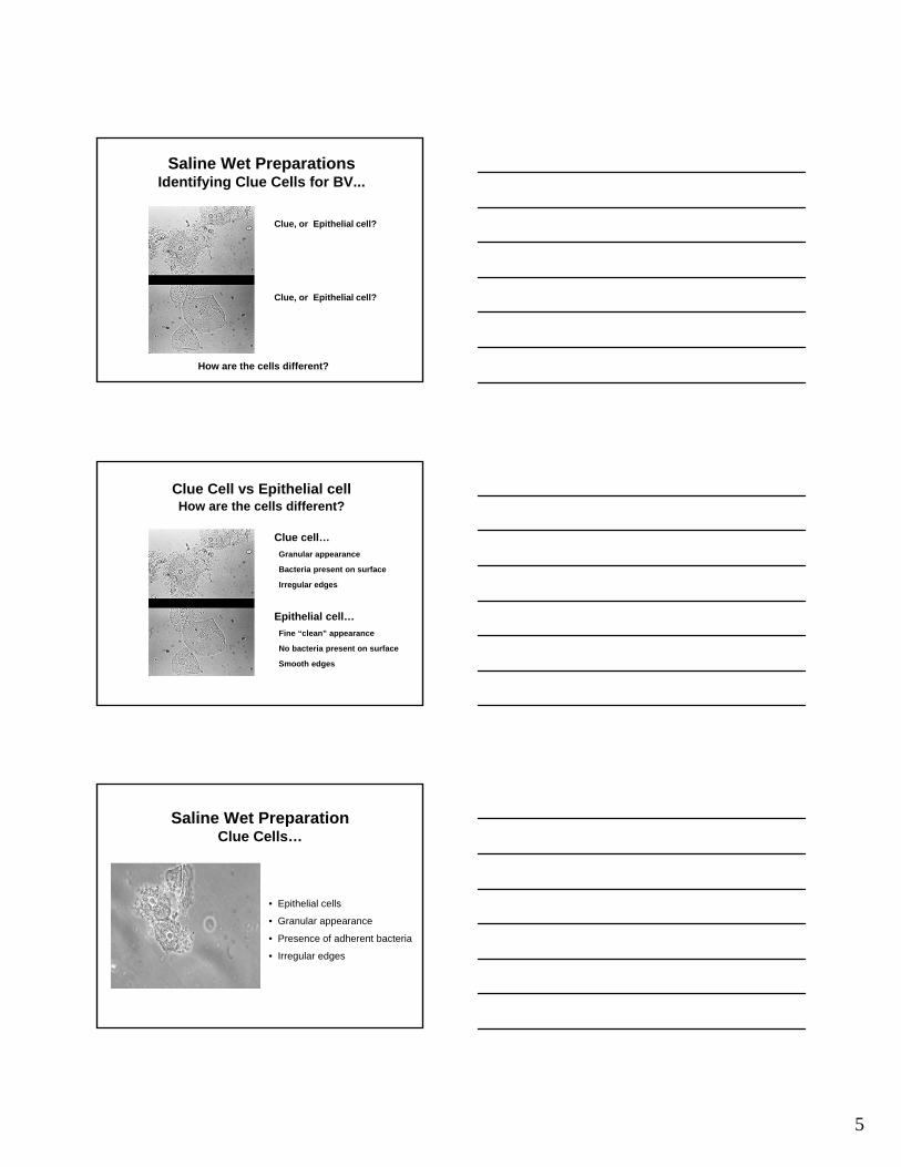
Saline Wet PreparationsIdentifying Clue Cells for BV...
Clue, or Epithelial cell?
How are the cells different?
Clue, or Epithelial cell?
Clue Cell vs Epithelial cellHow are the cells different?
Epithelial cell…
Fine “clean” appearance
No bacteria present on surface
Smooth edges
Clue cell…
Granular appearance
Bacteria present on surface
Irregular edges
Saline Wet PreparationClue Cells…
• Epithelial cells
• Granular appearance
• Presence of adherent bacteria
• Irregular edges
6
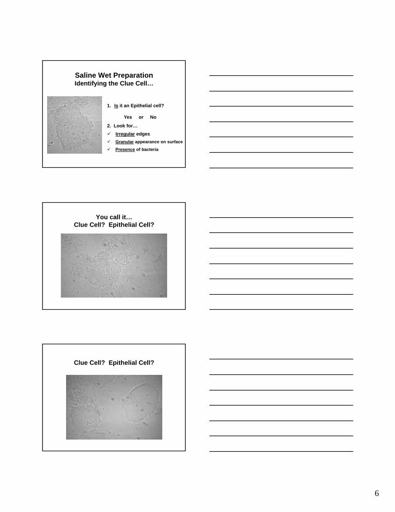
Saline Wet PreparationIdentifying the Clue Cell…
1. Is it an Epithelial cell?
Yes or No
2. Look for…
Irregular edges
Granular appearance on surface
Presence of bacteria
You call it… Clue Cell? Epithelial Cell?
Clue Cell? Epithelial Cell?

7
Clue Cell(Gram-Stained Specimen)
Gram-negative adherent bacteria present.
(Gram-positive bacteria may also be present.)
Troubleshooting: Problem #1
1. What do you observe in this specimen?
2. How would you eliminate this problem?
Troubleshooting: Solution #1
Observation: Oil Droplets
Recommended Action:
• Check cover slip for oil
• Check objective for oil
• Clean 40X objective
• Prepare and examine a second specimen
8
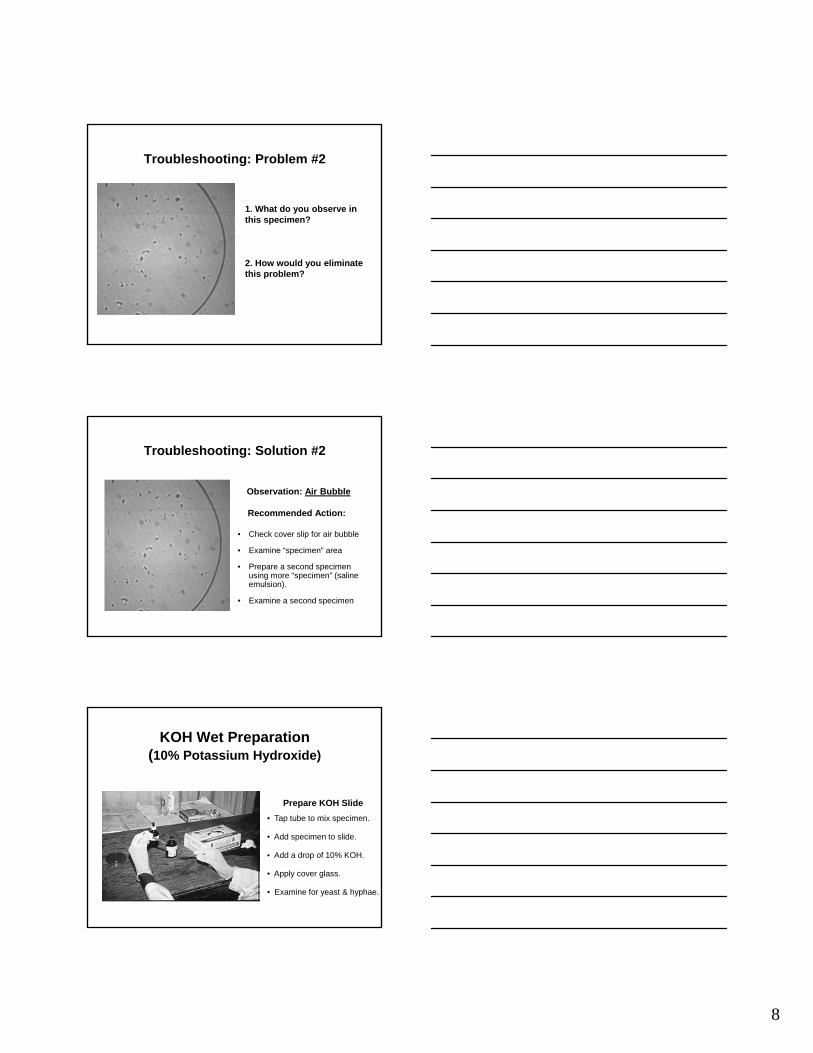
Troubleshooting: Problem #2
1. What do you observe in this specimen?
2. How would you eliminate this problem?
Troubleshooting: Solution #2
Observation: Air Bubble
Recommended Action:
• Check cover slip for air bubble
• Examine “specimen” area
• Prepare a second specimen using more “specimen” (saline emulsion).
• Examine a second specimen
KOH Wet Preparation(10% Potassium Hydroxide)
Prepare KOH Slide
• Tap tube to mix specimen.
• Add specimen to slide.
• Add a drop of 10% KOH.
• Apply cover glass.
• Examine for yeast & hyphae.

9
KOH Wet PreparationCandida albicans
• Oval Budding Yeast Cells
• Branching Hyphae
KOH Wet PreparationBudding Yeast Cells
Branching Hyphae
10
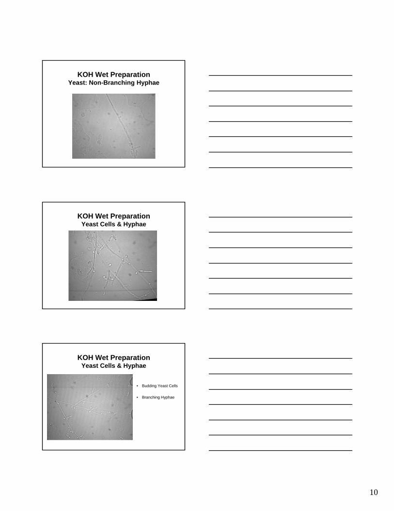
KOH Wet PreparationYeast: Non-Branching Hyphae
KOH Wet PreparationYeast Cells & Hyphae
KOH Wet PreparationYeast Cells & Hyphae
• Budding Yeast Cells
• Branching Hyphae
11
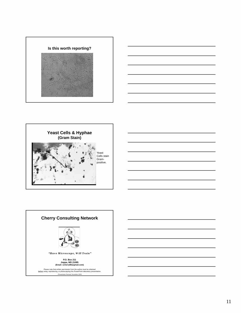
Is this worth reporting?
Yeast Cells & Hyphae(Gram Stain)
Yeast Cells stain Gram-positive.
Cherry Consulting Network
P.O. Box 211Joppa, MD 21085
(Email: [email protected])
Please note that written permission from the author must be obtained before using, reproducing, or photocopying this PowerPoint laboratory presentation.
(Presentation Revised: November 2006)
“Have Microscope, Will Train”


