
CALCIFIC UREMIC ARTERIOLOPATHY
10.28.14
Sushma Bhusal Renal Fellow

CASE • 45 Hispanic Female
• Chief Complaints: • Confusion x 1 day • Fatigue, sore throat, painful oral ulcerations x1 week
• Increased bilateral LE swelling x 1-2 weeks • Worsening emesis, poor appetite, lethargy x 2 weeks

CASE: HPI
Well known to renal clinic since 2011, h/o CKD due to diabetic nephropathy (with diabetic retinopathy) and a question of PKD Last seen in clinic in March 2014 AVF created in April 2014, patient had gone to Mexico after
that Symptoms started in Mexico, present for about a month

CASE: PMH/ PSH
• CKD Stage 5 • DM2 X 12 years • HTN • PKD • Hypothyroidism • Morbid Obesity • AVF creation April 2014 • Cholecystectomy, tubal ligation

MEDICATIONS
• Amlodipine 10 mg po d • Synthroid 125 mcg po d • HCTZ 25 mg po d • Lantus 10 units bid sc • ASA 81 mg po d

HISTORY
•Family: Father and 2 daughters with PKD, no one with ESRD
•Social : no toxic habits

PHYSICAL EXAM
• Vitals: T 96.3, 83, 18, 134/53, 100% RA
• Gen: Somnolent , arousable and responding to commands
• HEENT: tongue coated with whitish, shallow ulcerations lateral edges of tongue, bleeding +
• CVS: S1S2+, RRR, 3/6 SM LUSB/LLSB, no rubs
• Resp: Transmitted upper airway sounds, otherwise clear
• Abd: Obese, soft, non tender, non distended, +BS
• Ext: varicosities, edema 1+ to b/l shins, tenderness over LE (legs and thighs bilateral, subcut nodules), L AVF palpable thrill and bruit

LABS
20.2 19.8
6.5 499
< 10
89
5.5
123
10.9
238
LFTs: ALT: 26, AST: 17, AlkP: 164, Tbili: 0.1, Dbili: < 0.1, TP: 6.6, Alb: 3.1 Rapid Strep test: Negative
Urine Studies: UA: Bld 3+, pH: 7.5, Protein: > 300 mg / dL, WBC: Packed, RBC: 5 – 10, Bacteria: Many Urine Prot / Cr: 2 g / g
Phos: 13.6, iPTH: 315.4 Vit D: 15
Ca: 6.3
Glu: 109

IMAGING • US Abdomen
• RK 16.2, LK 13.3 cm • Increased echogenicity compatible with intrinsic renal
disease • Both kidneys with multiple, numerous cysts, largest on
RK upper pole 3x2.7x3.1, LK mid to lower pole 2.5x2.3x2.5 cm
• No hydronephrosis
• CXR: clear lungs
HOSPITAL COURSE
• Patient was started on Hemodialysis • Developed progressive pain over LE with violaceous tender plaques which later ulcerated

Day 7


DIFFERENTIAL DIAGNOSIS
• Calciphylaxis • Cellulitis • Panniculitis • Erythema Nodosum • Cryoglobulinemia

ADDITIONAL LABS
• HIV neg • ANA, RF, Hep B, C panel, APLA panel, Cryo: negative
• dsDNA, anti Smith, anti RNP: all negative

HOSPITAL COURSE
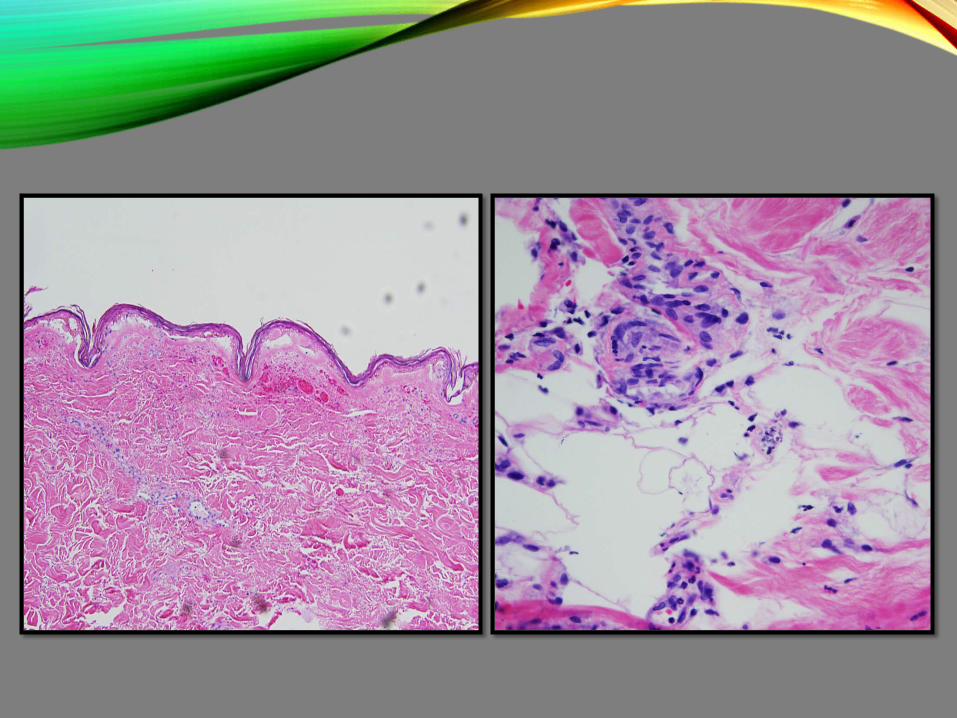
• Biopsy taken of a lesion over R medial thigh
• LLE X-ray

R Medial thigh lesion biopsy

R Medial thigh lesion biopsy


MANAGEMENT
• Started on Sodium thiosulfate • D/C Ca containing binders, vitamin D • Frequent HD • Sevelamer, Cincalcet • Local wound care with Silver sulfadiazine • Pain control

HOSPITAL COURSE
• Progressively worsening ulcers • Development of similar lesions over lower abdomen


FURTHER HOSPITAL COURSE • Also complicated by septicemia and AMS
• Blood cultures with Coagulase negative staph
• Started on antibiotics
• AMS improved after 2 days
• Progression of ulcers

CALCIFIC UREMIC
ARTERIOLOPATHY

HISTORY
• Rare disorder with mortality around 80%
• 1962: Hans Selye coined “Calciphylaxis” to describe skin necrosis in rats exposed to PTH, Vit D and associated cutaneous calcification
• Histopathology different from syndrome described in uremic patients, no vascular calcifications
• Coates suggested the term Calcific Uremic Arteriolopathy P. Yerram at al. The Ochsner Journal 1 4: 201 4 Branderburg et al Ped Nephrol Jan 201 4

EPIDEMIOLOGY • Exact prevalence unknown
• Small studies report 4% prevalence in HD patients and 1.3-4.5 per patient years in ESRD
• Several registries: Germany, UK, Kansas University Med Center
P. Yerram at al. The Ochsner Journal 1 4: 201 4 Registries
PATHOPHYSIOLOGY
• Previously thought to be a passive process described under “Metastatic Calcification”
• Vascular calcification now known to result from active cellular processes and not just passive mineral precipitation
• Cutaneous arterial calcification and vascular thrombosis both required to produce clinical lesions
Weenig et al J Am Acad Dermat 2008 Jablonski et al Hemodialysis Int Oct 2013 Cuo-Cheng et al Scientific World Journal July 2014

PATHOPHYSIOLOGY
Active Process Transformation of VSMCs to Osteogenic/Chrondrogenic
Phenotype
Passive Process Mineral precipitation of ECF
surrounding VSMCs
• Weenig et al J Am Acad Dermat 2008 • Jablonski et al Hemodialysis Int Oct 2013 • Cuo-Cheng et al Scientific World Journal July 2014

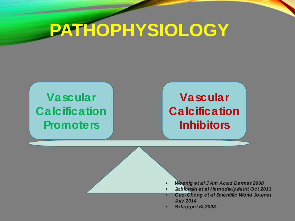
PATHOPHYSIOLOGY
Vascular Calcification
Promoters
Vascular Calcification
Inhibitors
• Weenig et al J Am Acad Dermat 2008 • Jablonski et al Hemodialysis Int Oct 2013 • Cuo-Cheng et al Scientific World Journal
July 2014 • Schoppet KI 2008

Promoters
Ca/P04 homeostasis dysregulation in CKD
FGF23/Klotho BMP2/RANK/RANKL Inflammation/ROS
Inhibitors
• MGP • Fetuin A • Pyrophosphate • OPG • BMP 7
Adapted from Kuo-Cheng et al Scientific World Journal July 2014
PATHOPHYSIOLOGY
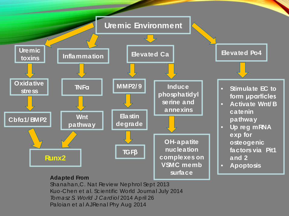
Uremic toxins
Oxidative stress
Cbfα1/BMP2
Uremic Environment
Inflammation
TNFα
Wnt pathway
Runx2
Elevated Ca Elevated Po4
MMP2/9
Elastin degrade
TGFβ
Induce phosphatidyl
serine and annexins
OH-apatite nucleation
complexes on VSMC memb
surface Adapted From Shanahan,C. Nat Review Nephrol Sept 2013 Kuo-Chen et al. Scientific World Journal July 2014 Tomasz S. World J Cardiol 2014 April 26 Paloian et al AJRenal Phy Aug 2014
• Stimulate EC to form μparticles
• Activate Wnt/B catenin pathway
• Up reg mRNA exp for osteogenic factors via Pit1 and 2
• Apoptosis

FGF 23 AND KLOTHO
FGF 23 (Phosphatonin): • Synthesized by osteocytes • Regulator of Po4 and Vit D metabolism • FGFR expressed ubiquitously
Klotho:
• Expressed mainly in PTH gland and Kidneys • Coreceptor for FGF 23
Olauson et al Curr Opin Nephrol HTN 2013 Jimbo et al KI 2013 Tomasz S. World J Cardiol 2014 April 26


Smith et al. Annals of Clinical Biochem 2014, Vol. 51(2) 203–227

MARKER OF VASCULAR CALCIFICATION IN CKD
• FGF23 early marker in CKD
• In response to phosphate, klotho decline, pathomechanistic relation to CKD progression
• Epidemiological studies reported association between high EGF 23 and poor outcome in CKD
• Klotho Independent studies: • Landmark study by Faul et al, LVH in mice • Phos induced vascular calcification in aortic rings from
uremic rats (Jimbo et al, KI 2014) Smith et al. Annals of Clinical Biochemistry 2014, Vol. 51(2) 203–227

PROPOSED MODEL FGF 23 AND KLOTHO DYSREGULATION IN CKD

INHIBITORS
• Binds Ca: inhibits supersaturation and crystallization within vessel walls
• Binds OH apatite crystals, inhibit growth
• Inactivates BMP 2 • Needs Vit K for
carboxylation
MGP Pyrophosphate Fetuin A
Adapted From Shanahan,C. Nat Review Nephrol Sept 2013 Danziger J. CJASN 2008
• Blocks hydroxyapatite nucleation and crystal growth
• Prevents further phosphate attachment
• Degraded by ALP
• Calciprotein particles
• Loaded into matrix vesicles of synthetic VSMCs

Shanahan,C. Nat Review Nephrol Sept 201

CLINICAL FEATURES
• Painful nodules or violaceous mottling
• Acral lesions and proximal
• Progress and expand, hemorrhagic, ulcerate, eschars form
Llach KI 2003 J AM Dermatol 2008 Yerram The Oschner J 2014

DIAGNOSIS
• Clinical picture • Skin biopsy • Bone scintigraphy
Llach KI 2003 J AM Dermatol 2008 Yerram The Oschner J 2014

TREATMENT

CJASN July 2013
MECHANISM OF ACTION
• Exact mechanism elusive
• Calcium chelation hypothesis challenged
• ?Direct extracellular effects of inhibiting calcification independent of Ca binding
• ?Antioxidant and vasodilatory mechanisms

MATERIALS AND METHODS
• Systematic observational study: 172 patients undergoing maintenance HD at FMCNA between Aug 2006-Jun 2009
• Study data obtained from FMCNA clinical information system
• Lab related and weight related data collected 90 days before,
during and 90 days after STS
• 2 part questionnaire for survey

OUTCOMES
• Study outcome of CUA: Based on survey responses and mortality data
• Safety outcomes

RESULTS
• 172 patients: 147 completed STS therapy, 53 were “surveyed”
• Baseline Characteristics: Avg age 55 yrs, 74% females, 56% white
• CUA confirmed with skin biopsy in 47% of surveyed patients, legs (60%), abd (23%), buttocks (9%)
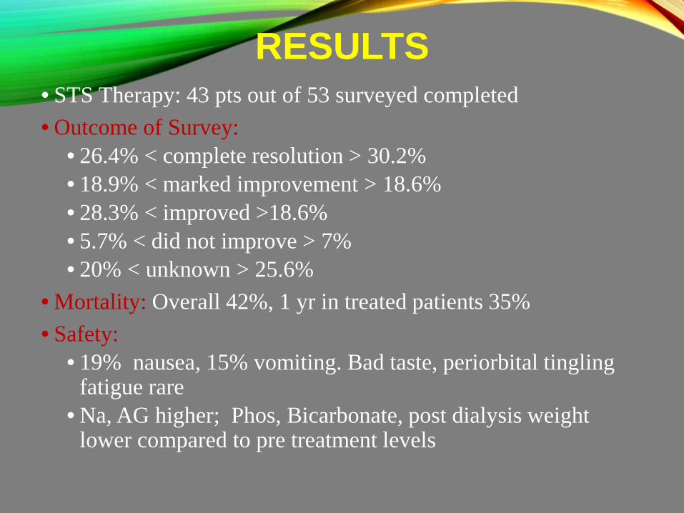
RESULTS • STS Therapy: 43 pts out of 53 surveyed completed • Outcome of Survey:
• 26.4% < complete resolution > 30.2% • 18.9% < marked improvement > 18.6% • 28.3% < improved >18.6% • 5.7% < did not improve > 7% • 20% < unknown > 25.6%
• Mortality: Overall 42%, 1 yr in treated patients 35% • Safety:
• 19% nausea, 15% vomiting. Bad taste, periorbital tingling fatigue rare
• Na, AG higher; Phos, Bicarbonate, post dialysis weight lower compared to pre treatment levels
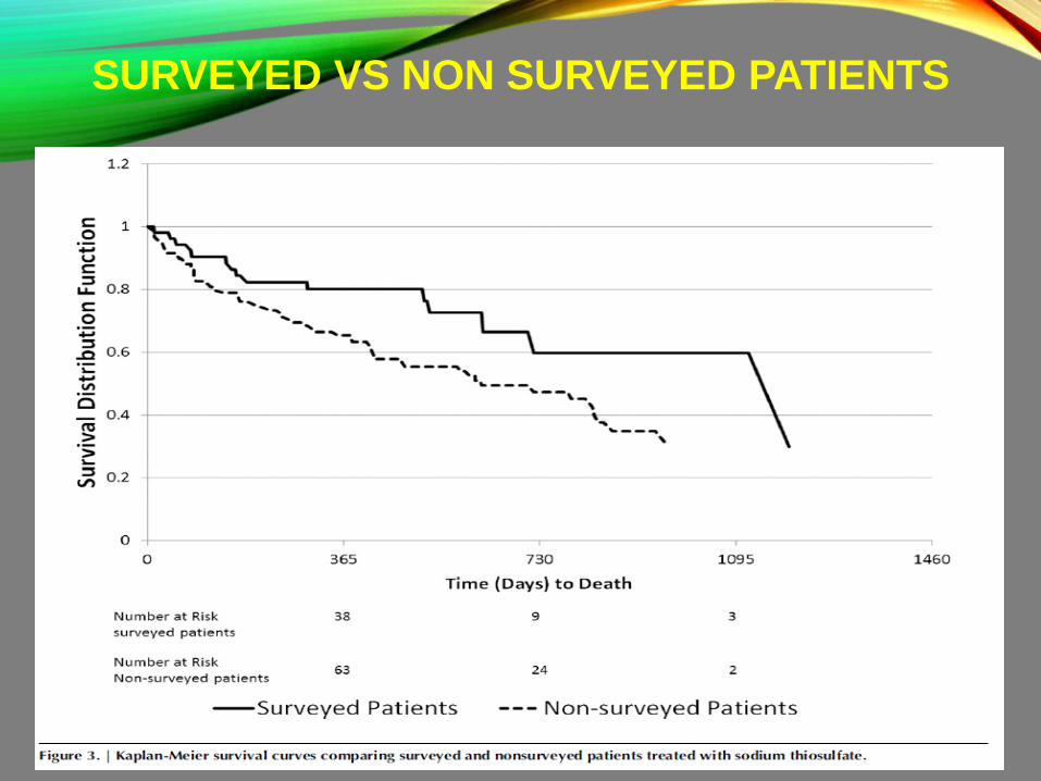
SURVEYED VS NON SURVEYED PATIENTS

CONCLUSIONS
• STS reasonably safe during Rx of CUA in maintenance HD patients
• Majority demonstrated clinical improvement

LIMITATIONS
• No control group
• Histological diagnosis available in less than half
• Selection bias, potential exclusion of hospitalized patient
• No data on concurrent risk factors (warfarin)


OTHER THERAPIES • Supportive: Aggressive wound care
• Decrease high CaxP: HD, discontinue calcium products, Vit D, Calcimimetics
• Avoid VKAs

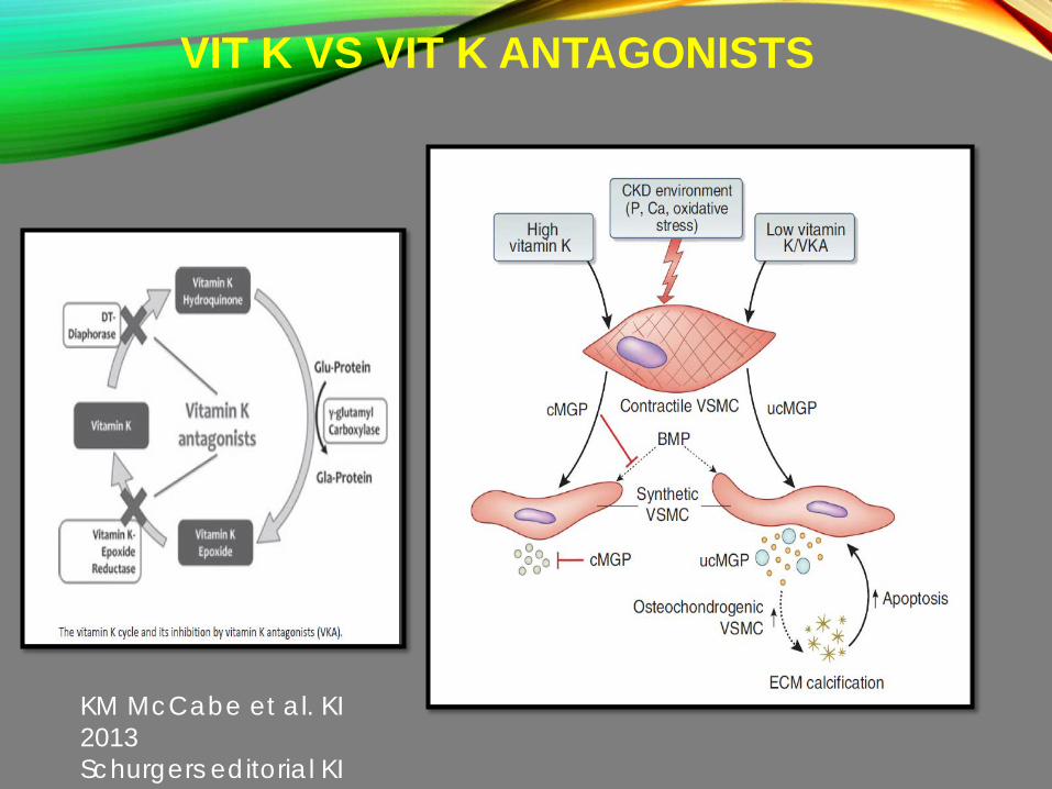
VIT K VS VIT K ANTAGONISTS
KM McCabe et al. KI 2013 Schurgers editorial KI

M Schoppet et al.KI (2008) 73, 384–390


