Download - Capnography During Sedation - Welcome to for

Capnography During Sedation
Bhavani Shankar Kodali MD Associate Professor, Harvard Medical School,
Vice Chair, Clinical Affairs Brigham and Women’s Hospital
New Standards of Monitoring
• American Society of Anesthesiologists (ASA) and the Association of Anaesthestists of Great Britain and Ireland (AAGBI) have revised standards in 2011 to monitor ventilation with capnography for all procedural sedation cases requiring moderate to deep sedation.
Bhavani Shankar Kodali M.D. www.capnography.com

Why were they introduced?
• It is difficult to predict how an individual will respond to an administered sedative.
• Incidence of hypoxia is less if capnography was used to monitor ventilation.
• Capnography when used in conjunction with pulse oximetry and visual inspection of chest detected respiratory depression 17 times more often than without capnography.
• Capnography forewarns of impending hypoxia by about 5 to 240 seconds.
• Capnography triggers early intervention and decreases the incidence of oxygen desaturation.
• Administration of supplemental oxygen delays the onset of desaturation following apnea and therefore relying on pulse oximetry alone will delay intervention.
Bhavani Shankar Kodali M.D. www.capnography.com

Objectives
• After reviewing this brief clinical concept, the clinician/nurse will understand the basic physiology of capnography.
• The clinician/nurse will be able to interpret capnography in monitoring ventilation during sedation more effectively and safely
Bhavani Shankar Kodali M.D. www.capnography.com

Definitions
• PETCO2: The maximum partial pressure of CO2 at the end of a breath. It is about 36- 40 mm Hg in healthy adults.
• PACO2: Partial pressure of CO2 in the alveoli.
• Capnogram: A plot of PCO2 versus time (time capnogram), or expired volume (volume capnogram). Time capnogram is common in clinical practice
Bhavani Shankar Kodali M.D. www.capnography.com

Basic Physiology
• When you inhale CO2 free air and exhale, and measure partial pressure of CO2 at the mouth, you get the following trace. Normal values of PETCO2 vary between 35 and 40 mm Hg
Bhavani Shankar Kodali M.D. www.capnography.com

Basic Physiology
The expiratory segment is divided into three phases phase I, II, and III
Phase I: Dead space gases Phase II: Dead space gases mix with alveolar gases resulting in the rise of PETCO2
Phase III: Represent CO2 evolving from alveoli Bhavani Shankar Kodali M.D.
www.capnography.com
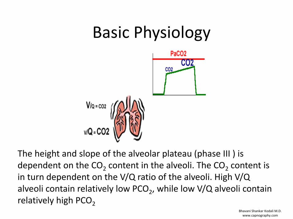
Basic Physiology
The height and slope of the alveolar plateau (phase III ) is dependent on the CO2 content in the alveoli. The CO2 content is in turn dependent on the V/Q ratio of the alveoli. High V/Q alveoli contain relatively low PCO2, while low V/Q alveoli contain relatively high PCO2
Bhavani Shankar Kodali M.D. www.capnography.com

Basic Physiology
Hence, it can be concluded that the height and the slope of the alveolar plateau is dependent on ventilation, cardiac output and more importantly on V/Q relationship.
Bhavani Shankar Kodali M.D. www.capnography.com

Basic Physiology
For example in COPD: the V/Q perfusion abnormalities result in a sloping phase II and phase III
Bhavani Shankar Kodali M.D. www.capnography.com

In the acute settings, for a given ventilation, PETCO2 is a function of cardiac output (pulmonary perfusion). This is the basic principle of directing the uses of capnography during CPR
Basic Physiology
Bhavani Shankar Kodali M.D. www.capnography.com

Basic Physiology
Hypoventilation
Hyperventilation
Bhavani Shankar Kodali M.D. www.capnography.com
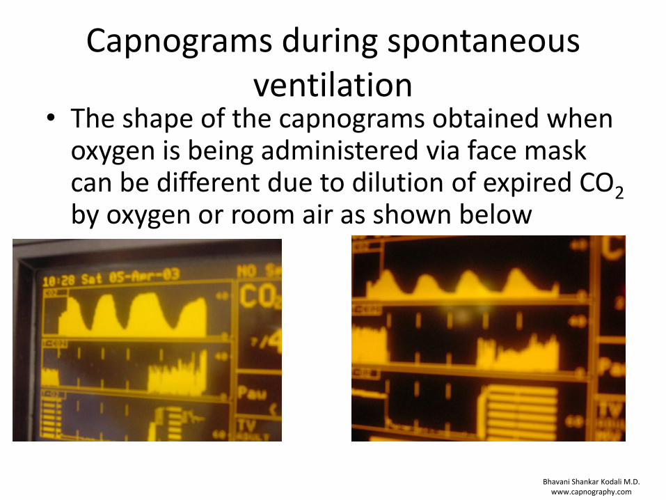
Capnograms during spontaneous ventilation
• The shape of the capnograms obtained when oxygen is being administered via face mask can be different due to dilution of expired CO2 by oxygen or room air as shown below
Bhavani Shankar Kodali M.D. www.capnography.com

Capnograms during sedation
The important concept here is to determine changes from baseline capnograms
Bhavani Shankar Kodali M.D. www.capnography.com

Capnograms during spontaneous ventilation
Look for three important changes from baseline capnograms • Respiratory rate: A decreased rate indicates respiratory
depression. Increased rate suggests stimulation from the procedure
• If the PETCO2 increases, it suggests hypoventilation • If the PETCO2 decreases, it suggests hyperventilation,
hypoventilation, or upper airway obstruction depending on the cause. Observe the patient carefully for evidence of respiratory obstruction. If present, give jaw thrust, PETCO2 increases as obstruction is relieved. If there is no improvement in PETCO2, it most likely suggests a central depression. Hyperventilation is indicated by an increased respiratory rate.
Bhavani Shankar Kodali M.D. www.capnography.com

Over sedation
Respiratory rate is decreased
Bhavani Shankar Kodali M.D. www.capnography.com

Over sedation
Height of the capnogram PETCO2 decreased
Bhavani Shankar Kodali M.D. www.capnography.com

Hypoventilation
Height of the capnogram (PETCO2) is increased
Bhavani Shankar Kodali M.D. www.capnography.com

Apnea, or Respiratory obstruction
A flat line indicates that there is apnea, or total respiratory obstruction. It may also indicate disconnection of the CO2 sampling system, or inability to sample the expired air. These circumstances call for immediate examination of the patient for apnea, or respiratory obstruction, or sampling issues.
Bhavani Shankar Kodali M.D. www.capnography.com

Sampling devices to monitor PETCO2
Here, PETCO2 is being sampled with a nasal sampling cannula (arrow), oxygen is being administered via an oxygen mask. An endoscope is inserted via a flap cut in the mask. This ensures adequate oxygenation and sampling of CO2
Bhavani Shankar Kodali M.D. www.capnography.com

Some Examples of Devices for CO2 sampling

Bhavani Shankar Kodali M.D. www.capnography.com

Bhavani Shankar Kodali M.D. www.capnography.com

Bhavani Shankar Kodali M.D. www.capnography.com

Bhavani Shankar Kodali M.D. www.capnography.com

Conclusion
• Place the sampling device and obtain best PETCO2 values and waveforms
• Look for changes from baseline values and shape of capnograms
• Hypoventilation is suggested by decreased respiratory rate, increased PETCO2 values, or decreased PETCO2 values (depending on cause).
• Hyperventilation is indicated by an increase in respiratory rate
• Look for evidence of respiratory obstruction when you begin to see changes in PETCO2 values, or shape.
• Lifting the jaw forwards relieves upper airway obstruction and increased PETCO2 values
Bhavani Shankar Kodali M.D. www.capnography.com

Differential Diagnosis of Capnography during Sedation
PETCO2
Hypoventilation
Hypoventilation Partial Respiratory
Obstruction Hyperventilation
Hyperventilation
Hypoventilation Partial Respiratory
Obstruction
If relieved, then respiratory obstruction
If not relieved, then hypoventilation
Increased
Decreased
Increased Respiratory Rate
Jaw Thrust
No CO2
Apnea Total Respiratory
Obstruction Sampling Issue
Observe patient to differentiate
Bhavani Shankar Kodali M.D. www.capnography.com
Pulse Oximetry can be used as an adjunct in differential diagnosis . Capnography should help to diagnose the problem before changes in SPO2

References
• Br J Anaesth 2011; 106: 617-31 • Acad Emerg Med 2000; 7: 228-35 • Acad Emerg Med 2007; 14: 41-6 • Am J Gastroenterol 2007; 102: 738-43 • Curr Opin Anaesthesiol 2007; 20: 347-51 • Ann Emerg Med 2010; 55: 258-64 • J Clin Anesth 2011; 23: 189-96 • Am J Gastroenterol 2012 May 29. doi:
10.1038/ajg.2012.136. [Anesthesiology 2009; 110: 759-65
• Anesthesiology 2006; 104: 228-34
Bhavani Shankar Kodali M.D. www.capnography.com

Question 1
• Normal PETCO2 values in healthy adults breathing normally with good sampling
(a) 80 mm Hg
(b) 40 mm Hg
(c) 20 mm Hg
(d) 25 mm Hg

(b)

Question 2
• During capnography monitoring for sedation, it is important to determine PETCO2 changes from baseline capnograms
True
False

True

Question 3
• If PETCO2 increases during sedation, it suggests
(a) Hyperventilation
(b) Hypoventilation
(c) Respiratory obstruction
(d) Low cardiac output

(b)

Question 4
• If PETCO2 decreases during sedation, it suggests
(a) Hyperventilation
(b) Respiratory obstruction
(c) Hypoventilation
(d) All of the above

(d)

Question 5
• Generally, capnography changes like hypoventilation, or respiratory obstruction occur before oxygen desaturation
• True
• False
• Neither

True


