
Carbon Nanotube-enhanced Thermal Destruction ofCancer Cells in a Noninvasive Radiofrequency Field
Christopher J. Gannon, MD1
Paul Cherukuri, PhD1,2
Boris I. Yakobson, PhD3,4
Laurent Cognet, PhD5,6
John S. Kanzius7
Carter Kittrell, PhD3,8
R. Bruce Weisman, PhD3,5
Matteo Pasquali, PhD3,5,8,9
Howard K. Schmidt, PhD5,8
Richard E. Smalley, PhD3,5,8y
Steven A. Curley, MD1,4
1 Department of Surgical Oncology, The Univer-sity of Texas M. D. Anderson Cancer Center,Houston, Texas.2 Department of Experimental Therapeutics, TheUniversity of Texas M. D. Anderson Cancer Cen-ter, Houston, Texas.
3 Department of Chemistry, Rice University,Houston, Texas.
4 Mechanical Engineering and Materials Science,Rice University, Houston, Texas.
5 Center for Biological and Environmental Nano-technology, Rice University, Houston, Texas.
6 Center of Optical and Hertzian MolecularPhysics, National Center for Scientific Research,Bordeaux University, Bordeaux, France.
7 ThermMed LLC, Erie, Pennsylvania.
8 Carbon Nanotechnology Laboratory, Rice Uni-versity, Houston, Texas.
9 Department of Chemical and Biomolecular En-gineering, Rice University, Houston, Texas.
BACKGROUND. Single-walled carbon nanotubes (SWNTs) have remarkable physi-
cochemical properties that may have several medical applications. The authors
have discovered a novel property of SWNTs—heat release in a radiofrequency
(RF) field—that they hypothesized may be used to produce thermal cytotoxicity
in malignant cells.
METHODS. Functionalized, water-soluble SWNTs were exposed to a noninvasive,
13.56-megahertz RF field, and heating characteristics were measured with infra-
red thermography. Three human cancer cell lines were incubated with various
concentrations of SWNTs and then treated in the RF field. Cytotoxicity was mea-
sured by fluorescence-activated cell sorting. Hepatic VX2 tumors in rabbits were
injected with SWNTs or with control solutions and were treated in the RF field.
Tumors were harvested 48 hours later to assess viability.
RESULTS. The RF field induced efficient heating of aqueous suspensions of
SWNTs. This phenomenon was used to produce a noninvasive, selective, and
SWNT concentration-dependent thermal destruction in vitro of human cancer
cells that contained internalized SWNTs. Direct intratumoral injection of SWNTs
in vivo followed by immediate RF field treatment was tolerated well by rabbits
bearing hepatic VX2 tumors. At 48 hours, all SWNT-treated tumors demonstrated
complete necrosis, whereas control tumors that were treated with RF without
SWNTs remained completely viable. Tumors that were injected with SWNTs but
were not treated with RF also were viable.
CONCLUSIONS. The current results suggested that SWNTs targeted to cancer cells
may allow noninvasive RF field treatments to produce lethal thermal injury to the
malignant cells. Now, the authors are developing SWNTs coupled with cancer cell-
targeting agents to enhance SWNT uptake by cancer cells while limiting uptake by
normal cells. Cancer 2007;110:2654–65. � 2007 American Cancer Society.
KEYWORDS: carbon nanotubes, radiofrequency, thermal cytotoxicity, cancer cells.
A noninvasive approach with the potential to treat many types of
cancers effectively with minimal or no toxic effects to normal
cells would be highly beneficial. Toward this objective, we combined
a radiofrequency (RF) field-generating system with single-walled
carbon nanotubes (SWNTs), which act efficiently to convert RF irra-Supported by American Association of CancerResearch Littlefield Grant (to C.J.G. and S.A.C.),by the National Aeronautics and Space Adminis-tration and the Alliance for NanoHealth (to P.C.,R.E.S., and H.K.S.), by the National ScienceFoundation and the Center for Biological andEnvironmental Nanotechnology (to B.I.Y., L.C.,M.P., R.B.W., and H.K.S.), and by the FulbrightFoundation (L.C.).
J.S.K. is the inventor of the radiofrequency fieldusing nanoparticles.
The first two authors contributed equally to thisarticle.
yDeceased.
We thank K. Ash for assistance in preparing thearticle.
Address for reprints: Steven A. Curley, MD,Department of Surgical Oncology, Unit 444, The
University of Texas M. D. Anderson Cancer Cen-ter, 1400 Holcombe Boulevard, Houston, TX77030; Fax: (713) 745-5235; E-mail: [email protected]
Received August 20, 2007; accepted September19, 2007.
ª 2007 American Cancer SocietyDOI 10.1002/cncr.23155Published online 24 October 2007 in Wiley InterScience (www.interscience.wiley.com).
2654

diation into heat. Although, today, RF ablation (RFA)
is used in clinical practice to treat some malignant
tumors, it suffers from serious drawbacks.1 RFA cur-
rently is an invasive treatment that requires the
insertion of needle electrodes directly into the
tumor(s) to be treated; incomplete tumor destruction
occurs in from 5% to 40% of the treated lesions; the
treatment is nonspecific and induces thermal necro-
sis in both malignant and normal tissues surround-
ing the needle electrode; complications arise in up to
10% of patients related to thermal injury to normal
tissues; and invasive RFA is limited to the treatment
of tumors in only a few organ sites (liver, kidney,
breast, lung, bone). Conversely, it is known that the
tissue penetration by RF energy fields is excellent.2
Thus, noninvasive RF treatment of malignant tumors
at any site in the body should be possible if agents
that convert RF energy into heat can be delivered to
the malignant cells.
Biomedical applications for SWNTs are being
investigated actively because of their useful combi-
nation of size and physicochemical properties.3–8 In
cancer patients, SWNTs have potential roles in deli-
vering pharmacologic agents, diagnostic imaging
agents, DNA, silent interfering RNA, oligonucleotides,
and proteins to detect or treat cancer cells.9–11
Roughly 66% of SWNTs are high-mobility, direct
band-gap seimiconductors7 that fluoresce in the near
infrared (NIR);8 the rest are metallic conductors
capable of carrying exceptional current densities.5
Given the unique electrical and chemical properties of
SWNTs, we hypothesized that exposure to a focused
external RF field would lead to significant heat release
by the SWNTs, allowing them to serve directly as an
anticancer therapeutic agent. We report herein that
exposure of intracellular SWNTs in vitro and intratu-
moral SWNTs in vivo to a noninvasive, focused RF
field produces lethal heating of malignant cells.
MATERIALS AND METHODSFunctionalized SWNTsSterile CoMoCAT SWNTs functionalized with Kentera
(Zyvex Corp, Richardson, Tex) were obtained at a
standard concentration of 500 mg/L. Kentera is a
polymer based on polyphenylene ethynylene (PPE).
The phenyl groups of the PPE are linked to form a
rigid, conjugated polymer that functionalizes SWNTs
through p–p stacking (noncovalent interaction).
Sterility of the SWNT solutions was assured by a 20-
minute exposure to ultraviolet irradiation. Character-
ization of the Kentera SWNT solution to assess for
individual versus clusters of SWNTs was performed
by atomic force microscopy (AFM) at 633 nm. A 20-
lL aliquot of Kentera SWNTs was spun cast onto
mica for AFM. Purity of the SWNT solutions and
analysis of metal content was performed by induc-
tively coupled plasma-mass spectroscopy. The mass
spectroscopy analysis of the Kentera SWNTs used for
all of our studies shows Mo, 58 parts per million
(ppm) (0.0058%); Co, 3ppm; Fe, 0.2 ppm; and Ti,
0.1 ppm. Raman spectroscopy was performed and
confirmed the presence of the signal intensity peaks
that were observed with individual SWNTs (data not
shown). In all suspensions, the mass ratio of SWNTs
to Kentera was 1:1.
External RF Generator/Coupling SystemA variable power 0- to 2-kilowatt, 13.56-megahertz
(MHz) RF signal generator (Therm Med LLC, Erie,
Pa) was built to specifications for use in these experi-
ments. The RF generator is connected to a high Q
coupling system (Therm Med LLC) that has a trans-
mitting (Tx) head, a receiving (Rx) head, and cou-
pling circuitry. The Tx and Rx heads and the
contained coupling circuitry are mounted on a swivel
bracket, allowing the RF field to be oriented in either
a horizontal or vertical direction. The distance
between the heads is adjustable, and the RF field is
generated between the Tx and Rx heads. The coaxial
coupling circuitry in the Tx and Rx heads produces a
focused electromagnetic field with a useful diameter
of 30 cm and with a peak intensity of 7 cm from the
central axis of the Tx and Rx heads. Each time the RF
field is activated, the heads are checked, and the
coupling circuit is fine-tuned to assure that there is no
power reflected back through the coupling circuit.
The electromagnetic field strength between the
Tx head and Rx head was established in a Farraday-
shielded room to exclude any interference from exter-
nal RF sources. The field strength was measured at
low power using an isotropic field monitor and probe
(models FM2004 and FP2000, respectively; Amplifier
Research Inc., Souderton, Pa); a Hewlett-Packard
Spectrum Analyzer (model 8566B; Agilent, Santa Clara,
Calif) was connected concomitantly to the system
through a directional coupler to measure transmitted
power accurately. The field strengths measured at low
generator output powers were then used to estimate
field strengths at the various RF generator output
powers that were used in our experimental studies.
Thermal Effect of RF Field on Functionalized SWNTsSerial dilutions of Kentera SWNTs were used to
assess concentration dependency to heat-deionized
water. Several concentrations of functionalized
SWNTs were diluted serially in deionized water and
placed in a 1.5-mL, circular quartz cuvette (height,
Nanotube Heating Kills Cancer Cells/Gannon et al. 2655

15 mm; diameter, 10 mm) located between the Tx
and Rx heads of the RF generator using a nonmetal-
lic, nonconductive Teflon holder. The samples were
located at the mid plane of the working volume and
were positioned radially to capture the maximum
electric field potential. The Tx and Rx heads were
spaced with a 7.5-cm air gap for these experiments.
Temperature in the Kentera SWNT solutions, in solu-
tions of Kentera without SWNTs, and in water alone
were measured continuously before activating the RF
field, during the entire period of RF field activation,
and for 2 minutes after RF field shutdown by using
a thermoelectrically cooled, Indium antimonide,
focal plane array IR camera (Amber Engineering,
Goleta, Calif). IR video was captured at 30 frames
per second by using iMovie software (Apple, Cuper-
tino, Calif), and all temperature measurements were
performed in triplicate.
Human Cancer Cell LinesHepG2 and Hep3B hepatocellular cancer cells and
Panc-1 pancreatic adenocarcinoma cells (American
TypeCulture Collection, Bethesda, Md) were cultured
in Dulbecco modified Eagle medium (DMEM) with
10% fetal calf serum (FCS) plus penicillin/streptomy-
cin at 378C under standard atmospheric conditions.
For experiments, each cell line was used only from
passages 2 through 9. For cell proliferation assays,
cells were grown to confluence in 96-well culture
plates. For cytotoxicity studies with or without RF
field treatment, cells were grown to near confluence
in 60-mm glass culture dishes.
Cell Proliferation AssayThe 3 human cancer cell lines were grown to near
confluence in 96-well plates. Media was aspirated
from the cells, and media containing various concen-
trations of functionalized SWNTs or control media
was added to the cell lines. Cell culture plates were
returned to the incubator for 24 hours at 378C. Fiftymicroliters of 3-(4,5-dimethylthiazol-2-yl)-2, 5-diphe-
nyltetrazolium bromide (MTT) (Sigma-Aldrich Corp.,
St. Louis, Mo) were added to each of the 96 wells,
and the plates were incubated for an additional 4
hours at 378C. The cell culture plates were then cen-
trifuged at 1500 revolutions per minute (rpm) for 5
minutes, the supernatant fluids were aspirated, and
the cells were then solubilized by adding 100 lL of
dimethyl sulfoxide to each well. Absorbance was read
on an automated, multiwell, plate-scanning spectro-
photometer at 570 nm and recorded. Each concen-
tration was repeated in triplicate with 5 wells in each
group.
Brightfield and NIR MicroscopyCells were grown on glass coverslips in 60-mm glass
dishes for NIR microscopy. Twenty-four hours after
addition of SWNTs to the human cancer cell cultures,
the media was aspirated from the cultures, and the
cells were washed with phosphate-buffered saline
(PBS) and resuspended in fresh media without
SWNTs. To confirm intracellular localization of
SWNTs, acid washing (pH 2.0) was performed on cell
cultures to quench external SWNT emission and to
remove any SWNTs adsorbed on the cell surfaces.
Acidic media was aspirated and discarded, and fresh
media was added for fluorescence imaging. Cover-
slips were mounted in thermoregulated slide holders
to maintain a temperature of 378C during fluores-
cence imaging. The cellular location of SWNTs was
measured by both brightfield and NIR microscopy.
NIR images of SWNTs from within cancer cells were
measured by using a cryogenically cooled 320 3 256
pixel image Indium gallium arsenide detector array
(model OMA-V 2D; Roper Scientific, Trenton, NJ). A
360 oil-immersion objective was used for obtaining
images, and the laser spot size was focused from
50 to 100 lm in greatest dimension (100–200 pixels
under 360 magnification). An excitation laser at
658 nm was used with a 946-nm long pass-blocking
filter in the detection path.
Assessment of CytotoxicityThe 3 human cancer cell lines were grown to near
confluence in 60-mm culture dishes. Media was aspi-
rated; fresh media containing various concentrations
of Kentera SWNTs, Kentera alone, or media alone
was added to the cells, and they were incubated for
an additional 24 hours at 378C. After 24 hours, the
media was aspirated, and the cells were washed 3
times with PBS to remove any Kentera SWNTs or
Kentera that was not taken up by the cells. Three
milliliters of fresh media without SWNTs were added
to each culture plate, and then the culture dishes
were placed individually on a nonconductive Teflon
holder between the Tx and Rx heads of the RF gener-
ator. The heads were oriented in a vertical direction
with a 7.5-cm space between the heads. Then, the
cells were exposed to a 13.56-MHz external RF field
at 800 W for 1 or 2 minutes. After the prescribed pe-
riod of RF field exposure, the culture dishes were
returned to the incubator for an additional 18 hours
at 378C. Cells were harvested from the culture dishes
by trypsinization, washed with PBS, and centrifuged
at 1500 rpm for 5 minutes. The cell pellet was resus-
pended during gentle vortexing and while adding 5
mL of 95% ethanol. Cells were fixed overnight at
room temperature and then stored at 48 until they
2656 CANCER December 15, 2007 / Volume 110 / Number 12

were ready for staining. For staining with propidium
iodide (PI) (Sigma-Aldrich Corp.), cells were centri-
fuged in the fixative at 1500 rpm for 5 minutes and
then resuspended in a Tris buffer solution (stock so-
lution: 24 g Tris, 12 g NaCl, 168 mL 1N HCl in a total
volume of 2 L [pH 7.4]). The cell pellets were resus-
pended in 500 lL of Tris buffer and 1 mL of PI (500
lg/mL in water were added). Thirty minutes before
flow cytometric analysis, 100 lL of RNase (1 mg/mL
and DNase-free; Sigma-Aldrich Corp.) were added,
and the fixed cells were incubated at 378C. Cell via-bility was assessed by adding the PI-stained cells to a
fluorescence-activated cell sorter (FACS) Calibur unit
(BD Biosciences, San Jose, Calif), and cell cycle pro-
portions and cell viability were assessed with Cell
Quest Pro software (BD Biosciences). All experiments
were performed in quadruplicate.
Tumor-bearing Animal ModelAll animal experiments were performed under a pro-
tocol that was reviewed and approved by the Institu-
tional Animal Care and Utilization Committee at the
University of Texas M. D. Anderson Cancer Center.
VX2 tumor cells were grown in DMEM plus 10% FCS
to confluence. The cells were harvested, washed, and
resuspended in sterile PBS. One milliliter of the VX2
tumor solution was injected into each flank of 2
adult New Zealand white rabbits (3.0–3.5 kg). When
tumors in the flanks of the donor rabbits reached a
greatest dimension of 2.5 to 3.0 cm, the tumors were
harvested under sterile condition and cut into 1-mm
cubes. Under general endotracheal anesthesia, a
2-cm upper midline laparotomy incision was made in
adult New Zealand white rabbits. An incision 1 mm
long was made in the capsule of a right liver segment
with a No. 11 scalpel blade, and a single cube of VX2
tumor was inserted into the liver parenchyma. The
abdominal wall incisions were then closed under
sterile conditions, and the animals were recovered
from anesthesia. Two weeks later, under general en-
dotracheal anesthesia, a repeat midline laparotomy
procedure was performed, and the resulting intrahe-
patic VX2 tumor (greatest dimension, 1.0–1.3 cm) was
injected directly with either a 1.0-mL solution of Ken-
tera SWNTs (500 mg/L) with 100 lg of epinephrine in
4 animals or with 1.0 mL of control solution consist-
ing of Kentera polymer (500 mg/L) and 100 lg of epi-
nephrine (no SWNTs) in 4 animals. An additional 4
animals underwent intratumoral injection of Kentera
SWNTs (500 mg/L) with epinephrine and served as a
control group that was not treated with RF. Epineph-
rine was added to the SWNT and control solutions to
produce transient intratumoral vasoconstriction to
reduce diffusion of SWNTs or control solution out of
the tumor. A final control group of 4 animals under-
went RF field treatment of the liver tumors without
injection of SWNTs or control solution into the
tumors. The 12 animals that were to receive RF treat-
ment were placed individually on a nonmetallic, non-
conductive Teflon platform between the Tx and Rx
heads of the RF generator with 10 cm of spacing
between the heads. The Tx and Rx heads were ori-
ented in a vertical configuration. Each animal was
positioned so the hepatic tumor was located at the
mid plane of the working volume to capture the max-
imum electric field potential. The RF field was acti-
vated and tuned, and each animal was treated for 2
minutes with RF at 600 W; then, the laparotomy inci-
sion was closed by using sterile technique, and the
animals were allowed to recover from the treatment.
Forty-eight hours later, the animals were euthanized,
and the liver and other tissues were harvested and
evaluated to assess for any evidence of thermal injury.
The liver and VX2 tumor were fixed in 10% formalin.
Tissue samples were cut, embedded in paraffin, then
prepared for histologic assessment with hematoxylin
and eosin staining and with terminal deoxynucleoti-
dyl transferase biotin-deoxyuridine triphosphate
nick-end labeling for apoptosis and viability accord-
ing to the standard protocol provided by Promega
(Promega Corp., Madison, Wis).
Statistical AnalysisDifferences between experimental groups (with
experiments performed in triplicate) were deter-
mined by using an analysis of variance with Statistica
software (StatSoft Inc, Tulsa, Okla). A significant dif-
ference between groups was defined as P < .05.
RESULTSRF-induced Heating of SWNT SuspensionsWe used SWNTs functionalized noncovalently with
Kentera (Zyvex; Richardson, Tex), which is a nonionic,
biocompatible polymer that does not substantially al-
ter the electronic properties of the nanotube.12 We
first characterized the heating rates and field depend-
ence of the SWNTs, and we tested 5 different con-
centrations of aqueous SWNT suspensions (5 mg/L,
50 mg/L, 125 mg/L, 250 mg/L, and 500 mg/L) in a
1.5-mL quartz cuvette. Each suspension was placed
in an experimental, focused, 13.56-MHz RF signal
generator (ThermMed LLC, Erie, Pa; US Patent nos.
US2006/0190063 A1, US2005/02511233 A1, and
US2005/0251234 A1 and World Intellectual Property
Organization WO 2007/027614) that included cou-
pling circuitry housed in transmission (Tx) and
Nanotube Heating Kills Cancer Cells/Gannon et al. 2657
receiving (Rx) heads. This RF system produces a con-
centrated electromagnetic field between the Tx head
and the Rx head. A 13.56-MHz signal generator
was used because it is an industrial, scientific, and
medical working frequency allocated by the Federal
Communications Commission; furthermore, it was
demonstrated previously that this frequency pro-
duces minimal tissue heating related to the specific
absorption rate of electromagnetic radiation in mam-
malian testing.13
We observed that these noncovalently suspended
SWNTs were very effective at converting RF energy
into heat. With our instrument, output powers of
400 W, 600 W, 800 W, and 1000 W were used, yielding
a maximum estimated electric field strength 2.5 cm
from the Tx head of 10.1 kilovolts (kV)/m, 12.4 kV/
m, 14.3 kV/m, and 16.0 kV/m, respectively. We deter-
mined that the lowest SWNT concentration that pro-
duced enhanced bulk heating, compared with a
reference sample of deionized water alone, was
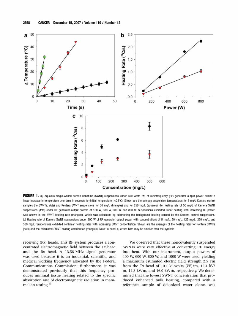
FIGURE 1. (a) Aqueous single-walled carbon nanotube (SWNT) suspensions under 600 watts (W) of radiofrequency (RF) generator output power exhibit alinear increase in temperature over time in seconds (s) (initial temperature, �258C). Shown are the average suspension temperatures for 5 mg/L Kentera controlsamples (no SWNTs; dots) and Kentera SWNT suspensions for 50 mg/L (triangles) and for 250 mg/L (squares). (b) Heating rate of 50 mg/L of Kentera SWNT
suspensions (dots) under RF generator output powers of 100 W, 300 W, 600 W, and 800 W. Suspensions exhibited linear heating with increasing RF power.
Also shown is the SWNT heating rate (triangles), which was calculated by subtracting the background heating caused by the Kentera control suspensions.
(c) Heating rate of Kentera SWNT suspensions under 600 W of RF generator output power with concentrations of 5 mg/L, 50 mg/L, 125 mg/L, 250 mg/L, and
500 mg/L. Suspensions exhibited nonlinear heating rates with increasing SWNT concentration. Shown are the averages of the heating rates for Kentera SWNTs
(dots) and the calculated SWNT heating contribution (triangles). Note: In panel c, errors bars may be smaller than the symbols.
2658 CANCER December 15, 2007 / Volume 110 / Number 12
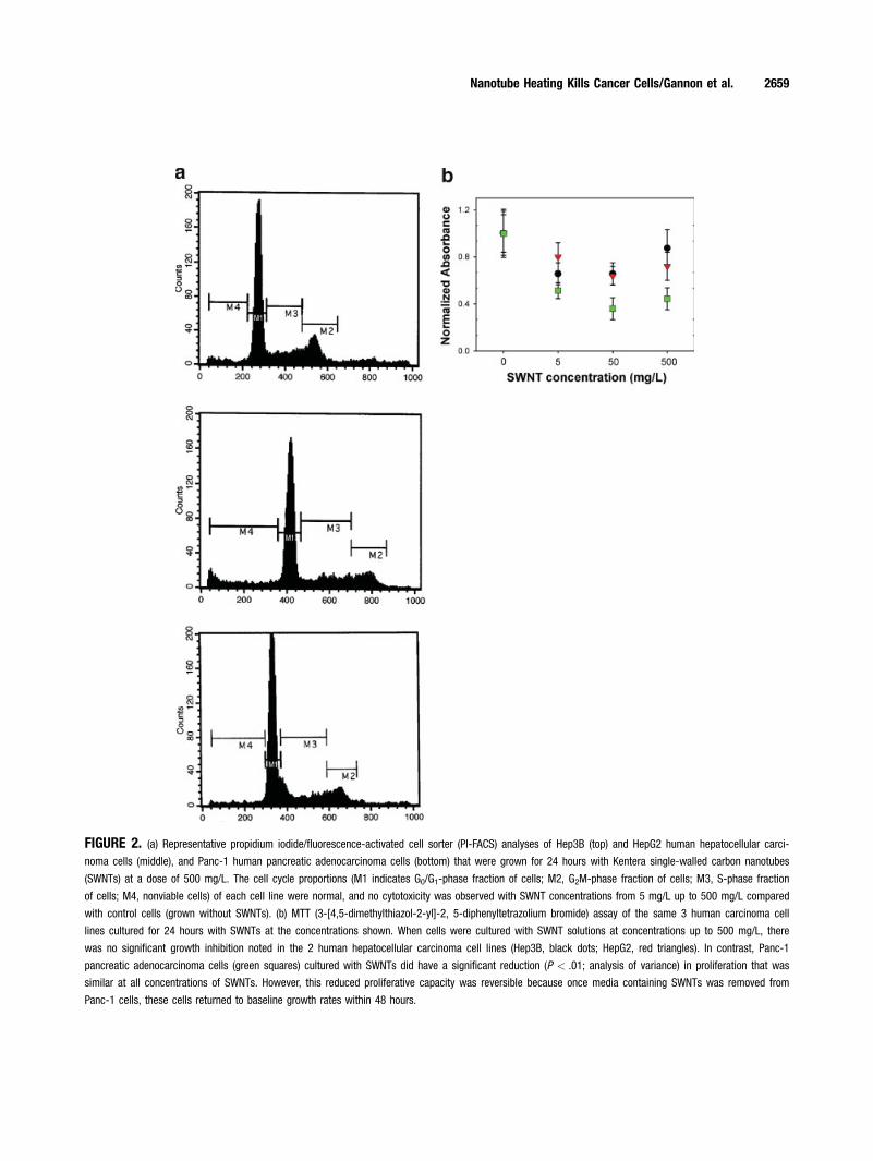
FIGURE 2. (a) Representative propidium iodide/fluorescence-activated cell sorter (PI-FACS) analyses of Hep3B (top) and HepG2 human hepatocellular carci-noma cells (middle), and Panc-1 human pancreatic adenocarcinoma cells (bottom) that were grown for 24 hours with Kentera single-walled carbon nanotubes
(SWNTs) at a dose of 500 mg/L. The cell cycle proportions (M1 indicates G0/G1-phase fraction of cells; M2, G2M-phase fraction of cells; M3, S-phase fraction
of cells; M4, nonviable cells) of each cell line were normal, and no cytotoxicity was observed with SWNT concentrations from 5 mg/L up to 500 mg/L compared
with control cells (grown without SWNTs). (b) MTT (3-[4,5-dimethylthiazol-2-yl]-2, 5-diphenyltetrazolium bromide) assay of the same 3 human carcinoma cell
lines cultured for 24 hours with SWNTs at the concentrations shown. When cells were cultured with SWNT solutions at concentrations up to 500 mg/L, there
was no significant growth inhibition noted in the 2 human hepatocellular carcinoma cell lines (Hep3B, black dots; HepG2, red triangles). In contrast, Panc-1
pancreatic adenocarcinoma cells (green squares) cultured with SWNTs did have a significant reduction (P < .01; analysis of variance) in proliferation that was
similar at all concentrations of SWNTs. However, this reduced proliferative capacity was reversible because once media containing SWNTs was removed from
Panc-1 cells, these cells returned to baseline growth rates within 48 hours.
Nanotube Heating Kills Cancer Cells/Gannon et al. 2659
5 mg/L. At any given concentration of SWNTs, the
heating of the solutions was linear over time (Fig.
1a), and the heating rate increased linearly with RF
generator output power (Fig. 1b). However, the heat-
ing rate of SWNT suspensions increased nonlinearly
with increasing concentrations at any given RF
power (Fig. 1c). Control experiments indicated that
the heating rate of deionized water alone was 0.2 K
per second, and the heating rate of Kentera polymer
solutions alone was 0.7 K per second. Thus, the frac-
tional heating provided by the SWNTs at the 50 mg/L
concentration was 0.9 K per second, and the heating
rate of Kentera SWNTs was 1.6 K per second. We
observed that, at all concentrations of Kentera SWNT
solutions, the Kentera polymer contributed �42% of
the heating in the RF field. In all suspensions, the
mass ratio of SWNTs to Kentera was 1:1.
Assessment of SWNT Toxicity In VitroBefore determining the in vitro response of cells that
had been incubated with SWNTs and treated with RF,
we investigated potential cytotoxic effects of growing
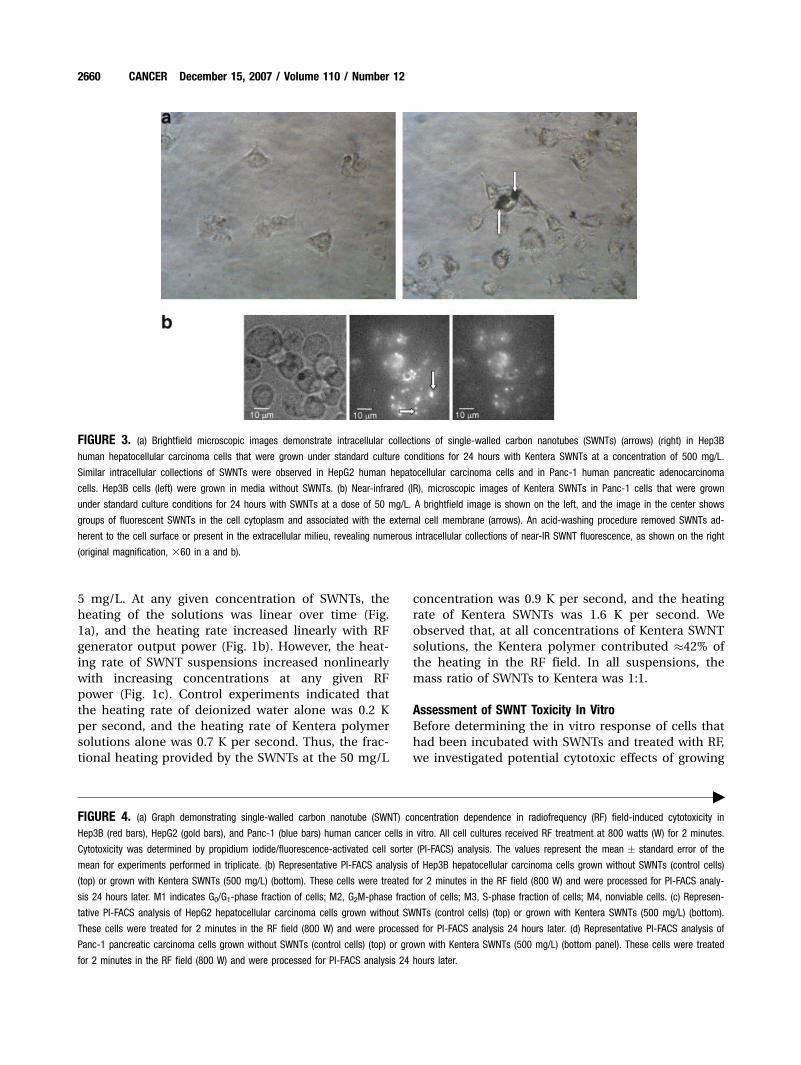
FIGURE 3. (a) Brightfield microscopic images demonstrate intracellular collections of single-walled carbon nanotubes (SWNTs) (arrows) (right) in Hep3Bhuman hepatocellular carcinoma cells that were grown under standard culture conditions for 24 hours with Kentera SWNTs at a concentration of 500 mg/L.
Similar intracellular collections of SWNTs were observed in HepG2 human hepatocellular carcinoma cells and in Panc-1 human pancreatic adenocarcinoma
cells. Hep3B cells (left) were grown in media without SWNTs. (b) Near-infrared (IR), microscopic images of Kentera SWNTs in Panc-1 cells that were grown
under standard culture conditions for 24 hours with SWNTs at a dose of 50 mg/L. A brightfield image is shown on the left, and the image in the center shows
groups of fluorescent SWNTs in the cell cytoplasm and associated with the external cell membrane (arrows). An acid-washing procedure removed SWNTs ad-
herent to the cell surface or present in the extracellular milieu, revealing numerous intracellular collections of near-IR SWNT fluorescence, as shown on the right
(original magnification, 360 in a and b).
"FIGURE 4. (a) Graph demonstrating single-walled carbon nanotube (SWNT) concentration dependence in radiofrequency (RF) field-induced cytotoxicity inHep3B (red bars), HepG2 (gold bars), and Panc-1 (blue bars) human cancer cells in vitro. All cell cultures received RF treatment at 800 watts (W) for 2 minutes.
Cytotoxicity was determined by propidium iodide/fluorescence-activated cell sorter (PI-FACS) analysis. The values represent the mean � standard error of the
mean for experiments performed in triplicate. (b) Representative PI-FACS analysis of Hep3B hepatocellular carcinoma cells grown without SWNTs (control cells)
(top) or grown with Kentera SWNTs (500 mg/L) (bottom). These cells were treated for 2 minutes in the RF field (800 W) and were processed for PI-FACS analy-
sis 24 hours later. M1 indicates G0/G1-phase fraction of cells; M2, G2M-phase fraction of cells; M3, S-phase fraction of cells; M4, nonviable cells. (c) Represen-
tative PI-FACS analysis of HepG2 hepatocellular carcinoma cells grown without SWNTs (control cells) (top) or grown with Kentera SWNTs (500 mg/L) (bottom).
These cells were treated for 2 minutes in the RF field (800 W) and were processed for PI-FACS analysis 24 hours later. (d) Representative PI-FACS analysis of
Panc-1 pancreatic carcinoma cells grown without SWNTs (control cells) (top) or grown with Kentera SWNTs (500 mg/L) (bottom panel). These cells were treated
for 2 minutes in the RF field (800 W) and were processed for PI-FACS analysis 24 hours later.
2660 CANCER December 15, 2007 / Volume 110 / Number 12
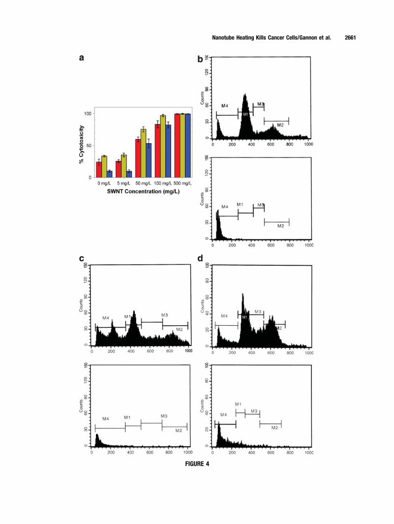
FIGURE 4
Nanotube Heating Kills Cancer Cells/Gannon et al. 2661

human cancer cells in the presence of various con-
centrations of SWNTs. There was no cytotoxicity in
Hep3B, HepG2, or Panc-1 cells cultured for 6 to 48
hours with SWNTs in concentrations from 5 mg/L to
500 mg/L (Fig. 2a) compared with control cell cul-
tures grown in the absence of SWNTs. MTT assays
revealed no significant reduction in the proliferation
of Hep3B or HepG2 cells grown with SWNTs in this
same range of concentrations (Fig. 2b). However,
there was a significant reduction (P<.01) in the pro-
liferation of Panc-1 cells at all concentrations of
SWNTs from 5 mg/L to 500 mg/L (Fig. 2b). This
reduction in proliferation was reversible, because the
Panc-1 cells that had been cultured in the presence
of SWNTs demonstrated growth rates identical to
control cells (no SWNT exposure) within 48 hours of
removing SWNTs from the culture media. Brightfield
microscopy revealed cytoplasmic collections of
SWNTs in cells that had been incubated for 12 to
24 hours with SWNT concentrations of 50 mg/L, 100
mg/L, or 500 mg/L (Fig. 3a). Near-IR fluorescence
microscopy confirmed the characteristic near-IR flu-
orescence of semiconducting SWNTs from the sur-
face and the cytoplasm of the cells (Fig. 3b).
RF-induced Cytotoxicity of Cancer Cells In VitroGiven these pronounced heating rates recorded in
aqueous suspensions of SWNTs in an RF field, we
tested the use of RF-induced SWNT heating to pro-
duce cancer cell cytotoxicity on 2 human hepatocel-
lular cancer cell lines and 1 human pancreatic
carcinoma cell line. In contrast to control conditions
with no RF treatment, we observed SWNT concentra-
tion-dependent cellular cytotoxicity in vitro in all 3
cancer cell lines after 2 minutes of RF field exposure
at 800 W of generator output power (Fig. 4a). At high
concentrations of SWNTs (500 mg/L), cytotoxicity
essentially was 100% in all 3 cell lines that had been
exposed to the RF field for 2 minutes (Fig. 4b-d). It is
worthy noting that control cell cultures containing
only media or Kentera alone with no SWNTs had
measurable rates of cell death (cytotoxicity rate, 11–
35%) (Fig. 4a) that were significantly lower (P < .01)
compared with the rates in cultures that containing
SWNTs. No significant difference in cytotoxicity was
observed when we compared the 2 groups of control
cells (media alone or media with Kentera at concen-
trations up to 500 mg/L). However, SWNT-free cells
that had been treated with RF at a generator output
of 800 W for only 1 minute were completely viable.
In contrast, 1 minute of RF exposure at 800 W in the
3 cancer cell lines containing SWNTs at 100 mg/L or
500 mg/L yielded cytotoxicity rates from 42% to 68%
(P < .01 vs control cells). Thus, both SWNT concentra-
tion and duration of RF exposure are controlling fac-
tors for inducing cytotoxicity in vitro. The observed
heating of the solute-rich cell culture media empha-
sizes the impact of nonspecific ionic stimulation
with resultant heat production in a powerful RF field.
RF-induced Cytotoxicity of Malignant Liver TumorsIn VivoAdult New Zealand white rabbits bearing VX2 tumors
ranging in size from 1.0 cm to 1.3 cm in greatest
dimension underwent a direct intratumoral injection
of water-soluble SWNTs or control solutions. Imme-
diately after injection of the SWNTs, the rabbits
received 2 minutes of continuous RF treatment. After
RF treatment, all animals were allowed to recover
from anesthesia, and they were killed 48 hours later.
The animals demonstrated no toxic effects from the
injection of SWNTs or the RF treatment. Histopathol-
ogy sections from tumors injected with SWNTs
revealed complete thermal necrosis of the tumor
tissue with a surrounding 2 mm to 5 mm zone of
thermal injury to the liver (Fig. 5a). The remaining
liver and all other organs that were assayed had no
evidence of thermal injury or other abnormalities. In
contrast, tumors that received injection of control
solutions (Kentera alone, no SWNTs) followed by RF
treatment had no evidence of tumor cell death in the
histologic specimens (Fig. 5b). Tumors that had been
injected with SWNTs but not treated with RF also
were completely viable, as were tumors that had not
been injected with SWNTs or control solutions and
had been treated with RF alone (data not shown).
DISCUSSIONThe substantial RF-induced heating rates of aqueous
SWNT suspensions raises a critical question: How
does a relatively small concentration of nanotubes
(5–500 mg/L) significantly enhance RF-induced heat-
ing of the sample or the cancer cells? Because the RF
wavelength (approximately 22 m for the frequency
13.56 MHz) greatly exceeds the nanotube length by
approximately 300 nm to 1 lm, RF fields appear far
off from any conceivable resonance,14 and RF energy
is much too small to excite electronic transitions in
the semiconducting SWNTs. A possible simple expla-
nation may be based on the resistive conductivity of
the SWNTs and their high aspect ratios (length of
individual SWNT/greatest dimension of individual
SWNT).15 Individual SWNTs, which typically measure
1 nm in greatest dimension, have an aspect ratio of
approximately 300 to 1000. Thus, for example, for
SWNTs that measure 1 lm in length at a concentra-
tion of 50 mg/L with a peak RF field strength of
2662 CANCER December 15, 2007 / Volume 110 / Number 12

approximately 15 kV/m, the predicted heating rate is
0.4 K per second, which is of the same order as the
observed thermal contribution from SWNTs at this
concentration and field strength in our system of
approximately 0.9 K per second. One explanation for
the larger observed (compared with the predicted)
heating rate may be attributed to the dynamic self-
assembly of the SWNTs into longer nanoantennae
microns in length (effectively, greater length) under
the influence of the RF field. The formation of self-
assembled antennae of suspended SWNTs in an RF
field is not the only potential explanation for higher
than predicted heating rates in solutions, but at least
a partial role for such a mechanism is supported by
our observation of linear assembly of nonfunctiona-
lized SWNT dispersions along the axis of the RF field
(data not shown). The unique combination of high-
field RF and aqueous SWNT suspensions used in the
current study produced thermal effects that, to our
knowledge, have not been reported previously.
Our results demonstrate that SWNTs can be used
as a therapeutic agent to treat malignant tumors
through RF-induced thermoablation, not just as a
vector for the delivery of anticancer agents. From a
clinical perspective, it is important that mammalian
cancer cells generally are more sensitive to heat-
induced damage and apoptosis than normal cells.16
This advantage must be exploited when using this
high-field external RF system to assure lethal thermal
injury in malignant cells while sparing normal cells.
We studied RF-induced heating of SWNTs in 2
human hepatocellular cell lines (Hep3B and HepG2)
and in 1 pancreatic cancer cell line (Panc-1) in vitro.
These cell lines were chosen in this initial study for 2
reasons: First, the liver is a common site for treat-
ment with invasive RFA approaches; and, second,
these types of cancer are particularly aggressive and
resistant to standard cancer therapies, with locally
advanced disease precluding potentially curative
treatment in most patients.17,18 Thus, these types of
FIGURE 5. (a) Photomicrographs of hepatic VX2 tumors from rabbits that received intratumoral injection of Kentera single-walled carbon nanotubes (SWNTs)followed immediately by 2 minutes of radiofrequency (RF) field treatment. The top left photomicrograph demonstrates necrotic tumor cells, inflammatory cells,
and long collections of coalesced SWNTs (black strands, arrow; standard H & E stain; original magnification, 3400); and the bottom left photomicrograph
demonstrates the characteristic brown staining observed with apoptotic and necrotic cells (stained with terminal deoxynucleotidyl transferase biotin-deoxyuridine
triphosphate nick-end labeling [TUNEL]; original magnification, 3250). (b) Photomicrographs of hepatic VX2 tumors from rabbits that received intratumoral injec-
tion of Kentera alone (no SWNTs) followed immediately by 2 minutes of RF field treatment. The top right photomicrograph demonstrates completely viable-
appearing tumor cells with numerous mitotic bodies (arrows; standard H&E stain; original magnification, 3400), and the bottom right photomicrograph demon-
strates the viable cells with only a rare brown apoptotic cell (arrow; stained with TUNEL; original magnification, 3400). The rate of apoptosis in untreated VX2
tumors was from 2% to 3%, and the control tumors treated with RF but no SWNTs had a similar 2% to 3% incidence of apoptotic cells.
Nanotube Heating Kills Cancer Cells/Gannon et al. 2663

cancer may be ideal for external RF therapy com-
bined with the targeted delivery of SWNTs.
Studies that measure the concentrations of intra-
cellular SWNTs needed for selective RF destruction
of cancer cells will be critical in determining SWNT
dosing schemes for targeted therapies. Because our
data indicate that RF-induced heating of intracellular
SWNTs is dose-dependent, methods to enhance cell-
specific delivery and uptake of SWNTs will be crucial.
SWNTs most likely enter normal and malignant cells
through endocytosis.8 SWNTs can be functionalized
with several types of molecules to maintain stable
hydrophilicity, but pegylation or use of noncovalent
linkers such as Kentera offer the advantage of provid-
ing multiple binding sites for potential tumor target-
ing molecules.19 Ideally, functionalized SWNTs can
be targeted to cancer cells by covalently or noncova-
lently linking antibodies, peptides, carbohydrates, or
pharmacologic agents directed at target molecules
that are expressed or over expressed on cancer cells,
as reported recently.19
Direct intratumoral injection of SWNTs or other
agents in vivo is a minimally invasive but nonspecific
approach, with RF-induced thermal injury to the
tumor and adjacent normal tissue a predictable out-
come. We observed thermal damage to normal liver
parenchyma cells in a 3- to 5-mm zone around the
VX2 tumors that were injected with SWNTs and then
treated in the RF field. Nonspecific thermal injury to
normal (nonmalignant) tissues and structures is
associated with complications that arise in approxi-
mately 10% of patients who undergo invasive RFA
treatment for malignant liver tumors.20 Incomplete
thermal destruction of the malignant cells also is
possible with intratumoral injection because of
uncontrollable heterogeneous SWNT concentrations
throughout the tumor microenvironment. This again
underscores the importance of developing cancer
cell-specific targeting methods to minimize damage
to normal cells while maximizing thermal-induced
cancer cell cytotoxicity.
The absence of SWNT-related toxicity and no or
minimal growth inhibition in the 3 human cancer
cell lines we studied is consistent with other reports,
in which no toxic irreversible effects were observed,
even at high concentrations, from aqueous SWNT
formulations in a variety of normal and malignant
cell types.8,10,21 Although an absence of acute toxicity
after intravenous dosing of SWNTs has been docu-
mented in preclinical studies,22,23 the complete safety
of SWNTs in animals or humans cannot be assumed.
Long-term studies to evaluate possible chronic toxi-
cities associated with intravenous dosing of SWNTs
must be performed in preclinical models.
Other investigators are evaluating the use of
SWNTs and other nanoparticles as therapeutic tar-
gets to produce thermal injury to cancer cells. It is
known that SWNTs absorb NIR light (range, 700–
1600 nm), and continuous exposure to NIR light
causes heat release by the SWNTs.10 It has been
demonstrated that this thermal property produced
death in vitro in cancer cells with internalized
SWNTs that were exposed to 2 minutes of continu-
ous 808-nm NIR light at approximately 3.5 W/cm.2,10
Unfortunately, this treatment approach is limited for
use primarily in superficial malignant tumors
because of the minimal tissue penetration (depth,
<2–3 cm) by NIR-wavelength light.24 This limitation
is shared with treatments that are based on NIR
heating of gold-silica nanoshell particles.25 Electro-
magnetic heating of targeted nanoparticles avoids
this issue; indeed, deep tissue tumor thermoablation
injecting from 10 to 20 nm iron oxide particles (mag-
netite) has been reported.26 It may be possible to
achieve nonspecific targeting of malignant tumors by
using magnetic focusing of these particles; however,
some magnetite particles also may deposit in normal
peritumoral tissue, leading to thermal destruction of
both malignant and normal cells. A key metric for
particle-based thermoablation treatments is the ther-
mal power deposited per gram of receptor material.
The highest value previously reported was 500 W/g
for 15 nm maghemite (g-Fe203) colloids stimulated
with 410 kilohertz AC magnetic fields at 11 kA/m.27
We observed that 50 mg/L Kentera SWNT suspen-
sions heated at a rate of 1.6 K per second when they
were excited with 600 W of RF generator output
power at 13.56 MHz in our system, yielding a rather
remarkable total thermal power deposition of
approximately 130,000 W/g. The heating rate attrib-
utable to the SWNTs in our experimental conditions
was approximately 0.9 K per second, as noted above;
this represents a thermal power deposition of
approximately 75,000 W/g.
The results of the current study strongly suggest
that the development of targeted delivery of SWNTs
to malignant cells in vitro and in preclinical models
should be investigated rigorously to determine
whether there is a role for noninvasive RF treatment
to effect the thermal destruction of malignant cells.
Ultimately, we hope that such research will lead to
our objective of initiating clinical trials using this
approach in cancer patients.
REFERENCES1. Haemmerich D, Laeseke PF. Thermal tumour ablation:
devices, clinical applications and future directions. Int J
Hyperthermia. 2005;21:755–760.
2664 CANCER December 15, 2007 / Volume 110 / Number 12

2. Bernardi P, Cavagnaro M, Pisa S, Piuzzi E. Specific absorp-
tion rate and temperature elevation in a subject exposed in
the far-field of radio-frequency sources operating in the
10–900-MHz range. IEEE Trans Biomed Eng. 2003;50:295–
304.
3. Iijima S. Helical microtubules of graphitic carbon. Nature.
1991;354:56–58.
4. Baughman RH, Zakhidov AA, de Heer WA. Carbon nano-
tubes—the route toward applications. Science. 2002;297:
787–792.
5. Bachtold A, Fuhrer MS, Plyasunov S, et al. Scanned probe
microscopy of electronic transport in carbon nanotubes.
Phys Rev Lett. 2000;84(26 pt 1):6082–6085.
6. McEuen PL, Fuhrer MS, Park H. Single-walled carbon
nanotube electronics. IEEE Trans Nanotech. 2002;1:78–85.
7. Durkop T, Getty SA, Cobas E, Fuhrer MS. Extraordinary
mobility in semiconducting carbon nanotubes. Nano Lett.
2004;4:35–39.
8. Bachilo SM, Strano MS, Kittrell C, et al. Structure-assigned
optical spectra of single-walled carbon nanotubes. Science.
2002;298:2361–2366.
9. Zhang Z, Yang X, Zhang Y, et al. Delivery of telomerase
reverse transcriptase small interfering RNA in complex
with positively charged single-walled nanotubes suppresses
tumor growth. Clin Cancer Res. 2006;12:4933–4939.
10. Kam NW, O’Connell M, Wisdom JA, Dai H. Carbon nano-
tubes as multifunctional biological transporters and near-
infrared agents for selective cancer cell destruction. Proc
Natl Acad Sci USA. 2005;102:11600–11605.
11. Kam NW, Liu Z, Dai H. Functionalization of carbon nano-
tubes via cleavable disulfide bonds for efficient intracellu-
lar delivery of siRNA and potent gene silencing. J Am
Chem Soc. 2005;127:12492–12493.
12. Chen J, Liu H, Weimer WA, Halls MD, Waldeck DH, Walker
GC. Noncovalent engineering of carbon nanotube surfaces
by rigid, functional conjugated polymers. J Am Chem Soc.
2002;124:9034–9035.
13. Durney CH, Massoudi H, Iskander MF. Radiofrequency
Radiation Dosimetry Handbook. 4th ed. Brooks AFB,
Tex: U.S. Air Force School of Aerospace Medicine Press;
1986.
14. Sandler JKW, Kirk JE, Kinloch IA, Shaffer MSP, Windle AH.
Ultra-low electrical percolation threshold in carbon-nano-
tube-epoxy composites. Polymer. 2003;44:5893–5899.
15. Landau LD, Lifshitz EM. Electrodynamics of Continuous
Media. Oxford, United Kingdom: Elsevier; 2004.
16. Kampinga HH. Cell biological effects of hyperthermia
alone or combined with radiation or drugs: a short intro-
duction to newcomers in the field. Int J Hyperthermia.
2006;22:191–196.
17. Engstrom PF, Sigurdson ER, Evans A, Lewis N. Primary
neoplasms of the liver. In: Kufe DW, Bast RC, Hait WN,
et al., editors. Cancer Medicine. Volume 7. Hamilton, On-
tario: B.C. Decker; 2006:1292–1301.
18. Wolff RA, Crane CH, Li D, Abruzzese JL, Evans DB. Neo-
plasms of the exocrine pancreas. In: Kufe DW, Bast RC,
Hait WN, et al., editors. Cancer Medicine. Volume 7. Hamil-
ton, Ontario: B.C. Decker; 2006:1331–1358.
19. Liu Z, Cai W, He L, et al. In vivo biodistribution and highly
efficient tumor targeting of carbon nanotubes in mice. Nat
Nanotech. 2007;2:47–52.
20. Curley SA, Marra P, Beaty K, et al. Early and late complica-
tions after radiofrequency ablation of malignant liver
tumors in 608 patients. Ann Surg. 2004;239:450–458.
21. Dumortier H, Lacotte S, Pastorin G, et al. Functionalized
carbon nanotubes are non-cytotoxic and preserve the func-
tionality of primary immune cells. Nano Lett. 2006;6:1522–
1528.
22. Cherukuri P, Gannon CJ, Leeuw TK, et al. Mammalian
pharmacokinetics of carbon nanotubes using intrinsic
near-infrared fluorescence. Proc Natl Acad Sci USA. 2006;
103:18882–18886.
23. Singh R, Pantarotto D, Lacerda L, et al. Tissue biodistribu-
tion and blood clearance rates of intravenously adminis-
tered carbon nanotube radiotracers. Proc Natl Acad Sci
USA. 2006;103:3357–3362.
24. Huang X, El-Sayed IH, Qian W, El-Sayed MA. Cancer cell
imaging and photothermal therapy in the near-infrared
region by using gold nanorods. J Am Chem Soc. 2006;128:
2115–2120.
25. Zhu TC, Finlay JC, Hahn SM. Determination of the distri-
bution of light, optical properties, drug concentration, and
tissue oxygenation in-vivo in human prostate during motex-
afin lutetium-mediated photodynamic therapy. J Photochem
Photobiol B. 2005;79:231–241.
26. Hilger I, Hergt R, Kaiser WA. Towards breast cancer treat-
ment by magnetic heating. J Magnetism Magnetic Materi-
als. 2005;293:314–319.
27. Hergt R, Hiergeist R, Hilger I, et al. Maghemite nanoparti-
cles with very high AC-losses for application in RF-mag-
netic hyperthermia. J Magnetism Magnetic Materials. 2004;
270:345–357.
Nanotube Heating Kills Cancer Cells/Gannon et al. 2665


