
Hindawi Publishing CorporationArthritisVolume 2012, Article ID 371909, 17 pagesdoi:10.1155/2012/371909
Review Article
Cardiovascular Disease in Rheumatoid Arthritis:A Systematic Literature Review in Latin America
Juan Camilo Sarmiento-Monroy, Jenny Amaya-Amaya, Juan Sebastian Espinosa-Serna,Catalina Herrera-Dıaz, Juan-Manuel Anaya, and Adriana Rojas-Villarraga
Center for Autoimmune Diseases Research (CREA), School of Medicine and Health Sciences,Universidad del Rosario, 111221 Bogota, Colombia
Correspondence should be addressed to Adriana Rojas-Villarraga, [email protected]
Received 31 July 2012; Accepted 27 August 2012
Academic Editor: Claudio Galarza-Maldonado
Copyright © 2012 Juan Camilo Sarmiento-Monroy et al. This is an open access article distributed under the Creative CommonsAttribution License, which permits unrestricted use, distribution, and reproduction in any medium, provided the original work isproperly cited.
Background. Cardiovascular disease (CVD) is the major predictor of poor prognosis in rheumatoid arthritis (RA) patients. Thereis an increasing interest to identify “nontraditional” risk factors for this condition. Latin Americans (LA) are considered as aminority subpopulation and ethnically different due to admixture characteristics. To date, there are no systematic reviews of theliterature published in LA and the Caribbean about CVD in RA patients. Methods. The systematic literature review was done bytwo blinded reviewers who independently assessed studies for eligibility. The search was completed through PubMed, LILACS,SciELO, and Virtual Health Library scientific databases. Results. The search retrieved 10,083 potential studies. A total of 16 articlesconcerning cardiovascular risk factors and measurement of any cardiovascular outcome in LA were included. The prevalence ofCVD in LA patients with RA was 35.3%. Non-traditional risk factors associated to CVD in this population were HLA-DRB1shared epitope alleles, rheumatoid factor, markers of chronic inflammation, long duration of RA, steroids, familial autoimmunity,and thrombogenic factors. Conclusions. There is limited data about CVD and RA in LA. We propose to evaluate cardiovascular riskfactors comprehensively in the Latin RA patient and to generate specific public health policies in order to diminish morbi-mortalityrates.
1. Introduction
RA is the most common inflammatory arthropathy world-wide with a prevalence of 0.5–1.0% in industrialized coun-tries [1]. The annual incidence is highly variable (12 to1,200 per 100,000 population) and is dependent on avariety of factors, including sex, ethnicity, and age [2]. RAis a chronic, multiorganic, and complex disease with anautoimmune basis. The disease is three times more frequentin women than men [1]. RA can damage virtually anyextraarticular tissue due to a systemic proinflammatory state.Cardiovascular disease (CVD) is considered an extraartic-ular manifestation (EAM) [3] and a major predictor ofpoor prognosis [2]. Several studies have documented ahigh prevalence of CVD in many autoimmune diseases(ADs) [2, 4–14]. Several traditional risk factors such as
obesity, dyslipidemia, type 2 diabetes mellitus (T2DM),metabolic syndrome (MetS), hypertension, physical inac-tivity, advanced age, male gender, family history of CVD,hyperhomocysteinemia, and tobacco have been associatedwith CVD in RA patients [15–20]. In fact, seropositive RAmay, like diabetes, act as an independent risk factor forCVD [21]. A proinflammatory state [7], insulin resistance[22], hyperhomocysteinemia [23], and oxidative stress [24]are common characteristics of both RA and atherogenesis.Nevertheless, excessive cardiovascular events observed inRA individuals are not fully explained by these traditionalrisk factors [7, 24]. Hence, there is an increasing interestin identifying “nontraditional” [4, 5] novel risk factors(i.e., genetic polymorphisms, autoantibodies, medication,duration of RA, high disease activity, development of EAMand many others) in order to explain the development

2 Arthritis
of early endothelial dysfunction, increased intima-medialthickness (IMT), and finally, accelerated atherosclerosis [25].The finding and understanding of these predisposing factorswill allow us to better describe cardiovascular subpheno-types including hypertension, stroke, coronary artery disease(CAD), angina, myocardial infarction (MI), arrhythmias,ventricular diastolic dysfunction [26, 27], congestive heartfailure (CHF), thrombosis, and peripheral arterial disease[16, 28].
Life expectancy of patients with RA is three to ten yearsless than that of the general population [29]. Although itis well established that cardiovascular mortality is higherin RA, the reasons for this remain elusive [30]. Currently,ischemic heart disease (IHD) secondary to atherosclerosisis the most prevalent cause of death associated with CVDin patients with RA [31]. CVD accounts for 30–50% of alldeaths in RA patients [3]. Thus, RA added to CVD as theleading cause of death around the world [32, 33] requiresus to take these diseases more seriously. Therefore, doctorsneed to be more committed to assessing, monitoring, andtreating cardiovascular risk factors in the early stages as wellas to promoting lifestyle changes in order to diminish morbi-mortality rates in RA individuals.
Hispanics are considered a minority group due to amixed ethnicity (so called mestizos) that is mainly derivedfrom a European and Amerindian inheritance [34]. There-fore, they represent a unique population. So far, somestudies of RA have documented differences in health status,disease prevalence, treatment outcomes, and healthcare useamong different ethnic groups [35, 36] which suggest thatminority health disparities influence RA. Moreover, CVDis still one of the most important comorbidities in thissubpopulation due to augmented mortality secondary toaccelerated atherosclerosis, systemic inflammation, and MIor stroke [37–39].
RA is not uncommon in LA, the geographical areadefined by Mexico, Central America, South America, andthe islands of the Caribbean [1]. Overall,RA affects 0.5%of LA [40]. In Argentina, Spindler et al. [41] reported anoverall prevalence ratio (per 1,000) of 1.97 (95% CI: 1.8–2)for both sexes, 0.6 (95% CI: 0.49–0.73) for men and 3.2 (95%CI: 2.9–3.5) for women. Pelaez-Ballestas et al. [42] found aprevalence of 0.7–2.8% in Mexican patients. In an isolatedAfrican Colombian population, a prevalence of 0.01% wasreported [43]. However, CVD has not been systematicallyassessed in LA and only a few studies have evaluatedsome of the traditional and nontraditional risk factors,cardiovascular subphenotypes, and mechanisms underlyingthe accelerated atherosclerosis that is characteristic of thispopulation. Therefore, in this study, a systematic review ofCVD in LA patients with RA was done.
2. Material and Methods
2.1. Search Strategy. A systematic literature review of articleson CVD and RA in LA was carried out in the followingdatabases: PubMed, LILACS, SciELO, and Virtual HealthLibrary (VHL). It included articles published between Jan-uary 1947 and May 2012. Two reviewers did the search
independently (SMJC and HDAC) while applying the sameselection criteria described below. The search results werecompared and disagreements were resolved by consensus.The Preferred Reporting Items for Systematic Reviews andMeta-Analyses (PRISMA) guidelines were followed in dataextraction, analysis, and reporting [44].
The search was done in PubMed, using the followingMedical Subject Headings (MeSH terms): “Arthritis,Rheumatoid,” “Latin America,” “Ethnic Groups,” “MinorityGroups,” “Latin America/Epidemiology,” “Latin America/Ethnology,” “Brazil,” “Mexico,” “Colombia,” “Chile,” “Cuba,”“Panama,” “Venezuela,” “Bolivia,” “Peru,” “Argentina,”“Uruguay,” “Paraguay,” “Ecuador,” “Nicaragua,” “Surinam,”“French Guiana,” “Guatemala,” “Honduras,” “Belize,” “CostaRica,” “El Salvador,” “Puerto Rico,” “Dominican Republic,”and “Haiti.” Each one of them was cross-referenced withthe following MeSH terms: “Cardiovascular Diseases,”“Hypertension,” “Thrombosis,” “Stroke,” “MyocardialInfarction,” and “Coronary Artery Disease.” Each term wascross-referenced for the greatest number of results. No limitsregarding language, period of publication, or publicationtype were used. In a quality control assessment of the firstsystematic search, it was evident that some publicationswere missed when only MeSH terms were used. Therefore, asecond search was done by implementing key words. In thesecond search, also without limits, MeSH terms (“HispanicAmericans” and some of the previously described terms suchas “Arthritis, Rheumatoid;” “Latin America” and “MinorityGroups”) and key words (Rheumatoid Arthritis was matchedwith every country and Hispanics with RA) were included.
A similar strategy was followed for the other databases.Each MeSH term was translated into DeCS (Health SciencesDescriptors) in order to explore sources of information inPortuguese, Spanish, and English through SciELO, LILACSand VHL databases. The following terms were selected:“Artritis Reumatoide,” “America Latina,” “Salud de Mino-rias,” “Grupos Etnicos,” “Brasil,” and “Haitı” (24 countries,as well as PubMed). Then each of the terms was cross-referenced with the following: “Enfermedades Cardiovascu-lares,” “Hipertension,” “Embolia y Trombosis,” “AccidenteCerebrovascular,” “Infarto del Miocardio,” and “EnfermedadCoronaria” for the first search. Each term was cross-referenced for the greatest number of results. Once again, nolimits were used. For the second search in SciELO, some ofthe DeCS terms and keywords included were Artritis Reuma-toid, America Latina, Salud de Minorias, Grupos Etnicos,“Enfermedades Cardiovasculares,” “Hipertension,” “Emboliay Trombosis,” “Accidente Cerebrovascular,” “Infarto del Mio-cardio,” and “Enfermedad Coronaria.” Both Spanish (ArtritisReumatoide) and English (Rheumatoid Arthritis) key wordswere matched with every country (Brazil to Haiti). “ArtriteReumatoide” was included as an additional term for Brazil inthe search for articles published about CVD in this country.Likewise, in two remaining databases—LILACS and VHL (allsources)—both Spanish (Artritis Reumatoide) and English(Rheumatoid Arthritis) key words were matched with everycountry (Brazil to Haiti). As in SciELO, “Artrite Reumatoide”was included as an additional term for Brazil.
Arthritis 3
2.2. Study Selection, Data Extraction, and Quality Assessment.A study was included if (a) the abstract was available, (b) itcontained original data, and (c) it used accepted classificationcriteria for RA and measured cardiovascular risk factors(traditional, nontraditional) and/or any of the cardiovascularsubphenotypes. Articles were excluded from the analysis ifthey dealt with juvenile idiopathic arthritis or were done onanimal models (i.e., murine models) instead of RA patients.Studies were also excluded if they were reviews or casereports, if they discussed topics not related to CVD, and/orwere not done on an LA population. Those references fromthe articles that seemed to be relevant for the present paperwere hand-searched and were included in the discussion.Abstracts and full text articles were reviewed to find eligiblestudies. Duplicate papers were excluded.
Three blinded reviewers (SMJC, AAJC, and HDAC)organized selected articles on the basis of publication source,author, cardiovascular outcome, and traditional and nontra-ditional cardiovascular risk factors as well as subphenotypesevaluated. Moreover, a descriptive analysis from these datawas completed. Articles were not included in the analysiswhen there was a lack of inclusion criteria, insufficientdata, and statistical significance. A database with pertinentinformation from these studies which included authors,name of study, country, language, study design, numberof patients, objective, cardiovascular outcome, method ofhypothesis testing, results, limits/bias of the study, andreference was created. Disagreements between the reviewerswere resolved by consensus. Each record was classified basedon the quality score of the studies that was assigned byapplying the levels established by the Oxford Centre forEvidence-based Medicine 2011 in order to evaluate the riskof bias [45].
3. Results
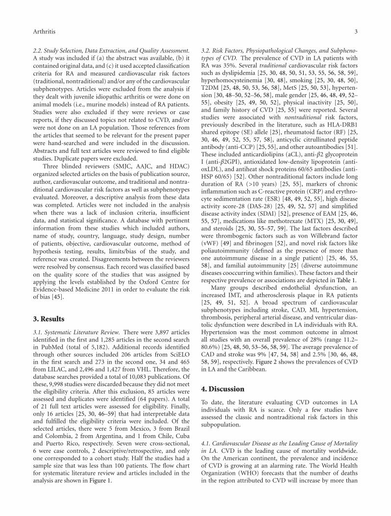
3.1. Systematic Literature Review. There were 3,897 articlesidentified in the first and 1,285 articles in the second searchin PubMed (total of 5,182). Additional records identifiedthrough other sources included 206 articles from SciELOin the first search and 273 in the second one, 34 and 465from LILAC, and 2,496 and 1,427 from VHL. Therefore, thedatabase searches provided a total of 10,083 publications. Ofthese, 9,998 studies were discarded because they did not meetthe eligibility criteria. After this exclusion, 85 articles wereassessed and duplicates were identified (64 papers). A totalof 21 full text articles were assessed for eligibility. Finally,only 16 articles [25, 30, 46–59] that had interpretable dataand fulfilled the eligibility criteria were included. Of theselected articles, there were 5 from Mexico, 3 from Braziland Colombia, 2 from Argentina, and 1 from Chile, Cubaand Puerto Rico, respectively. Seven were cross-sectional,6 were case controls, 2 descriptive/retrospective, and onlyone corresponded to a cohort study. Half the studies had asample size that was less than 100 patients. The flow chartfor systematic literature review and articles included in theanalysis are shown in Figure 1.
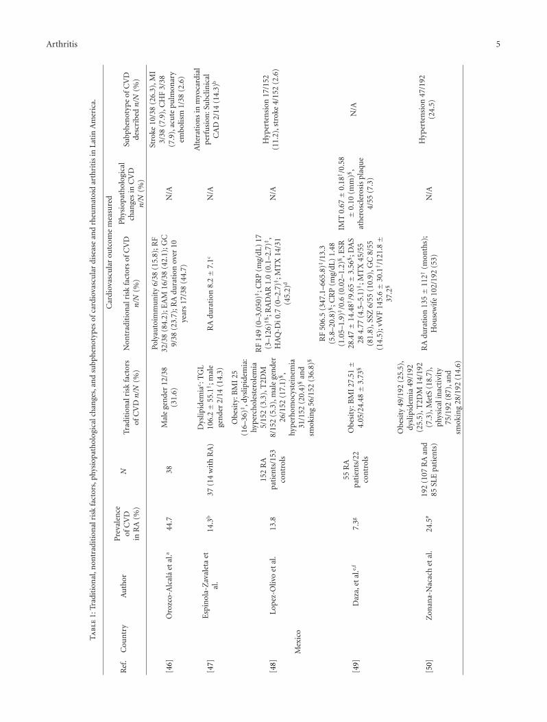
3.2. Risk Factors, Physiopathological Changes, and Subpheno-types of CVD. The prevalence of CVD in LA patients withRA was 35%. Several traditional cardiovascular risk factorssuch as dyslipidemia [25, 30, 48, 50, 51, 53, 55, 56, 58, 59],hyperhomocysteinemia [30, 48], smoking [25, 30, 48, 50],T2DM [25, 48, 50, 53, 56, 58], MetS [25, 50, 53], hyperten-sion [30, 48–50, 52–56, 58], male gender [25, 46, 48, 49, 52–55], obesity [25, 49, 50, 52], physical inactivity [25, 50],and family history of CVD [25, 55] were reported. Severalstudies were associated with nontraditional risk factors,previously described in the literature, such as HLA-DRB1shared epitope (SE) allele [25], rheumatoid factor (RF) [25,30, 46, 49, 52, 55, 57, 58], anticyclic citrullinated peptideantibody (anti-CCP) [25, 55], and other autoantibodies [51].These included anticardiolipins (aCL), anti-β2 glycoproteinI (anti-β2GPI), antioxidated low-density lipoprotein (anti-oxLDL), and antiheat shock proteins 60/65 antibodies (anti-HSP 60/65) [52]. Other nontraditional factors include longduration of RA (>10 years) [25, 55], markers of chronicinflammation such as C-reactive protein (CRP) and erythro-cyte sedimentation rate (ESR) [48, 49, 52, 55], high diseaseactivity score-28 (DAS-28) [25, 49, 52, 57] and simplifieddisease activity index (SDAI) [52], presence of EAM [25, 46,55, 57], medications like methotrexate (MTX) [25, 30, 49],and steroids [25, 30, 55–57, 59]. The last factors describedwere thrombogenic factors such as von Willebrand factor(vWF) [49] and fibrinogen [52], and novel risk factors likepoliautoimmunity (defined as the presence of more thanone autoimmune disease in a single patient) [25, 46, 55,58], and familial autoimmunity [25] (diverse autoimmunediseases cooccurring within families). These factors and theirrespective prevalence or associations are depicted in Table 1.
Many groups described endothelial dysfunction, anincreased IMT, and atherosclerosis plaque in RA patients[25, 49, 51, 52]. A broad spectrum of cardiovascularsubphenotypes including stroke, CAD, MI, hypertension,thrombosis, peripheral arterial disease, and ventricular dias-tolic dysfunction were described in LA individuals with RA.Hypertension was the most common outcome in almostall studies with an overall prevalence of 28% (range 11.2–80.6%) [25, 48, 50, 53–56, 58, 59]. The average prevalence ofCAD and stroke was 9% [47, 54, 58] and 2.5% [30, 46, 48,58, 59], respectively. Figure 2 shows the prevalences of CVDin LA and the Caribbean.
4. Discussion
To date, the literature evaluating CVD outcomes in LAindividuals with RA is scarce. Only a few studies haveassessed the classic and nontraditional risk factors in thissubpopulation.
4.1. Cardiovascular Disease as the Leading Cause of Mortalityin LA. CVD is the leading cause of mortality worldwide.On the American continent, the prevalence and incidenceof CVD is growing at an alarming rate. The World HealthOrganization (WHO) forecasts that the number of deathsin the region attributed to CVD will increase by more than

4 Arthritis
Iden
tifi
cati
onSc
reen
ing
Elig
ibili
tyIn
clu
ded
Records indentified through database searching
(PubMed)
First search: only (MeSH terms), n = 3,897
Second search: (MeSH terms) and key words,
n = 1 ,285
(n = 5 ,182)
Additional records identified through other sources(ScIELO) first search: DeCS terms, n = 206
Second search: DeCS terms and key words, n = 273
(LILACs) first search: DeCS terms, n = 34
Second search: only key words, n = 465
(VHL) first search: DeCS/MeSH terms, n = 2,496
Second search: only key words,n = 1,427(n = 4 ,901)
Potential articles were found
(n = 10 ,083) Excluded
Not human, not RA patients, JIA,
review, case report, other topic, not
about CVD outcomes, not made in
Latin American population
(n = 9,998)First screen, after exclusion
(n = 85)
Duplicated
(n = 64)
Full text articles assessed for
(n = 21)
Not about CVD outcomes
(n = 5)
Studies evaluating cardiovasculardisease and rheumatoid arthritis in
Latin America selected for
methodological analysis
(n = 16)
eligibility
Figure 1: Flow chart of the systematic literature review; VHL: virtual health library; RA: rheumatoid arthritis; JIA: juvenile idiopathicarthritis; CVD: cardiovascular disease.
60% between 2000 and 2020 unless preventive measuresare taken [60]. Thus, this chronic disease is one of themajor causes of death around the world [33]. Thanks tothe CARMELA initiative study, many traditional factorshave been described in LA population such as hypertension,dyslipidemia, obesity, smoking, T2DM, and MetS [61].
Table 2, which was adapted from the Pan AmericanHealth Organization report [62], shows the mortality rates ofCVD in the Americas as of 2007–2009 in terms of IHD andcerebrovascular disease. The data on this table is organizedby country and region thus making it possible to contrastmortality rates from these two diseases in the United Statesof America (USA) and Canada with LA and the Caribbean.Generally, high rates of death were mostly observed indeveloped countries such as USA and Canada 136.3/100,000people. Incidence of mortality in LA and the Caribbeandue to IHD and cerebrovascular disease is 55.8/100,000and 44.8/100,000 people, respectively. Individuals living indeveloped countries have more risk factors, for example,
inappropriate life styles, that contribute to a higher rate ofdeath from CVD. Thus, it is important to promote healthyhabits among the general population and in patients withan early diagnosis of RA in order to prevent CVD. Inspecific LA countries, numbers show high rates of IHD incountries such as Cuba (140.1/100,000 people) and PuertoRico (100.7/100,000 population). The importance of thenumbers lies in the fact that they can be analyzed from theperspective of increased risk of CVD in RA in comparisonto the general population. Therefore, it is important todiscriminate mortality CVD rates by patients with chronicinflammatory diseases (i.e., RA).
LA has a growing population and it is a very dynamicregion with an estimated population of 515 million. Asmentioned before, the RA prevalence reported in LA isconsidered to be less than 0.5% [63, 64]. The heterogeneityacross LA is expected due to the high degree of admixturebetween subpopulations. Hispanic/Latino populations arethe result of a two-way admixture between Amerindian
Arthritis 5
Ta
ble
1:Tr
adit
ion
al,n
ontr
adit
ion
alri
skfa
ctor
s,ph
ysio
path
olog
ical
chan
ges,
and
subp
hen
otyp
esof
card
iova
scu
lar
dise
ase
and
rheu
mat
oid
arth
riti
sin
Lati
nA
mer
ica.
Ref
.C
oun
try
Au
thor
N
Car
diov
ascu
lar
outc
ome
mea
sure
dP
reva
len
ceof
CV
Din
RA
(%)
Trad
itio
nal
risk
fact
ors
ofC
VDn/N
(%)
Non
trad
itio
nal
risk
fact
ors
ofC
VD
n/N
(%)
Phy
siop
ath
olog
ical
chan
ges
inC
VD
n/N
(%)
Subp
hen
otyp
eof
CV
Dde
scri
bedn/N
(%)
[46]
Oro
zco-
Alc
ala
etal
.a44
.738
Mal
ege
nde
r12
/38
(31.
6)
Poly
auto
imm
un
ity
6/38
(15.
8);R
F32
/38
(84.
2);E
AM
16/3
8(4
2.1)
;GC
9/38
(23.
7);R
Adu
rati
onov
er10
year
s17
/38
(44.
7)
N/A
Stro
ke10
/38
(26.
3),M
I3/
38(7
.9),
CH
F3/
38(7
.9),
acu
tepu
lmon
ary
embo
lism
1/38
(2.6
)
[47]
Esp
inol
a-Z
aval
eta
etal
.14
.3b
37(1
4w
ith
RA
)D
yslip
idem
iac :T
GL
106.
2±
55.1† ;
mal
ege
nde
r2/
14(1
4.3)
RA
dura
tion
8.2±
7.1c
N/A
Alt
erat
ion
sin
myo
card
ial
perf
usi
on:S
ubc
linic
alC
AD
2/14
(14.
3)b
[48]
Mex
ico
Lop
ez-O
livo
etal
.13
.815
2R
Apa
tien
ts/1
53co
ntr
ols
Obe
sity
:BM
I25
(16–
36)‡
,dys
lipid
emia
:hy
per
chol
este
role
mia
5/15
2(3
.3),
T2D
M8/
152
(5.3
),m
ale
gen
der
26/1
52(1
7.1)§ ,
hype
rhom
ocys
tein
emia
31/1
52(2
0.4)§
and
smok
ing
56/1
52(3
6.8)§
RF
149
(0–3
,050
)‡;C
RP
(mg/
dL)
17(3
–126
)ठ;
RA
DA
R1.
0(0
.1–2
.7)‡
,H
AQ
-Di0
.7(0
–2.7
)‡;M
TX
14/3
1(4
5.2)
d
N/A
Hyp
erte
nsi
on17
/152
(11.
2),s
trok
e4/
152
(2.6
)
[49]
Daz
a,et
al.e,
f7.
3g55
RA
pati
ents
/22
con
trol
s
Obe
sity
:BM
I27
.51±
4.05
/24.
48±
3.73
§
RF
506.
5(3
47.1
–665
.8)‡
/13.
3(5
.8–2
0.8)§ ;
CR
P(m
g/dL
)1.
48(1
.05–
1.9)‡ /
0.6
(0.0
2–1.
2)§ ,
ESR
28.4
7±
14.4
8†/9
.65±
3.56
§ ;D
AS
284.
77(4
.5–5
.1)‡
;MT
X45
/55
(81.
8),S
SZ6/
55(1
0.9)
,GC
8/55
(14.
5);v
WF
145.
6±
30.1† /
121.
8±
37.2§
IMT
0.67±
0.18
† /0.
58±
0.10
(mm
)§,
ath
eros
cler
osis
plaq
ue
4/55
(7.3
)
N/A
[50]
Zon
ana-
Nac
ach
etal
.24
.5#
192
(107
RA
and
85SL
Epa
tien
ts)
Obe
sity
49/1
92(2
5.5)
,dy
slip
idem
ia49
/192
(25.
5),T
2DM
14/1
92(7
.3),
Met
S(1
8.7)
,ph
ysic
alin
acti
vity
75/1
92(8
7),a
nd
smok
ing
28/1
92(1
4.6)
RA
dura
tion
135±
112†
(mon
ths)
;H
ouse
wif
e10
2/19
2(5
3)N
/AH
yper
ten
sion
47/1
92(2
4.5)

6 Arthritis
Ta
ble
1:C
onti
nu
ed.
Ref
.C
oun
try
Au
thor
N
Car
diov
ascu
lar
outc
ome
mea
sure
dP
reva
len
ceof
CV
Din
RA
(%)
Trad
itio
nal
risk
fact
ors
ofC
VDn/N
(%)
Non
trad
itio
nal
risk
fact
ors
ofC
VD
n/N
(%)
Phy
siop
ath
olog
ical
chan
ges
inC
VD
n/N
(%)
Subp
hen
otyp
eof
CV
Dde
scri
bedn/N
(%)
[52]
Pere
ira
etal
.i14
.1g
71R
Apa
tien
ts/5
1co
ntr
ols
Mal
ege
nde
rj7/
71(9
.9)
CR
Pk
(mg/
L):
8.64±
8.27
† /19
.75±
25.0
8,E
SRk:2
4.1±
14.6† /
34.0
7±
23.5
4;D
AS
28:4
.24±
1.02
† /4.
64±
1.05
;SD
AIk
:35.
54±
12.3
4/48
.5±
30.2
8§;M
TX
k(m
g/w
):20±
5/19
.09
±5.
74,G
Ck
(mg/
day)
:7.2
5±
2.75
/7.7
2±
3.59
;fibr
inog
enk
326.
04±
113.
56† /
371.
94±
121.
08§
IMT
j :0.7
2±
0.17
/0.6
7±
0.15
(mm
).C
arot
idpl
aqu
esj(
IMT>
1.5
mm
)10
/71
(14.
1)/1
(1.9
)§
N/A
[51]
Bra
zil
Pere
ira
etal
.l14
.1g
71R
Apa
tien
ts/5
3co
ntr
ols
Dys
lipid
emia
k:C
T24
3.3
±31
.2/1
91.5
4±
36.2
1§,
mal
ege
nde
rj(9
.9/1
3.9)
Sam
eda
tafr
om[5
2]an
dau
toan
tibo
dies
:RFk
:195
.10±
281.
71† /
308.
13±
584.
46,a
nti
-CC
P(7
9/1.
9),a
CL
(IgG
5.6/
3.8,
IgM
7/3.
8),a
nti
-B2G
PI
(IgG
1.4/
1.9,
IgM
4.2/
1.9)
,an
ti-H
SP60
(14.
1/7.
5),a
nti
-HSP
65(3
6.6/
17),
anti
-LP
Lan
tibo
dies
(2.8
/1.9
)
IMT
j :0.7
2±
0.17
/0.6
7±
0.15
(mm
).C
arot
idpl
aqu
esj(
IMT>
1.5
mm
)10
/71
(14.
1)/1
(1.9
)§
N/A
[53]
deC
un
ha
etal
.80
.6#
283
RA
pati
ents
/226
con
trol
s
Obe
sity
:BM
I26
.6±
5.1†
/26.
8±
4.3,
dysl
ipid
emia
:HD
L58
.9±
16.4† /
52.7±
12.1§ ,
LDL
109.
9±
33.2
/122
.8±
37.7§ ,
T2D
M32
/283
(11.
3)-6
/226
(2.7
)§,
Met
S11
1/28
3(3
9.2)
-44/
226
(19.
5)§ ,
mal
ege
nde
r50
(17.
7)/3
4(1
5)
N/A
N/A
Hyp
erte
nsi
onm
228
(80.
6)/9
5(4
2)

Arthritis 7
Ta
ble
1:C
onti
nu
ed.
Ref
.C
oun
try
Au
thor
N
Car
diov
ascu
lar
outc
ome
mea
sure
dP
reva
len
ceof
CV
Din
RA
(%)
Trad
itio
nal
risk
fact
ors
ofC
VDn/N
(%)
Non
trad
itio
nal
risk
fact
ors
ofC
VD
n/N
(%)
Phy
siop
ath
olog
ical
chan
ges
inC
VD
n/N
(%)
Subp
hen
otyp
eof
CV
Dde
scri
bedn/N
(%)
[54]
Pin
eda
etal
.n32
.441
Mal
ege
nde
r12
/41
(29)
N/A
N/A
Hyp
erte
nsi
on(2
4.2)
,C
AD
9/41
(8.2
)
[25]
Col
ombi
aR
ojas
-Vill
arra
gaet
al.o
41#
140
Obe
sity
23/1
40(1
6),
dysl
ipid
emia
49/1
40(3
5),T
2DM
6/14
0(4
),M
etS
61/1
40(4
4),
phys
ical
inac
tivi
ty11
9/14
0(8
5),m
ale
gen
der
16/(
23),
fam
ilyh
isto
ryof
CH
D22
/140
(16)
,eve
rsm
okin
g61
/140
(44)§ ,
and
his
tory
ofh
orm
one
repl
acem
ent
ther
apy
10/1
40(7
)
Poly
-au
toim
mu
nit
y31
/140
(22)
;fa
mily
his
tory
ofau
toim
mu
nit
y29
/140
(21)§ ;
HLA
-DR
B1
SE60
/136
(46)§ ;
RF
85/1
34(6
3)§ ,
anti
-CC
Pan
tibo
dies
73/9
4(7
8);C
RP
5.9±
15† ,
ESR
38.9±
25.1† ;
DA
S28
4.4
±1.
4†,H
AQ
1.7±
0.7†
;EA
M60
/140
(43)
;MT
X13
1/14
0(9
4),G
C13
1/14
0(9
4);R
Adu
rati
on13
.8±
8.5§
Ear
lyen
doth
elia
ldy
sfu
nct
ion
44/1
40(3
1)§ ,
incr
ease
dIM
T75
/140
(54)§ ,
ath
eros
cler
osis
plaq
ue
10/1
40(7
)§
Hyp
erte
nsi
on57
/140
(41)
[55]
Ort
ega-
Her
nan
dez
etal
.o32
538
Dys
lipid
emia
64/5
34(1
0),m
ale
gen
der
80/5
38(1
5)
Poly
auto
imm
un
ity
48/5
38(9
);R
F24
6/38
5(6
4)§ ,
anti
-CC
Pan
tibo
dies
146/
183
(80)
;CR
P8.
65±
20.2
1†,
ESR
†38
.86±
25.9
3§;E
AM
113/
538
(21)
;GC
39/4
86(8
);R
Adu
rati
on12
.53±
8.08
§
N/A
Hyp
erte
nsi
on12
8/53
4(2
4),t
hro
mbo
sis
43/5
34(8
)
[56]
Arg
enti
na
Larr
oude
etal
.13
.9#
137
Dys
lipid
emia
89/1
37(6
5),T
2DM
2/13
7(1
.45)
,an
dm
ale
gen
der
N/A
11/1
37(8
)
GC
71/1
37(5
1.8)
N/A
Hyp
erte
nsi
on19
/137
(13.
9)
[57]
Lasc
ano
etal
.p47
32R
Apa
tien
ts/3
2co
ntr
ols
N/A
RF
27/3
2(8
4);E
SR28±
15† ;
DA
S28
4.3±
1.4†
;EA
M8/
32(2
5);G
C21
/32
(66)
;RA
dura
tion
10.2±
8.4†
N/A
Ven
tric
ula
rdi
asto
licdy
sfu
nct
ion
15/3
2(4
7)
[30]
Ch
ileC
iste
rnas
etal
.q46
.454
RA
pati
ents
/32
con
trol
s
Obe
sity
:BM
I26
(18–
39)‡
,dys
lipid
emia
18/5
4(3
3),m
ale
gen
der
7/54
(13)
,fam
ilyh
isto
ryof
CV
D9/
54(1
7),
hype
rhom
ocys
tein
emia
38/5
4(7
0)§ ,
and
smok
ing
21/5
4(3
9)
RF
50/5
4(9
2),a
CL‡
IgM
3(0
.53–
23)/
1.6
(0.2
1–10
.6)
IgG
4.3
(0.3
–85)
/2.5
(0–1
2.3)
;CR
P0.
73(0
.04–
5.96
)‡/0
.31
(0.0
5–2.
88)§
,ESR
27(3
–99)‡ ;
MT
X41
/54
(75)
,GC
42/5
4(7
7);R
Adu
rati
on9.
5(0
.2–3
2)‡
N/A
Hyp
erte
nsi
on21
/54
(39)
,st
roke
2/54
(3.7
),st
able
angi
na
2/54
(3.7
)

8 Arthritis
Ta
ble
1:C
onti
nu
ed.
Ref
.C
oun
try
Au
thor
N
Car
diov
ascu
lar
outc
ome
mea
sure
dP
reva
len
ceof
CV
Din
RA
(%)
Trad
itio
nal
risk
fact
ors
ofC
VDn/N
(%)
Non
trad
itio
nal
risk
fact
ors
ofC
VD
n/N
(%)
Phy
siop
ath
olog
ical
chan
ges
inC
VD
n/N
(%)
Subp
hen
otyp
eof
CV
Dde
scri
bedn/N
(%)
[58]
Cu
baA
cost
aet
al.r
34.1
172
Dys
lipid
emia
10/1
72(5
.8),
T2D
M16
/172
(9.3
),an
dm
ale
gen
der
29/1
72(1
6.9)
Poly
auto
imm
un
ity
2/17
2(1
.1);
RF
52/8
5(6
1.1)
N/A
Hyp
erte
nsi
on46
/172
(26.
7),s
trok
e1/
172
(0.5
),C
AD
8/17
2(4
.6),
and
per
iph
eral
vasc
ula
rdi
seas
e4/
172
(2.3
)
[59]
Pu
erto
Ric
oSa
nti
ago-
Cas
aset
al.s
55.9
214
Dys
lipid
emia§
(9.1
)-(5
2.7)
-(58
.4)
T2D
M§
(9.1
)-(5
2.7)
-(58
.4)
Met
S(1
8.2)
-(39
.6)-
(43.
4)Sm
okin
g(4
.5)-
(11.
0)-(
7.9)
RF
(52.
4)-(
52.9
)-(5
7.1)
;ESR
(81.
0)-(
92.2
)-(9
1);S
tero
ids§
(54.
5)-(
78.0
)-(8
2.2)
;RA
dura
tion
‡
(3.4±
2.9)
-(9.
5±
8.2)
-(13
.6±
10.7
)
N/A
Hyp
erte
nsi
on(1
3.6)
-(40
.7)-
(76.
2),M
I(0
)-(2
.2)-
(9.1
),an
gin
ape
ctor
is(0
)-(1
.1)-
(4.0
),st
roke
(0)-
(1.1
)-(8
.0),
per
iph
eral
arte
rydi
seas
e(0
)-(1
.1)-
(5.0
),an
dC
HF
(0)-
(1.1
)-(5
.0)
CV
D:
card
iova
scu
lar
dise
ase;
RA
:rh
eum
atoi
dar
thri
tis;
RF:
rheu
mat
oid
fact
or;
EA
M:
extr
aart
icu
lar
man
ifes
tati
ons;
GC
:gl
uco
cort
icoi
ds;
N/A
:n
otav
aila
ble;
MI:
myo
card
ial
infa
rcti
on;
CH
F:co
nge
stiv
eh
eart
failu
re;T
GL
:tri
glyc
erid
es;C
AD
:cor
onar
yar
tery
dise
ase;
BM
I:bo
dym
ass
inde
x;T
2DM
:typ
e2
diab
etes
mel
litu
s;C
RP
:C-r
eact
ive
prot
ein
;RA
DA
R:r
apid
asse
ssm
ent
ofdi
seas
eac
tivi
tyin
rheu
mat
olog
y;H
AQ
-Di:
hea
lth
asse
ssm
ent
ques
tion
nai
redi
sabi
lity
inde
x;M
TX
:m
eth
otre
xate
;E
SR:
Ery
thro
cyte
Sedi
men
tati
onR
ate;
DA
S-28
:D
isea
seA
ctiv
ity
Scor
e-28
;SS
Z:
sulf
asal
azin
e;vW
F:vo
nW
illeb
ran
dFa
ctor
;IM
T:
inti
ma-
med
ial
thic
knes
s;M
etS:
met
abol
icsy
ndr
ome;
SDA
I:si
mpl
ified
dise
ase
acti
vity
inde
x;T
C:
tota
lch
oles
tero
l;an
ti-C
CP
:an
ti-c
yclic
citr
ulli
nat
edpe
ptid
ean
tibo
dies
;aC
L:
anti
card
iolip
ins
anti
bodi
es;
anti
-B2G
PI:
anti
-β2
glyc
opro
tein
Ian
tibo
dies
;an
ti-H
SP60
/65:
anti
-hea
tsh
ock
prot
ein
s60
/65
anti
bodi
es;a
nti
-LP
L:a
nti
Lip
opro
tein
lipas
ean
tibo
dies
;HD
L:h
igh
-den
sity
lipop
rote
inch
oles
tero
l;LD
L:lo
w-d
ensi
tylip
opro
tein
chol
este
rol.
a On
lyde
scri
ptiv
est
udy
,wh
ich
eval
uat
edca
use
sof
mor
talit
yin
adu
ltpa
tien
tsw
ith
RA
.bB
yec
hoc
ardi
ogra
man
dga
mm
agra
phy.
c Dat
afr
ompa
tien
tsw
ith
RA
14/3
7(3
7.8)
.dD
ata
from
pati
ents
wit
hhy
perh
omoc
yste
inem
ia(>
15μ
mol
/L).
e Exc
lusi
oncr
iter
ia:p
atie
nt
wit
htr
adit
ion
alca
rdio
vasc
ula
rri
skfa
ctor
s.f O
nly
fem
ale
wer
ein
clu
ded,
each
wit
hat
leas
t5
year
sof
dura
tion
ofth
edi
seas
ean
dbe
twee
n35
and
54ye
ars
ofag
e.gN
otC
VD
subp
hen
otyp
em
easu
red.
Pre
vale
nce
rega
rdin
gpr
esen
ceof
ath
eros
cler
osis
plaq
ue.
hO
nly
fem
ale
wer
ein
clu
ded.
i Exc
lusi
oncr
iter
ia:s
mok
ing,
diab
etes
and
hype
rten
sion
preg
nan
cy,r
enal
failu
re,c
hro
nic
hep
atop
athy
,nep
hro
tic
syn
drom
e,hy
poth
yroi
dism
and
use
ofst
atin
s/fi
brat
es.
j RA
pati
ents
vers
us
con
trol
s.l E
xclu
sion
crit
eria
:sm
okin
g,di
abet
es,a
nd
hyp
erte
nsi
on.
mH
igh
bloo
dpr
essu
rew
asde
fin
edab
ove
130/
85m
mH
g.n
Th
eob
ject
ive
was
toan
alyz
eca
use
san
ddi
rect
cost
sof
hos
pita
lizat
ion
ofC
olom
bian
pati
ents
wit
hR
A.
oSa
mpl
epo
pula
tion
was
orig
inal
lyfr
omN
orth
wes
tern
Col
ombi
a.T
hey
are
con
side
red
eth
nic
ally
diff
eren
t.pE
xclu
sion
crit
eria
:any
sym
ptom
sof
hea
rtdi
seas
eor
risk
fact
ors
for
CV
D.
qSu
bjec
tsov
er60
year
sw
ere
excl
ude
d.r O
nly
coh
ort,
6ye
ars
follo
wu
p.Lo
wm
orta
lity
rate
9/32
(5.2
%).
s Th
ree
age
grou
p(<
40y)
-(40
–59
y)-(>
60y)
.Eld
erp
eopl
e(>
60y)
hav
em
ore
prob
abili
tyto
deve
lop
CV
Din
depe
nde
nt
ofR
A.
† Mea
n±
stan
dard
devi
atio
n.
‡ Med
ian
(in
terq
uar
tile
ran
ge).
#P
reva
len
ceof
CV
Dre
gard
ing
the
only
subp
hen
otyp
ede
scri
bed.
§ Pva
lues
<0.
05w
ere
con
side
red
sign
ifica
nt.

Arthritis 9
Mexico: 20.9%
Cuba
Puerto Rico
Brazil: 47.4%
Colombia: 35.1%
Chile
Argentina: 33.5%
CA: 20.9%
C: 45%44.7% (46)a
14.3% (47)b
13.8% (48)c
7.3% (49)d
24.5% (50)a,e
34.1% (58)l
55.9% (59)m
41% (25)g
32.4% (54)h
32% (55)i
SA: 39.8%
46.4% (30)k
LA: 35.3%
13.9% (56)e
47% (57) j
14.1% (51, 52)d,f
80.6% (53)e
Figure 2: Cardiovascular disease in rheumatoid arthritis in Latin America and the Caribbean; LA: Latin America; CA: Central America;SA: South America; C: Caribbean. aGeneral cause of death was evaluated. CVD was the highest. bSubclinical coronary artery disease.cHypertension and stroke. dNot CVD subphenotype measured. Prevalence regarding presence of atherosclerosis plaque. eHypertensionf References [48, 49] report data from the same cohort of patients. Hence, the prevalence of CVD and risk factors is identical. gHypertensionand atherosclerosis plaque. hHypertension and coronary artery disease. iHypertension and thrombosis. jVentricular diastolic dysfunction.kHypertension, stroke, and stable angina. lHypertension, stroke, coronary artery disease, peripheral vascular disease. mHypertension,myocardial infarction, angina pectoris, stroke, and peripheral vascular disease, and congestive heart failure.
and European populations or of three-way admixture ofAmerindian, European, and West African populations [65].
Some studies have documented differences in the healthstatus of, disease prevalence in, treatment outcome in, andhealthcare use by different ethnic groups. Yazici et al. [35]compared patients from different ethnic groups with earlyRA using disease activity measures, identifying possibledifferences in patterns of clinical severity. They found thatHispanic patients with RA scored the worst in all self-reportmeasures compared to Caucasians and African Americanswith statistically significant differences in the Modified
Health Assessment Questionnaire (MHAQ) functional score,psychological distress, and morning stiffness [35]. In a studyof RA patients, Bruce et al. [36] demonstrated disparitiesbetween Caucasians and African Americans and Hispanicsin disability, pain, and global health. Pain was worse in thelatter two groups and global health was worse in Hispanics.The results of this exploratory study suggest that in arelatively similar cohort of patients with RA, minority healthdisparities exist [36]. Moreover, the prevalence of MI is highin Hispanics living in the USA, and coronary events arepresented by people younger than in other minorities [48].

10 Arthritis
Table 2: Cardiovascular disease mortality in the Americas∗.
RegionAnnual deaths average Mortality rate from IHDa,b Mortality rate from cerebrovascular diseasea
(thousands)a Total Total
Americas 6,447.2 87.4 45.1
North America 2,885 136 45
Canada 262.8 109 41.4
United States of America 2,621.7 139 45.4
Latin America and the Caribbean 3,562.2 55.8 44.8
Latin America 3,510.8 56 44.9
Mexico 549.4 54.1 27.5
Central American Isthmus 226.1 41.9 24.4
Belize 1.2 30.9 25.7
Costa Rica 20.4 48.4 21.3
El Salvador 41 56 22.4
Guatemala 80.5 25.5 16.4
Honduras 37.5 N/A N/A
Nicaragua 27.5 54.2 32.8
Panama 18.1 57.2 51.5
Latin Caribbean 270.5 N/A N/A
Cuba 83.9 140 80.6
Dominican Republic 60.1 N/A N/A
French Guiana 0.9 N/A N/A
Haiti 90 N/A N/A
Puerto Rico 29.1 101 40.1
Andean Area 722.5 58.7 35.7
Bolivia 72.9 N/A N/A
Colombia 260.6 74.1 38.7
Ecuador 74.5 25.6 34.1
Peru 161.4 27.8 26.6
Venezuela 153.1 81.4 41
Brazil 1.261.1 60.4 62.2
Southern Cone 481.3 49.1 51.2
Argentina 315.6 46.8 48.2
Chile 98.2 47.1 46.8
Paraguay 36.1 50.3 55.5
Uruguay 31.3 85.4 103
Non-Latin Caribbean 51.3 63.4 63.8
Guyana 4.4 80.9 70.3
Suriname 3.8 47 72∗
Adapted from [62]. The values were obtained from “Corrected Mortality” data. These values were computed by applying a correction algorithm for mortalityunderregistration and a redistribution algorithm for deaths from ill-defined causes. The methodology used is presented in Health Statistics from the Americas.2006 edition (http://www.paho.org/HSA2006).aValues are expressed in incidence rates/100.000 population (2007–2009).bIHD: ischemic heart disease.N/A: not available.
Nevertheless, only two studies in LA assessed mortalityin RA patients. Orozco-Alcala et al. [46] showed that therewere no differences between RA patients and the generalpopulation concerning causes of death. Acosta et al. [58]
demonstrated a mortality rate of 5.2% in a six-year followup.For both, the most frequent cause of death was CVD in44.7% and 22.2% of the cases, respectively. In the otherselected articles, a wide range of prevalence for CVD was
Arthritis 11
reported (13.8–80.6%). The highest prevalence was indicatedby Santiago-Casas et al. [59] in Puerto Rican patients(55.9%) when the demographic characteristics, clinicalmanifestations, comorbidities, pharmacological profile, andfunctional status of different age groups were determined.Nevertheless, the fact that elderly people (>60 years) havea higher probability of developing CVD whether or notthey have RA had to be taken into account for calculatingthe prevalence of CVD in Puerto Rico. Cisternas et al. [30]evaluated cardiovascular risk factors in Chilean patients withRA and reported a prevalence of 46.4% for CVD. For Brazil[51, 53], Colombia [25, 54, 55], and Argentina [56, 57],a similar prevalence was indicated (47.4, 35.1 and 30.5%,resp.). In Mexico, five studies [46–50] reported an overallprevalence of 20.9% for CVD in RA patients.
4.2. Traditional Risk Factors, CVD, and RA. RA is a relativelyfrequent AD, which is chronic in nature, and these patientsare doubly at risk of developing any CVD subphenotypewith respect to the non-RA population [66, 67]. In fact, IHDsecondary to atherosclerosis is the most prevalent cause ofdeath associated with CVD in patients with RA [30]. Theworldwide prevalence of hypertension in RA is between 49and 77% [5]. It is considered the most common comorbiditiyin Hispanic patients with RA. The most frequent classic riskfactor for CVD in this systematic literature review (withmore than 2,000 RA patients included) was hypertensionas well. Nevertheless, a lower prevalence (27.9%) than thatreported previously in other countries was found. Manyof these predisposing factors have been described in LAstudies: hypertension [30, 36, 53–55, 58, 59, 61, 68, 69],T2DM [25, 48, 50, 53, 56, 58], dyslipidemia [25, 58, 59, 70],MetS [17, 25, 50, 53, 68, 69, 71], and hyperhomocysteinemia[22, 25, 48, 72]. For details, see Table 3.
4.3. Nontraditional Risk Factors, CVD, and RA. Since thereis no classification system for nontraditional risk factors,we would like to propose one. Our recommendation isto divide them into genetic, AD associated, and others.The genetic group includes both HLA and non-HLA genes.HLA-DRB1 SE alleles are related to chronic inflammation,endothelial dysfunction, premature death, and CVD itself[25, 73–80]. The non-HLA genes include polymorphisms inthe endothelin-1 and methylene tetrahydrofolate reductasegenes. Endothelin-1 enhances CVD by endothelial dys-function and hypertension [81]. Methylene tetrahydrofolatereductase has been related to atherosclerosis and the clinicalresponse to some Disease-Modifying Antirheumatic Drugs(DMARDs) [82]. Others genes are TNFA rs1800629 andNFKB1-94ATTG ins/del polymorphisms. These are associ-ated with predisposition to cardiovascular complications inpatients with RA, as subclinical and accelerated atheroscle-rosis [83, 84]. However, other gene polymorphisms placedoutside the HLA region and not strongly associated withsusceptibility to RA and CVD. Rodrıguez-Rodrıguez et al.[85] showed a potential influence of the CCR5Δ32 deletionon the risk of CV disease among patients with RA. This may
be due to a protective effect of this allelic variant against thedevelopment of vascular endothelial dysfunction.
The AD associated factors include a broad spectrum ofautoantibodies as well as RA characteristics. The autoanti-bodies include RF [25, 49, 86], anti-CCP, aCL, anti-B2GPI,anti-HSP 60/65 [25, 30, 51, 55], and anti-oxLDL [30, 87,88]. The RA characteristics are inflammatory basis [39, 89,90], high disease activity [91], long duration [25], systemicinvolvement [56, 76, 92], treatment (systemic steroids) [93–95], and others, recently described, such as polyautoimmu-nity [25, 46, 55, 58] and familial autoimmunity [25].
Other issues, such as thrombogenic factors, whichinclude vWF and fibrinogen levels, are related to CVD aswell [49, 96, 97]. Several new cardiovascular risk factors inRA have received only modest attention and the differentstudies have shown contradictory results in LA patients.Each of these factors contribute to an impaired endothelialfunction, increased IMT, accelerated atherosclerosis, andfinally, manifest CVD. For details, see Table 3.
4.4. Discovering Novel Nontraditional Risk Factors. Despite ofall the traditional risk factors that have been associated withCVD in RA patients, the literature on it with respect to LAand the Caribbean is still scarce. Even though it has beengenerally accepted that systemic activity is related to chronicinflammation and accelerated pathogenic processes leadingto cardiovascular compromise, it is important to assess othernovel factors in patients that may also contribute. Therefore,we believe further research is needed in order to establishother factors that are not currently taken into account. Todate, there are no systematic reviews of literature involvingLA patients as a minority group.
After the systematic search was done, 2,119 RA patientsfrom different LA countries were included and evaluated forcardiovascular outcomes in studies ranging from 1993 to2012 (see Supplementary Table 1 in Supplementary Mate-rial available online at doi:1155/2012/371909). Commonlimiting factors in the sixteen studies analyzed included alack of prospective follow up of RA patients and a generallimitation on sample sizes. Most of the studies were eithercross-sectional or case-control which in terms of evidenceplace them at level 4 [45]. Moreover, 50% of the studiesincluded in the analysis had sample sizes of more than100 RA patients. The rest of them had limited numbersof patients included, which was another common limit orbias found in the retrieved studies. Furthermore, the lack ofadequate statistical methods and hypothesis testing in someof the studies should be noted. This was the case for fourof the studies, which were descriptive or did not calculate Pvalues, adjusted odds ratio or confidence intervals.
There is insufficient literature regarding CVD in LApatients with RA. Although the number of patients assessedis not negligible, when the geographical area of LA, the diver-sity, and the admixture of the population are considered,there is a need to include true cohorts to ensure more decisiveconclusions.

12 Arthritis
Table 3: Traditional and non-traditional risk factors associated with cardiovascular disease and rheumatoid arthritis in Latin America.
Risk factor associated with CVD Comments Reference(s)
Traditional
HypertensionIncreases the risk to suffer IHD or stroke with an important impact on mortality inpatients with RA
[16]
T2DMPatients with RA have a similar risk of developing CVD when compared to thesame risk in patients with T2DM. Unfortunately, when there is a coexistence ofboth diseases, this risk is increased by three times
[69]
DyslipidemiaAltered lipid profiles in RA patients are related with higher probability of IHD byaccelerating atherosclerosis
[25, 70]
Is characterized for an alteration in production/secretion of proinflammatoryadipokines and leads to increased activity of RA and accelerating atherosclerosis
[68, 71]
MetSStudies about the prevalence of MetS in LA patients have not achieved definitiveconclusions, although its presence has been directly associated with a worseprognosis
[53]
In RA patients, was related with pain and functional status, suggesting diseaseactivity. Therefore, a better control of disease activity may reduce CVD risk
[50]
Hyperhomocysteinemia
Homocysteine is considered as biomarker for atherosclerosis and a risk factorrelated with CAD and stroke
[22, 72]
There is still controversy about whether hyperhomocysteinemia is a causative agentof cardiovascular damage or only an epiphenomenon of inflammation
[48]
A high prevalence of this biomarker in Mexican patients with RA had a statisticalassociation with male gender and higher radiological damage
[48]
High homocysteine concentration can be an important risk marker for CVD inChilean patients with RA, as it was significantly associated
[30]
Nontraditional
Related with chronic inflammation, endothelial dysfunction, and premature deathfor CVD
[73–75]
Genetic HLA-DRB1 SE allelesAssociated with severe RA and with more EAM, high activity, and systemicinflammation
[74–77, 79]
Being a carrier of a single copy of HLA-DRB1 SE were significantly associated withan increased risk of atherosclerotic plaque in RA Colombian patients
[25]
PolyautoimmunitySome articles included patients with poliautoimmunity, but no correlation withCVD subphenotypes was described
[25, 46, 55, 58]
Familial autoimmunity Was associated with presence of atherosclerotic plaque in RA Colombian patients.[25]
High titers have been established to be a predictor of CVD due to immune complexformation and tissue injury. It has been shown that such immune complexes fromRF can be deposited in the endothelium and through inflammatory reactionsgenerate endotelial disfunction and atherosclerotic process
[86]
RF positivityRF seropositivity was significantly associated with an increased risk of endothelialdysfunction in RA Colombian patients
[25]
A statistical association between increased IMT, atherosclerosis plaque, andpresence of RF was described in Mexican population with RA
[49]
anti-oxLDL
Promote instability and rupture of the atheromatous plaque within the coronaryarteries
[24, 88]
Only one LA study evaluated this antibodies, but no correlation with CVD wasfound
[30]
Other autoantibodies
The presence of plaques was higher in Brazilian patients with RA, but nocorrelation between IMT or plaques and autoantibodies were found
[51]
ADassociated
Other autoantibodies were assessed in LA population, such as aCL, anti-β2GPI,anti-HSP 60/6, and anti-CCP antibodies with no association regarding CVDoutcomes
[25, 30, 51, 55]
Inflammatory markers
The association of inflammatory pathways with CVD is complex and is composedof several intermediate factors, including dyslipidemia, homocysteinemia, insulinresistance, and endothelial dysfunction
[89]

Arthritis 13
Table 3: Continued.
Risk factor associated with CVD Comments Reference(s)
May accelerate atherogenic processes, either by the accentuation of knownpathways of plaque formation or by the onset of additional immune pathways
[90]
Disease activityThe lipid profile in RA depends on disease activity. Higher disease activity leads todepressed levels of total cholesterol. However, HDL cholesterol levels are even moredepressed, resulting in a more unfavourable atherogenic index
[90]
Long duration of RA(>10 years)
Implies more time for chronic inflammatory process to generate sequelae such asatherosclerosis and endothelial dysfunction
[39]
Were significantly associated with an increased risk of atherosclerotic plaque in RAColombian patients
[25]
EAMIs an indirect indicator of disease severity and systemic compromise.Patients are considered to have three times higher risk to develop CVD
[55, 76]
GC
Could enhance cardiovascular risk owing to their potentially deleterious effects onlipids, glucose tolerance, insulin production and resistance, blood pressure, andobesity. On the other hand, it may actually decrease the risk of atherosclerosis andCVD by suppressing inflammation, which paradoxically may improve glucoseintolerance and dyslipidaemia
[93]
Others Thrombogenic factors
vWF has been recognized to induce a procoagulant stateRepresent a biomarker of endothelial dysfunction
[96, 97]
The measurements of the IMT together with the vWF serum levels could givevaluable information about the artery status and the atherosclerosis process in earlystages in Mexican patients with RA without cardiovascular risk factors
[49]
CVD: cardiovascular disease; IHD: ischemic heart disease; RA: rheumatoid arthritis; T2DM: type 2 diabetes mellitus; LA: Latin America; MetS: metabolicsyndrome; SE: shared epitope; RF: rheumatoid factor; IMT: intima-medial thickness; anti-oxLDL: anti-oxidized low-density lipoprotein antibodies; aCL:anticardiolipins antibodies; anti-B2GPI: anti-β2 glycoprotein I antibodies; anti-HSP 60/65: antiheat shock proteins 60/65 antibodies; anti CCP: anti-cycliccitrullinated peptide antibodies; HDL: high-density lipoprotein cholesterol; EAM: extra-articular manifestations; GC: glucocorticoids; vWF: von Willebrandfactor.
4.5. Assessing CVD in RA Patients. Heart disease in patientswith RA is a major concern. Rheumatologists often face thequestion of how to treat and prevent CVD. To appropriatelydo so, we need to answer three important questions. (1)How do we estimate the risk of CVD in RA? Unfortu-nately, neither the Framingham Risk Score nor Reynold’sRisk Score were designed to estimate risk in RA patients.The European League Against Rheumatism published theirrecommendation on estimating cardiovascular risk in RA;however, this has not been validated yet. (2) Which actionsdecrease CVD risk? Eating a well-balanced diet, exercisingon a regular basis, quitting smoking, and maintaining ahealthy weight have a positive impact on cardiovascularhealth. Targets based on the individual risk profile of everypatient also have to be set. Well-established risk factorssuch as blood pressure, LDL levels, and hemoglobin A1Cneed to be considered. Treatments that reduce these riskfactors include angiotensin-converting enzyme inhibitors,statins, and, in some patients, metformin. (3) What shouldbe the target of all these efforts? That question raises morequestions. Inflammation in RA is a risk factor for CVD whichcan be treated effectively, but can targeting “inflammation”decrease CVD risk in RA? Should the target be remission,a low CRP level, or lack of swollen joints? Is targetingspecific inflammatory pathways more effective for reducingcardiovascular risk than other therapies? There are manyunanswered questions and a lot of controversy about how
to best address cardiovascular risk in patients with RA.Therefore, a comprehensive multidisciplinary approach isthe first step towards addressing this complex issue and tooptimize patient outcomes [98].
5. Conclusions
RA and CVD share common pathophysiology mechanisms(i.e., systemic and chronic inflammation) with secondaryaccelerated atherosclerosis that can explain the high mor-tality rates and augmented risk of ischemic events inthese patients. Therefore, early or subclinical atherosclerosisshould be assessed in every patient through the measurementof IMT in carotid arteries and other inflammatory markerson a regular clinical basis.
LA patients are ethnically different from other pop-ulations and have a worse disease course due to theirdifferent genetic burden that could be the cause of a higherprevalence of EAM. Trying to extrapolate previous resultsfrom countries with patients from a different ethnic groupto our subpopulation could be a mistake.
Although there is an evident association of traditionalrisk factors and cardiovascular compromise in RA patients,they do not completely explain the high rates of CVD inthese patients. Thus, novel risk factors which are related toautoimmunity are now becoming a more important focus

14 Arthritis
of attention. This is the reason why we propose to separatetraditional and nontraditional risk factors and evaluate themcomprehensively and in a multidisciplinary fashion.
There is a lack of literature about CVD in Hispanicpatients as demonstrated by this systematic search. Tomake matters worse, literature evaluating nontraditionalrisk factors is scarce. This should be a challenge to therheumatologist to do research in these fields in order toelucidate the underlying mechanisms involved for the benefitof the patient.
Unfortunately, LA patients receive lower quality diagnos-tic assessment and treatment choices than Caucasian patientsdue to difficulties in access to health services and delayeddiagnosis. Cardiovascular compromise in RA patients is atherapeutic challenge and doctors need to be committedto assessing, monitoring, and treating cardiovascular riskfactors in the early stages as well as generating effective publichealth policies in developing LA countries so that morbi-mortality rates can be decreased promptly.
Abbreviations
CVD: Cardiovascular diseaseRA: Rheumatoid arthritisLA: Latin AmericaEAM: ExtraarticularmanifestationsT2DM: Type 2 diabetes mellitusMetS: Metabolic syndromeAD: Autoimmune diseaseIMT: Intima-medial thicknessCAD: Coronary artery diseaseMI: Myocardial infarctionCHF: Congestive heart diseaseIHD: Ischemic heart diseaseVHL: Virtual health librarySE: Shared epitopeRF: Rheumatoid factorAnti CCP: Anti-cyclic citrullinatedpeptide
antibodiesaCL: Anticardiolipin antibodiesAnti-B2GPI: Anti-β2 glycoprotein I antibodiesAnti-oxLDL: Antioxidized low-density lipoprotein
antibodiesAnti-HSP 60/65: Antiheat shock proteins 60/65
antibodiesCRP: C-Reactive ProteinESR: Erythrocyte sedimentation rateDAS-28: Disease activity score-28SDAI: Simplified disease activity indexMTX: MethotrexatevWF: von Willebrand factorWHO: World Health OrganizationMHAQ: Modified Health Assessment
QuestionnaireDMARDs: Disease nodifying anti rheumatic drugs.
Conflict of Interests
The authors declare no conflict of interests.
Acknowledgments
The authors are grateful to the all the members of Centerfor Autoimmune Diseases Research (CREA) for their fruitfuldiscussions and contributions to this paper. This work wassupported by the School of Medicine and Health Sciences ofUniversidad del Rosario in Bogota, Colombia.
References
[1] D. L. Scott, F. Wolfe, and T. W. J. Huizinga, “Rheumatoidarthritis,” The Lancet, vol. 376, no. 9746, pp. 1094–1108, 2010.
[2] A. N. DeMaria, “Relative risk of cardiovascular events inpatients with rheumatoid arthritis,” American Journal ofCardiology, vol. 89, no. 6, pp. 33D–38D, 2002.
[3] A. Sandoo, J. J. C. S. Veldhuijzen van Zanten, G. S. Metsios, D.Carroll, and G. D. Kitas, “Vascular function and morphologyin rheumatoid arthritis: a systematic review,” Rheumatology,vol. 50, pp. 2125–2139, 2011.
[4] P. Sarzi-Puttini, F. Atzeni, R. Gerli et al., “Cardiac involvementin systemic rheumatic diseases: an update,” AutoimmunityReviews, vol. 9, no. 12, pp. 849–852, 2010.
[5] A. Farzaneh-Far and M. J. Roman, “Accelerated atherosclerosisin rheumatoid arthritis and systemic lupus erythematosus,”International Journal of Clinical Practice, vol. 59, no. 7, pp.823–824, 2005.
[6] H. R. Kramer and J. T. Giles, “Cardiovascular disease risk inrheumatoid arthritis: progress, debate, and opportunity,”Arthritis Care & Research, vol. 63, no. 4, pp. 484–499, 2011.
[7] E. Myasoedova and S. E. Gabriel, “Cardiovascular diseasein rheumatoid arthritis: a step forward,” Current Opinion inRheumatology, vol. 22, no. 3, pp. 342–347, 2010.
[8] N. Sattar and I. B. McInnes, “Vascular comorbidity inrheumatoid arthritis: potential mechanisms and solutions,”Current Opinion in Rheumatology, vol. 17, no. 3, pp. 286–292,2005.
[9] S. M. A. Toloza, A. G. Uribe, G. McGwin et al., “Systemic lupuserythematosus in a multiethnic US cohort (LUMINA): XXIII.Baseline predictors of vascular events,” Arthritis and Rheuma-tism, vol. 50, no. 12, pp. 3947–3957, 2004.
[10] Y. Sherer and Y. Shoenfeld, “Antiphospholipid syndrome,antiphospholipid antibodies, and atherosclerosis,” CurrentAtherosclerosis Reports, vol. 3, no. 4, pp. 328–333, 2001.
[11] K. Veres, G. Lakos, A. Kerenyi et al., “Antiphospholipidantibodies in acute coronary syndrome,” Lupus, vol. 13, no.6, pp. 423–427, 2004.
[12] R. Cervera, “Coronary and valvular syndromes and antiphos-pholipid antibodies,” Thrombosis Research, vol. 114, no. 5-6,pp. 501–507, 2004.
[13] S. Guiducci, R. Giacomelli, and M. M. Cerinic, “Vascularcomplications of scleroderma,” Autoimmunity Reviews, vol. 6,no. 8, pp. 520–523, 2007.
[14] M. Perez-De-Lis, M. Akasbi, A. Siso et al., “Cardiovascular riskfactors in primary Sogren’s syndrome: a case-control study in624 patients,” Lupus, vol. 19, no. 8, pp. 941–948, 2010.
[15] A. Stavropoulos-Kalinoglou, G. S. Metsios, V. F. Panoulas etal., “Associations of obesity with modifiable risk factors forthe development of cardiovascular disease in patients withrheumatoid arthritis,” Annals of the Rheumatic Diseases, vol.68, no. 2, pp. 242–245, 2009.
[16] V. F. Panoulas, G. S. Metsios, A. V. Pace et al., “Hypertension inrheumatoid arthritis,” Rheumatology, vol. 47, no. 9, pp. 1286–1298, 2008.

Arthritis 15
[17] R. M. R. Pereira, J. F. de Carvalho, and E. Bonfa, “Metabolicsyndrome in rheumatological diseases,” AutoimmunityReviews, vol. 8, no. 5, pp. 415–419, 2009.
[18] V. F. Panoulas, K. M. J. Douglas, H. J. Milionis et al., “Preva-lence and associations of hypertension and its control inpatients with rheumatoid arthritis,” Rheumatology, vol. 46, no.9, pp. 1477–1482, 2007.
[19] M. J. Kaplan, “Cardiovascular disease in rheumatoid arthritis,”Current Opinion in Rheumatology, vol. 18, no. 3, pp. 289–297,2006.
[20] G. D. Kitas and S. E. Gabriel, “Cardiovascular disease inrheumatoid arthritis: state of the art and future perspectives,”Annals of the Rheumatic Diseases, vol. 70, pp. 8–14, 2011.
[21] N. J. Goodson, N. J. Wiles, M. Lunt, E. M. Barrett, A. J. Silman,and D. P. M. Symmons, “Mortality in early inflammatory pol-yarthritis: cardiovascular mortality is increased in seropositivepatients,” Arthritis and Rheumatism, vol. 46, no. 8, pp. 2010–2019, 2002.
[22] N. Sattar, D. W. McCarey, H. Capell, and I. B. McInnes,“Explaining how ”high-grade” systemic inflammation accel-erates vascular risk in rheumatoid arthritis,” Circulation, vol.108, no. 24, pp. 2957–2963, 2003.
[23] S. L. Whittle and R. A. Hughes, “Folate supplementation andmethotrexate treatment in rheumatoid arthritis: a review,”Rheumatology, vol. 43, no. 3, pp. 267–271, 2004.
[24] S. H. Kim, C. K. Lee, Y. L. Eun et al., “Serum oxidized low-density lipoproteins in rheumatoid arthritis,” RheumatologyInternational, vol. 24, no. 4, pp. 230–233, 2004.
[25] A. Rojas-Villarraga, O. D. Ortega-Hernandez, L. F. Gomez etal., “Risk factors associated with different stages of atheroscle-rosis in colombian patients with rheumatoid arthritis,” Sem-inars in Arthritis and Rheumatism, vol. 38, no. 2, pp. 71–82,2008.
[26] F. Levendoglu, A. Temizhan, H. Ugurlu, A. Ozdemir, andM. Yazici, “Ventricular function abnormalities in activerheumatoid arthritis: a Doppler echocardiographic study,”Rheumatology International, vol. 24, no. 3, pp. 141–146, 2004.
[27] C. Gonzalez-Juanatey, A. Testa, A. Garcia-Castelo et al.,“Echocardiographic and doppler findings in long-term treatedrheumatoid arthritis patients without clinically evident car-diovascular disease,” Seminars in Arthritis and Rheumatism,vol. 33, no. 4, pp. 231–238, 2004.
[28] A. E. Voskuyl, “The heart and cardiovascular manifestationsin rheumatoid arthritis,” Rheumatology, vol. 45, supplement4, pp. iv4–iv7, 2006.
[29] V. R. da Cunha, C. V. Brenol, J. C. T. Brenol, and R. M. Xavier,“Rheumatoid arthritis and metabolic syndrome,” RevistaBrasileira de Reumatologia, vol. 51, no. 3, pp. 260–268, 2011.
[30] M. Cisternas, M. A. Gutierrez, J. Klaassen, A. M. Acosta, and S.Jacobelli, “Cardiovascular risk factors in Chilean patients withrheumatoid arthritis,” Journal of Rheumatology, vol. 29, no. 8,pp. 1619–1622, 2002.
[31] D. H. Solomon, E. W. Karlson, E. B. Rimm et al., “Cardio-vascular morbidity and mortality in women diagnosed withrheumatoid arthritis,” Circulation, vol. 107, no. 9, pp. 1303–1307, 2003.
[32] M. Chan, Global Status Report on Noncommunicable Diseases,World Health Organization, 2010.
[33] D. Yach, C. Hawkes, C. L. Gould, and K. J. Hofman, “Theglobal burden of chronic diseases: overcoming impedimentsto prevention and control,” Journal of the American MedicalAssociation, vol. 291, no. 21, pp. 2616–2622, 2004.
[34] A. L. Price, N. Patterson, F. Yu et al., “A genomewide admixturemap for latino populations,” American Journal of HumanGenetics, vol. 80, no. 6, pp. 1024–1036, 2007.
[35] Y. Yazici, H. Kautiainen, and T. Sokka, “Differences inclinical status measures in different ethnic/racial groups withearly rheumatoid arthritis: implications for interpretation ofclinical trial data,” Journal of Rheumatology, vol. 34, no. 2, pp.311–315, 2007.
[36] B. Bruce, J. F. Fries, and K. N. Murtagh, “Health status dispar-ities in ethnic minority patients with rheumatoid arthritis: across-sectional study,” Journal of Rheumatology, vol. 34, no. 7,pp. 1475–1479, 2007.
[37] Y. Shoenfeld, R. Gerli, A. Doria et al., “Accelerated atheroscle-rosis in autoimmune rheumatic diseases,” Circulation, vol.112, no. 21, pp. 3337–3347, 2005.
[38] I. Avalos, Y. H. Rho, C. P. Chung, and C. M. Stein,“Atherosclerosis in rheumatoid arthritis and systemic lupuserythematosus,” Clinical and Experimental Rheumatology, vol.26, no. 5, pp. S5–S13, 2008.
[39] L. E. Full, C. Ruisanchez, and C. Monaco, “The inextricablelink between atherosclerosis and prototypical inflammatorydiseases rheumatoid arthritis and systemic lupus erythemato-sus,” Arthritis Research & Therapy, vol. 11, no. 2, p. 217, 2009.
[40] L. Massardo, B. A. Pons-Estel, D. Wojdyla, M. H. Cardiel,C. M. Galarza-Maldonado, M. P. Sacnun et al., “Earlyrheumatoid arthritis in Latin America: low socioeconomicstatus related to high disease activity at baseline,” Arthritis Care& Research, vol. 64, pp. 1135–1143, 2012.
[41] A. Spindler, V. Bellomio, A. Berman et al., “Prevalenceof rheumatoid arthritis in Tucuman, Argentina,” Journal ofRheumatology, vol. 29, no. 6, pp. 1166–1170, 2002.
[42] I. Pelaez-Ballestas, L. H. Sanin, J. Moreno-Montoya et al.,“Epidemiology of the rheumatic diseases in Mexico. A studyof 5 regions based on the COPCORD methodology,” Journalof Rheumatology, supplement, vol. 86, pp. 3–6, 2011.
[43] J. M. Anaya, P. A. Correa, R. D. Mantilla, F. Jimenez, T.Kuffner, and J. M. McNicholl, “Rheumatoid arthritis inAfrican Colombians from Quibdo,” Seminars in Arthritis andRheumatism, vol. 31, no. 3, pp. 191–198, 2001.
[44] A. Liberati, D. G. Altman, J. Tetzlaff, C. Mulrow, P. C.Gøtzsche, J. P. A. Ioannidis et al., “The PRISMA statementfor reporting systematic reviews and meta-analyses of studiesthat evaluate health care interventions: explanation andelaboration,” Journal of Clinical Epidemiology, vol. 62, pp. e1–34, 2009.
[45] OCEBM Levels of Evidence Working Group, “The Oxford2011 Levels of Evidence,” Oxford Centre for Evidence-BasedMedicine, 2011.
[46] J. Orozco-Alcala, T. Gomez-Ocegueda, and L. Garcia-Benavides, “Causas de muerte en pacientes con artritisreumatoide del adulto,” Revista Mexicana de Reumatologıa,vol. 8, pp. 195–201, 1993.
[47] N. E. Zavaleta, E. Alexanderson, M. E. Soto, M. Flores,and M. C. Amigo, “Analysis of the ulsefulnes of contrastechocardiography and nuclear medicine in cardiovascularaffection due to autoimmune diseases,” Archivos de Cardiologiade Mexico, vol. 75, no. 1, pp. 42–48, 2005.
[48] M. A. Lopez-Olivo, L. Gonzalez-Lopez, A. Garcia-Gonzalezet al., “Factors associated with hyperhomocysteinaemia inMexican patients with rheumatoid arthritis,” ScandinavianJournal of Rheumatology, vol. 35, no. 2, pp. 112–116, 2006.
[49] L. Daza, M. Aguirre, M. Jimenez, R. Herrera, and J. J.Bollain, “Common carotid intima-media thickness and vonWillebrand factor serum levels in rheumatoid arthritis female
16 Arthritis
patients without cardiovascular risk factors,” Clinical Rheuma-tology, vol. 26, no. 4, pp. 533–537, 2007.
[50] A. Zonana-Nacach, E. Santana-Sahagun, F. J. Jimenez-Balderas, and A. Camargo-Coronel, “Prevalence and factorsassociated with metabolic syndrome in patients with rheuma-toid arthritis and systemic lupus erythematosus,” Journal ofClinical Rheumatology, vol. 14, no. 2, pp. 74–77, 2008.
[51] I. Pereira, I. Laurindo, R. Burlingame et al., “Auto-antibodiesdo not influence development of atherosclerotic plaques inrheumatoid arthritis,” Joint Bone Spine, vol. 75, no. 4, pp. 416–421, 2008.
[52] I. A. Pereira, I. M. M. Laurindo, A. F. Zimmermann, G. R. W.Castro, F. Mello, and E. F. Borba, “Single measurements of C-reactive protein and disease activity scores are not predictors ofcarotid at herosclerosis in rheumatoid arthritis patients,” ActaReumatologica Portuguesa, vol. 34, no. 1, pp. 58–64, 2009.
[53] V. da Cunha, C. Brenol, J. Brenol, S. Fuchs, E. Arlindo, I.Melo et al., “Metabolic syndrome prevalence is increased inrheumatoid arthritis patients and is associated with diseaseactivity,” Scandinavian Journal of Rheumatology, vol. 41, pp.186–191, 2012.
[54] R. Pineda, G. Arcila, P. Restrepo, and J. M. Anaya, “Impacto dela enfermedad cardiovascular en los costos de hospitalizacionde pacientes con artritis reumatoidea,” Biomedica, vol. 24, pp.366–374, 2004.
[55] O. D. Ortega-Hernandez, R. Pineda-Tamayo, A. L. Pardo, A.Rojas-Villarraga, and J. M. Anaya, “Cardiovascular disease isassociated with extra-articular manifestations in patients withrheumatoid arthritis,” Clinical Rheumatology, vol. 28, no. 7,pp. 767–775, 2009.
[56] M. Larroude and A. Romanowicz, “Artritis Reumatoidea yaterosclerosis,” Revista Argentina de Reumatologıa, vol. 14, pp.16–24, 2003.
[57] C. Lascano, P. Alba, C. Gobbi, F. Videla, F. Campos, H. Sosa etal., “Disfuncion diastolica ventricular izquierda en la artritisreumatoidea,” Revista de la Facultad de Ciencias Medicas, vol.66, pp. 58–65, 2009.
[58] R. R. Acosta, C. Castell, M. Hernandez, and A. Pernas,“Comorbilidad y mortalidad en una cohorte de pacientescubanos con artritis reumatoide,” Revista Cubana de Medicina,vol. 48, pp. 1–12, 2009.
[59] Y. Santiago-Casas, T. C. Gonzalez-Rivera, L. E. Castro-Santanaet al., “Impact of age on clinical manifestations and outcomein Puerto Ricans with rheumatoid arthritis,” Ethnicity &Disease, vol. 20, supplement 1, pp. S1–S191, 2010.
[60] A. Barcelo, “Cardiovascular diseases in Latin America and theCaribbean,” The Lancet, vol. 368, no. 9536, pp. 625–626, 2006.
[61] H. Schargrodsky, R. Hernandez-Hernandez, B. M. Cham-pagne et al., “CARMELA: assessment of cardiovascular risk inseven latin American cities,” American Journal of Medicine, vol.121, no. 1, pp. 58–65, 2008.
[62] B. Indicators, “Basic Indicators 2011,” 2011.[63] A. M. Delgado-Vega and J. M. Anaya, “Meta-analysis of
HLA-DRB1 polymorphism in Latin American patients withrheumatoid arthritis,” Autoimmunity Reviews, vol. 6, no. 6, pp.402–408, 2007.
[64] A. Delgado-Vega, J. Martın, J. Granados, and J. M. Anaya,“Epidemiologıa genetica de la artritis reumatoide: que esperarde America Latina?” Biomedica, vol. 26, no. 4, pp. 562–584,2006.
[65] X. Mao, A. W. Bigham, R. Mei et al., “A genomewideadmixture mapping panel for hispanic/latino populations,”American Journal of Human Genetics, vol. 80, no. 6, pp. 1171–1178, 2007.
[66] J. A. Avina-Zubieta, H. K. Choi, M. Sadatsafavi, M. Etminan, J.M. Esdaile, and D. Lacaille, “Risk of cardiovascular mortalityin patients with rheumatoid arthritis: a meta-analysis ofobservational studies,” Arthritis Care and Research, vol. 59, no.12, pp. 1690–1697, 2008.
[67] J. Lindhardsen, O. Ahlehoff, G. H. Gislason, O. R. Madsen,J. B. Olesen, J. H. Svendsen et al., “Risk of atrial fibrillationand stroke in rheumatoid arthritis: danish nationwide cohortstudy,” British Medical Journal, vol. 344, article, 2012.
[68] S. A. Karvounaris, P. I. Sidiropoulos, J. A. Papadakis et al.,“Metabolic syndrome is common among middle-to-olderaged Mediterranean patients with rheumatoid arthritis andcorrelates with disease activity: a retrospective, cross-sectional,controlled, study,” Annals of the Rheumatic Diseases, vol. 66,no. 1, pp. 28–33, 2007.
[69] E. Gremese and G. Ferraccioli, “The metabolic syndrome: thecrossroads between rheumatoid arthritis and cardiovascularrisk,” Autoimmunity Reviews, vol. 10, pp. 582–589, 2011.
[70] T. E. Toms, V. F. Panoulas, and G. D. Kitas, “Dyslipidaemia inrheumatological autoimmune diseases,” The Open Cardiovas-cular Medicine Journal, vol. 5, pp. 64–75, 2011.
[71] C. P. Chung, A. Oeser, J. F. Solus et al., “Prevalence of themetabolic syndrome is increased in rheumatoid arthritis and isassociated with coronary atherosclerosis,” Atherosclerosis, vol.196, no. 2, pp. 756–763, 2008.
[72] S. Van Doornum, G. McColl, and I. P. Wicks, “Acceleratedatherosclerosis: an extraarticular feature of rheumatoid arthri-tis?” Arthritis and Rheumatism, vol. 46, no. 4, pp. 862–873,2002.
[73] M. A. Gonzalez-Gay, C. Gonzalez-Juanatey, M. J. Lopez-Diazet al., “HLA-DRB1 and persistent chronic inflammation con-tribute to cardiovascular events and cardiovascular mortalityin patients with rheumatoid arthritis,” Arthritis Care andResearch, vol. 57, no. 1, pp. 125–132, 2007.
[74] M. A. Gonzalez-Gay, C. Gonzalez-Juanatey, and W. E. Ollier,“Endothelial dysfunction in rheumatoid arthritis: influence ofHLA-DRB1 alleles,” Autoimmunity Reviews, vol. 3, no. 4, pp.301–304, 2004.
[75] T. M. Farragher, N. J. Goodson, H. Naseem et al., “Associationof the HLA-DRB1 gene with premature death, particu-larly from cardiovascular disease, in patients with rheuma-toid arthritis and inflammatory polyarthritis,” Arthritis andRheumatism, vol. 58, no. 2, pp. 359–369, 2008.
[76] C. Turesson, R. L. McClelland, T. J. H. Christianson, and E.L. Matteson, “Severe extra-articular disease manifestations areassociated with an increased risk of first ever cardiovascularevents in patients with rheumatoid arthritis,” Annals of theRheumatic Diseases, vol. 66, no. 1, pp. 70–75, 2007.
[77] C. Turesson, W. M. O’Fallon, C. S. Crowson, S. E. Gabriel, andE. L. Matteson, “Occurrence of extraarticular disease mani-festations is associated with excess mortality in a communitybased cohort of patients with rheumatoid arthritis,” Journal ofRheumatology, vol. 29, no. 1, pp. 62–67, 2002.
[78] P. Soltesz, G. Kerekes, H. Der et al., “Comparative assessmentof vascular function in autoimmune rheumatic diseases:considerations of prevention and treatment,” AutoimmunityReviews, vol. 10, no. 7, pp. 416–425, 2011.
[79] C. Gonzalez-Juanatey, A. Testa, A. Garcia-Castelo et al., “HLA-DRB1 status affects endothelial function in treated patientswith rheumatoid arthritis,” American Journal of Medicine, vol.114, no. 8, pp. 647–652, 2003.
[80] C. Gonzalez-Juanatey, J. Llorca, J. Martin, and M. A. Gonzalez-Gay, “Carotid intima-media thickness predicts the develop-ment of cardiovascular events in patients with rheumatoid

Arthritis 17
arthritis,” Seminars in Arthritis and Rheumatism, vol. 38, no.5, pp. 366–371, 2009.
[81] V. F. Panoulas, K. M. J. Douglas, J. P. Smith et al., “Polymor-phisms of the endothelin-1 gene associate with hypertensionin patients with rheumatoid arthritis,” Endothelium, vol. 15,no. 4, pp. 203–212, 2008.
[82] R. Palomino-Morales, C. Gonzalez-Juanatey, T. R. Vazquez-Rodriguez et al., “A1298C polymorphism in the MTHFR genepredisposes to cardiovascular risk in rheumatoid arthritis,”Arthritis Research and Therapy, vol. 12, no. 2, article R71, 2010.
[83] L. Rodrıguez-Rodrıguez, C. Gonzalez-Juanatey, R. Palomino-Morales et al., “TNFA -308 (rs1800629) polymorphism isassociated with a higher risk of cardiovascular disease inpatients with rheumatoid arthritis,” Atherosclerosis, vol. 216,no. 1, pp. 125–130, 2011.
[84] R. Lopez-Mejıas, M. Garcıa-Bermudez, C. Gonzalez-Juanatey,S. Castaneda, J. A. Miranda-Filloy, C. Gomez-Vaquero etal., “NFKB1-94ATTG ins/del polymorphism (rs28362491)is associated with cardiovascular disease in patients withrheumatoid arthritis,” Atherosclerosis, vol. 224, no. 2, pp. 426–429, 2012.
[85] L. Rodrıguez-Rodrıguez, C. Gonzalez-Juanatey, M. Garcıa-Bermudez, T. R. Vazquez-Rodrıguez, J. A. Miranda-Filloy, B.Fernandez-Gutierrez et al., “CCR5Δ32 variant and cardiovas-cular disease in patients with rheumatoid arthritis: a cohortstudy,” Arthritis Research & Therapy, vol. 13, article R133,2011.
[86] M. Heliovaara, K. Aho, P. Knekt, A. Aromaa, J. Maatela,and A. Reunanen, “Rheumatoid factor, chronic arthritis andmortality,” Annals of the Rheumatic Diseases, vol. 54, no. 10,pp. 811–814, 1995.
[87] S. H. Kim, C. K. Lee, Y. L. Eun et al., “Serum oxidized low-density lipoproteins in rheumatoid arthritis,” RheumatologyInternational, vol. 24, no. 4, pp. 230–233, 2004.
[88] A. M. B. Medeiros, C. A. Von Muhlen, M. A. Gidlund, R.Bodanese, M. G. V. Gottlieb, and L. C. Bodanese, “Antibodiesagainst oxLDL and acute coronary syndrome,” ArquivosBrasileiros de Cardiologia, vol. 95, no. 1, pp. 47–54, 2010.
[89] M. H. Snow and T. R. Mikuls, “Rheumatoid arthritis andcardiovascular disease: the role of systemic inflammationand evolving strategies of prevention,” Current Opinion inRheumatology, vol. 17, no. 3, pp. 234–241, 2005.
[90] F. Ingegnoli, F. Fantini, E. G. Favalli et al., “Inflammatoryand prothrombotic biomarkers in patients with rheumatoidarthritis: effects of tumor necrosis factor-α blockade,” Journalof Autoimmunity, vol. 31, no. 2, pp. 175–179, 2008.
[91] B. J. Radovits, D. A. Popa-Diaconu, C. Popa et al., “Diseaseactivity as a risk factor for myocardial infarction in rheuma-toid arthritis,” Annals of the Rheumatic Diseases, vol. 68, no. 8,pp. 1271–1276, 2009.
[92] H. Maradit-Kremers, P. J. Nicola, C. S. Crowson, K. V. Ball-man, and S. E. Gabriel, “Cardiovascular death in rheumatoidarthritis: a population-based study,” Arthritis and Rheuma-tism, vol. 52, no. 3, pp. 722–732, 2005.
[93] M. J. L. Peters, D. P. M. Symmons, D. McCarey et al., “EULARevidence-based recommendations for cardiovascular riskmanagement in patients with rheumatoid arthritis and otherforms of inflammatory arthritis,” Annals of the RheumaticDiseases, vol. 69, no. 2, pp. 325–331, 2010.
[94] V. F. Panoulas, K. M. J. Douglas, A. Stavropoulos-Kalinoglouet al., “Long-term exposure to medium-dose glucocorticoidtherapy associates with hypertension in patients with rheuma-toid arthritis,” Rheumatology, vol. 47, no. 1, pp. 72–75, 2008.
[95] P. H. Dessein, B. I. Joffe, A. E. Stanwix, B. F. Christian, and M.Veller, “Glucocorticoids and insulin sensitivity in rheumatoidarthritis,” Journal of Rheumatology, vol. 31, no. 5, pp. 867–874,2004.
[96] W. Foster, D. Carruthers, G. Y. H. Lip, and A. D. Blann,“Inflammation and microvascular and macrovascularendothelial dysfunction in rheumatoid arthritis: effect oftreatment,” Journal of Rheumatology, vol. 37, no. 4, pp.711–716, 2010.
[97] A. Sodergren, K. Karp, K. Boman et al., “Atherosclerosis inearly rheumatoid arthritis: very early endothelial activationand rapid progression of intima media thickness,” ArthritisResearch and Therapy, vol. 12, no. 4, article R158, 2010.
[98] K. P. Liao, “Cardiovascular disease in rheumatoid arthritis,”The Rheumatologist, July 2012.

Submit your manuscripts athttp://www.hindawi.com
Stem CellsInternational
Hindawi Publishing Corporationhttp://www.hindawi.com Volume 2014
Hindawi Publishing Corporationhttp://www.hindawi.com Volume 2014
MEDIATORSINFLAMMATION
of
Hindawi Publishing Corporationhttp://www.hindawi.com Volume 2014
Behavioural Neurology
EndocrinologyInternational Journal of
Hindawi Publishing Corporationhttp://www.hindawi.com Volume 2014
Hindawi Publishing Corporationhttp://www.hindawi.com Volume 2014
Disease Markers
Hindawi Publishing Corporationhttp://www.hindawi.com Volume 2014
BioMed Research International
OncologyJournal of
Hindawi Publishing Corporationhttp://www.hindawi.com Volume 2014
Hindawi Publishing Corporationhttp://www.hindawi.com Volume 2014
Oxidative Medicine and Cellular Longevity
Hindawi Publishing Corporationhttp://www.hindawi.com Volume 2014
PPAR Research
The Scientific World JournalHindawi Publishing Corporation http://www.hindawi.com Volume 2014
Immunology ResearchHindawi Publishing Corporationhttp://www.hindawi.com Volume 2014
Journal of
ObesityJournal of
Hindawi Publishing Corporationhttp://www.hindawi.com Volume 2014
Hindawi Publishing Corporationhttp://www.hindawi.com Volume 2014
Computational and Mathematical Methods in Medicine
OphthalmologyJournal of
Hindawi Publishing Corporationhttp://www.hindawi.com Volume 2014
Diabetes ResearchJournal of
Hindawi Publishing Corporationhttp://www.hindawi.com Volume 2014
Hindawi Publishing Corporationhttp://www.hindawi.com Volume 2014
Research and TreatmentAIDS
Hindawi Publishing Corporationhttp://www.hindawi.com Volume 2014
Gastroenterology Research and Practice
Hindawi Publishing Corporationhttp://www.hindawi.com Volume 2014
Parkinson’s Disease
Evidence-Based Complementary and Alternative Medicine
Volume 2014Hindawi Publishing Corporationhttp://www.hindawi.com


