Chapter 2
Islet and Pancreas Transplantation
Davide Mineo, Gaetano Ciancio, George W. Burke,
Rodolfo Alejandro, and Camillo Ricordi
Abstract Islet allotransplantation for patients with brittle type 1 diabetes melli-
tus (T1DM) is a minimally invasive and relatively safe procedure that can induce
sustained, normalized glucose control and restore C-peptide secretion, with reduc-
tion of hypoglycemic episodes, stabilization or delay of chronic complications,
and better quality of life. Current immunosuppressive protocols have significantly
improved short-term outcomes, whereas long-term results are still inadequate (from
80% to 10% insulin-independence from 1 to 5 years post-transplant). Principal
limitations include: imperfections in the islet isolation process, auto- and alloimmu-
nity, allosensitization, immunosuppression-related toxicity, and unsuitability of the
intrahepatic implantation site. More efficient isolation methods, safer and more effi-
cient immunosuppressive agents in tolerogenic strategies, and alternative transplant
site(s) may resolve these limitations in the near future. Simultaneous pancreas–
kidney (SPK) transplantation is the optimal treatment for patients with T1DM
with end-stage renal disease. Restoration of normoglycemia after pancreas trans-
plant, as well as of renal function after kidney transplant, results in significant
improvement of neuropathy, retinopathy, and nephropathy. Novel immunosuppres-
sive therapies, improvements in surgical techniques, and better understanding of
postoperative recipient care have improved results of SPK transplants consistently
over the past decade. Future directions include optimization of immunosuppression,
allowing freedom from insulin injection therapy while maintaining normoglycemia,
and avoidance of chronic transplant glomerulopathy, with durable normalization of
kidney function, thus improving quality of life as well as extending patient survival.
2.1 Type 1 Diabetes Mellitus
Type 1 diabetes mellitus (T1DM) is a cell-specific autoimmune disease triggered
by environmental factors (e.g., viral infections, toxins, diet nutrients or anti-
gens) in genetically predisposed individuals [e.g., human leukocyte antigen (HLA)
D. Mineo (B)Diabetes Research Institute, University of Miami, Miami, FL, USAe-mail: [email protected]
41S. Efrat (ed.), Stem Cell Therapy for Diabetes, Stem Cell Biology and RegenerativeMedicine, DOI 10.1007/978-1-60761-366-4_2,C© Humana Press, a part of Springer Science+Business Media, LLC 2010

42 D. Mineo et al.
class II DR/DQ, insulin-VNTR, and CTLA4 genes], primarily children and young
adults. This chronic process leads to selective destruction of the insulin-producing
β cells within the pancreatic islets. The resultant complete deficit of insulin,
the main hormone regulating glucose as well as lipid and protein metabolism,
causes hyperglycemia, which leads to acute (ketoacidosis) and chronic (retinopa-
thy, nephropathy, neuropathy) complications, hypercoagulability, dyslipidemia, and
accelerated atherosclerosis, with poorer quality of life, increased cardiovascular dis-
ease, and reduced life expectancy (The Diabetes Control and Complications Trial
Research Group 1993; Zimmet et al., 2001; Leroith et al., 2003).
T1DM represents 5–10% of all cases of DM. It is estimated that in 2010 the
worldwide prevalence of T1DM will be 0.1–0.5% of the general population, more
than 6 million patients (1 out of 100–300 newborns), and its incidence will be 30–
50 new patients per each 100,000 individuals, with a 3% increase yearly, mainly in
developing nations acquiring a western lifestyle and diet. In addition to racial and
regional differences involving the genetic background and environmental triggers,
possible reasons for such an increase in T1DM frequency are the rise in childhood
obesity and increasing sedentary lifestyle, which cause metabolic stress by devel-
opment of insulin resistance and inflammatory injury to β cells with functional
exhaustion, thus accelerating the onset and progression of the disease (accelera-
tor theory) (The Diabetes Control and Complications Trial Research Group, 1993;
Zimmet et al., 2001; Leroith et al., 2003; Yoon and Jun, 2005; Daneman, 2006;
Wilkin, 2008).
T1DM-related micro- and macro-vasculopathy are the main causes of blindness,
chronic end-stage renal disease requiring dialysis, and peripheral limb amputa-
tions and deformities, together with associated disabilities, comorbidities, and death.
Their impact involves some 10% of total health-care expense in Western countries,
with over $100 billion spent every year in United States and over $200 billion world-
wide. Daily exogenous insulin is the treatment of choice in association with tailored
diet and physical exercise programs. Novel insulin formulations (e.g., glargine
and lispro analogues) together with infusion-pump and glucose-sensor technolo-
gies have significantly improved metabolic control, with lower rates of side effects,
prevention or reduction of chronic complications, and better quality of life (The
Diabetes Control and Complications Trial Research Group 1993; Zimmet et al.,
2001; Leroith et al., 2003; Daneman, 2006).
2.2 Pancreatic Islet Allotransplantation
Intensive insulin treatment in T1DM has been associated with increasing severe
hypoglycemic episodes, which can associate with cardiovascular accidents and dete-
rioration of glucose control. Up to 10–20% of long-standing T1DM patients cannot
achieve stable metabolic control or avoid life-threatening hypoglycemia and pro-
gressive complications, owing primarily to diabetic neuropathy with hypoglycemia
unawareness and a concomitant alteration of the contraregulatory mechanisms.

2 Islet and Pancreas Transplantation 43
Attempting tight glycemic control is of major importance in view of the high mor-
tality rate among such subjects while they wait for over 4 years for a pancreas
transplant. In this subgroup of T1DM patients, β-cell replacement therapy by allo-
geneic pancreatic islet transplantation (IT) might be an attractive, less invasive, and
safer option than pancreas transplantation. Despite the fact that it improves glu-
cose control, chronic complications, and quality of life, and provides longer graft
survival and function, pancreas transplantation has an higher risk of perioperative
morbidity and mortality (The Diabetes Control and Complications Trial Research
Group 1993, 1997; Zimmet et al., 2001; Gruessner et al., 2004; Gruessner and
Sutherland, 2005; Ryan et al., 2006; Lipshultz and Wilkinson, 2007; Gerstein et al.,
2008; McCrimmon, 2008).
The pancreatic islets of Langerhans, which contain the insulin-producing β cells,
are functionally complex endocrine structures that detect minimal changes in blood
levels of glucose and other metabolites and maintain metabolic homeostasis through
a fine real-time secretion of specific hormones. IT is an alternative therapy that can
restore physiological glucose sensing and insulin delivery in patients with unstable
T1DM (Cabrera et al., 2006; Leibiger and Berggren, 2008).
Clinical indications for IT include T1DM patients with basal or stimulated C-
peptide of less than 0.3 ng/ml and imminent or current end-stage renal disease who
will receive a kidney transplant, namely simultaneous islet–kidney (SIK) transplants
from the same donor, or who already had a kidney transplant and will receive an
islet-after-kidney (IAK) transplant from a different donor. IT alone (ITA) is a valid
option for T1DM patients with normal or minimally altered renal function and fre-
quent acute and severe metabolic complications requiring urgent medical care (e.g.,
life-threatening hypoglycemic episodes, severe hyperglycemia, or recurrent ketoaci-
dosis); and/or with incapacitating physical and emotional problems with insulin
therapy; and/or with failure of insulin management to prevent chronic complications
(Ryan et al., 2006; Marzorati et al., 2007).
The main goal of IT is to achieve stable, normalized glycemic con-
trol and absence of severe hypoglycemic episodes, thus improving quality of
life, preventing long-term diabetic complications, and reducing procedure- and
immunosuppression-related side effects. Insulin independence, although desirable,
is not necessarily the primary goal of IT, although a significant reduction in insulin
requirements and the restoration of C-peptide secretion are desirable and have some
beneficial effects (Ryan et al., 2006; Leitao et al., 2008a).
2.2.1 Islet Transplantation Procedures
2.2.1.1 Recipient and Donor Selection
Selection of IT recipients is based primarily on the following criteria: patients who
have had T1DM for at least 5 years, are 18–65 years of age, with a body mass index
(BMI) less than 26 kg/m2, and have one or more of the following conditions: severe,
incapacitating hypoglycemic episodes with lack of awareness (based on Clarke or

44 D. Mineo et al.
Hypo scores); poor, labile glucose control [according to mean amplitude glucose
excursion (MAGE) or lability index], with hemoglobin-A1c (HbA1c) greater than
8.0% despite intensive insulin therapy and care; and progressive diabetic compli-
cations. Exclusion criteria include: nephropathy [creatinine > 1.6 mg/dl, estimated
glomerular filtration rate (eGFR) < 80 ml/min, and albuminuria >300 mg/24 h],
unstable diabetic retinopathy or neuropathy, or any condition limiting islet engraft-
ment and survival or immunosuppression (e.g., hepatitis) (Ryan et al., 2004, 2006;
Marzorati et al., 2007).
Criteria for selection of multiorgan, brain-deceased, and heart-beating donors
include: subjects of 25–45 years of age, with BMI greater than 25 kg/m2, and neg-
ative record or evidence of DM or other severe or chronic illness, transmissible
infective agent or disease, under toxic substance or drug abuse. Several donor char-
acteristics may positively influence the outcomes of the isolation process and the
islet yield: age 16–40, BMI > 27, male gender; traumatic death; normoglycemia
while hospitalized; use of steroids and vasopressors, especially pitressin, with hemo-
dynamic stability; shorter duration of cardiac arrest and hypotension; and a larger
organ size with surface integrity and no edema (Lakey et al., 1996; Nano et al., 2005;
Ryan et al., 2006; Marzorati et al., 2007; Ponte et al., 2007; Hanley et al., 2008).
Donor–recipient ABO compatibility is required, together with negative lympho-
cyte cross-match and panel reactive antibody (PRA) of less than 20%. In SIK,
HLA-matching is quite strict in order to guarantee kidney graft survival, whereas in
ITA and IAK histocompatibility is not required. This strategy, although limiting the
recurrence of autoimmunity, which relies on intrinsic β-cell antigenicity, increases
the risk of HLA-dependent allorejection (Roep et al., 1999; Bosi et al., 2001).
2.2.1.2 Pancreas Procurement, Islet Isolation, and Transplantation
The pancreatic islets of Langerhans are tight mixed clusters of different endocrine
cells scattered throughout the pancreas, each type secreting a specific hormone: α
cells (glucagon), β cells (insulin and amylin), δ cells (somatostatin), and PP cells
(pancreatic polypeptide). It is estimated that the number of islets in a normal human
pancreas is about 1 million, but significant variations can occur depending on donor
age, sex, or weight and organ size and integrity (Ricordi, 1992; Leroith et al., 2003;
Cabrera et al., 2006; Leibiger and Berggren, 2008).
The islet isolation process is designed to obtain an adequate yield of integral and
functional islets from donor pancreata. Pancreas procurement and preservation are
key steps for a successful isolation, requiring a short (<10 min) warm ischemia time
(interval between uncontrolled non-heart-beating up to resumption of heart activity),
organ recovery by an expert surgical team (preferably from the same IT program),
pancreas storage in standard iced-chilled preservation solution, and short (<12 h)
cold ischemia time (interval between pancreas harvesting and the islet isolation)
(Ricordi, 1992; Lakey et al., 1996; Lee et al., 2004; Ponte et al., 2007; Porrett et al.,
2007; Hanley et al., 2008).
Despite an increase in organ donations, rates of pancreas recovery remain unsat-
isfactory and much lower than those of other solid organs. Indeed, from more than

2 Islet and Pancreas Transplantation 45
8000 multiorgan donors available in the United States in 2006, only 2000 pancreata
were recovered, and only 1440 were used for transplantation, with the remaining
not being retrieved because of poor organ and donor quality (63%, mainly owing
to altered exocrine and/or endocrine function), placement-related issues and time
constraints (9%), or other undefined causes (28%). Furthermore, IT centers receive
a pancreas only after it has not been accepted for whole organ transplantation at the
local, regional, or national level, often when the cold ischemia time has exceeded
the ideal. A recent pancreas allocation scheme attempts to minimize this time, plac-
ing organs from donors over 50 years or BMI of more than 30 kg/m2 directly for
IT, but it may include older subjects with reduced islet function and mass or bor-
derline diabetics with higher islet mass but lower insulin secretion capacity. A poor
utilization of potential “islet donor pancreata” has also been reported. In fact, in the
United States in the period 2000–2004, from the overall pool of pancreata available,
only 22.3% were used for whole organ transplantation (“optimal” glands); of the
remaining ones, 48.5% were considered “suitable islet donors” (11% “optimal” and
89% “standard”), but only 2.1% of them (only 8.7% of the “optimal” donors) were
used for IT. There is a wide margin for improvement in pancreas allocation and
utilization, including the use of “optimal” donors and a fair noncompetitive organ
distribution between IT and pancreas transplantation programs, which might ful-
fill the demand of the small T1DM population requiring β-cell replacement (Lakey
et al., 1996; Deng et al., 2004; Ihm et al., 2006; Porrett et al., 2007; Stegall et al.,
2007; Hanley et al., 2008).
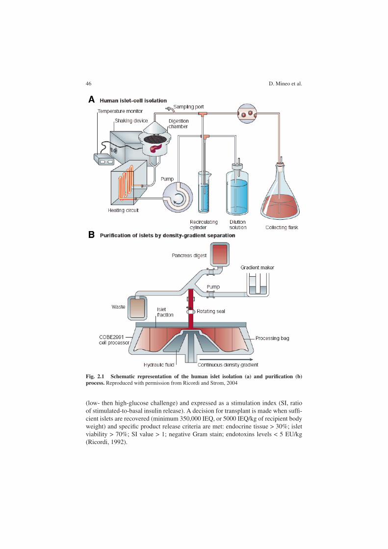
A semiautomated method of mechanically enhanced enzymatic pancreas disso-
ciation in a digestion-dissociation chamber (Ricordi chamber), with different blends
of lytic enzymes (e.g., collagenases and proteases), is used to release the islets
from the surrounding interstitial-connective and exocrine tissues. A semiautomated
purification technique in a computerized centrifuge system (COBE 2991), with
various density gradient solutions (e.g., glucose-based), is performed thereafter to
separate the endocrine from the exocrine cells (Fig. 2.1). Finally, a small volume
(<5 ml) of highly purified islet product is recovered and undergoes a 2-day cul-
ture for cell recovery from the traumatic isolation process. The cell culture medium
is enriched with trophic and antioxidant substances (e.g., insulin, nicotinamide, L-
glutathione) to prevent oxidative stress and apoptosis, preserving β-cell function and
survival (Ricordi et al., 1988; Ricordi, 1992; Ichii et al., 2006).
This interval also allows for assessment of islet survival, content, and function
prior to transplantation, thus determining product clinical suitability by FDA-
approved tests. Islet counting is performed at optical microscope from final product
samples using diphenylthiocarbazone (DTZ) staining (selectively binding to zinc–
insulin granules with red coloring). The islet mass is calculated using an algorithm
whereby islets are scored according to their diameter and counted as the num-
ber of islet equivalents (IEQ) based on a standard islet size of 150 µm. Product
purity is evaluated as a percentage of DTZ-stained endocrine cells compared
to unstained exocrine cells. Islet viability is assessed by fluorescent inclusion–
exclusion dyes selectively binding to viable or necrotic cells. Islet function is deter-
mined in vitro by measuring glucose-mediated insulin release in static incubation
46 D. Mineo et al.
Fig. 2.1 Schematic representation of the human islet isolation (a) and purification (b)
process. Reproduced with permission from Ricordi and Strom, 2004
(low- then high-glucose challenge) and expressed as a stimulation index (SI, ratio
of stimulated-to-basal insulin release). A decision for transplant is made when suffi-
cient islets are recovered (minimum 350,000 IEQ, or 5000 IEQ/kg of recipient body
weight) and specific product release criteria are met: endocrine tissue > 30%; islet
viability > 70%; SI value > 1; negative Gram stain; endotoxins levels < 5 EU/kg
(Ricordi, 1992).

2 Islet and Pancreas Transplantation 47
Despite significant progress, even in the most experienced centers fewer than
50% of the pancreata processed with the intent to transplant provide a sufficient
number of islets; moreover, more than 50% of the pancreatic islet content is lost in
the process, as a result of donor brain-death related events, suboptimal organ preser-
vation, deficient isolation process, and inadequate β-cell cytoprotection. Overall,
these conditions limit the chances of a satisfactory islet yield from a single pan-
creas, so that frequently more than one donor is required to collect the number of
islets needed to normalize glucose control or achieve insulin independence (Nano
et al., 2005; Pileggi et al., 2006, Ponte et al., 2007).
IT occurs via microembolization into the hepatic portal venous system, with islet
entrapping in the peripheral branches, at the presinusoid level due to size restriction,
followed by their engraftment and revascularization from the hepatic vasculature,
with immediate function and sustained survival. The transplant is performed by
gravity infusion from a closed-bag system containing the heparinized islet product
in the main portal vein through percutaneous transhepatic catheterization, under flu-
oroscopic and ultrasound guidance, using local anesthesia and conscious sedation,
with close monitoring of portal pressure. This minimally invasive interventional
radiologic procedure lasts approximately 1 h and patients are discharged from the
hospital within 24–48 h, once clinically stable and if no complications arise. In SIK,
or if there are contraindications to this approach (e.g., risk of hemorrhage, anatomi-
cal anomalies), cannulation of a tributary of the portal vein, such as the mesenteric
or umbilical vein, is performed by laparotomy or laparoscopy (Baidal et al., 2003;
Pileggi et al., 2006; Goss et al., 2003).
2.2.2 Clinical Protocols
2.2.2.1 Historical Protocols
Following the first case of a functioning allogeneic IT reported in 1980, several tri-
als in patients with T1DM were performed in late 1980s, mainly as SIK and IAK or
in combination with other solid organ transplants. Variable numbers of pancreatic
islets, purified from cadaver single-donors, were injected into the liver during the
main organ transplant or through a transient intraportal catheter as a post-transplant
percutaneous procedure. The immunosuppressive regimens were those tradition-
ally used in solid organ transplants, combining corticosteroids (prednisolone or
methylprednisolone), purine antagonist azathioprine or calcineurin inhibitor (CNI)
cyclosporine A, with lymphodepleting polyclonal antibodies added at induction in
a few trials [e.g., diverse animal-derived antithymocyte globulin (ATG)] (Largiadr
et al., 1980; Mintz et al., 1988).
The first promising results in IT were reported in the context of multiorgan
transplants in the early 1990s using the new CNI tacrolimus, with greater immuno-
suppressive effect and fewer side effects than cyclosporine A, as a maintenance
drug. Later on, mycophenolate mofetil (MMF), a prodrug of mycophenolate acid
(MPA), a purine synthesis inhibitor, was launched as a maintenance drug with equal
48 D. Mineo et al.
immunosuppressive efficacy but lower nephrotoxicity than CNI. At the same time,
more efficient induction strategies were tested, and the two monoclonal antibod-
ies daclizumab and basiliximab, targeting the IL2 receptor/CD25 on T-lymphocytes
with functional and proliferative inhibition, were used with significant reduction of
acute rejection episodes. In contrast, muromonab-OKT3, targeting the T-cell sur-
face marker CD3 with profound lymphodepletion, was tested but soon abandoned
owing to severe cytokine release. In a few trials, bone marrow cells (BMCs) or
hematopoietic stem cells (HSCs) from the same single-donors were coinfused, using
lymphodepleting nonmyeloablative conditioning, in the attempt to induce recipient
hematopoietic chimerism and islet graft tolerance, but islet graft survival was not
maintained after discontinuation of immunosuppressive drugs (Tzakis et al., 1990;
Ricordi et al., 1992; Gores et al., 1993; Alejandro et al., 1997; Secchi et al., 1997;
Oberholzer et al., 2000; Pileggi et al., 2004).
The overall results of this first decade of IT trials were encouraging but not
satisfactory, and limited islet graft survival, high rates of primary nonfunction,
only transient insulin independence, and relevant immunosuppressive side effects
were often observed. Indeed, post-transplant reduction of insulin requirements and
improvement in glycemic control rarely lasted long term, with only 10% of islet
recipients maintaining insulin independence at 1 year (Bretzel et al., 1999).
A main obstacle in achieving consistent positive results was the diabetogenic
effect of corticosteroids and CNIs on β-cell function and survival, as well as on
the development of peripheral insulin resistance. Post-transplantation DM occurs in
more than 50% of solid organ transplant recipients, including pancreas, with inci-
dence increasing with dose and duration of immunosuppressive therapy. Moreover,
drug-dependent increment of lipids is associated with increased allograft loss and
toxicity. Glucolipotoxicity may cause β-cell dysfunction and loss (Subramanian and
Trence, 2007; Vantyghem et al., 2007; Poitout and Robertson, 2008).
Corticosteroids (dose > 5 mg/day) can induce hyperglycemia by decreasing
insulin-mediated glucose uptake in peripheral tissues, with insulin resistance, and
by inhibiting insulin production and secretion, with β-cell dysfunction and possi-
bly apoptosis. Increased hepatic gluconeogenesis, reduced glycogen synthesis, and
lipolysis also occur. Hyperlipidemia is due to increased VLDL synthesis and down-
regulation of LDL receptor and lipoprotein lipase activity, resulting in increased
LDL cholesterol and triglycerides and reduced HDL cholesterol. Both metabolic
alterations may result in overall increased cardiovascular risk after transplant
(Poitout and Robertson, 2008).
CNIs frequently cause hyperglycemia and hyperlipidemia. High-dose tacrolimus
(trough levels > 6 ng/ml) is more diabetogenic but less deleterious for lipids
than cyclosporine A (trough levels > 300 ng/ml). Hyperglycemia is consequent
to decreased insulin synthesis and secretion. Morphological anomalies are present,
including reduced β-cell density, loss of secretory granules, cytoplasmatic swelling
and vacuolization, and possibly apoptosis. Such alterations seem to be dose-
dependent and reversible by drug discontinuation, with no chronic cumulative
toxicity on β cells. Effects on insulin sensitivity are still being debated, with
some animal and clinical studies reporting increased hyperinsulinemia and insulin
2 Islet and Pancreas Transplantation 49
resistance. Dyslipidemia, with increased LDL cholesterol and impaired VLDL
and LDL clearance, also occurs. Increased LDL oxidation and lipoprotein levels
with accelerated atherosclerosis, as well as increased vascular tone and resis-
tance with hypertension, contribute to a greater cardiovascular risk (Vantyghem
et al., 2007).
2.2.2.2 Current Protocols
In late 1990s, new immunosuppressants, such as mTOR inhibitors, sirolimus and
everolimus, and novel anti-inflammatory agents, such as TNFα blockers inflix-
imab (chimeric monoclonal antibody) and etanercept (recombinant fusion protein),
allowed avoidance of corticosteroids and reduction of tacrolimus dosage in specifi-
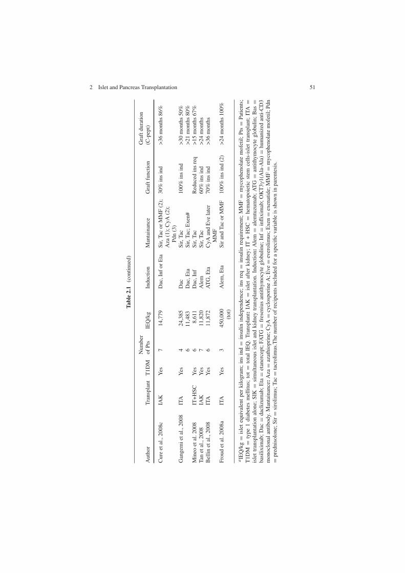
cally designed ITA protocols (Table 2.1) (Mineo et al., 2008c).
In 2000, the Edmonton group reported remarkable results from a steroid-free
protocol including daclizumab at induction and high-dose sirolimus (trough lev-
els 12–15 ng/ml during the first trimester and then 10–12 ng/ml) plus low-dose
tacrolimus (trough levels 3–6 ng/ml) at maintenance. After 1 year, virtually all
recipients were insulin-free, with normalized HbA1c and absence of severe hypo-
glycemia. Insulin independence was obtained infusing more than 10,000 IEQ/kg or
more than 700,000 IEQ total (full islet mass), from two or more fresh islet transplant
infusions (Shapiro et al., 2000; Ryan et al., 2002).
Subsequently, the Miami group successfully introduced a 2-day culture stage
in supplemented medium prior to transplant, to allow β-cell recovery from the
isolation process, thus increasing islet viability while preserving islet mass. This
time period permits the administration of induction strategies that can prevent
acute rejection episodes and improve long-term outcomes. It also allows the ship-
ment of islet products to remote facilities for transplantation. The same group
also attempted to achieve recipient hematopoietic chimerism and islet graft tol-
erance infusing HSCs from the same single-donor, without any conditioning, but
neither recipient chimerism nor islet graft function persisted after discontinua-
tion of immunosuppression 1 year after transplantation (Froud et al., 2005; Mineo
et al., 2008a).
Later on, the Minneapolis group showed that a more potent lymphodepletion at
induction, using rabbit ATG (rATG) or a modified humanized OKT3 (hOKT3γ1
ala-ala), together with an IT-specific anti-inflammatory strategy using etanercept,
achieved insulin independence from a single-donor with less than 10,000 IEQ/kg
(marginal islet mass). Sirolimus and low-dose tacrolimus or MMF were used at
maintenance (Hering et al., 2004, 2005).
Since the year 2000 many groups have adopted similar immunosuppressive
strategies in IT, for a total of more than 700 transplants in about 400 recipients
at some 50 centers worldwide, according to data from the Clinical Islet Transplant
Registry (CITR), with comparable results in terms of prolonged improvement of
glucose metabolism and rate of insulin independence at 1 year, steady at about
70–80% among the most experienced groups (Shapiro et al., 2006, Alejandro et al.,
2008).

50 D. Mineo et al.
Tab
le2.1
Mainclinicalisletallotransplantationtrialsaftertheyear2000(adaptedfrom
Marzoratietal.,2007)a
Author
Transplant
T1DM
Number
ofPts
IEQ/kg
Induction
Mantainance
Graftfunction
Graftduration
(C-pept)
Shapiroetal.,2000
ITA
Yes
711,547
Dac
Sir,Tac
100%
insind
>12months67%
Hirshbergetal.,2003
ITA
Yes
6>
10,000
Dac
Sir,Tac
50%
insind
>22months83%
Heringetal.,2004
ITA
Yes
6>
10,300
OKT3γ1(A
la-
Ala)
Sir,Tac
67%
insind
>12months83%
Franketal.2004
ITA
IAK
Yes
9 415,475
Dac
Sir,Tac
100%
insind
>26months57%
>26months20%
Goss
etal.,2004
ITA
Yes
10
>10,000
Dac
Sir,Tac
50%
insind
>18months90%
Lehmannetal.,2004
SIK
Yes
916,172
Dac
Sir,Tac
84%
insind
>12months89%
Heringetal.,2005
ITA
Yes
87,271
ATG,Dac,Eta
Sir,Tac
laterMMF
100%
insind
>12months62%
Froudetal.,2005
ITA
Yes
16
13,552
Dac,Inf
Sir,Tac
100%
insind
>26months80%
Kem
pfetal.,2005
ITA,SIK
,IA
KYes
22
>10,000
Dac;Bas
Sir,Tac;Eve,CyA
83%
insind
>12months100%
Ryan
etal.,2005
ITA
Yes
65
11,910
Dac,Inf(10);
Alem
(9)
Sir,Tac
100%
insind
>60months80%
Warnock
etal.,2005
ITA
Yes
10
13,806
ATG,then
Dac
Sir,Tac,orMMF(2)
100%
insind
6–21months100%
Toso
etal.,2006
IAK
Yes
812,530
Dac
Sir,Tac
71%
insind
>12months
O’Connelletal.,2006
ITA
Yes
617,958
Dac
Sir,Tac
50%
insind
>18months83%
Shapiroetal.,2006
ITA
Yes
23
13,473
Dac
Sir,Tac
58%
insind
>24months70%
Ghofailietal.,2007
ITA
Yes
11
14,312
Dac
Tac,MMForSir(1);
Exen
73%
insind
4–30months100%
Badetetal.,2007
ITA
Yes
10
11,089
Dac
Sir,Tac
80%
insind
>24months80%
Maffietal.,2007
ITA
Yes
19
11,477
Dac
Sir,Tac
orMMF(6),
CyA(1)
65%
insind
>24moths33%
Gillard
etal.,2008
ITA
Yes
5 54,700
6,400
ATG
Sir
Sir,Tac
Reducedinsreq
60%
insind
>30months40%
>24months60%
Gerber
etal.,2008
SIK
Yes
13
345,000
(tot)
Dac
Sir,Tac
31%
insind
>48months40%
2 Islet and Pancreas Transplantation 51
Tab
le2.1
(continued)
Author
Transplant
T1DM
Number
ofPts
IEQ/kg
Induction
Mantainance
Graftfunction
Graftduration
(C-pept)
Cure
etal.,2008c
IAK
Yes
714,779
Dac,InforEta
Sir,Tac
orMMF(2);
Aza
(1);CyA(2);
Pdn(3)
30%
insind
>36months86%
Gangem
ietal.,2008
ITA
Yes
4 624,385
11,483
Dac
Dac,Eta
Sir,Tac
Sir,Tac;Exen#
100%
insind
>30months50%
>21months80%
Mineo
etal.2008
IT+HSC
Yes
68,611
Dac,Inf
Sir,Tac
Reducedinsreq
>15months67%
Tan
etal.,2008
IAK
Yes
711,820
Alem
Sir,Tac
60%
insind
>24months
Bellinetal.,2008
ITA
Yes
611,872
ATG,Eta
CyAandEvelater
MMF
70%
insind
>36months
Froudetal.2008a
ITA
Yes
3450,000
(tot)
Alem,Eta
SirandTac
orMMF
100%
insind(2)
>24months100%
aIEQ/kg
=isletequivalentper
kilogram;insind
=insulinindependence;insreq
=insulinrequirem
ent;MMF
=mycophenolate
mofetil;Pts
=Patients;
T1DM
=type1diabetes
mellitus;
tot=
totalIEQ.Transplant:IA
K=
isletafterkidney;IT
+HSC
=hem
atopoieticstem
cells-islettransplant;ITA
=
islettransplantationalone;
SIK
=simultaneousisletandkidney
transplantation.Induction
:Alem
=alem
tuzumab;ATG
=antithymocyte
globulin;Bas
=
basilixim
ab;Dac
=daclizumab;Eta
=etanercept;FATG
=freseniusantithymocyteglobuline;Inf=
inflixim
ab;OKT3γ1(A
la-A
la)=
humanized
anti-C
D3
monoclonalantibody.Mantainance
:Aza
=azathioprine;CyA
=cyclosporineA;Eve
=everolimus;Exen
=exenatide;MMF
=mycophenolatemofetil;Pdn
=prednisolone;Sir
=sirolimus;Tac
=tacrolimus.Thenumber
ofrecipentsincluded
foraspecificvariableisshownin
parentesis.
52 D. Mineo et al.
New immunosuppressive or immunomodulatory drugs are being tested to reduce
side effects while attempting to use single-donor islet infusion, in order to avoid
recipient allosensitization and overcome organ shortage. Preliminary promising
results show significant improvements in short-term islet function and survival.
The groups in Miami and Edmonton are using alemtuzumab, an anti-CD52 lym-
phodepleting monoclonal antibody, as an induction agent, while including MMF
at maintenance, rather than tacrolimus or sirolimus. Similarly, the group in San
Francisco is using rATG and etanercept at induction, with sirolimus plus efalizumab,
an anti-LFA1/CD11a leukocyte antiadhesion monoclonal antibody, for maintenance
(Froud et al. 2008; Posselt et al., 2008a; Shapiro et al., 2008).
Recently, in order to improve islet function, and possibly survival, as well as to
prevent long-term graft exhaustion, the glucagon-like peptide-1 (GLP1) synthetic
analogue exenatide, administered subcutaneously at meals, has been given from the
time of the first islet infusion (University of Illinois group) or after islet graft dys-
function (Miami and Vancouver groups). The Miami group also reported patients
receiving islet retransplants who had been under chronic exenatide treatment prior
to the supplemental infusion. Overall, exenatide therapy seems to improve islet
engraftment as well as islet graft function and survival, normalizing glucose con-
trol and favoring insulin independence (Ghofaili et al., 2007; Faradji et al., 2008;
Froud et al., 2008; Gangemi et al., 2008; Faradji et al., 2009).
The Edmonton protocol has also been tested in several IAK and SIK trials.
In both settings, resulting rates of insulin independence were not always com-
parable with ITA, varying from 30 to 70% at 1-year post-transplant, but similar
stable, normalized glucose control and sustained C-peptide secretion were achieved,
also significantly improving function and longevity of kidney grafts without either
increasing the risk of kidney rejection or inducing premature decline in its function.
Recently, a report from one SIK trial showed successful islet engraftment and func-
tion using alemtuzumab and an Edmonton regimen, with 60% insulin independence
at 1 year and 100% kidney graft survival for more than 2 years (Toso et al., 2006;
Cure et al., 2008a; Gerber et al., 2008; Tan et al., 2008).
2.2.3 Results
2.2.3.1 Clinical Outcomes
Long-term results from different groups have shown that the rate of insulin indepen-
dence using the Edmonton protocol declines post-transplant to 50% at 2 years, 30%
at 3 years, and 10% at 5 years, although 70–80% of recipients have detectable C-
peptide (>0.5 ng/ml), with 50–60% reduction in insulin requirement and normalized
HbA1c (<6.5%). This progressive islet allograft loss and exhaustion seem mainly
due to auto- and allorejection, immunosuppressant-related islet graft toxicity and
implantation-site related unsuitability. Recently, the group in Minneapolis reported
the achievement of 60% insulin independence for more than 3 years post-transplant,
using rATG and etanercept as induction together with mTOR inhibitors plus CNIs
(later changed for MMF) at maintenance (Ryan et al., 2005; Bellin et al., 2008).
2 Islet and Pancreas Transplantation 53
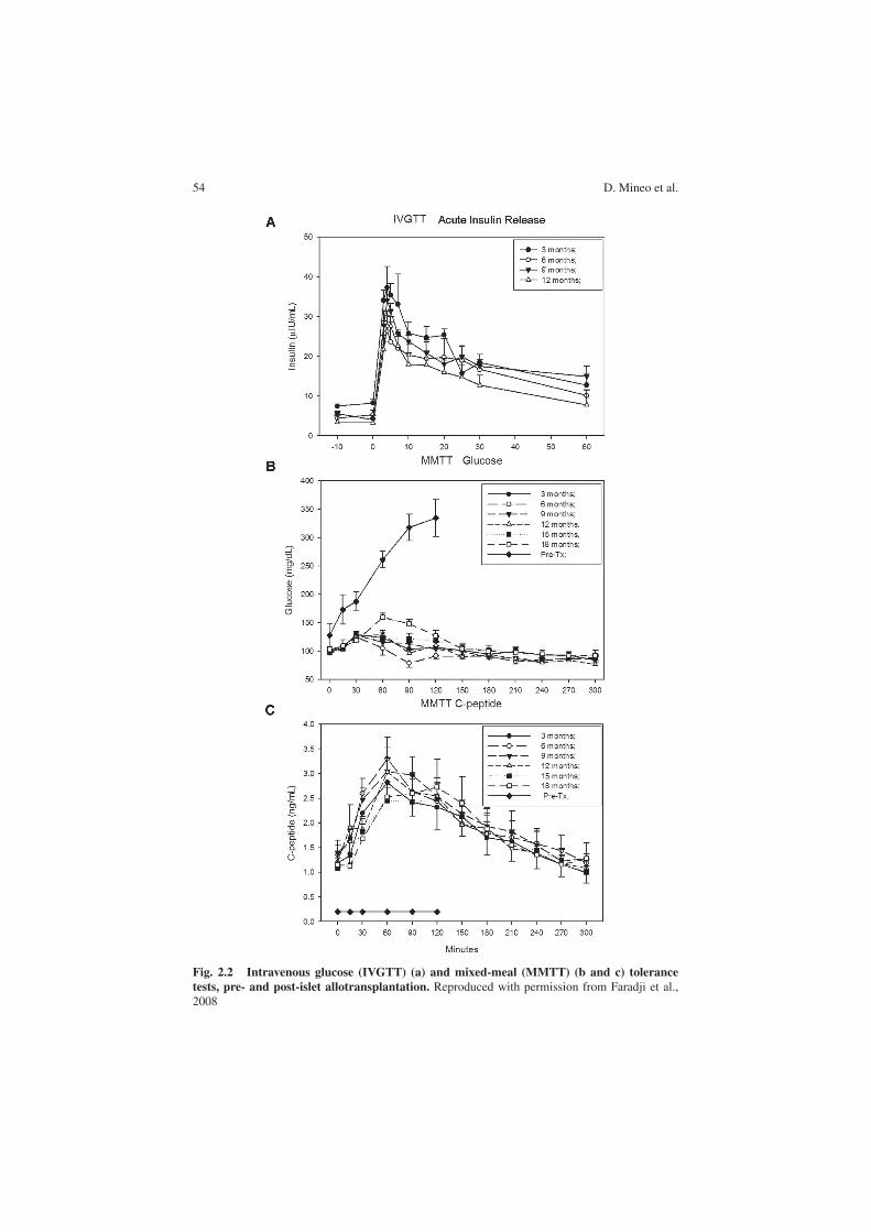
Significant metabolic improvements are achieved and maintained after IT, even
with only partial islet graft function, including stability of glucose control with
normalized HbA1c and corrected substrata metabolism, amelioration of insulin sen-
sitivity with reduced insulin requirements, absence of severe hypoglycemia with
restored awareness, and improved quality of life. In particular, both the first-phase
insulin secretion after an intravenous glucose tolerance test (IVGTT) and the area-
under-the-curve (AUC) of C-peptide secretion after an oral mixed-meal tolerance
test (MMTT) appear to be restored, with normalization of glucose levels and reduc-
tion of glucose excursion at the subcutaneous glucose monitoring system (CGMS)
(Figs. 2.2 and 2.3). Notably, glucagon response to hypo- and hyperglycemia appears
partially restored, with recovery of sympathoadrenal response and reduced hepatic
glucose output, respectively, thus contributing to improved metabolic control after
transplant (Luzi et al., 2001; Paty et al., 2002; Rickels et al., 2005a; Meier et al.,
2006; Poggioli et al., 2006; Rickels et al., 2006a; Rickels et al., 2007; Gorn et al.,
2008; Leitao et al., 2008b; Poggioli et al., 2008; Tharavanij et al., 2008).
Beneficial effects of IT are also evident for long-term diabetic complications,
with stabilization or reduced progression of retinopathy and even improvement of
neuropathy, with reduced nerve expression of receptor of advanced glycated end-
product (RAGE) and increased nerve conduction. The effects on renal function are
discordant, with some reports showing a decline in renal function after a long period
subsequent to transplantation, whereas others show stability. Acceleration of the
diabetic nephropathy as well as renal toxicity per se have been ascribed to immuno-
suppressive therapy. Prompt implementation of antihypertensive nephroprotective
therapies and appropriate recipient selection, especially in ITA, including T1DM
patients with virtually normal renal function and presumably slow progression of
the diabetic nephropathy, are recommended for limiting post-transplant renal side
effects. Results primarily from IAK recipients indicate that IT can induce improve-
ments in cardiovascular and endothelial function (e.g., improved diastolic function,
increased nitric oxide production), atherothrombotic profile (e.g., reduced lipid oxi-
dation, delayed intimal media thickening), with fewer cardiovascular events and
better survival in IT recipients (90 vs. 50% at 7 years). Overall, together with
the improvement in glycemic control, IT seems to be protective for kidney graft
function and to increase its longevity. The prolonged C-peptide secretion may con-
tribute to such beneficial effects by reducing nerve dysfunction and increasing blood
flow in cardiac and renal districts, with myocardial and glomerular vasodilatation,
improving cardiovascular and kidney function, and slowing the progression of dia-
betic macro- and microangiopathy (Johansson et al., 2000; Wahren et al., 2000;
Hansen et al., 2002; Fiorina et al., 2003a, b; Fiorina et al., 2005a, b; Lee et al.,
2005; Venturini et al., 2006; Ryan et al., 2006; Del Carro et al., 2007; Fung et al.,
2007; Maffi et al., 2007; Senior et al., 2007; Thompson et al., 2008; Warnock et al.,
2008, Leitao et al., 2009).
At islet graft dysfunction, long- and short-acting insulin analogues (e.g., glargine
and lispro), and/or the incretin-mimetic exenatide, are gradually started. The lat-
ter seems to have direct effects on β cells (increased glucose-dependent insulin
secretion, restored first-phase secretion, better insulin processing, and higher amylin
54 D. Mineo et al.
Fig. 2.2 Intravenous glucose (IVGTT) (a) and mixed-meal (MMTT) (b and c) tolerance
tests, pre- and post-islet allotransplantation. Reproduced with permission from Faradji et al.,2008

2 Islet and Pancreas Transplantation 55
Fig. 2.3 Continuous glucose monitoring system (CGMS) profiles pre- (a) and post-islet (b)
allotransplantation, and at islet graft dysfunction (c). Different lines represent different days ofglucose monitoring. Reproduced with permission from Gorn et al., 2008

56 D. Mineo et al.
synthesis) and indirect effects on glucose metabolism (reduced glucagon secre-
tion, lower hepatic gluconeogenesis, reduced gastric empting, and delayed glucose
absorption). Whether reduction of apoptosis or regeneration of β cells can occur, as
observed in experimental models, is not yet clear. Exenatide may also aid in pro-
tecting β cells from immunosuppression-related toxicity. Several side effects (e.g.,
vomiting, nausea), the risk of pancreatitis, and the possible worsening of preexisting
diabetic gastroparesis may limit its use (D’Amico et al., 2005; Ranta et al., 2006;
Cure et al., 2008b; Ranganath, 2008).
2.2.3.2 Islet Graft Monitoring
The clinical management of islet transplant recipients relies on the combina-
tion of several immune responses and metabolic parameters together with blood
trough levels of immunosuppressants and recipient clinical status, including
immunosuppressive-related side effects and toxicity symptoms.
The immune alloresponse is monitored principally by mixed lymphocyte allore-
action (MLR) and panel reactive alloantibody (PRA) assays for cellular and
humoral reactivity, respectively. Evaluation of cytotoxic gene expression levels (e.g.,
granzyme B) or ATP production in in-vitro stimulated CD4+ T-lymphocytes may
represent helpful tools for confirming the clinical picture and the islet graft course,
together with cytokine measurement and characterization or other soluble markers.
Recurrent autoimmunity can be detected by reappearance of T1DM-specific autoan-
tibodies (e.g., anti-GAD65, anti-IA2, and anti-insulin) and seems to be associated
with lower insulin-independence rates and shorter islet graft survival. Histological
signs of selective destruction of β-cell allograft as well as autoreactive cytotoxic and
memory T cells against specific β-cell epitopes have been also described (Stegall
et al., 1996; Bosi et al., 2001; Han et al., 2004; Pinkse et al., 2005; Huurman et al.,
2008; Huurman et al., 2009; Mineo et al., 2008b; Monti et al., 2008, Saini et al.,
2008).
Monitoring islet graft function for detection or prediction of β-cell dysfunction
or failure is based on insulin requirements and blood HbA1c, glucose, C-peptide,
and insulin levels measured in the fasting state or after stimulation testing (e.g.,
intravenous arginine tolerance test, IVGTT, and MMTT). Several indices of islet
graft function are derived from these measurements (e.g., acute insulin or C-peptide
release, fasting C-peptide/glucose ratio, 90-min glucose). Composite indices are
also calculated based on insulin requirements, HbA1c, and the number of infused
IEQ, such as the beta score. The use of CGMS or of the MAGE index derived
from daily glucose measurements with finger-sticks can help detect early graft
dysfunction. Unfortunately, none of these indices is completely reliable or standard-
ized, resulting in detection of metabolic alterations when it is too late to intervene
with modifications of the immunosuppressive therapy for rescuing the islet graft
(Teuscher et al., 1998; Geiger et al., 2005; Rickels et al., 2005b; Faradji et al., 2007b;
Rickels et al., 2007b; Gorn et al., 2008; Baidal et al., 2009).
To date, limited imaging methods are clinically available for visualizing or
monitoring the islet graft in vivo. Luciferase-transduced bioluminescence optical
2 Islet and Pancreas Transplantation 57
imaging, despite high sensitivity, has limited depth penetration and is not applicable
to human studies. High-sensitivity (e.g., 3-tesla) magnetic resonance imaging of
islets labeled with different tracers (e.g., superparamagnetic iron nanoparticles)
is being tested in animal settings with promising results for clinical applica-
tion. Positron emission tomography with 18-fluorodeoxy-D-glucose has been used
recently in human setting to assess intrahepatic islet engraftment and survival in the
immediate postinfusion period. Percutaneous hepatic biopsy is not routinely used
owing to procedure-related risks (e.g., bleeding) and lack of certainty of retrieval of
islet graft tissue (Eich et al., 2007; Medarova and Moore, 2008).
2.2.4 Complications and Limitations
2.2.4.1 Recipient- and Graft-Related Complications
Acute complications during the islet infusion procedure are rare (<2–6%), and
include: intraabdominal bleeding, pleural or abdominal effusions, peripheral portal
vein branches thrombosis, and transient transaminitis. Novel radiological tech-
niques, intracatheter-tract coagulants, and recipient peritransplant antithrombotic
prophylaxis have reduced their incidence. Intrahepatic focal steatosis and amyloid
deposits may follow IT, but their effect on islet graft function and survival is still
unclear (Bhargava et al., 2004; Froud et al., 2004; Barshes et al., 2005; Hafiz et al.,
2005; Westermark et al., 2008).
The extended period of the islet allograft survival in recent protocols has involved
long-term immunosuppression-related side effects in virtually all recipients, primar-
ily common or opportunistic infections (mainly skin, respiratory, and urinary tracts),
and direct immunosuppressive toxicity (Table 2.2). Several serious adverse events
have been observed that required hospitalization and specific therapy (e.g., profound
neutropenia, pneumonia, ovarian cysts), but only one death could be attributed to
immunosuppression (viral meningitis). Extremely rare are viral reactivations (e.g.,
EBV, CMV) or de novo malignancies, with only 13 neoplasms reported (two pap-
illary thyroid carcinomas, six squamous and two basal-cell skin carcinomas, one
ovarian and one breast cancer, one pulmonary nodule) in approximately 400 IT
recipients according to data of CITR (Cure et al., 2004; Hafiz et al., 2004; 2005;
Faradji et al., 2007a, Alejandro et al., 2008; Cure et al., 2008c).
Sirolimus has opposing effects on insulin secretion and action, which appear to
be cell- , species- , and dose-dependent, and act by inhibition of insulin-receptor
signal transduction and of the kinases regulating the β-cell cycle. Beta-cell dys-
function and reduction of insulin secretion seem to occur only at doses higher than
those used in the clinical setting, whereas increased basal and glucose-stimulated
insulin levels with reduced apoptosis have been seen at therapeutic concentrations.
In skeletal muscle and adipose cells, long-term exposure seems to reduce insulin-
dependent glucose uptake and insulin sensitivity, whereas in the short term opposite
effects have been observed. Reversible, dose-dependent dyslipidemia also occurs
(Subramanian and Trence, 2007; Vantyghem et al., 2007).

58 D. Mineo et al.
Tab
le2.2
Immunosuppression-related
sideeffectsin
isletallotransplantation(adaptedfrom
Leitaoetal.,2008a)
a
Side-effect
Agent
Treatment
Commentary
Gastrointestinal
Oralulcers(>70%)
Sirolimus
Topicalsymptomatic
–Diarrhea
(>60%)
Sirolimus
MMF
Switch
toMMF
Switch
toenteric-coated
MS
Excludeinfections(C
lost
rid
ium
dif
fici
le/CMV)
Nausea/vomiting(˜50%)
Sirolimus/tacrolimus
Symptomatic
Excludedrugtoxicity
Hem
atological
Anem
ia(>90%)
Sirolimus/tacrolimus/MMFor
irondeficiency
Ironsupplement,recombinant
eritropoetin
Mildleucopenia(˜100%)
Sirolimus/MMF
–Norm
alizingwithin
3months
Neutropenia<500/µl(>20%)
Sirolimus/MMF/
Cotrim
azol/valganciclovir/
CMVinfection
GCSFif<500/µlor<1000/µl
withfever
<500/µlwithfever
(rare,1/26
recipients)requires
hospitalizationand
broad-spectrum
antibiotic
therapy
Severelymphopenia(>10%)
Alemtuzumab
andthymoglobulin
Frequentmonitoringfor
infections
Desirableeffect,canlastupto
oneyear
Mildthrombocytopenia(>60%)
Sirolimus
–Spontaneousremission.ITPin
alem
tuzumab
users-nocases
inislettransplantrecipients
Dermatological
Acne(>50%)
Sirolimus
Topicaltreatm
ent
–Folliculitis(>20%)
Sirolimus
Topicaltreatm
ent
–Eczem
a(>10%)
Tacrolimus
Topicaltreatm
ent
Can
besevere,requiringdrug
discontinuation
Metabolic
Dyslipidem
ia(>50%)
Sirolimus
Statins
Increase
inLDL-cholesterol
Impairedinsulinsecretion
(variable)
Tacrolimus/sirolimus
–See
immunosuppressivebetacell
toxicity
Hypophosphatem
ia(>90%)
Hypomagnesem
ia(>60%)
Sirolimus/tacrolimus/MMF
Oralreplacement.
Mild

2 Islet and Pancreas Transplantation 59
Tab
le2.2
(continued)
Side-effect
Agent
Treatment
Commentary
Gonadal
Ovariancysts(>60%)
Altered
menses(>60%)
Sirolimus
Horm
onal
Surgeryin
selected
cases
Oligo-oram
enorrhea.
Gonadothropinsdysregulation.
Nopolycysticpattern
atultrasound
Cardiovascular
Transienthypotension(rare)
Alemtuzumab
Symptomatic
–Increase
inbloodpressure
(>30%)
Tacrolimus/sirolimus
Initiation/increase
inantihypertensivemedication
–
Peripheraledem
a(>50%)
Sirolimus
Diuretics
–Renal
Proteinuria/albuminuria,decrease
inglomerularfiltrationrate
(variable)
Tacrolimus/sirolimus
ACEi/ARB,statins
Stableifadequatetreatm
entof
diabeticnephropathyclassical
risk
factors(i.e.,
LDL-cholesterolandblood
pressure)isprovided
Neurological
Tremors,paresthesias,headache,
milddepression(>10%)
Tacrolimus
Switch
toMMF
Can
besevere:disablingpain
syndromeand
leukoencephalopathy
aACEi=
angiotensin-converting-enzymeinhibitors;ARB
=angiotensinogen
receptors
blockers;CMV
=cytomegalovirus;GCSF
=granulocyte
colony-
stim
ulatingfactor;ITP
=im
munethrombocytopenicpurpura;LDL
=low-density
lipoprotein;MS
=mycophenolatesodium.
60 D. Mineo et al.
MPA may also have a detrimental effect on β cells, by reducing insulin secretion
and inducing apoptosis, as well as on peripheral insulin sensitivity, with most of such
data coming from experimental settings, whereas lipid metabolism is not affected.
The enteric-coated formulation mycophenolate sodium has recently shown better
gastrointestinal tolerability and absorption than MMF and is increasingly used to
avoid toxicity from other immunosuppressive drugs (Havrdova et al., 2005; Gao
et al., 2007; Subramanian and Trence, 2007; Park et al., 2009).
All the immunosuppressive agents can interfere with islet engraftment and β-
cell self-renewal. Indeed, sirolimus has antiproliferative and antiangiogenic effects
on duct and islet cells that may impair β-cell engraftment and neovascularization as
well as viability and regeneration. Tacrolimus andMPA also have negative effects on
duct and islet cell proliferation and differentiation, preventing β-cell neogenesis or
replication. No negative effects of everolimus, a newly introduced mTOR inhibitor,
on glucose metabolism, have yet been reported, although it can induce dyslipidemia
(Bussiere et al., 2006; Cantaluppi et al., 2006a, b; Marcelli-Tourvieille et al., 2007;
Nir et al., 2007; Zahr et al., 2007).
Renal toxicity is still a major side effect of immunosuppressive therapy.
Tacrolimus may cause acute vasomotor vasculopathy with tubular necrosis and/or
chronic fibrotic vasculopathy with glomerulosclerosis and interstitial fibrosis.
Moreover, sirolimus may induce acute renal dysfunction and/or chronic proteinuria
by increasing glomerular permeability and injury or by suppressing the compen-
satory renal cell proliferation and repair capacity. Their combined use in IT can
have synergic negative effects on renal function per se or may cause the progression
of diabetic nephropathy, especially in the presence of pretransplant abnormalities
(e.g., microalbuminuria, reduced eGFR), whereas the alternative use of MPA-based
regimens could prevent renal injury (Rangan, 2006; Williams and Haragsim, 2006).
Supportive therapy is normally used to counteract systemic immunosuppressant-
related side effects, such as angiotensin converting enzyme inhibitors (ACEi)
or angiotensinogen receptor blockers (ARB), statins or ezetimibe, together with
bone marrow stimulants (e.g., granulocyte-colony stimulating factor, erythropoi-
etin), anti-infective prophylaxes, and dietary supplements (e.g., iron). In most
cases prompt treatment of complications minimizes recipient morbidity without any
sequela (Hafiz et al., 2005; Faradji et al. 2007).
2.2.4.2 Transplant-Related Limitations
Similarly to the pretransplantation period, many factors can contribute to a signif-
icant post-transplantation islet loss especially during the early postinfusion phase,
reducing the effective number of functioning islets available during the follow-up
period. Because of that often a second or third donor islet infusion is required to
achieve insulin independence and durable normalization of glucose control in the
recipients.
In particular, during islet infusion, an intravascular instant blood-mediated
inflammatory reaction (IBMIR) seems responsible for destroying 50–70% of the
infused β cells. An upregulation of tissue factor and other molecules on islet
2 Islet and Pancreas Transplantation 61
cell surface after the isolation process is capable of triggering innate immunity
via activation of coagulation, complement, inflammation, and natural antibod-
ies, destroying the islets. Peritransplant anticoagulant prophylaxis with heparin
can counteract this reaction (Moberg et al., 2002; Johansson et al., 2005; Eich
et al., 2007).
A progressive intrahepatic islet graft dysfunction and loss also occurs owing
to: poor revascularization, chronic hypoxia, absent reinnervations, proinflammatory
milieu, drug toxicity, glucolipotoxicity, fat and amyloid deposition, islet functional
overload, premature apoptosis, and lack of regeneration. Several ongoing experi-
ments are aimed at identifying alternative and less hostile implantation sites for islet
allograft, with the omental pouch, the thymus, or the bone marrow being the most
attractive. Recently, islet autotransplantation in the forearm muscle of a child with
genetically determined pancreatitis has shown a prolonged (over 2 years) restora-
tion of insulin secretion and normalized glucose control, while requiring minimal
exogenous insulin therapy (Desai et al., 2003; Bhargava et al., 2004; Pileggi et al.,
2006; Huang et al., 2008; Merani et al., 2008; Rafael et al., 2008; Westermark et al.,
2008; Lau and Carlsson, 2009).
Finally, a major concern of IT is the recipient-wide allosensitization from the
multiple HLA-mismatched donor infusions performed to achieve insulin indepen-
dence, hypothetically jeopardizing the chances of receiving future organ transplants
(e.g., kidney or pancreas). Pretransplant PRA levels higher than 15–20% and
donor-specific antibodies (DSA) seem associated with reduced islet graft survival.
Post-transplant positive PRA levels and de novo DSA may occur after drug dose
reduction for persistent or serious side effects (e.g., infections) but their impact on
islet graft loss is still unclear. Allosensitization seems absent or minimal under the
recommended trough levels of immunosuppression, which also seem able to con-
tain low PRA levels (<5–15%), but it occurs constantly when immunosuppression
is discontinued, such as after islet graft failure. High PRA levels (>50%) with DSA
and cross-reacting non-DSA may persist for a long time. A slower immunosup-
pressive tapering could minimize or prevent sudden and massive antigen exposition
from residual islet graft (Mohanakumar et al., 2006; Rickels et al., 2006b; Campbell
et al., 2007a, b; Cardani et al., 2007).
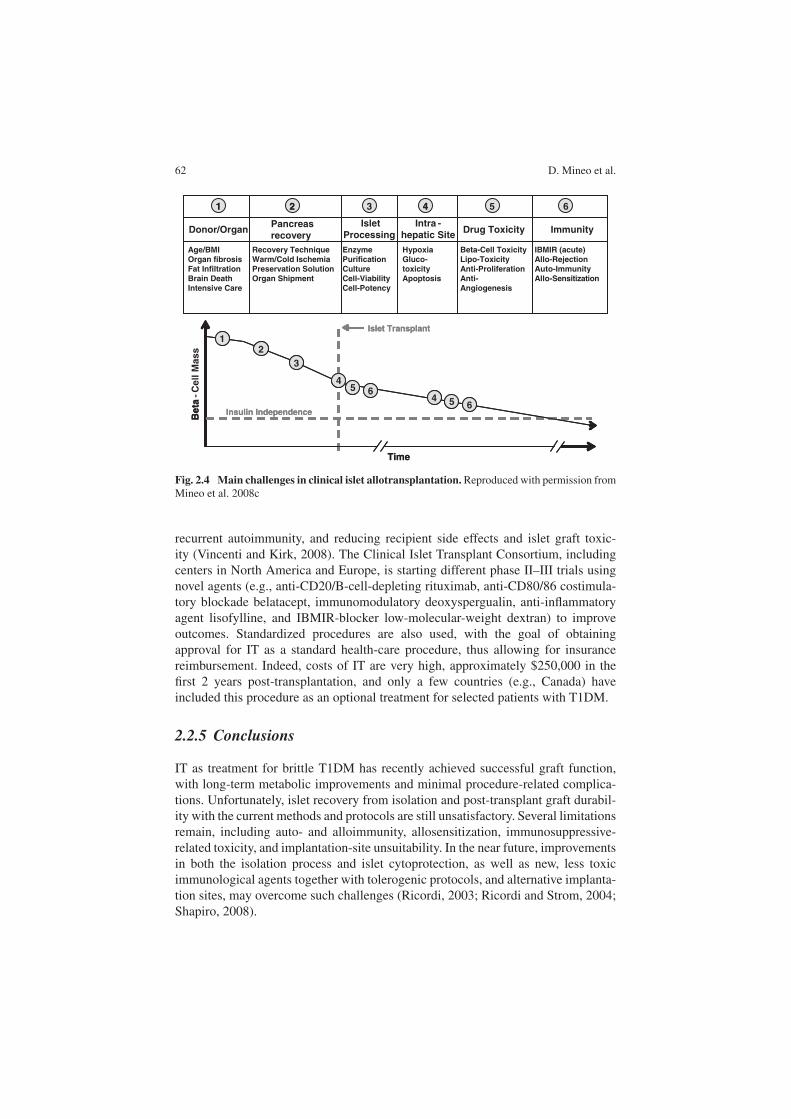
2.2.4.3 Current Challenges and Future Perspectives
Several technical and clinical limitations still persist in IT (Fig. 2.4). Various cyto-
protective strategies and agents are currently under investigation to improve organ
preservation and islet yield and survival through the isolation process, such as perflu-
orocarbons in a two-layer method, new lytic enzyme blends or purification methods,
and JNK or caspase inhibitors (Kin et al., 2006; Barbaro et al., 2007; Emamaullee
et al., 2007; Sabek et al., 2008; Varona-Santos et al., 2008).
New immunological and possibly tolerogenic strategies, including more selec-
tive lymphodepleting drugs, costimulatory blockade, and anti-inflammatory agents,
are being tested for increasing islet allograft longevity, preventing allorejection and
62 D. Mineo et al.
Drug Toxicity ImmunityIntra -
hepatic Site
Islet
Processing Pancreas
recovery Donor/Organ
1 2 3 4 5 6
Insulin Independence
12
Time
Beta
Islet Transplant
654
4
3
5 6
Beta-Cell Toxicity
Lipo-Toxicity
Anti-Proliferation
Anti-
Angiogenesis
IBMIR (acute)
Allo-Rejection
Auto-Immunity
Allo-Sensitization
Hypoxia
Gluco-
toxicity
Apoptosis
Enzyme
Purification
Culture
Cell-Viability
Cell-Potency
Recovery Technique
Warm/Cold Ischemia
Preservation Solution
Organ Shipment
Age/BMI
Organ fibrosis
Fat Infiltration
Brain Death
Intensive Care
1 2 4
Insulin Independence
12
Time
Beta
- C
ell
Ma
ss
Islet Transplant
654
4
3
5 6
Fig. 2.4 Main challenges in clinical islet allotransplantation. Reproduced with permission fromMineo et al. 2008c
recurrent autoimmunity, and reducing recipient side effects and islet graft toxic-
ity (Vincenti and Kirk, 2008). The Clinical Islet Transplant Consortium, including
centers in North America and Europe, is starting different phase II–III trials using
novel agents (e.g., anti-CD20/B-cell-depleting rituximab, anti-CD80/86 costimula-
tory blockade belatacept, immunomodulatory deoxyspergualin, anti-inflammatory
agent lisofylline, and IBMIR-blocker low-molecular-weight dextran) to improve
outcomes. Standardized procedures are also used, with the goal of obtaining
approval for IT as a standard health-care procedure, thus allowing for insurance
reimbursement. Indeed, costs of IT are very high, approximately $250,000 in the
first 2 years post-transplantation, and only a few countries (e.g., Canada) have
included this procedure as an optional treatment for selected patients with T1DM.
2.2.5 Conclusions
IT as treatment for brittle T1DM has recently achieved successful graft function,
with long-term metabolic improvements and minimal procedure-related complica-
tions. Unfortunately, islet recovery from isolation and post-transplant graft durabil-
ity with the current methods and protocols are still unsatisfactory. Several limitations
remain, including auto- and alloimmunity, allosensitization, immunosuppressive-
related toxicity, and implantation-site unsuitability. In the near future, improvements
in both the isolation process and islet cytoprotection, as well as new, less toxic
immunological agents together with tolerogenic protocols, and alternative implanta-
tion sites, may overcome such challenges (Ricordi, 2003; Ricordi and Strom, 2004;
Shapiro, 2008).
2 Islet and Pancreas Transplantation 63
2.3 Simultaneous Pancreas–Kidney Transplantation
Simultaneous pancreas–kidney transplantation (SPK) is considered the best treat-
ment option for patients with T1DM and end-stage renal disease (ESRD). The pan-
creas transplant can restore euglycemia, providing long-term insulin independence;
increase patient survival; stabilize or improve diabetic retinopathy and neuropathy;
and, in combination with the kidney transplant, eliminate the need for long-term
dialysis (Gruessner and Sutherland, 2005; Leichtman et al., 2008).
More potent immunosuppression agents, improvements in surgical techniques,
and better understanding of postoperative complications have led to consistent
improvement in SPK transplantation results over the past decade. Drainage of the
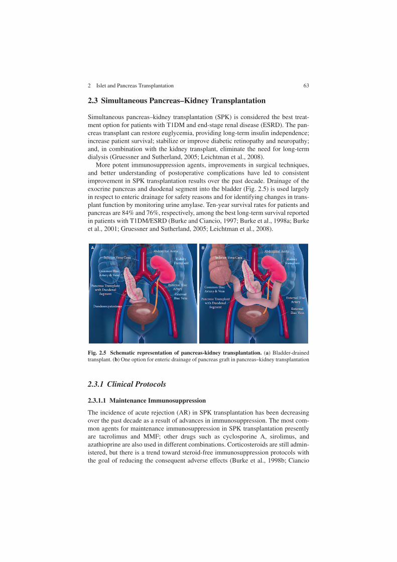
exocrine pancreas and duodenal segment into the bladder (Fig. 2.5) is used largely
in respect to enteric drainage for safety reasons and for identifying changes in trans-
plant function by monitoring urine amylase. Ten-year survival rates for patients and
pancreas are 84% and 76%, respectively, among the best long-term survival reported
in patients with T1DM/ESRD (Burke and Ciancio, 1997; Burke et al., 1998a; Burke
et al., 2001; Gruessner and Sutherland, 2005; Leichtman et al., 2008).
Fig. 2.5 Schematic representation of pancreas-kidney transplantation. (a) Bladder-drainedtransplant. (b) One option for enteric drainage of pancreas graft in pancreas–kidney transplantation
2.3.1 Clinical Protocols
2.3.1.1 Maintenance Immunosuppression
The incidence of acute rejection (AR) in SPK transplantation has been decreasing
over the past decade as a result of advances in immunosuppression. The most com-
mon agents for maintenance immunosuppression in SPK transplantation presently
are tacrolimus and MMF; other drugs such as cyclosporine A, sirolimus, and
azathioprine are also used in different combinations. Corticosteroids are still admin-
istered, but there is a trend toward steroid-free immunosuppression protocols with
the goal of reducing the consequent adverse effects (Burke et al., 1998b; Ciancio
64 D. Mineo et al.
et al., 2000a; Burke et al., 2004b; Gruessner and Sutherland, 2005; Cantarovich and
Vistoli, 2009; Mineo et al., 2008c; Singh and Stratta, 2008).
2.3.1.2 Induction Therapy
The recent therapeutic protocols in kidney and kidney–pancreas transplantation
attempt to reduce the incidence and severity of AR as well as prevent long-term
chronic (vascular) allograft dysfunction (CAD). The methodologies include reduc-
tion of CNIs and of their short- and long-term nephrotoxicity, reduction or avoidance
of corticosteroids, use of adjunctive maintenance antiproliferative agents (e.g.,
mTOR inhibitors), and utilization of new agents, such as nonlymphodepleting mon-
oclonal antibodies (daclizumab or basiliximab), or lymphodepleting monoclonal
(alemtuzumab) and polyclonal (e.g., rATG) antibodies. The percentage of patients
treated with induction therapy has been increasing and was more than 75% in the
most recently reported data from the International Pancreas Transplant Registry
(IPTR) 2004 (Gruessner and Sutherland, 2005).
Daclizumab
A series of studies has been published analyzing the safety and efficacy of
daclizumab as induction therapy in SPK transplant recipients (Bruce et al. 2000;
Burke et al., 2001; Lo et al., 2001a, b; Stratta et al., 2001; Burke et al., 2002a,
b; Stratta et al., 2002). The results of a multicenter survey using daclizumab as
induction therapy showed a low incidence of AR when used in combination with
tacrolimus, MMF, and corticosteroids in SPK transplant recipients (Bruce et al.,
2001). The survey reported experience with 71 SPK transplant recipients receiving
4–5 daclizumab doses (n = 45) or 1–3 doses (n = 26). There were no differences
in patient and kidney graft survival rates, 98 vs. 96% and 92 vs. 92%, respec-
tively. However, there was a trend toward improved pancreas graft survival rates
in the group receiving 4–5 doses, compared with 1–3 doses (96 vs. 85%, p =
0.07). Although more patients receiving 1–3 doses had rejection (54%) than patients
receiving 4–5 doses (24%), there was no dose–response relationship between the
total number of doses or the adjusted total milligram/kilogram dose and time to
rejection. All patients with functioning grafts had good renal and pancreatic allo-
graft function at 6 and 12 months. The overall incidence of major infection was
27%, and there were no differences in the incidence of infection between the two
groups. No major adverse events were attributed to daclizumab use. In conclusion,
excellent short-term outcomes were noted in this retrospective, multicenter survey of
initial experience with daclizumab induction in combination with tacrolimus, MMF,
and corticosteroids in SPK transplant recipients.
The safety and efficacy of two dosing regimens of daclizumab as an adjunctive
immunosuppressive agent versus no antibody induction in SPK transplant recipients
receiving tacrolimus and MMF as primary immunosuppression were investigated
in a multicenter, open label, comparative trial (Stratta et al., 2002). SPK trans-
plant recipients were randomized to one of three groups: daclizumab 1 mg/kg every

2 Islet and Pancreas Transplantation 65
14 days for five doses (Group I), daclizumab 2 mg/kg every 14 days for two doses
(Group II), and no antibody induction (Group III). A total of 166 patients were ran-
domized into the three groups [Group I (n = 70), Group II (n = 74), Group III
(n = 22)]. At a minimum follow-up of 3 months, patient, kidney and pancreas graft
survival rates were similar among the three groups. However, the rates of acute renal
allograft rejection were 18% for Group I, 8% for Group II, and 36% for Group III
(p < 0.005). The probabilities of either kidney or pancreas allograft rejection were
22% for Group I, 8% for Group II, and 38% for Group III. At 3 months, the actuarial
event-free survival (no AR, allograft loss, or death) rates were 67%, 81%, and 50%
in Group I, II, and III, respectively. Although the follow-up was short, this study
emphasized the important role of induction antibodies in reducing AR.
Daclizumab in Combination with rATG
The use of new immunosuppressive agents continues to be associated with reduced
rates of AR episodes in SPK transplant recipients (Burke et al., 2002a, b). Forty-
two SPK transplant recipients were included in a prospective, randomized trial in
which they received rATG and daclizumab, tacrolimus, and corticosteroids as base-
line immunosuppression. They were then randomized to receive either MMF or
sirolimus in addition to baseline immunosuppression. Twenty-two patients received
MMF and 20 received sirolimus. There were three episodes of AR (7.1%). These
were in the MMF group, all in patients who were off either MMF (wound infection,
pneumonia) or corticosteroids. Each of these episodes was corticosteroid-resistant,
but responsive to antibody therapy (OKT3 or rATG). Actuarial patient, kidney, and
pancreas allograft survivals were 100%, 100%, and 95% in the sirolimus group and
100%, 100%, and 100% in the MMF group (Burke et al., 2002b).
A similar study (Gallon et al., 2007) reported the effect of two tacrolimus-based
maintenance regimens on long-term renal allograft function in SPK transplant recip-
ients [tacrolimus/MMF (n = 22) vs. tacrolimus/sirolimus (n = 20)] (Schaapherder
et al., 1993). All patients received rATG as induction therapy. The difference from
the previous study (Burke et al., 2002b) was that both regimens included prednisone-
free maintenance. Patient and pancreas graft survival rates at 6 years were the same,
but kidney allograft survival was higher in the tacrolimus/MMF group (90.7% vs.
70.7%, p = 0.09). The incidence of AR and rate of decline in eGFR were similar in
both groups (Gallon et al., 2007).
Alemtuzumab
A nonrandomized study of 75 pancreas–kidney and solitary pancreas recipients who
received alemtuzumab (four doses for induction and twelve doses within the first
year) and MMF (≥2 gr/day) for induction and maintenance therapy was reported
(Gruessner et al., 2005). Thirty milligrams of alemtuzumab was given intravenously
intraoperatively for induction as well as for maintenance dosing, the latter doses
administered when the absolute lymphocyte count increased to 200/mm3 or more;
the maximum number of alemtuzumab doses was limited to ten within the first
66 D. Mineo et al.
year. In a 6-month follow-up the results were compared with an historical group
of 266 consecutive pancreas recipients using rATG induction and tacrolimus main-
tenance. Patient survival at 6 months for SPK transplant recipients was 90%; for
pancreas-after-kidney (PAK) recipients 91%; and for pancreas transplant alone
(PTA) recipients 97% (p ≥ 0.4).
The patient survival rates were not different between the control group and the
three study groups (p ≥ 0.06). Pancreas graft survival at 6 months in the study
group for SPK transplant recipients (vs. historical control) was 81% (vs. 79%; p ≥
0.66); for PAK recipients 91% (vs. 85%; p ≥ 0.59); and for PTA recipients 71%
(vs. 84%; p ≥ 0.07). Kidney graft survival in the historical control versus the study
group at 6 months for SPK transplant recipients was 81% vs. 85%; (p ≥ 0.2). The
incidence of a first (reversible) rejection episode at 6 months in the study versus
the control group for SPK transplant recipients was 41% (vs. 9%; p ≥ 0.0003); for
PAK recipients 14% (vs. 10%; p ≥ 0.89); and for PTA recipients 19% (vs. 26%; p
≥ 0.36). In all three recipient categories the median “modification of renal disease”
level at 6 months was higher and the median serum creatinine concentration was
lower in the study versus control groups, but the differences did not reach statistical
significance. The conclusion was that the combination of alemtuzumab and MMF
was associated with an acceptable rejection rate (albeit higher than expected for SPK
transplants), and good (graft and native) kidney function; it eliminated undesired
CNI- and corticosteroid-related side effects, but a long-term follow-up is warranted.
More recently a single-center nonrandomized retrospective sequential study
was reported (Kaufman et al., 2006) comparing the effect of alemtuzumab (n
= 50) and rATG (n = 38) as an induction immunosuppression for recipients of
SPK transplant given a prednisone-free maintenance regimen in combination with
tacrolimus/sirolimus-based maintenance therapy. The overall 1-year patient and
graft survival rates were similar for the two treatment groups. The 1-year actual
patient survival rates for recipients who received alemtuzumab and rATG were 96%
and 100%, respectively (p = ns); the 1-year actual death-censored kidney graft
survival rates were 95% and 97.4%, respectively (p = ns); the 1-year actual death-
censored pancreas graft survival rates were 92% and 100%, respectively (p = ns);
the 12-month actual rejection rates were 6.1% and 2.6%, respectively (p = ns). At
12 months, the serum creatinine values for the alemtuzumab and rATG group were
1.45 ± 0.36 and 1.29 ± 0.43, respectively (p = ns). Viral infectious complications
were statistically significantly lower in the alemtuzumab group. Despite the study
limitation, both alemtuzumab and rATG induction were effective in facilitating a
prednisone-free maintenance protocol in SPK transplant recipients.
The use of alemtuzumab as induction therapy in SPK transplant recipients has
increased substantially. Lately, the impact of steroid-free maintenance immunosup-
pression in pancreas transplantation using alemtuzumab as induction therapy has
been evaluated in a single-center study (Muthusamy et al., 2008), where 102 pan-
creas transplantations were performed in 100 patients with tacrolimus and MMF,
with no maintenance corticosteroids. With a median follow-up of 17 months,
patient, pancreas and kidney graft survival (actuarial) was 97%, 89%, and 94%,
respectively. Overall incidence of rejection was 25%. The incidence of CMV
2 Islet and Pancreas Transplantation 67
and BKV infections was 6.8% and 3.8%, respectively. This experience suggested
that alemtuzumab is safe and effective. Furthermore, steroid-free maintenance was
achieved in 83% of the patients with a 25% incidence of rejection.
A cautious tone should be used in the context of corticosteroid-free immunosup-
pression, since a recent report from the Minnesota group showed that occurrence of
AR has a far greater impact on kidney graft survival (15 years actuarial) than the
development of new onset DM (NODM) (Matas et al., 2008). This may dampen
some of the enthusiasm for steroid-free protocols in which the high rate of AR may
well translate into worse long-term graft (and hence patient) survival.
In another study (Clatworthy et al., 2007) alemtuzumab was given subcuta-
neously in 21 SPK transplant recipients. The rate of AR was 14% at 1 year.
This route of administration was recommended because lymphocyte depletion was
comparable to that seen in patients receiving intravenous alemtuzumab. Recently,
alemtuzumab was compared with rATG (Farney et al. 2008) and basiliximab induc-
tion therapy (Magliocca et al., 2008). The use of alemtuzumab for induction therapy
after SPK transplants was found to be as safe and effective as rATG and basiliximab.
Furthermore, the outcome was not inferior to that of the other two induction thera-
pies. It is important to note that there was a higher incidence of CMV infections in
the alemtuzumab group and since then a single dose (rather than two) has been used.
2.3.2 Results
2.3.2.1 Patient and Graft Survival
Long- and short-term patient survival rates have improved steadily over the years.
Patient survival rates at 1 year have been higher than 90% since the earliest eras, and
are now more than 95% for SPK transplantations performed in 2002/2003. Overall,
5-year survival rates have also improved and are higher than 80%. Survival rates at
10 years are 69% for SPK transplantation. One-year pancreas graft survival rates are
85%, and 1-year kidney graft survival rates are 92%. The 5-year pancreas graft sur-
vival reached 69%, and the 5-year kidney graft survival was 77% for the 1998/1999
period. The 10-year pancreas and kidney graft survival rates for the 1992/1993 were
46% and 45%, respectively (Gruessner and Sutherland, 2005). These numbers are
similar to those in recent reports (Leichtman et al., 2008). At the University of
Miami 10-year survival rates for patients, pancreas, and kidney are 8%, 76%, and
51%, respectively (Burke et al., 2001).
2.3.2.2 Diabetic Nephropathy
The effects of pancreas transplantation on diabetic nephropathy are among the
most studied benefits of pancreas transplantation. A pivotal study demonstrated
that pancreas transplantation can reverse preexisting histological lesions of diabetic
nephropathy in the native kidneys, but reversal requires more than 5 years of nor-
moglycemia (Fioretto et al., 1998). Another study reported on 32 T1DM patients

68 D. Mineo et al.
that were evaluated before and 1 year after successful PTA and compared with
30 matched nontransplanted T1DM patients. Evidence for improvement of renal
function after pancreas transplantation was found, documented by the reduction
of urinary excretion of protein with stable creatinine concentration and clearances
(Coppelli et al., 2005).
2.3.2.3 Diabetic Retinopathy
Retinopathy is the most common microvascular complication of DM. The major-
ity of our patients who undergo SPK transplantation have already developed
some degree of retinopathy and most of them have received laser therapy
(LT). Patients with advanced retinopathy are less likely to benefit from a SPK
transplant.
A prospective study evaluated 33 PTA recipients and 35 patients with T1DM
who did not receive PTA. At baseline, 9% of PTA and 6% of non-PTA patients
had no diabetic retinopathy, 24% and 29% had nonproliferative diabetic retinopa-
thy (NPDR), and 67% and 66% had laser-treated and/or proliferative retinopathy
(LT/PDR), respectively. No new case of diabetic retinopathy (DR) occurred in either
group during at least 1 year of follow-up. In the NPDR PTA group, 50% of patients
improved by one grading and 50% showed no change. In the LT/PDR, stabilization
was observed in 86% of cases but worsening of retinopathy occurred in 14% of
patients. In the NPDR non-PTA group, DR improved in 20% of patients, remained
unchanged in 10%, and worsened in the remaining 70%. In the LT/PDR non-PTA
group, retinopathy did not change in 43% and deteriorated in 57% of patients.
Overall, the percentage of patients with improved or stabilized DR was signifi-
cantly higher in the PTA group (Giannarelli et al., 2006). Another report concluded
that advanced DR is present in a high proportion of SPK transplant recipients as
a consequence of the duration of T1DM (mean of 24.6 years) and the presence
of ESRD. More than 90% of patients have stable DR following transplantation
(Pearce et al., 2000).
2.3.2.4 Diabetic Neuropathy
Polyneuropathy is a very common (almost 100%) complication of both T1DM and
ESRD, and advanced motor, sensory, and autonomic neuropathies are very fre-
quent in patients undergoing SPK transplantation. Improvement or stabilization of
gastric function was observed in 12 out of 23 (52%) SPK transplant recipients ver-
sus 5 out of 12 (41.7%) T1DM recipients who underwent kidney transplant alone
(KTA) (Hathaway et al., 1994). SPK transplant recipients also demonstrated sig-
nificant improvement in postural adjustment ratio (Navarro et al., 1998). Sensory
and motor neuropathies, as measured by nerve conduction studies, have also shown
improvement in SPK transplant recipients (Muller-Felber et al. 1993; Navarro
et al., 1998).

2 Islet and Pancreas Transplantation 69
2.3.2.5 Quality of Life
Patients who received SPK transplants consistently reported an improvement in their
quality of life (Sureshkumar et al., 2005). SPK transplantation had a significant
positive effect on DM-related quality of life even though SPK transplantation is a
complex surgical procedure.
SPK transplantation has been viewed as a higher-cost and higher-risk surgical
procedure than kidney transplant, and it is unclear if SPK transplantation offers
better health and quality of life outcomes than insulin therapy plus KTA. A study
found that both SPK and KTA recipients report better health and quality of life, but
SPK transplant recipients also report greater improvement in physical health and in
areas that are DM-specific than those of KTA (Gross et al., 2002).
2.3.3 Complications and Limitations
Surgical complications are more common after pancreas transplantation, compared
to kidney transplantation. Nonimmunological complications of pancreas transplan-
tation (including thrombosis and graft pancreatitis) account for graft losses in
5–10% of cases. These usually occur within 6 months of transplantation and are as
important an etiology of pancreas graft loss in SPK transplantation as AR (Ciancio
et al. 1996b; Gruessner et al., 1996; Gruessner and Sutherland, 2001; Gruessner and
Sutherland, 2005).
2.3.3.1 Hypercoagulation in SPK
T1DM has been shown to result in hypercoagulation, as assessed by numer-
ous studies involving different components of the clotting cascade. In addition,
hyperlipidemia, commonly associated with DM, further contributes to such hyper-
coagulation. Subsequent to SPK transplantation, with restoration of euglycemia,
the lipid profile usually normalizes (Burke et al., 1998) or is treated, when nec-
essary, with lipid-lowering medications. Although the uremic effect on platelets
could offset the hypercoagulation associated with DM, our experience suggests that
it does not.
In our program, a thromboelastogram (TEG) performed at the time of transplan-
tation surgery has confirmed this hypercoagulable state in a remarkably consistent
pattern (Burke et al., 2004). Generally, rheologic assessment, including the combi-
nation of shortened prothrombin time (PT), partial thromboplastin time (PTT), and
elevated platelet count (>400,000/mm3), fibrinogen (>400 mg/dl), and hematocrit
(>40%), along with hyperlipidemia, are all features associated with hypercoagu-
lability and conceptually integrated into the TEG. As each of these factors can
vary over time, performing TEG at the time of surgery is the most helpful test in
determining the degree of hypercoagulability at transplant.
The pancreas transplant portion of SPK transplant has historically been more
prone to thrombosis than other solid organ transplants. This has been ascribed to
70 D. Mineo et al.
several factors, including: the degree of organ injury (i.e., the cumulative effect
of preterminal donor injury with hypotension and hypoperfusion) and ischemia–
reperfusion damage (Coppelli et al., 2005), which affect all solid organ transplants;
technical issues (e.g., the size of the vessels or the method of vascular recon-
struction) (Troppmann et al., 1996) [however, others have described low rates of
pancreas transplant thrombosis with similar vascular techniques (Sollinger, 1996)];
and the use of desmopressin in the therapy of diabetes insipidus in the donor. This
was associated with impaired microcirculation and subsequent pancreas transplant
thrombosis (Burke et al., 2004).
When viewed in the context of Virchow’s triad, thrombosis can in fact be pre-
dicted. Virchow’s triad incorporates hypercoagulability, endothelial damage, and
venous stasis as the criteria for venous thrombosis, and all three criteria are met
in solid organ pancreas transplant. The hypercoagulable state is clearly defined in
these patients with T1DM and ESRD by the TEG (Burke et al. 2004). The endothe-
lial damage is associated with all solid organ transplants undergoing a period of cold
and warm ischemia, with subsequent reperfusion injury after release of the vascular
clamps. Such events result in the well-described ischemia–reperfusion injury with
endothelial damage from numerous mediators, including cytokines, O2 radicals, and
nitric oxide. The venous stasis occurs when the spleen is removed from the tail of
the pancreas and the major source of blood flow through the splenic vein is lost. The
splenic vein remains with its high capacitance, but only with the limited flow from
the arterial side of the pancreas. The superior mesenteric vein similarly no longer
receives venous return from the small bowel, and is limited to small pancreatic
venous radicals to maintain flow. Furthermore, immunosuppression itself, mostly
CNIs (tacrolimus, cyclosporine A), can induce endothelial damage and hyperco-
agulability by enhancing secretion of procoagulant factors, for example, endothelin
(Burke et al., 1999). Thus, pancreas transplant fulfills Virchow’s triad for propensity
to venous thrombosis.
The TEG-demonstrated hypercoagulability of patients with DM and ESRD has
led us to use heparin intraoperatively when the degree of hypercoagulability is
matched with the degree of operative field hemostasis. The PT loss rate of 1% from
thrombosis shows that this has been an effective strategy, while reoperating from
bleeding is also low (2%) (Burke et al. 2004). Anticoagulation may also confer pro-
tection to the distal extremity, which suffers an ischemia–reperfusion injury itself
(typically subclinical) after cross-clamping the iliac artery and vein while transplan-
tation is being performed. Since pancreas transplantation can be performed without
anticoagulation with a similarly low rate of thrombosis, the demonstration of the
hypercoagulable state in T1DMwith ESRD by TEGmay be more important as a risk
factor for atherosclerosis (i.e., an issue for long-term patient survival). When seen
in the context of other risk factors for atherosclerosis (e.g., hypertension, obesity,
DM, insulin resistance, dyslipidemia, all components of the metabolic syndrome),
the greatest benefit of the demonstration of the hypercoagulable state may lie in
its subsequent therapy. Appropriate anticoagulation with aspirin therapy or other
medications, in addition to the correction of DM, hypertension, renal failure, and
dyslipidemia related to SPK transplantation, may result in reduced atherosclerosis.
2 Islet and Pancreas Transplantation 71
Although SPK transplantation prolongs patient survival (Burke et al., 2001), recog-
nition and treatment of the hypercoagulable state, along with new approaches to
inflammation and atherosclerosis, may allow further improvement.
2.3.3.2 Bladder-Drained Pancreas Transplant
Bladder-drained pancreas transplants (Fig. 2.5) are associated with multiple uro-
logical (Ciancio et al., 1995; Ciancio et al., 1996b) and metabolic complications,
requiring enteric conversion in 14–50% of most reported series, although only in
8% of our 390 consecutive SPK transplants.
Hematuria occurs frequently but generally resolves early after transplantation
with conservative measures. Late-occurring hematuria may be caused by formation
of a bladder stone on the staple or suture line. Approximately 5% of patients will
require interventions such as Foley catheter placement, irrigation, and cystoscopy
for evacuation of clots. Urinary tract infections are common; they occur in as many
as half of all cases. Although the urinary pH is generally alkaline and maintains
pancreatic proenzymes in an inactivated state, a urinary tract infection can reduce
the pH enough to activate these digestive enzymes. Enterokinase present in the brush
border of the duodenal mucosa may activate the proenzyme trypsinogen and thereby
initiate the pancreatic enzyme activation cascade. Other proteases, such as plasmin,
thrombin, and fibrolysin, as well as bacterial enzymes, may also activate trypsino-
gen to trypsin. The severe burning from urethritis is attributed to autodigestion by
the activated pancreatic enzymes trypsinogen and chymotrypsinogen. If untreated,
these symptoms may progress to urethral disruption and later stricture. Treatment
of urethral complications requires both enteric conversion and urological expertise
(Ciancio et al. 1996a). Fortunately, this complication has become less common with
the use of small duodenal segments.
Metabolic acidosis is caused by the excretion into the bladder of large quantities
of alkaline pancreatic secretions. Most patients take supplemental sodium bicarbon-
ate orally to minimize the degree of acidosis. With time, most of these patients are
able to decrease their oral sodium bicarbonate intake.
Fluid management can become problematic for these patients because of the
potential for relatively large volume losses. Patients are at risk for dehydration,
which can be exacerbated by poor intake as a result of gastric-motility problems
commonly associated with T1DM. The symptoms from dehydration can be further
compounded when patients have preexisting orthostatic hypotension related to dia-
betic autonomic neuropathy. Fluid balance can be improved in some patients by the
administration of fludrocortisone acetate. Out of our 390 patients who underwent
SPK transplantation with bladder drainage, 20% were readmitted within the first
year for correction of acidosis and/or dehydration. Their serum creatinine concen-
trations usually returned to baseline after the administration of intravenous fluids
with bicarbonate. Occasionally, patients experience a persistent rise in creatinine
associated with episodes of dehydration and require conversion to enteric drainage
of the pancreatic secretions.
72 D. Mineo et al.
Urine leaks owing to breakdown of the duodenal segment can occur years
after transplantation, but are usually encountered within the first 2–3 postopera-
tive months. The causes of early urine leaks are most often technical in nature and
generally require surgical correction with prolonged Foley catheter drainage. Late-
occurring leaks can be caused by high pressure in the duodenum during urination.
The onset of abdominal pain with elevated serum amylase, which can mimic reflux
pancreatitis or AR, is a typical presentation. Imaging studies utilizing a cystogram
or CT scanning may be necessary to confirm the diagnosis. Operative intervention
may be required, including reanastomosis to the bladder or to bowel.
Despite these complications, bladder drainage of the pancreas graft has many
advantages. Early and late complications may cause morbidity; however, these are
rarely lethal because enteroenterostomy and, hence, potential intraperitoneal enteric
spillage can be avoided.
Another advantage of bladder drainage is the ability to monitor the patient for
graft rejection. A decrease of more than 50% in urine amylase activity after pancreas
transplantation signals possible AR. The decrease in urinary amylase may be the
only clinical indication of a problem, with no change in the serum concentration
of creatinine or glucose or the activity of serum amylase or lipase. A biopsy of the
pancreas should be performed to confirm the diagnosis of rejection.
After the administration of rejection therapy with corticosteroids or antilym-
phocyte preparations, the need for repeat pancreatic biopsy can be determined by
measuring urine amylase activity. If low urine amylase activity persists after rejec-
tion therapy, pancreas biopsy is indicated. In contrast, if urine amylase activity is
restored to baseline and the blood-glucose concentration remains high after therapy,
the causative factor is steroid therapy with insulin resistance, rather than rejection,
and pancreatic biopsy is not necessary.
2.3.3.3 Enteric-Drained Pancreas Transplant
When pancreas transplantation was first performed in the early 1970s, the results of
enteric drainage (ED) were poor. The small-bowel drainage procedure fell into dis-
favor because of anastomotic leaks with abscess formation. Resultant sepsis caused
high rates of morbidity and mortality. Recently, more centers are experiencing suc-
cess with ED (Reddy et al., 1999; Stratta et al., 2000; Monroy-Cuadros et al.,
2006; Lipshultz and Wilkinson, 2007) because of improvement in donor manage-
ment, optimized surgical techniques during organ procurement, better preservation
solutions, improvement of the implantation procedure, and new immunosuppres-
sive drugs (Ciancio et al., 1997; 1998; Ciancio et al., 1999; Ciancio et al., 2000a;
Gruessner and Sutherland, 2005). Enteric drainage techniques (Fig. 2.5) vary in
bowel arrangement, level of anastomosis, site of the recipient small bowel, and
choice of either stapled or hand-sewn anastomosis (Di Carlo et al., 1998).
Of the pancreas transplantations performed in 2002/2003, 82% in the SPK, 72%
in the PAK, and 57% in the PTA categories were ED. Of the few ED transplants
carried out before 1996/1997, most were done with a Roux-en-Y limb of the recip-
ient bowel, but in the 2002/2003 ED pancreas transplantations only 29% used a
Roux-en-Y limb (Gruessner and Sutherland, 2005).

2 Islet and Pancreas Transplantation 73
The most serious complication of enteric-drained pancreas transplantation is
a leak from the anastomotic site. This serious problem occurs 1–6 months after
transplantation and results in fever, abdominal discomfort, and leukocytosis. CT
scans are helpful in diagnosing the problem. The mandatory treatment is surgical
exploration and repair of the enteric leak. Gastrointestinal bleeding may occur at the
duodenal–enteric suture line as a result of perioperative anticoagulation and inade-
quate homeostasis. When conservative management is not sufficient, reoperation is
necessary (Reddy et al., 1999; Stratta et al., 2000).
Enteric drainage has some advantages over bladder drainage. First, because
exocrine pancreas secretions are enterically directed, metabolic acidosis and dehy-
dration do not occur and bicarbonate supplementation is not needed. Second, this
procedure is not associated with urological complications, such as urinary infec-
tions, hematuria, bladder stones, and urinary leaks. Third, fewer laboratory tests
are required because there is no reason to monitor urinary activity. However, rejec-
tion episodes may progress undiagnosed before treatment is started, and this delay
increases the possibility of allograft loss.
2.3.4 Conclusions
The optimal treatment for T1DM in the context of ESRD, where the primary goal
is to restore normal glucose metabolism and then kidney function, is achieved by
whole pancreas and kidney allograft transplantation. The administration of lym-
phodepleting agents continues to increase, whereas use of IL2 receptor antagonists
is declining. The main goal of induction therapy is to provide a strong and long-
term immunosuppressive effect for protocols that include steroid avoidance, CNIs
minimization, or even monotherapy maintenance. The current trend is to reduce or
minimize the number of immunosuppressive drugs in order to prevent or avoid side
effects and adverse events. The challenge is to find the balance between benefit
(protection from AR and long-term graft function) and risk (side effects, infection,
cancers).
Acknowledgments We thank Mr. John Wilkes for assistance in reviewing and editing themanuscript.
For further information including transplant data and annual reportsU.S. Department of Health and Human Services (http://www.hhs.gov); Organ Procurement
and Transplantation Network (http://www.optn.org); Scientific Registry of TransplantRecipients (http://www.ustransplant.org); Health Resources and Services Administration(http://www.hrsa.gov); Collaborative Islet Transplant Registry (http://www.citregistry.org); andClinical Islet Transplant consortium (www.citisletstudy.org).
References
Alejandro R, Barton FB, Hering BJ, et al. (2008) Collaborative Islet Transplant RegistryInvestigators. 2008 Update from the Collaborative Islet Transplant Registry (CITR).Transplantation. 86:1783–1788.
Alejandro R, Lehmann R, Ricordi C, et al. (1997) Long-term function (6 years) of islet allograftsin type 1 diabetes. Diabetes. 46:1983–1989.
74 D. Mineo et al.
Badet L, Benhamou PY, Wojtusciszyn A, et al. (2007) Expectations and strategies regarding islet
transplantation: metabolic data from the GRAGIL 2 trial. Transplantation. 84:89–96.
Baidal DA, Faradji RN, Messinger S, et al. (2009) Early metabolic markers of islet allograft
dysfunction. Transplantation. 87:689–697.
Baidal DA, Froud T, Ferreira JV, et al. (2003) The bag method for islet cell infusion. Cell
Transplant. 12:809–813.
Barbaro B, Salehi P, Wang Y, et al. (2007) Improved human pancreatic islet purification with the
refined UIC-UB density gradient. Transplantation. 84:1200–1203.
Barshes NR, Lee TC, Goodpastor SE, et al. (2005) Transaminitis after pancreatic islet translanta-
tion. J Am Coll Surg. 200:353–361.
Bellin MD, Kandaswamy R, Parkey J, et al. (2008) Prolonged insulin independence after islet
allotransplants in recipients with type 1 diabetes. Am J Transplant. 8:2463–2470.
Bhargava R, Senior PA, Ackerman TE, et al. (2004) Prevalence of hepatic steatosis after islet
transplantation and its relation to graft function. Diabetes. 53:1311–1317.
Bosi E, Braghi S, Maffi P, et al. (2001) Autoantibody response to islet transplantation in type 1
diabetes. Diabetes. 50:2464–2471.
Bretzel RG, Brandhorst D, Brandhorst H, et al. (1999) Improved survival of intraportal pan-
creatic islet cell allografts in patients with type-1 diabetes mellitus by refined peritransplant
management. J Mol Med. 77:140–143.
Bruce DS, Sollinger HW, Humar A, et al. (2001) Multicenter survey of daclizumab induction in
simultaneous kidney-pancreas transplant recipients. Transplantation. 72:1637–1643.
Burke GW, Ciancio G. (1997) Critical care issues in the renal and pancreatic allograft recipient.
In: Civetta JM, Taylor RW, Kirby RR (eds). Critical Care, 3rd edn. Lippincott; Philadelphia.
Burke GW, Ciancio G, Alejandro R, et al. (1998a) Cholesterol control: long-term benefit
of pancreas-kidney transplantation with FK506 immunosuppression. Transplant Proc. 30:
513–514.
Burke GW, Ciancio G, Alejandro R, et al. (1998b) The use of tacrolimus and mycopheno-
late mofetil (MMF) for pancreas/kidney transplantation with or without OKT3 induction.
Transplant Proc. 30:1544–1545.
Burke GW, Ciancio G, Cirocco R, et al. (1999) Microangiopathy in kidney and simultane-
ous pancreas/kidney recipients treated with tacrolimus: evidence of endothelin and cytokine
involvement. Transplantation. 68:1336–1342.
Burke GW, Ciancio G, Figueiro J, et al. (2002a) Can acute rejection be prevented in SPK
transplantation. Transplant Proc. 34:1913–1914.
Burke GW, Ciancio G, Figueiro J, et al. (2002b) Steroid-resistant acute rejection following
SPK: importance of maintaining therapeutic dosing in a triple-drug regimen. Transplant Proc.
34:1918–1919.
Burke GW, Ciancio G, Figueiro J, et al. (2004a) Hypercoagulable state associated with kidney-
pancreas transplantation. Thromboelastogram-directed anti-coagulation and implications for
future therapy. Clin Transplant. 18:423–428.
Burke GW, Ciancio G, Olson L, et al. (2001) Ten-year survival after simultaneous pancreas/kidney
transplantation with bladder drainage and tacrolimus-based immunosuppression. Transplant
Proc. 33:1681–1683.
Burke GW, Ciancio G, Sollinger HW. (2004b) Advances in pancreas transplantation.
Transplantation. 77:S62–S67.
Bussiere CT, Lakey JR, Shapiro AM, et al. (2006) The impact of the mTOR inhibitor sirolimus
on the proliferation and function of pancreatic islets and ductal cells. Diabetologia. 49:
2341–2349.
Cabrera O, Berman DM, Kenyon NS, et al. (2006) The unique cyto-architecture of human pancre-
atic islets has implications for islet cell function. Proc Natl Acad Sci USA. 103:2334–2339.
Campbell PM, Salam A, Ryan EA, et al. (2007a) Pretransplant HLA antibodies are asso-
ciated with reduced graft survival after clinical islet transplantation. Am J Transplant.
7:1242–1248.
2 Islet and Pancreas Transplantation 75
Campbell PM, Senior PA, Salam A, et al. (2007b) High risk of sensitization after failed islet
transplantation. Am J Transplant. 7:2311–2317.
Cantaluppi V, Biancone L, Romanazzi GM, et al. (2006a) Antiangiogenic and immunomodulatory
effects of rapamycin on islet endothelium: relevance for islet transplantation. Am J Transplant.
6:2601–2611.
Cantaluppi V, Biancone L, Romanazzi GM, et al. (2006b) The impact of the mTOR inhibitor
sirolimus on the proliferation and function of pancreatic islets and ductal cells. Diabetologia.
49:2341–2349.
Cantarovich D, Vistoli F. (2009) Minimization protocols in pancreas transplantation. Transpl Int.
22:61–68.
Cardani R, Pileggi A, Ricordi C, et al. (2007) Allosensitization of islet allograft recipients.
Transplantation. 84:1413–1427.
Ciancio G, Burke GW,Miller J. (2000a) Current treatment practices in immunosuppression. Expert
Opin Pharmacother. 1:1307–1330.
Ciancio G, Burke GW, Nery J, et al. (1995) Urologic complications following simultaneous
pancreas-kidney transplantation. Transplant Proc. 27:3125–3126.
Ciancio G, Burke GW, Nery JR, et al. (1996a) Urethritis/dysuria after simultaneous pancreas-
kidney transplantation. Clin Transplantation. 10:67–70.
Ciancio G, Burke GW, Roth D, et al. (1997) Tacrolimus. Curr Opin Transplant. 2:62–67.
Ciancio G, Burke GW, Roth D, et al. (1998) Tacrolimus and Mycophenolate Mofetil as primary
immunosuppression for renal allograft recipients. In: Racusen LC, Solez K, Burdick J (eds).
Kidney Transplant Rejection: Diagnosis and Treatment. Marcel Dekker; New York.
Ciancio G, Burke GW, Roth D, et al. (1999) Tacrolimus and mycophenolate mofetil regimens in
transplantation: benefits and pitfalls. Biodrugs. 11:395–407.
Ciancio G, Burke GW, Viciana AL, et al. (1996b) Destructive allograft fungal arteritis following
simultaneous pancreas-kidney transplantation. Transplantation. 61:1172–1175.
Ciancio G, Lo Monte A, Buscemi G, et al. (2000b) Use of tacrolimus and mycophenolate mofetil
as induction and maintenance in simultaneous kidney pancreas transplantation. Transplant Int.
13:S191–S194.
Clatworthy MR, Sivaprakasan R, Butler AJ, et al. (2007) Subcutaneous administration of alem-
tuzumab in simultaneous pancreas-kidney transplantation. Transplantation. 84:1563–1567.
Coppelli A, Giannarelli R, Vistoli F, et al. (2005) The beneficial effects of pancreas transplant alone
on diabetic nephropathy. Diabetes Care. 28:1366–1370.
Cure P, Froud T, Leito CB, et al. (2008c) Late Epstein Barr virus reactivation in islet after kidney
transplantation. Transplantation. 86:1324–1325.
Cure P, Pileggi A, Alejandro R. (2008b) Exenatide and rare adverse events. N Engl J Med.
358:1969–1970.
Cure P, Pileggi A, Froud T, et al. (2004) Alterations of the female reproductive system in recipients
of islet grafts. Transplantation. 78:1576–1581.
Cure P, Pileggi A, Froud T, et al. (2008a) Improved metabolic control and quality of life in
seven patients with type 1 diabetes following islet-after-kidney transplantation. Transplantation.
85:801–812.
Daneman D. (2006) Type 1 diabetes. Lancet. 367:847–858.
Del Carro U, Fiorina P, Amadio S, et al. (2007) Evaluation of polyneuropathy markers in type 1
diabetic kidney transplant patients and effects of islet transplantation: neurophysiological and
skin biopsy longitudinal analysis. Diabetes Care. 30:3063–3069.
Deng S, Vatamaniuk M, Huang X, et al. (2004) Structural and functional abnormalities in the islets
isolated from type 2 diabetic subjects. Diabetes. 53:624–632.
Desai NM, Goss JA, Deng S, et al. (2003) Elevated portal vein drug levels of sirolimus
and tacrolimus in islet transplant recipients: local immunosuppression or islet toxicity?
Transplantation. 76:1623–1625.
Di Carlo V, Castoldi R, Cristallo M, et al. (1998) Techniques of pancreas transplantation through
the world: An IPITA Center Survey. Transplant Proc. 30:231–241.
76 D. Mineo et al.
D’Amico E, Hui H, Khoury N, et al. (2005) Pancreatic β-cells expressing GLP-1 are resistant to
the toxic effects of immunosuppressive drugs. J Mol Endocrinol. 34:377–390.
Eich T, Eriksson O, Lundgren T. (2007) Nordic Network for Clinical Islet Transplantation.
Visualization of early engraftment in clinical islet transplantation by positron-emission tomog-
raphy. N Engl J Med. 356:2754–2755.
Emamaullee JA, Stanton L, Schur C, et al. (2007) Caspase inhibitor therapy enhances marginal
mass islet graft survival and preserves long-term function in islet transplantation. Diabetes.
56:1289–1298.
Faradji RN, Cure P, Ricordi C, et al. (2007a) Care of the islet transplant recipient: immunosup-
pressive management and complications. (Chapter 3) In: Shapiro AMJ, Shaw J (eds). Islet
Transplantation and β cell Replacement Therapy. Informa Healthcare, 147–148, London, UK.
Faradji RN, Froud T, Messinger S, et al. (2009) Long-term metabolic and hormonal effects of
exenatide on islet transplant recipients with allograft dysfunction. Cell Transplant. (in press).
Faradji RN, Monroy K, Messinger S, et al. (2007b) Simple measures to monitor β-cell mass and
assess islet graft dysfunction. Am J Transplant. 7:303–308.
Faradji RN, Tharavanij T, Messinger S, et al. (2008) Long-term insulin independence and improve-
ment in insulin secretion after supplemental islet infusion under exenatide and etanercept.
Transplantation. 86:1658–1665.
Farney A, Sundeberg A, Moore P, et al. (2008) A randomized trial of alemtuzumab vs. anti-
thymocyte globulin induction in renal and pancreas transplantation. Clin Transplant. 22:41–49.
Fioretto P, Steffes MW, Sutherland DE, et al. (1998) Reversal of lesions of diabetic nephropathy
after pancreas transplantation. N Engl J Med. 339:69–75.
Fiorina P, Folli F, Bertuzzi F, et al. (2003a) Long-term beneficial effect of islet transplantation
on diabetic macro-/micro-angiopathy in type 1 diabetic kidney-transplanted patients. Diabetes
Care. 26:1129–1136.
Fiorina P, Folli F, Maffi P, et al. (2003b) Islet transplantation improves vascular diabetic compli-
cations in patients with diabetes who underwent kidney transplantation: a comparison between
kidney-pancreas and kidney-alone transplantation. Transplantation. 75:1296–1301.
Fiorina P, Gremizzi C, Maffi P, et al. (2005a) Islet transplantation is associated with an improve-
ment of cardiovascular function in type 1 diabetic kidney transplant patients. Diabetes Care.
28:1358–1365.
Fiorina P, Venturini M, Folli F, et al. (2005b) Natural history of kidney graft survival, hypertro-
phy, and vascular function in end-stage renal disease of type 1 diabetic kidney-transplanted
patients beneficial impact of pancreas and successful islet cotransplantation. Diabetes Care. 28:
1303–1310.
Frank A, Deng S, Huang X, et al. (2004) Transplantation for type I diabetes: comparison of
vascularized whole-organ pancreas with isolated pancreatic islets. Ann Surg. 240:631–640.
Froud T, Baidal DA, Faradji R, et al. (2008a) Islet transplantation with Alemtuzumab induction
and calcineurin-free maintenance immunosuppression results in improved short and long-term
outcomes. Transplantation. 86:1695–1701.
Froud T, Faradji RN, Pileggi A, et al. (2008b) The use of exenatide in islet transplantation recipi-
ents with chronic allograft dysfunction: safety, efficacy and metabolic effects. Transplantation.
86:36–45.
Froud T, Ricordi C, Baidal DA, et al. (2005) Islet transplantation in type 1 diabetes mellitus
using cultured islets and steroid-free immunosuppression: Miami experience. Am J Transplant.
5:2037–2046.
Froud T, Yrizarry JM, Alejandro R, et al. (2004) Use of D-STAT to prevent bleeding following
percutaneous transhepatic intraportal islet transplantation. Cell Transplant. 13:55–59.
Fung MA, Warnock GL, Ao Z, et al. (2007) The effect of medical therapy and islet cell
transplantation on diabetic nephropathy: an interim report. Transplantation. 84:17–22.
Gallon LG, Winoto J, Chhabra D, et al. (2007) Long-term renal transplantation function in recip-
ient of simultaneous kidney and pancreas transplant maintained with two prednisone-free
maintenance immunosuppressive combinations: Tacrolimus/Mycophenolate Mofetil versus
Tacrolimus/Sirolimus. Transplantation. 83:1324–1329.
2 Islet and Pancreas Transplantation 77
Gangemi A, Salehi P, Hatipoglu B, et al. (2008) Islet transplantation for brittle type 1 diabetesthe
UIC Protocol. Am J Transplant. 8:1250–1261.
Gao R, Ustinov J, Korsgren O, et al. (2007) Effects of immunosuppressive drugs on in vitro neo-
genesis of human islets: mycophenolate mofetil inhibits the proliferation of ductal cells. Am J
Transplant. 7:1021–1026.
Geiger MC, Ferreira JV, Hafiz MM, et al. (2005) Evaluation of metabolic control using a
continuous subcutaneous glucose monitoring system in patients with type 1 diabetes mel-
litus who achieved insulin independence after islet cell transplantation. Cell Transplant. 14:
77–84.
Gerber PA, Pavlicek V, Demartines N, et al. (2008) Simultaneous islet-kidney vs pancreas-kidney
transplantation in type 1 diabetes mellitus: a 5 year single centre follow-up. Diabetologia.
51:110–119.
Gerstein HC, Miller ME, Byington RP, et al. (2008) Action to control cardiovascular risk in dia-
betes study group: effects of intensive glucose lowering in type 2 diabetes. N Engl J Med.
358:2545–2559.
Ghofaili KA, Fung M, Ao Z, et al. (2007) Effect of exenatide on β cell function after islet
transplantation in type 1 diabetes. Transplantation. 83:24–28.
Giannarelli R, Copelli A, Sartini MS, et al. (2006) Pancreas transplant alone has beneficial effects
on retinopathy in type 1 diabetic patients. Diabetologia. 49:2977–2982.
Gillard P, Ling Z, Mathieu C, et al. (2008) Comparison of sirolimus alone with sirolimus plus
tacrolimus in type 1 diabetic recipients of cultured islet cell grafts. Transplantation. 85:
256–263.
Gores PF, Najarian JS, Stephanian E, et al. (1993) Insulin independence in type I diabetes after
transplantation of unpurified islets from single donor with 15-deoxyspergualin. Lancet. 341:
19–21.
Gorn L, Faradji RN, Messinger S, et al. (2008) Impact of islet transplantation on glycemic control
as evidenced by a continuous glucose monitoring system. J Diab Sci Tech. 2:221–228.
Goss JA, Goodpastor SE, Brunicardi FC, et al. (2004) Development of a human pancreatic islet-
transplant program through a collaborative relationship with a remote islet-isolation center.
Transplantation. 77:462–466.
Goss JA, Soltes G, Goodpastor SE, et al. (2003) Pancreatic islet transplantation: the radiographic
approach. Transplantation. 76:199–203.
Gross CR, Limwattanannon C, Matthees B, et al. (2002) Impact of transplantation on quality of
life in patients with diabetes and renal dysfunction. Transplantation. 70:1736–1746.
Gruessner RW, Burke GW, Stratta R, et al. (1996) Multicenter analysis of the first experience
with FK506 for induction and rescue therapy after pancreas transplantation. Transplantation.
61:261–273.
Gruessner RW, Kandaswamy R, Humar A, et al. (2005) Calcineurin inhibitor and steroid-free
immunosuppression in pancreas-kidney and solitary pancreas transplantation. Transplantation.
79:1184–1189.
Gruessner AC, Sutherland DE. (2001) Report from the International Pancreas Transplant Registry
2000. Transplant Proc. 33:1643–1646.
Gruessner AC, Sutherland DE. (2005) Pancreas transplant outcomes for United States (US)
and non-US cases as reported to the United Network for Organ Sharing (UNOS) and
the International Pancreas Transplant Registry (IPTR) as of June 2004. Clin Transplant.
19:433–455.
Gruessner RW, Sutherland DE, Gruessner AC. (2004) Mortality assessment for pancreas trans-
plants. Am J Transplant. 4:2018–2026.
HafizMM, Faradji RN, Froud T, et al. (2005) Immunosuppression and procedure-related complica-
tions in 26 patients with type 1 diabetes mellitus receiving allogeneic islet cell transplantation.
Transplantation. 80:1718–1728.
Hafiz MM, Poggioli R, Caulfield A, et al. (2004) Cytomegalovirus prevalence and transmission
after islet allograft transplant in patients with type 1 diabetes mellitus. Am J Transplant.
4:1697–1702.

78 D. Mineo et al.
Han D, Xu X, Baidal D, et al. (2004) Assessment of cytotoxic lymphocyte gene expression in
the peripheral blood of human islet allograft recipients. Elevation precedes clinical evidence of
rejection. Diabetes. 53:2281–2290.
Hanley SC, Paraskevas S, Rosenberg L. (2008) Donor and isolation variables predicting human
islet isolation success. Transplantation. 85:950–955.
Hansen A, Johansson BL, Wahren J, et al. (2002) C-peptide exerts beneficial effects on myocardial
blood flow and function in patients with type 1 diabetes. Diabetes. 51:3077–3082.
Hathaway DK, Abell T, Cardoso S, et al. (1994) Improvement in autonomic and gastric function
following pancreas-kidney versus kidney-alone transplantation and the correlation with quality
of life. Transplantation. 27:816–822.
Havrdova T, Saudek F, Boucek P, et al. (2005) Metabolic effect of sirolimus versus mycopheno-
late mofetil on pancreatic graft function in the early posttransplant period. Transplant Proc.
37:3544–3545.
Hering BJ, Kandaswamy R, Ansite JD, et al. (2005) Single-donor, marginal-dose islet transplanta-
tion in patients with type 1 diabetes. J Am Med Assoc. 293:830–835.
Hering BJ, Kandaswamy R, Harmon JV, et al. (2004) Transplantation of cultured islets from two-
layer preserved pancreases in type 1 diabetes with anti-CD3 antibody. Am J Transplant. 4:
390–401.
Hirshberg B, Rother KI, Digon BJ 3rd , et al. (2003) Benefits and risks of solitary islet transplan-
tation for type 1 diabetes using steroid-sparing immunosuppression: the National Institutes of
Health experience. Diabetes Care. 26:3288–3295.
Huang X, Moore DJ, Ketchum RJ, et al. (2008) Resolving the conundrum of islet transplantation
by linking metabolic dysregulation, inflammation, and immune regulation. Endocr Rev. 29:
603–630.
Huurman VA, Hilbrands R, Pinkse GG, et al. (2008) Cellular islet autoimmunity associates with
clinical outcome of islet cell transplantation. PLoS ONE. 3:e2435.
Huurman VA, Velthuis JH, Hilbrands R, et al. (2009) Allograft-specific cytokine profiles associate
with clinical outcome after islet cell transplantation. Am J Transplant. 9:382–388.
Ichii H, Wang X, Messinger S, et al. (2006) Improved human islet isolation using nicotinamide.
Am J Transplant. 6:2060–2068.
Ihm SH, Matsumoto I, Sawada T, et al. (2006) Effect of donor age on function of isolated human
islets. Diabetes. 55:1361–1368.
Johansson BL, Borg K, Fernqvist-Forbes E, et al. (2000) Beneficial effects of C-peptide on
incipient nephropathy and neuropathy in patients with Type 1 diabetes mellitus. Diabet Med.
17:181–189.
Johansson H, Lukinius A, Moberg L, et al. (2005) Tissue factor produced by the endocrine cells of
the islets of Langerhans is associated with a negative outcome of clinical islet transplantation.
Diabetes. 54:1755–1762.
Kaufman DB, Leventhal JR, Gallon LG, et al. (2006) Alemtuzumab induction and prednisone-
free maintenance immunotherapy in simultaneous pancreas-kidney transplantation comparison
with rabbit antithymocyte globulin induction—long term results. Am J Transplant. 6:331–339.
Kempf MC, Andres A, Morel P, et al. (2005) Logistics and transplant coordination activity in
the GRAGIL Swiss-French multicenter network of islet transplantation. Transplantation. 79:
1200–1205.
Kin T, Mirbolooki M, Salehi P, et al. (2006) Islet isolation and transplantation outcomes of pancreas
preserved with University of Wisconsin solution versus two-layer method using preoxygenated
perfluorocarbon. Transplantation. 82:1286–1290.
Lakey JR, Warnock GL, Rajotte RV, et al. (1996) Variables in organ donors that affect the recovery
of human islets of Langerhans. Transplantation. 61:1047–1053.
Largiadr F, Kolb E, Binswanger U. (1980) A long-term functioning human pancreatic islet
allotransplant. Transplantation. 29:76–77.
Lau J, Carlsson PO. (2009) Low revascularization of human islets when experimentally trans-
planted into the liver. Transplantation. 87:322–325.

2 Islet and Pancreas Transplantation 79
Lee TC, Barshes NR, Brunicardi FC, et al. (2004) Procurement of the human pancreas for
pancreatic islet transplantation. Transplantation. 78:481–483.
Lee TC, Barshes NR, O’Mahony CA, et al. (2005) The effect of pancreatic islet transplantation on
progression of diabetic retinopathy and neuropathy. Transplant Proc. 37:2263–2265.
Lehmann R, Weber M, Berthold P, et al. (2004) Successful simultaneous islet-kidney trans-
plantation using a steroid-free immunosuppression: two-year follow-up. Am J Transplant.
4:1117–1123.
Leibiger IB, Berggren PO. (2008) Insulin signaling in the pancreatic β-cell. Annu Rev Nutr.
28:233–251.
Leichtman AB, Cohen D, Keith D, et al. (2008) Kidney and pancreas transplantation in the United
States, 1997–2006: The HRSA breakthrough collaboratives and the 58 DSA challenge. Am J
Transplant. 8:946–957.
Leitao CB, Cure P, Messinger S, et al. (2009) Stable renal function after islet transplantation:
importance of patient selection and aggressive clinical management. Transplantation. 87:
681–688.
Leitao CB, Cure P, Tharavanij T, et al. (2008a) Current challenges in islet transplantation. Curr
Diab Rep. 8:324–331.
Leitao CB, Tharavanij T, Cure P, et al. (2008b) Restoration of hypoglycemia awareness after islet
transplantation. Diabetes Care. 31:2113–2115.
Leroith D, Taylor SI, Olefky JM (eds). (2003) Diabetes Mellitus. A Fundamental and Clinical Text,
3rd edn. Lippincott, Williams Wilkins; Philadelphia.
Lipshultz GS, Wilkinson AH. (2007) Pancreas-kidney and pancreas transplantation for the
treatment of diabetes mellitus. Endocrinol Metab Clin North Am. 36:1015–1038.
Lo A, Stratta RJ, Alloway RR, et al. (2001a) Initial clinical experience with interleukin-2 receptor
antagonist induction in combination with tacrolimus, mycophenolate mofetil and steroids in
simultaneous kidney-pancreas transplantation. Transplant Int. 14:396–404.
Lo A, Stratta RJ, Alloway RR, et al. (2001b) Limited benefits of induction with mono-
clonal antibody to interleukin-2 receptor in combination with tacrolimus, mycophenolate
mofetil, and steroids in simultaneous kidney-pancreas transplantation. Transplant Proc. 33:
1701–1703.
Luzi L, Perseghin G, Brendel MD, et al. (2001) Metabolic effects of restoring partial β-cell function
after islet allotransplantation in type 1 diabetic patients. Diabetes. 50:277–282.
Maffi P, Bertuzzi F, De Taddeo F, et al. (2007) Kidney function after islet transplant alone in
type 1 diabetes: impact of immunosuppressive therapy on progression of diabetic nephropathy.
Diabetes Care. 30:1150–1155.
Magliocca JF, Odorico JS, Pirsch JD, et al. (2008) Comparison of alemtuzumab with basil-
iximab induction in simultaneous pancreas-kidney transplantation. Am J Transplant. 8:
1702–1710.
Marcelli-Tourvieille S, Hubert T, Moerman E, et al. (2007) In vivo and in vitro effect of sirolimus
on insulin secretion. Transplantation. 83:532–538.
Marzorati S, Pileggi A, Ricordi C. (2007) Allogeneic islet transplantation. Expert Opin Biol Ther.
7:1627–1645.
Matas AJ, Gillingham KJ, Humar A, et al. (2008) Posttransplant diabetes mellitus and acute
rejection: impact on kidney transplant outcome. Transplant Int. 85:338–343.
McCrimmon R. (2008) The mechanisms that underlie glucose sensing during hypoglycaemia in
diabetes. Diabet Med. 25:513–522.
Medarova Z, Moore A. (2008) Non-invasive detection of transplanted pancreatic islets. Diabetes
Obes Metab. 10(Suppl 4):88–97.
Meier JJ, Hong-McAtee I, Galasso R, et al. (2006) Intrahepatic transplanted islets in humans
secrete insulin in a coordinate pulsatile manner directly into the liver. Diabetes. 55:
2324–2332.
Merani S, Toso C, Emamaullee J, et al. (2008) Optimal implantation site for pancreatic islet
transplantation. Br J Surg. 95:1449–1461.

80 D. Mineo et al.
Mineo D, Cure P, Baidal DA, et al. (2008b) Defining the immune response levels in islet
allotransplantation by the immune cell function assay Immuknow-Cylex. Transplantation.
86(Suppl 2449).
Mineo D, Ricordi C, Xu X, et al. (2008a) Combined islet and hematopoietic stem cell allotrans-
plantation: a clinical pilot trial to induce chimerism and graft tolerance. Am J Transplant.
8:1262–1274.
Mineo D, Sageshima J, Burke GW, et al. (2008c) Minimization and withdrawal of steroids in
pancreas and islet transplantation. Transplant Int. 22:20–37.
Mintz DH, Alejandro R, Miller J. (1988) Human islet transplantation; the University of Miami
experience. In: van Schilfgaarde R, HardyMA (eds). Transplantation of the Endocrine Pancreas
in Diabetes Mellitus. Elsevier Science; Philadelphia, PA.
Moberg L, Johansson H, Lukinius A, et al. (2002) Production of tissue factor by pancreatic islet
cells as a trigger of detrimental thrombotic reactions in clinical islet transplantation. Lancet.
360:2039–2045.
Mohanakumar T, Narayanan K, Desai N, et al. (2006) A significant role for histocompatibility in
human islet transplantation. Transplantation. 82:180–187.
Monroy-Cuadros M, Salazar A, Yilmaz S, et al. (2006) Bladder vs enteric drainage in simultaneous
pancreas-kidney transplantation. Nephrol Dial Transplant. 21:483–487.
Monti P, Scirpoli M, Maffi P, et al. (2008) Islet transplantation in patients with autoimmune dia-
betes induces homeostatic cytokines that expand autoreactive memory T cells. J Clin Invest.
118:1806–1814.
Muthusamy ASR, Vaidya AC, Sinha S, et al. (2008) Alemtuzumab induction and steroid-free
maintenance immunosuppression in pancreas transplantation. Am J Transplant. 8:2126–2131.
Müller-Felber W, Landgraf R, Scheuer R, et al. (1993) Diabetic neuropathy 3 years after successful
pancreas and kidney transplantation. Diabetes. 42:1482–1486.
Nano R, Clissi B, Melzi R, et al. (2005) Islet isolation for allotransplantation: variables associated
with successful islet yield and graft function. Diabetologia. 48:906–912.
Navarro X, Sutherland DE, Kennedy WR. (1998) Long-term effects of pancreas transplantation on
diabetic neuropathy. Ann Neurol. 42:727–736.
Nir T, Melton DA, Dor Y. (2007) Recovery from diabetes in mice by β cell regeneration. J Clin
Invest. 117:2553–2561.
Oberholzer J, Triponez F, Mage R, et al. (2000) Human islet transplantation: lessons from 13
autologous and 13 allogeneic transplantations. Transplantation. 69:1115–1123.
O’Connell PJ, Hawthorne WJ, Holmes-Walker DJ, et al. (2006) Clinical islet transplantation in
type 1 diabetes mellitus: results of Australia’s first trial. Med J Aust. 184:221–225.
Park YJ, Ahn HJ, Chang HK, et al. (2009) The RhoGDI-alpha/JNK signaling pathway plays a
significant role in mycophenolic acid-induced apoptosis in an insulin-secreting cell line. Cell
Signal. 21:356–364.
Paty BW, Ryan EA, Shapiro AM, et al. (2002) Intrahepatic islet transplantation in type 1 diabetic
patients does not restore hypoglycemic hormonal counterregulation or symptom recognition
after insulin independence. Diabetes. 51:3428–3434.
Pearce IA, Ilango B, Sells RA, et al. (2000) Stabilisation of diabetic retinopathy following
simultaneous pancreas and kidney transplant. Br J Ophthalmol. 84:736–740.
Pileggi A, Cobianchi L, Inverardi L, et al. (2006) Overcoming the challenges now limiting islet
transplantation: a sequential, integrated approach. Ann NY Acad Sci. 1079:383–398.
Pileggi A, Ricordi C, Kenyon NS, et al. (2004) Twenty years of clinical islet transplantation at the
Diabetes Research Institute-University of Miami. Clin Transpl:177–204.
Pinkse GG, Tysma OH, Bergen CA, et al. (2005) Autoreactive CD8 T cells associated with β cell
destruction in type 1 diabetes. Proc Natl Acad Sci USA. 102:18425–18430.
Poggioli R, Enfield G, Messinger S, et al. (2008) Nutritional status and behavior in subjects with
type 1 diabetes, before and after islet transplantation. Transplantation. 85:501–506.
Poggioli R, Faradji RN, Ponte G, et al. (2006) Quality of life after islet transplantation. Am J
Transplant. 6:371–378.
2 Islet and Pancreas Transplantation 81
Poitout V, Robertson RP. (2008) Glucolipotoxicity: fuel excess and β-cell dysfunction. Endocr Rev.
29:351–366.
Ponte GM, Pileggi A, Messinger S, et al. (2007) Toward maximizing the success rates of human
islet isolation: influence of donor and isolation factors. Cell Transplant. 16:595–607.
Porrett PM, Yeh H, Frank A, et al. (2007) Availability of suitable islet donors in the United States.
Transplantation. 84:280–282.
Posselt A, Szot G, Frassetto L, et al. (2008) Islet allograft survival in type 1 diabetics using a
calcineurin-free based on Efalizumab and Sirolimus. Am J Transplant. 8(Suppl 2):S235.
Rafael E, Tibell A, Rydn M, et al. (2008) Intramuscular autotransplantation of pancreatic islets in
a 7-year-old child: a 2-year follow-up. Am J Transplant. 8:458–462.
Rangan GK. (2006) Sirolimus-associated proteinuria and renal dysfunction. Drug Saf. 29:
1153–1161.
Ranganath LR. (2008) The entero-insular axis: implications for human metabolism. Clin Chem
Lab Med. 46:43–56.
Ranta F, Avram D, Berchtold S, et al. (2006) Dexamethasone induces cell death in insulin-secreting
cells, an effect reversed by exendin-4. Diabetes. 55:1380–1390.
Reddy KS, Stratta RJ, Shokouh-Amiri MH, et al. (1999) Surgical complications after pancreas
transplantation with portal-enteric drainage. J Am Col Surg. 189:305–313.
Rickels MR, Kearns J, Markmann E, et al. (2006b) HLA sensitization in islet transplantation. Clin
Transpl. 77:413–420.
Rickels MR, Naji A, Teff KL. (2006a) Insulin sensitivity, glucose effectiveness, and free fatty
acid dynamics after human islet transplantation for type 1 diabetes. J Clin Endocrinol Metab.
91:2138–2144.
Rickels MR, Naji A, Teff KL. (2007b) Acute insulin responses to glucose and arginine as
predictors of β-cell secretory capacity in human islet transplantation. Transplantation. 84:
1357–1360.
Rickels MR, Schutta MH, Markmann JF, et al. (2005b) {β}-Cell function following human islet
transplantation for type 1 diabetes. Diabetes. 54:100–106.
Rickels MR, Schutta MH, Mueller R, et al. (2005a) Islet cell hormonal responses to hypoglycemia
after human islet transplantation for type 1 diabetes. Diabetes. 54:3205–3211.
Rickels MR, Schutta MH, Mueller R, et al. (2007a) Glycemic thresholds for activation of coun-
terregulatory hormone and symptom responses in islet transplant recipients. J Clin Endocrinol
Metab. 92:873–879.
Ricordi C. (1992) Pancreatic islet cell transplantation 1892–1992. In: Ricordi C (ed). One Century
of Transplantation for Diabetes. Landes; Austin.
Ricordi C. (2003) Islet transplantation: a brave new world. Diabetes. 52:1595–1603.
Ricordi C, Lacy PE, Finke EH, et al. (1988) Automated method for isolation of human pancreatic
islets. Diabetes. 37:413–420.
Ricordi C, Strom TB. (2004) Clinical islet transplantation: advances and immunological chal-
lenges. Nat Rev Immunol. 4:259–268.
Ricordi C, Tzakis AG, Carroll PB, et al. (1992) Human islet isolation and allotransplantation in 22
consecutive cases. Transplantation. 53:407–414.
Roep BO, Stobbe I, Duinkerken G, et al. (1999) Auto- and alloimmune reactivity to human islet
allografts transplanted into type 1 diabetic patients. Diabetes. 48:484–490.
Ryan EA, Bigam D, Shapiro AM. (2006) Current indications for pancreas or islet transplant.
Diabetes Obes Metab. 8:1–7.
Ryan EA, Lakey JR, Paty BW, et al. (2002) Successful islet transplantation: continued insulin
reserve provides long-term glycemic control. Diabetes. 15:2148–2157.
Ryan EA, Paty BW, Senior PA, et al. (2005) Five-year follow-up after clinical islet transplantation.
Diabetes. 54:2060–2069.
Ryan EA, Shandro T, Green K, et al. (2004) Assessment of the severity of hypoglycemia and
glycemic lability in type 1 diabetic subjects undergoing islet transplantation. Diabetes. 53:
955–962.
82 D. Mineo et al.
Sabek OM, Cowan P, Fraga DW, et al. (2008) The effect of isolation methods and the use of
different enzymes on islet yield and in vivo function. Cell Transplant. 17:785–792.
Saini D, Ramachandran S, Nataraju A, et al. (2008) Activated effector and memory T cells con-
tribute to circulating sCD30: potential marker for islet allograft rejection. Am J Transplant.
8:1798–1808.
Schaapherder AFM, van Oosterhout ECCA, Bode PJ, et al. (1993) Pancreatic graft survival after
arterial thrombosis in simultaneous renal-pancreatic transplantation. Clin Transplant. 7:37–42.
Secchi A, Socci C, Maffi P, et al. (1997) Islet transplantation in IDDM patients. Diabetologia.
40:225–231.
Senior PA, Zeman M, Paty BW, et al. (2007) Changes in renal function after clinical islet
transplantation: four-year observational study. Am J Transplant. 7:91–98.
Shapiro AMJ. (2008) Islet transplantation: the imperative need for continued clinical trials. Nat
Clin Pract Nephrol. 4:662–663.
Shapiro AMJ, Koh A, Kin T, et al. (2008) Outcomes following Alemtuzumab induction in clinical
islet transplantation. Am J Transplant. 8(Suppl 2):S213.
Shapiro AMJ, Lakey JR, Ryan EA, et al. (2000) Islet transplantation in seven patients with type
1 diabetes mellitus using a glucocorticoid-free immunosuppressive regimen. N Engl J Med.
343:230–238.
Shapiro AMJ, Ricordi C, Hering BJ, et al. (2006) International trial of the Edmonton protocol for
islet transplantation. N Engl J Med. 355:1318–1330.
Singh RP, Stratta RJ. (2008) Advances in immunosuppression for pancreas transplantation. Curr
Opin Organ Transplant. 13:79–84.
Sollinger HW. (1996) Pancreatic transplantation and vascular graft thrombosis (Editorial). J Am
Coll Surg. 182:362–363.
Stegall MD, Dean PG, Sung R, et al. (2007) The rationale for the new deceased donor pancreas
allocation schema. Transplantation. 83:1156–1161.
Stegall MD, Lafferty KJ, Kam I, et al. (1996) Evidence of recurrent autoimmunity in human
allogeneic islet transplantation. Transplantation. 61:1272–1274.
Stratta RJ, Alloway RR, Hodge E, et al. (2002) A multicenter, open-label, comparative trial
of two daclizumab dosing strategies versus no antibody induction in combination with
tacrolimus, mycophenolate mofetil, and steroids for the prevention of acute rejection in simul-
taneous kidney-pancreas transplant recipients: 6-month interim analysis. Transplant Proc.
34:1903–1905.
Stratta RJ, Alloway RR, Lo A, et al. (2001) A multicenter trial of two daclizumab dosing strategies
versus no antibody induction in simultaneous kidney-pancreas transplantation: interim analysis.
Transplant Proc. 33:1692–1693.
Stratta RJ, Gaber AO, Shokouh-Amiri MH, et al. (2000) A prospective comparison of systemic-
bladder versus portal-enteric drainage in vascularized pancreas transplantation. Surgery.
127:217–226.
Subramanian S, Trence DL. (2007) Immunosuppressive agents: effects on glucose and lipid
metabolism. Endocrinol Metab Clin North Am. 36:891–905.
Sureshkumar KK, Patel BM, Markatos A, et al. (2005) Quality of life after organ transplantation
in type 1 diabetics with end-stage renal disease. Clin Transplant. 20:19–25.
Tan J, Yang S, Cai J, et al. (2008) Simultaneous islet and kidney transplantation in seven patients
with type 1 diabetes and end-stage renal disease using a glucocorticoid-free immunosuppres-
sive regimen with alemtuzumab induction. Diabetes. 57:2666–2671.
Teuscher AU, Kendall DM, Smets YF, et al. (1998) Successful islet autotransplantation in humans:
functional insulin secretory reserve as an estimate of surviving islet cell mass. Diabetes.
47:324–330.
Tharavanij T, Betancourt A, Messinger S, et al. (2008) Improved long-term health-related quality
of life after islet transplantation. Transplantation. 86:1161–1167.
The Diabetes Control and Complications Trial Research Group. (1993) The effect of intensive
treatment of diabetes on the development and progression of long-term complications in
insulin-dependent diabetes mellitus. N Engl J Med. 329:977–986.

2 Islet and Pancreas Transplantation 83
The Diabetes Control and Complications Trial Research Group. (1997) Hypoglycemia in thediabetes control and complications trial. Diabetes. 46:271–286.
Thompson DM, Begg IS, Harris C, et al. (2008) Reduced progression of diabetic retinopathyafter islet cell transplantation compared with intensive medical therapy. Transplantation. 85:1400–1405.
Toso C, Baertschiger R, Morel P, et al. (2006) Sequential kidney/islet transplantation: efficacyand safety assessment of a steroid-free immunosuppression protocol. Am J Transplant. 6:1049–1058.
Troppmann C, Gruessner AC, Benedetti E, et al. (1996) Vascular graft thrombosis after pancreatictransplantation: univariate and multivariate operative and operative risk factor analysis. J AmColl Surg. 182:285–316.
Tzakis AG, Ricordi C, Alejandro R, et al. (1990) Pancreatic islet transplantation after upperabdominal exenteration and liver replacement. Lancet. 336:402–405.
Vantyghem MC, Marcelli-Tourvielle S, Pattou F, et al. (2007) Effects of non-steroid immunosup-pressive drugs on insulin secretion in transplantation. Ann Endocrinol. 68:21–27.
Varona-Santos JL, Pileggi A, Molano RD, et al. (2008) c-Jun N-terminal kinase 1 is deleterious tothe function and survival of murine pancreatic islets. Diabetologia. 51:2271–2280.
Venturini M, Fiorina P, Maffi P, et al. (2006) Early increase of retinal arterial and venous bloodflow velocities at color Doppler imaging in brittle type 1 diabetes after islet transplant alone.Transplantation. 81:1274–1277.
Vincenti F, Kirk AD. (2008) What’s next in the pipeline. Am J Transplant. 8:1972–1981.Wahren J, Ekberg K, Johansson J, et al. (2000) Role of C-peptide in human physiology. Am J
Physiol Endocrinol Metab. 278:759–768.Warnock GL, Meloche RM, Thompson D, et al. (2005) Improved human pancreatic islet isolation
for a prospective cohort study of islet transplantation vs best medical therapy in type 1 diabetesmellitus. Arch Surg. 140:735–744.
Warnock GL, Thompson DM, Meloche RM, et al. (2008) A multi-year analysis of islet trans-plantation compared with intensive medical therapy on progression of complications in type 1diabetes. Transplantation. 86:1762–1766.
Westermark GT, Westermark P, Berne C, et al. (2008) Nordic Network for Clinical IsletTransplantation. Widespread amyloid deposition in transplanted human pancreatic islets. NEngl J Med. 359:977–979.
Wilkin TJ. (2008) Diabetes 1 and 2, or one and the same? Progress with the accelerator hypothesis.Pediatr Diabetes. 9:23–32.
Williams D, Haragsim L. (2006) Calcineurin nephrotoxicity. Adv Chronic Kidney Dis. 13:47–55.Yoon JW, Jun HS. (2005) Autoimmune destruction of pancreatic β cells. Am J Ther. 12:580–591.Zahr E, Molano RD, Pileggi A, et al. (2007) Rapamycin impairs in vivo proliferation of islet
β-cells. Transplantation. 84:1576–1583.Zimmet P, Alberti KG, Shaw J. (2001) Global and societal implications of the diabetes epidemic.
Nature. 414:782–787.


