
177
Chapter 7
HDAC2 in COPD
7.1 Introduction
As already described in detail in Chapter 2, COPD is characterized by progressive
inflammation of the airways (and destruction of the lung parenchyma) mediated by
increased expression of inflammatory genes, in response to noxious particles, gases
and especially tobacco smoke (Barnes, 2003). In COPD a specific pattern of airway
inflammation is mainly characterized by increased numbers of neutrophils,
macrophages and T-lymphocytes, predominantly cytotoxic (CD8+) cells (Barnes et
al., 2005; Saetta et al., 1993; Saetta et al., 1998; Saetta et al., 1997; Saetta, Turato,
Maestrelli, Mapp, & Fabbri, 2001). The increased expression of these inflammatory
genes is regulated by acetylation of core histones around which DNA is wound,
allowing access of pro-inflammatroy transcription factors to transcription-regulatory
sites. On the other hand these activated genes are switched off by deacetylation of
these histones (Chapter 2) (Barnes et al., 2005). Examples of the transcription factors
involved in this regulatory system in airway diseases are nuclear factor-Kappa B
(NF-κB), activator protein-1 (AP-1) and also the activated glucocorticoid receptors
(Barnes, 2006d). These are all at least partly controlled by this histone
acetylation/deacetylation mechanism.
COPD responds relatively poorly to therapeutic corticosteroids, even though steroids
have been shown to have effects both in the short term and long term use. As
discussed in Chapter 2 there is emerging evidence that corticosteroids resistance in
COPD is due to decrease in histone deacetylase -2 (or HDAC-2) (Barnes, 2006c; Ito
et al., 2001). Though exact mechanisms are still not clearly understood, it is
suggested that it may involve modulation of HDACs by nitrosylation on distinct
tyrosine residues in response to tobacco smoke (Barnes et al., 2005).
Since 1960, it has been known that acetylation of DNA-associated histone proteins
and remodelling of the tightly packed chromatin structure is associated with

178
induction of genes (Littau et al., 1965). Histone and chromatin remodelling is central
to gene expression and regulation through the process of acetylation, deacetylation
and also methylation, though this is even less understood (Barnes et al., 2005; Rice &
Allis, 2001). Research regarding the exact role of histone acetylation and
deacetylation in chronic inflammatory disease is only in its infancy, and even more
so in COPD, where the picture has probably been made rather over-simplistic, as
indeed the degree of absolute ICS insensitivity has been exaggerated. However, there
is significant body of literature (as explained in Chapter 2) suggesting that expression
and activity of anti-inflammatory HDAC2 are reduced in COPD lungs, airways and
alveolar macrophages and becomes worse with severity of the disease (Barnes,
2006c; Ito et al., 2001).
The methodology that has been used was not comprehensive enough to identify the
changes in total cellularity in the lamina propria and largely dependent on molecular
RNA quantitation and protein analysis which could not take into account relative
differences in cellular profiles in airway tissues in different disease and control
groups, where biases could arise due to differences in total and differential
cellularity. There has been a serious lack of comprehensive biopsy studies to confirm
the extent of suppressed HDAC2 levels by immunostaining within the airways of
COPD; which can take into account cell numbers and type, its reversibility with ICS
or smoking cessation. Therefore, I designed a detailed cross-sectional study and used
material collected in a longitudinal study (D. W. Reid et al., 2008), for looking in
detail at the status of HDAC2 expression in COPD airways.

179
7.2 Hypothesis
I hypothesized that the current literature is correct in that HDAC2 is down-regulated
in COPD airways, and that these reduced HDAC2 levels are normalised by
aggressive ICS therapy and also smoking cessation in patients with COPD.
7.2.1 Aims
1. To confirm the extent of suppressed HDAC2 by immunostaining airway biopsies
from COPD current smokers and also from normal lung function smokers to
understand whether changes in HDAC2 are solely related to COPD or smoking
or both.
2. To confirm the potential of smoking cessation to raise HDAC2 expression by
comparing airway biopsies from COPD ex-smokers versus current smokers with
COPD, and the potential for HDAC2 expression to be normalised by aggressive
ICS therapy.
7.3 Methods and materials
7.3.1 Subjects and study design
The studies involved both detailed cross-sectional and longitudinal analyses. In the
cross-sectional study, 17 current smokers with established COPD (CS), 16 current
smokers with normal lung function (NS), 17 ex-smokers with COPD (ES) and 15
normal healthy, never-smoking controls (NC) were recruited for bronchoscopy and
airway biopsy. In longitudinal analysis the COPD subjects further entered a double
blind randomised controlled trial comparing fluticasone propionate (0.5 mg inhaled
twice daily) with placebo for 6 months (details given in Chapter 3).
7.3.2 Bronchoscopy
Details are given in Chapter 3.

180
7.3.3 Processing of biopsies and immunostaining
Sections were stained with monoclonal antibodies: anti-HDAC2, together with a
horseradish peroxidase (HRP) conjugated DAKO Envision + reagent for secondary
antibody binding and colour resolution using diaminobenzidine (DAB) (details given
in Chapter 3).
7.3.4 Biopsy analysis
Details are given in Chapter 3.
7.3.5 Statistical analysis
As data were not normally distributed (tested by kolmogorov smirnov & Shapiro
Wilk test), a non-parametric ANOVA (Kruskal Wallis Test) was undertaken, and
specific group differences then explored using the Mann Whitney U test. Wilcoxon
two related-samples test was used to test the effect of ICS and placebo in the
longitudinal study. Associations between variables were assessed using Spearman's
rank test. Statistical analyses were performed using SPSS 15.0 for Windows, 2003,
with a two-tailed P-value ≤ 0.05 being considered statistically significant (details are
given in Chapter 3).
.

181
7.4 Results (cross-sectional analysis)
The group demographics of subjects who participated in the study are the same as
presented Chapter 4.
7.4.1 HDAC2 expression in the airway epithelium
HDAC2 expression in the airway epithelium (as measured by percentage area of
epithelium stained for HDAC2) was not different between the groups (p=0.7, Figure
7.1 and 7.2).
7.4.2 Total cellularity of the lamina propria
There, was a significant reduction in total number of cells in lamina propria in
COPD-CS compared to NC (p=0.04) (Figure 7.3 and 7.1). This total cell change
strongly negatively correlated to smoking history (R = -0.8, p<0.003) (Figure 7.6).
No association was found with lung function measurements.
7.4.3 HDAC2 positive cells in the lamina propria
Compared to NC there was a significant reduction in HDAC2 positive cells in the
lamina propria in COPD-CS (p=0.02). Interestingly, normal lung function smokers
had significantly more HDAC2 positive cells in the lamina propria compared to
COPD-CS (p<001), ie smoking actually seems to be stimulating anti-inflammatory
HDAC2. There were significantly more HDAC2 positive cells in COPD-ES
compared to COPD-CS (p=0.04) (Figure 7.4 and 7.1); ie COPD-CS smokers were
essentially normalised. No association was found with lung function measurements.
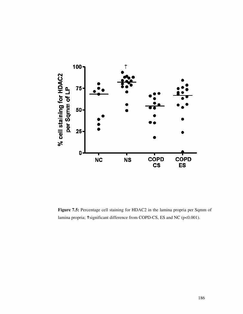
7.4.4 Percentage cell HDAC2 staining in the lamina propria
For percentage cell HDAC2 staining (ie taking into account total cell numbers)
normal lung function smokers were significantly different from all other groups
(p<0.001, with smoking again apparently stimulating anti-inflammatory HDAC2),
but COPD-CS, ES and NC were quite similar (p=0.42) (Figure 7.5). No association
was found with lung function measurements.

182
Figure 7.1: Bronchial biopsy section from (A) normal control; (B) normal lung
function smoker; (C) COPD current smoker; and (D) COPD ex-smokers: (black
arrows) brown staining indicates HDAC2 positive cells in the epithelium and in the
lamina propria, black arrows, also indicating decreased cellularity in the LP in (B)
and (C). Original magnification, x400.

183
Figure 7.2: Percentage of area of epithelium stained for HDAC2 in COPD current
smokers and ex-smokers, normal lung function smokers compared to normal controls
with no significant difference between groups. Horizontal bars represent the median
for each group.

184
Figure 7.3: Total number of cells in the lamina propria per Sqmm of lamina propria;
†significant difference from NC (p=0.04).

185
Figure 7.4: Number of HDAC2 positive cells in the lamina propria per Sqmm of
lamina propria; †significant difference from NC (p=0.02); ����significant difference
from COPD-CS (p<0.001); *significant difference from COPD-CS (p=0.04).
186
Figure 7.5: Percentage cell staining for HDAC2 in the lamina propria per Sqmm of
lamina propria; �significant difference from COPD-CS, ES and NC (p<0.001).

187
Figure 7.6: Correlation between total number of cells per Sqmm of lamina propria in
COPD-CS and smoking (pack years).

188
7.5 Longitudinal ICS intervention study
The group demographics of subjects who participated in the study are the same as
presented Chapter 6.
7.5.1 HDAC2 expression in the airway epithelium
Six months of treatment with fluticasone propionate (active arm; p=0.2, placebo;
p=0.5) made no difference on HDAC2 expression in the airway epithelium (Figure
7.7).
7.5.2 HDAC2 positive cells in the lamina propria
There was no difference observed before versus after treatment for HDAC2 positive
cells in the lamina propria (active arm, p=0.5 and placebo; p=0.3, Figure 7.8). I also
observed that there was no change in the total number of cells in the lamina propria
before and after treatment (active arm, p=0.5 and placebo; p=0.8, Figure 7.9). Thus,
percentage cell HDAC staining was also not different before and after treatment
(active arm, p=0.5 and placebo; p=0.8, Figure 7.10).

189
Figure 7.7: The percentage change in HDAC2 in the epithelium on treatment with
(A) fluticasone propionate and (B) placebo, the horizontal bars represent the
medium. There was no significant difference comparing before and after treatments.

190
Figure 7.8: Number of HDAC2 positive cells in the lamina propria per Sqmm of
lamina propria on treatment with (A) fluticasone propionate and (B) placebo: the
horizontal bars represent the mediums. There was no significant difference
comparing before and after treatments.

191
Figure 7.9: The total number of cells in the lamina propria per Sqmm of lamina
propria on treatment with (A) fluticasone propionate and (B) placebo: the horizontal
bars represent the mediums. There was no significant difference between before and
after treatments.

192
Figure 7.10: Percentage cell staining for HDAC2 in the lamina propria per Sqmm of
lamina propria; (A) with fluticasone propionate and (B) with placebo: the horizontal
bars represent the mediums. There was no significant difference comparing before
and after treatments.

193
7.6 Discussion
To the best of my knowledge, this is the first detailed biopsy immunostaining study
looking at the status of HDAC2 expression in COPD and its potential reversibility
with ICS or/and smoking cessation (albeit the latter in a cross-sectional study only at
this stage).
The airway epithelium showed strong positivity for HDAC2 staining/expression but,
interestingly this was not different between the groups. Although COPD current
smokers showed a slight decrease in HDAC2 staining in the epithelium (as
percentage of HDAC2 area of staining in the epithelial area measured), this was not
statistically significant when compared to normal controls. However the difference
between groups, even if real, hardly suggests that this is a major effect and HDAC2
is more variable between individuals than groups. The fact that HDAC2 expression
in the epithelium is well preserved in smokers (even if little less in current smokers
COPD subjects) suggest that ICS may still be effective in this tissue compartment, as
the CS-GCR complex should be able to access its transcription sites as CS-sensitive
genes.
In the lamina propria the data were very striking. I found that there was a decrease in
the total number of cells in the lamina propria in COPD current smokers compared to
normal controls. This total cell change was strongly related to smoking history and is
an effect not previously described. Normal lung function smokers also showed a
slight decrease in total cellularity whilst the COPD-ES group showed a normalisation
in total cellularity.
In the COPD subjects the plot clearly indicates that smoking is affecting the total
cellularity of the lamina propria. Notably, if I had used molecular techniques as used
by the Barnes group, etc I would have come to a false conclusion as I would not be
aware that there is a dramatic change in total cellularity in the lamina propria, largely
causing the apparent down regulation of HDAC2 mRNA and decreased protein
content. For example (as explained in Chapter 2 as well), Ito et al (Ito et al., 2001),
showed by using western blotting that HDAC2 protein content was reduced in
bronchial biopsies from normal lung function smokers compared to normal controls,

194
but looking now at the images they actually published in their paper (but which they
did not comment on), it is quite clear that total cellularity of the lamina propria in
their normal lung smokers was also substantially less than in their normal controls.
But, as stated above, their analysis did not take into account the overall decrease in
cellularity and therefore the consequential total protein available. ICS made no
difference to the total number of cells in lamina propria.
Our data for HDAC2 positive cells in the lamina propria was quite interesting as
well. It suggested that HDAC2 positive cells decrease in the lamina propria of
current smokers with COPD compared to normal controls and on the other hand
there was an increase in HDAC2 positive cells in the lamina propria of COPD ex-
smokers compared to COPD current smokers; essentially returning to normal. Data
on normal lung function smokers was quite remarkable; paradoxically they had
significantly higher numbers of HDAC2 positive cells in the lamina propria. This
suggests that smoking is actually stimulating anti-inflammatory HDAC2 levels,
although ICS made no difference to HDAC2 positive cells in lamina propria.
The data on percentage cell staining for HDAC2 further confirmed the above data on
normal lung function smokers that the percentage HDAC2 staining was also
significantly high in normal lung function smokers compared to other groups, but
COPD-CS, COPD-ES and NC were quite similar to each other. This strongly
confirms at least the possibility of smoking stimulating anti-inflammatory HDAC2
expression in chronic smokers. However, the decrease observed in COPD in relation
to HDAC2 does seem most likely due to changes in cellularity, and not HDAC2
itself. ICS made no difference to percentage cell HDAC2 staining.
Taken together, these data suggest that smoking itself stimulates the HDAC2
expression while COPD does not seems to be affecting the HDAC2 status itself, but
the apparent decrease in HDAC2 observed in COPD current smokers is due to
confounding by changes in total cellularity, which itself is strongly negatively related
to smoking history. Quitting does seem to have a potential for upregulating HDAC2
at a cell level as shown by an increase in number of cells positive for HDAC2 in the
lamina propria of COPD ex-smokers and further supported by slight increase in
percentage cells staining in COPD ex-smokers, although this was not statistically

195
significant when compared to COPD current smokers (type 2 error). Most of the
effect seems related to change in total cell numbers. However, we need long term
smoking cessation studies with larger cohorts to confirm and tease out these findings.
More precisely, the key finding is the difference in HDAC2 expression in normal
lung function smokers and decrease in cells in COPD current smokers. It probably
suggests that smoking stimulates anti-inflammatory HDAC2 in non-COPD smokers,
but in those who develop COPD then HDAC2 goes down but mainly due to decrease
in cellularity of lamina propria and not HDAC2 expression itself. Moreover the
status of HDAC2 is not different in the epithelium in either smokers or COPD. So
essentially smoking is stimulating HDAC2, but decreasing the total lamina propria
cellularity in COPD current smokers. It is possible that this difference between
smokers with and without COPD may be important in the aetiology of COPD ie if in
an individual smoking decreases cells more than stimulating anti-inflammatory
HDAC2, then COPD might ensue.
I cannot say at this stage which particular cell type(s) is decreased in the lamina
propria in smokers. For more information I would need double staining studies for
different cells in the lamina propria ie HDAC2 plus specific cell type marker. It is
also suggested that in COPD there is increased apoptosis but this is controversial and
is not clearly established. However, increased oxidative stress produced by tobacco
smoke and protease–antiprotease imbalance, and also genetic susceptibility, may
contribute to increased apoptosis in COPD airways (Park, Ryter, & Choi, 2007;
Plataki et al., 2006). In another parallel study in our group (Soltani A, 2009 ) we
found that there is a significant reduction in total number of vessels in the lamina
propria in smoking/COPD, so decreased vascular supply might be contributing to
decreased cellularity in COPD airways, but we need further studies to confirm that.
In summary, our data suggest that HDAC2 expression is increased in normal
smokers but reduced in current smokers with COPD, though the latter seems largely
due to confounding by changes in cellularity in the lamina propria but not in HDAC2
itself. Quitting does seem to have a real effect on up-regulating HDAC2 at a cell
level, but it is not affected by ICS. We need comprehensive immunohistochemical
studies to fully understand cellular changes in the lamina propria and prospective

196
long term smoking cessation studies with larger cohorts to confirm these findings.
Molecular methods must take such changes in the cellular environment into account
and cannot be taken on simple face value; indeed the current published data is likely
to be misleading because this was not taken into account.


