
1
CONSIDERING WIDER MYOFASCIAL INVOLVEMENT
AS POSSIBLE CONTRIBUTORS TO UPPER
EXTREMITY DYSFUNCTION FOLLOWING
TREATMENT FOR PRIMARY BREAST CANCER.
Fourie W J Nat. Dip. PT
Private practitioner, Johannesburg, RSA.
Tel: + 27 (0) 11 763 6990
Email: will [email protected]
Corresponding author
Willie Fourie, Private practitioner
P.O Box 209, Florida Hills, 1716, Roodepoort, Republic of South Africa.
Tel: + 27 (0) 11 763 6990
Fax no: + 27 (0) 866 180 179
Email: will [email protected]

2
ABSTRACT
BACKGROUND Breast cancer is the most common malignancy in w omen. Although scarring is
recognized as contributing to limited shoulder movements, compromised tissue gliding in a
wider range of fascial and connective tissue structures are under-recognized.
AIM To report on soft tissue patterns in patients w ith upper limb dysfunction after modif ied
radical mastectomy.
METHODS Tissue gliding w as assessed in the neck, chest w all, abdomen, axilla and upper
arm. Scarring, areas and directions of tightness w ere mapped on upper body charts.
RESULTS 18 shoulders w ere evaluated. All patients had combinations of restrictive tissue
gliding and shoulder movements. Four dominant restrictive areas were identif ied - surgical
scarring, axillary tightness radiating into the upper arm, lateral chest w all and posterior tightness
over the teres major muscle.
DISCUSSION Breast cancer treatment results in t issues losing its shearing and gliding ability.
Mapped restrictive tissue gliding clearly show w ider than reported restrictions. This pattern
needs further research and investigation.

3
BACKGROUND
Breast cancer is the most common malignancy in w omen in the w estern world and of rising
concern in developing countries. In the United States of America (US) breast cancer w ill be
diagnosed in 1 in 8 w omen (Kingsbury 2007), w hile the lifetime risk for w omen in the United
Kingdom (UK) is currently estimated at 1 in 9 (www.cancerresearchuk.org/ ).
Medical treatments for breast cancer may include surgery, chemotherapy, hormone
replacement therapy and radiation. Depending on the histology and the extent of the disease,
combinations of surgery and adjuvant therapy may be used. The level of surgery varies greatly,
from modif ied radical mastectomy (MRM) w ith removal of all the breast material as w ell as some
supporting fascial structures, to minimal invasive breast saving operations (BSO) and sentinel
lymph node biopsy (SLNB) from the axilla. Further surgery may include various breast
reconstruction procedures. Although effective in treatment of cancer, the above interventions
are often associated w ith side effects which may affect a patient’s function and quality of life
(Ghazinouri et al 2005).
Advances in medicine and technology, combined w ith improved therapy and earlier detection,
yields the largest group of cancer survivors in the US today (Lash & Silliman 2000). As a result,
cancer survival rates defined as a relative combined 5-year statistic in this patient group, have
increased steadily over the years. Survival rates are currently reported as above 50% (Fialka-
Moser et al 2003) in general, and up to 90% in certain population groups (Ghazinouri et al
2005). As a result of these factors, it is likely that progressively more patients w ith breast
cancer may develop upper extremity dysfunction and be referred for evaluation and treatment
by rehabilitat ion professionals (Ghazinouri et al 2005).

4
Quality of life strongly depends on physical function. With more w omen now living w ith their
disease, issues of survivorship, both physical and psychosocial, are of increasing importance.
Breast cancer treatment is often follow ed by a decline in upper body function, even at some
time distant from therapy (Karki et al 2005). Lash & Silliman (2000) conclude that ‘upper body
dysfunction may arise shortly after therapy and resolve, arise, and persist for at least 21
months, or arise at some time distant from therapy’. Post-treatment impairments consist, for
example, of upper limb oedema, decreased shoulder mobility, neural tissue injuries causing
sensory and motor dysfunction, and pain (Karki et al 2005).
For decades, reduced shoulder range of movement (ROM) and functional impairment have
been recognized as a problem after breast cancer surgery and treatment (Lash & Silliman 2000;
Box et al 2002; Ghazinouri et al 2005; Karki et al 2005; Lauridsen et al 2005; Wyrick et al 2006).
The presence of impaired shoulder ROM show s a lot of variability, ranging from a 1.5%
incidence to as high as 50% (Rietman et al 2002; Box et al 2005; Karki et al 2005; Wyrick et al
2006). A signif icant relationship is reported betw een oedema and restricted range of motion,
and impaired activities of daily living and influencing quality of life. Activities such as pulling a
sw eater over the head, fastening a bra, carrying a heavy bag, sleeping on the operated side,
reaching out, w orking w ith the ipsilateral arm, housew ork, leisure and sporting activities or
handicraft are reported as compromised (Rietman et al 2002). Shoulder-arm morbidity is a
complex syndrome w hich cannot be adequately described by single symptoms (Karki et al
2005), or rely on single-therapy treatment strategies for optimal results.
Breast and axillary scarring is commonly recognized as contributing to limited shoulder
movements and some of the impaired activit ies above (Lash & Silliman 2000; Karki et al 2005).
In contrast to reporting on impairments in body functions and structures, little is know n about

5
compromised t issue gliding and shearing ability betw een supporting fascial and connective
tissue structures as a possible source of restrictive musculoskeletal syndromes in this patient
group. Limited reporting on tissue glide could possibly be attributed to a lack of objective
information on the psychometric properties of palpating restrictions in fascial glide by manual
therapies.
AIM:
The primary aim of this study is to describe observed soft tissue mobility patterns in a group of
breast cancer patients w ith upper body dysfunction, subsequent to modif ied radical
mastectomy. We hope that this may add to understanding as to how restricted tissue gliding in
one area, may contribute to dysfunction in another area.
METHODS
Patients:
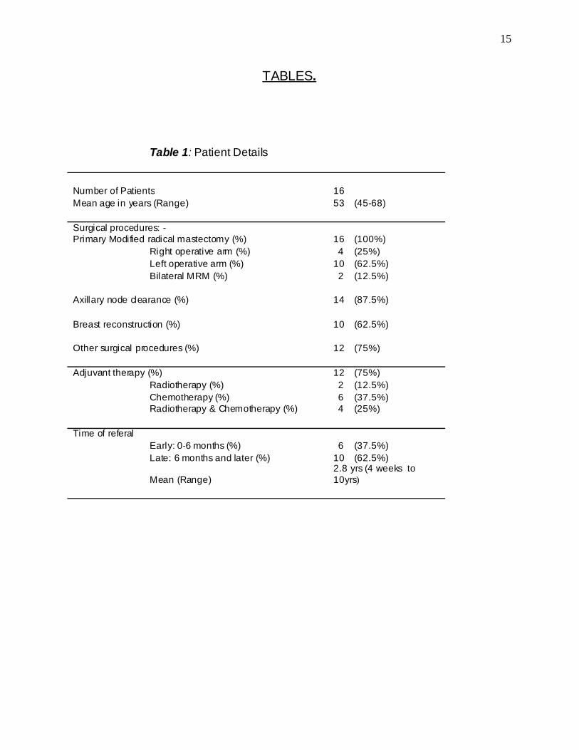
The clinical records and evaluation charts of 16 consecutively referred breast cancer patients
were review ed retrospectively. All patients had undergone a modif ied radical mastectomy
(MRM) as the primary surgical procedure for breast cancer, as well as varying combinations of
other medical treatments and procedures prior to referral to physiotherapy. In all cases, the
reason for referral w as for the management of upper body and shoulder impairments and
functional deficits. Referrals included patients from both the early post-operative period (0 to 6
months) to patients w ith established chronic dysfunction many years post breast cancer
treatment (table 1).

6
Recorded data: patient records:
Documented data included primary breast cancer treatment (surgery, chemotherapy,
radiotherapy, number of lymph nodes removed and breast reconstruction procedures), time of
f irst referral (0 to 6 months or 6 months +), shoulder range of motion (ROM), presence of
lymphoedema, pain, neck ache, methods of early rehabilitat ion and soft tissue mobility.
Recorded data w ere analysed for patterns of dysfunction, catagorised and grouped in table form
(tables 1-3).
Shoulder range of movement (ROM):
ROM w as assessed both actively and passively using abduction in the plane of the scapula as
the movement of choice for recording and measuring treatment progress. This w as recorded as
degrees of elevation of the arm. A limitation in recorded ROM data is that some of our early
referrals in this patient group w ere only measured subjectively by observation rather than w ith
the use of measuring equipment. Recorded ROM data (table 2) should therefore be interpreted
accordingly. During active shoulder movement, changes in scapulohumeral rhythm w ere noted
and recorded. Observations on internal rotation, external rotation and adduction of the
shoulder, as w ell as neck and thoracic spine movements w ere also recorded, but not included in
this study as it w as incompletely recorded for all cases.
Soft tissue mobility:
Soft tissue gliding w as manually assessed by applying gentle shearing stretch to the skin over
the neck, chest w all, abdomen, axilla and upper arm. Tissues in tested areas w ere taken to the
limits of its available range of gliding. Where soft tissue gliding w as felt to come to a premature
end, tissue mobility w ere considered as restricted. Areas where mobility w as restricted by
scarring or tethering and w here reduced tissue f lexibility could be palpated, w ere mapped on

7
upper body charts (Figure 1). Mapping included all surgical scars, areas of tissue tightness as
well as the directions of palpated tissue restriction. Tissue mobility w ould be graded either as
normal or restricted.
RESULTS
Our patient sample consisted of 16 consecutively referred patients betw een the ages of 45 and
68 (mean age: 53) w ith upper body morbidity after treatment of primary breast cancer. Patients
were from the same demographic area w ithin the Greater Johannesburg Metropolitan Council.
The sample period w as between January and December 2006. Time of f irst referral varied
betw een 4 w eeks post surgery to 10 years after the initial treatment (mean: 2.8 years). Only the
three patients referred w ithin the f irst 6 w eeks after surgery did not have additional treatment
procedures prior to referral. The remaining 13 patients had varying combinations of adjuvant
medical treatments and surgical procedures prior to referral for physiotherapy (Table 1).
Three factors were shared by all the patients, i.e. type of primary surgery (modif ied radical
mastectomy), upper body dysfunction and tissue restriction. Additional surgery, before or after
the breast cancer, were reported by 11 patients and 12 patients received adjuvant therapy after
the MRM (Table 1).
Eighteen shoulder girdles w ere evaluated in sixteen patients (tw o patients had bilateral
mastectomies). Fifteen patients had restricted shoulder movements and all sixteen presented
with changes in scapulohumeral rhythm w hen moving the arm. The average ROM, measuring
abduction in the plane of the scapula, w as 130 degrees (range, 40 – 180 degrees). Underlying
glenohumeral pathology w as evident in only one shoulder and confirmed and treated by her

8
orthopaedic surgeon. Although restrictions in shoulder movements w ere evident in 95% of the
study group, lymphoedema of the arm (8/16), neck ache (8/16) and pain in the chest, shoulder
and arm (11/16) co-existed w ith the restricted shoulder movements (Table 2).
Restrictions in normal gliding betw een the skin and subcutaneous tissue w ere evident in the
entire sample group (Table 3). Evaluation of tissue gliding on the neck, chest w all, abdomen,
axilla and upper arm identif ied tightness in a w ide range of tissues. The tw o most prominent
areas of identif ied restriction w ere the surgical scarring on the anterior chest w all and donor
sites for reconstructive surgery (14/18 = 77.7%) and axillary tightness radiating into the medial
upper arm (15/18 = 83%). Additional combinations of tightness on the lateral chest w all
radiating into the axilla (11/18 = 61%), from the drain sites (5/18 = 29%), the posterior axillary
border over the teres major and infraspinatus muscles (10/18 = 55%), above the clavicle (6/18 =
33%) and around other surgical sites (7/18 = 39%) w ere also found (Figure 1).
DISCUSSION
All the patients in this study had undergone a modif ied radical mastectomy as the primary
surgical procedure for breast cancer, while 12 patients received varying combinations of other
medical treatments and procedures prior to referral to physiotherapy. In all cases, the reason
for referral w as for the management of upper body and shoulder impairments and functional
deficits. Although restrictions in shoulder movements w ere evident in 95% of the study group,
this w as not alw ays the primary reason for referral. Co-existing problems such as
lymphoedema of the arm, neck ache and pain in the chest, shoulder and arm often dominated
as the more severe symptoms of upper body dysfunction. On evaluating of the upper body and

9
shoulder impairments, all patients shared varying degrees of restricted soft tissue mobility
contributing to dysfunction.
Treatment for breast cancer leads to a number of w ell know n and documented sequelae of
which restricted shoulder movements, pain and oedema are the most debilitating. Both
Rietman et al (2002) and Karki et al (2005) report that dysfunction may persist for extended
periods, w ith some patients experiencing a progressive decline in activities of daily living, w ork
and leisure activities follow ing mastectomy. It is therefore suggested that appropriate
interventions should be planned for at least 2 years after treatment for primary breast cancer
(Lash & Silliman 2000; Karki et al 2005). Patients in this study group w ere still experiencing
limitations in the above activit ies for as long as 10 years after primary treatment. This continued
long term dysfunction and impairment cause feelings of frustration and anger (personal
comments from patients). Some patients in the present study group even suggested that they
have stopped complaining to their doctors about the experienced limitations, pain and
dysfunction and that they are “just getting on w ith life, regardless of quality”.
Breast and axillary scar tightness remain one of the most common impairments after
mastectomy even at 12-month follow up (Karki et al 2005). Patients after mastectomy also
report higher levels of limited shoulder movements than patients after tissue preserving
procedures (Rietman et al 2002). Although breast saving operations are increasingly advocated
by surgeons, modif ied radical mastectomy is still performed more commonly in elderly patients
(Karki et al 2005) and accepted as the standard surgical treatment for breast cancer
(Pendergrast 1989). All the patients in our study group, except one, had varying degrees of
restricted shoulder movement upon referral. The major t ight areas w ere identif ied over the
anterior chest w all radiating into the axilla (78%), and medial upper arm (83%). This is
consistent w ith described tightness and scarring contributing to shoulder morbidity in literature

10
(Box 2002; Karki et al 2005). The tightness found in the lateral chest w all radiating into the
axilla in 11 (61%) of our patients could be added to the group w ith anterior chest w all scarring.
Understanding the extent of tissue dissection during surgery may explain the large percentage
of patients in our study w ith the above patterns of tissue restriction. Dur ing a modif ied radical
mastectomy, not only the breast tissue and lymph nodes in the axilla are removed, but important
supporting fascial structures as well. The surgical procedure involves the careful mobilisation,
dissection and removal of breast tissue and the underlying fascia/epimysium from the pectoralis
major muscle. Continuing around the lateral edge of pectoralis major, the fat pad and the lymph
nodes betw een pectoralis major and pectoralis minor are removed by sharp dissection. Further
surgical removal or sampling of lymph nodes involve careful dissection of varying numbers of
nodes from the axilla, together w ith its supporting fat and areolar connective tissue (Dao & Patel
1984). The fascia extending from the lateral edge of pectoralis major is continuous w ith the
deep fascia covering serratus anterior. Further posterior this fascial sheet splits to ensheathe
latissimus dorsi as deep fascia/epimysium (Warw ick & Williams 1976). Dissection for removal
of the axillary tail of the breast and the subscapular and lateral thoracic groups of lymph nodes
continues over the subscapularis muscle until the edge of the latissimus dorsi muscle is reached
(Dao & Patel 1984). Surgery can therefore be seen to contribute substantially to observed
tissue tightness – even in areas not directly visible as surgical scarring on the body surface.
A viable explanation as to w hy surgery creates such a w ide range of restrictions in tissue, even
at sites distant from the direct surgical scarring, could possibly be found in the fascial
arrangements betw een the pectoralis major muscle and the brachial fascia of the arm. The
fascia covering pectoralis major is continuous w ith the brachial fascia in tw o distinct w ays
(Stecco et al 2007):

11
• The fascia overlying the clavicular part of pectoralis major continues into the anterior
brachial fascia.
• The fascia covering the costal part is continuous w ith the axillary fascia and then w ith the
medial brachial fascia.
Furthermore, abundant amounts of fat and loose areolar connective tissue protect and bind the
numerous structures w ithin the axilla together. By virtue of its extensibility and elasticity, a
considerable amount of tissue movement is simultaneously allow ed for arm elevation (Warw ick
& Williams 1976). It could be reasoned that damage to the pectoralis major fascia and the
protective axillary connective tissue may contribute to limited arm and shoulder movement.
From our documented results, restrictions w ere however w ider than described above and
reported in the literature. This refers specif ically to the tightness found over the posterior
axillary w all and over the teres major muscle and the long head of triceps (Figure 2). The
relatively large number of shoulders w ith posterior axillary w all tightness (10 or 55%) w as
unexpected. This tissue restriction is felt as an area of distinct fascial tethering (Figure 3), and
not usually associated w ith surgical scarring. Only in tw o patients w ith breast reconstructions
using a latissimus dorsi f lap could tightness here be explained as part of direct surgical
procedures. The identif ied area of tethering corresponds to the teres major and minor trigger
spots according to Travell and Simons (1992) and the centre of coordination for humeral
extension as described by L Stecco (2004). Posterior axilla and teres major muscle tightness
may be associated w ith restricted shoulder elevation, changes in scapulohumeral rhythm and
changes in glenohumeral biomechanics.
Stecco et al. (2007) further describe a f ibrous lamina betw een the fascia of latissimus dorsi and
the triceps brachial fascia creating a thickening in the posterior axilla. This thickening is fan-
shaped, w ith the apex directed tow ards the axilla and the base tow ards the postero-medial side

12
of the brachial fascia. With the extent of dissection described during axillary clearance
extending to the edge of latissimus dorsi (Dao & Patel,1984), it seems feasible to assume that
direct anterior and lateral fascial damage may w ell inf luence the fascial structures as far as the
posterior axilla and teres major as w ell.
CONCLUSIONS.
Breast cancer treatment results in a range of supporting connective tissues losing its normal
shearing and gliding ability. In this limited sample, mapped restrictive tissue gliding clearly show
wider than reported restrictions. The w ider extent of limitation in tissue gliding w as somew hat
unexpected. This pattern needs further research and investigation.
Shoulder-arm morbidity is a complex syndrome w hich cannot be adequately described by single
symptoms, or rely on single-therapy treatment strategies for optimal results. In our practice,
adding mobilization of all tissue gliding restrictions resulted in improved upper limb function,
reduced pain and increased exercise tolerance. We therefore propose that the entire upper
quarter be assessed and treated for tissue gliding restrictions as part of the long term
rehabilitation plan for patients after treatment for breast cancer.

13
REFERENCES.
Box RC, Reul-Hirche HM, Bullock-Saxton JE, Furnival CM 2002 Shoulder movement after
breast cancer surgery: results of a randomized controlled study of postoperative physiotherapy.
Breast Cancer Res Treat 75:35-50
Cancer Research UK (2006). www.cancerresearchuk.org/cancerstats/types/breast/incidence/
(accessed 25.9.06).
Dao TL, Patel J 1984 Modif ied radical mastectomy. In: Nyhus LM, Baker RJ. (eds.) Mastery of
Surgery: Volume I. Brow n and Company, Boston/Toronto
Fialka-Moser V, Crevenna R, Korpan M, Quittan M 2003 Cancer rehabilitat ion. Particularly w ith
aspects on physical impairments. J Rehabil Med 35: 153-162
Ghazinouri R, Levy C, Ben-Porat L, Stubblefield MD 2005 Shoulder impairments in patients
with breast cancer: a retrospective review Rehabil Onc 23: 5-8
Kärki A, Simonen R, Mälkiä E, Selfe J 2005 Impairments, activity limitations and participation
restrictions 6 and 12 months after breast cancer operation. J Rehabil Med 37: 180-188
Kingsbury K 2007 The changing face of breast cancer, Time 170:36-43
Lash TL, Silliman RA 2000 Patient characteristics and treatments associated w ith decline in
upper-body function follow ing breast cancer therapy. J Clin Epidemiol, 53: 615-622

14
Lauridsen MC, Christiansen P, Hessov I 2005 The effect of physiotherapy on shoulder function
in patients surgically treated for breast cancer: a randomized study. Acta Oncol 44:449-457
Pendergrast WJ 1989 Surgical management of breast cancer. In: Lew is JR (Ed) The Art of
Aesthetic Plastic Surgery. Volume II. Little, Brow n and Company, Boston
Rietman JS, Dijkstra PU, Hoekstra HJ, et al 2002 Late morbidity after treatment of breast cancer
in relation to daily activities and quality of life: a systematic review . EJSO 29:229-238.
Stecco C, Gagey O, Macchi V, et al 2007 Tendinous muscular insertions onto the deep fascia
of the upper limb f irst part: anatomical study. Morphologie, doi:10.1016/j.morpho.2007.05.001
Stecco L 2004 Fascial Manipulation for Musculoskeletal Pain. Piccin Nuova Libraria S.p.A.,
Padova.
Travell J G, Simons D G 1992. Volume 2. Myofascial pain and dysfunction. The Trigger Point
Manual. The Low er Extremities.Williams and Wilkins, Baltimore.
Warw ick R, Williams PL 1976 ed. Gray’s Anatomy. 35th ed. Longman
Wyrick SL, Waltke LJ, Ng AV 2006 Physical therapy may promote resolution of lymphatic
cording in breast cancer survivors. Rehab Onc 24:29-34.
15
TABLES.
Table 1: Patient Details
Number of Patients 16 Mean age in years (Range) 53 (45-68) Surgical procedures: - Primary Modified radical mastectomy (%) 16 (100%)
Right operative arm (%) 4 (25%) Left operative arm (%) 10 (62.5%) Bilateral MRM (%) 2 (12.5%)
Axillary node clearance (%) 14 (87.5%)
Breast reconstruction (%) 10 (62.5%)
Other surgical procedures (%) 12 (75%) Adjuvant therapy (%) 12 (75%)
Radiotherapy (%) 2 (12.5%) Chemotherapy (%) 6 (37.5%) Radiotherapy & Chemotherapy (%) 4 (25%)
Time of referal
Early: 0-6 months (%) 6 (37.5%) Late: 6 months and later (%) 10 (62.5%)
Mean (Range) 2.8 yrs (4 weeks to 10yrs)

16
Table 2: Impairment and complaints (reason for referral)
Number of Shoulders 18
Shoulder ROM restrictions (%) 17 (95%)
Mean Abduction ROM (Range) 130° (40 - 180)
Changed Scapulo-humeral rhythm (%) 18 (100%)
Pain in chest, shoulder or arm (%) 11 (61%)
Neckache (%) 8 (44%)
Lymphoedema (%) 8 (44%)

17
Table 3: Sites of tisssue restriction.
Tissue gliding Restrictions (%) 18 (100%)
Surgical scar (%) 14 (78%)
Drain sites (%) 5 (29%)
Axilla and upper arm (%) 15 (83%)
Axilla and lateral chest wall (%) 11 (61%)
Posterior axilla / Scapula (%) 10 (55%)
Neck, above clavicle (%) 6 (33%)
Tightness from other surgical sites (%) 7 (39%)

18
FIGURES.
Figure 1: Body chart w ith scarring, area and direction of tissue tightness recorded.

19
Figure 2: Posterior axillary w all and teres major tightness

20
Figure 3: Fascial tethering over teres major muscle.


