Accepted Manuscript
Control of liver size by RNAi-mediated multiplex knockdown and its applica-tion for discovery of regulatory mechanisms
Hao Yin, Roman L. Bogorad, Carmen Barnes, Stephen Walsh, Iris Zhuang,Hidenori Nonaka, Vera Ruda, Satya Kuchimanchi, Lubomir Nechev, AkinAkinc, Wen Xue, Marino Zerial, Robert Langer, Daniel G. Anderson, VictorKoteliansky
PII: S0168-8278(15)00786-2DOI: http://dx.doi.org/10.1016/j.jhep.2015.11.028Reference: JHEPAT 5911
To appear in: Journal of Hepatology
Received Date: 17 June 2015Revised Date: 22 October 2015Accepted Date: 11 November 2015
Please cite this article as: Yin, H., Bogorad, R.L., Barnes, C., Walsh, S., Zhuang, I., Nonaka, H., Ruda, V.,Kuchimanchi, S., Nechev, L., Akinc, A., Xue, W., Zerial, M., Langer, R., Anderson, D.G., Koteliansky, V., Controlof liver size by RNAi-mediated multiplex knockdown and its application for discovery of regulatory mechanisms,Journal of Hepatology (2015), doi: http://dx.doi.org/10.1016/j.jhep.2015.11.028
This is a PDF file of an unedited manuscript that has been accepted for publication. As a service to our customerswe are providing this early version of the manuscript. The manuscript will undergo copyediting, typesetting, andreview of the resulting proof before it is published in its final form. Please note that during the production processerrors may be discovered which could affect the content, and all legal disclaimers that apply to the journal pertain.
1
Control of liver size by RNAi-mediated multiplex knockdown and its application for discovery of
regulatory mechanisms
Hao Yina*, Roman L. Bogorada*, Carmen Barnesb, Stephen Walsha, Iris Zhuanga,c, Hidenori Nonakad, Vera
Rudaa, Satya Kuchimanchib, Lubomir Nechevb, Akin Akincb, Wen Xuee, Marino Zeriald, Robert Langera,f,g,h,
Daniel G. Andersona,f,g,h,#, Victor Kotelianskyi,j,#
a David H. Koch Institute for Integrative Cancer Research, Massachusetts Institute of Technology,
Cambridge, MA, 02139, USA
b Alnylam Pharmaceuticals, Cambridge, MA, 02142, USA
c Department of Biology, Massachusetts Institute of Technology, Cambridge, MA, 02139, USA
d Max Planck Institute of Molecular Cell Biology and Genetics (MPI-CBG), Dresden, 01307, Germany
e RNA Therapeutics Institute and Program in Molecular Medicine, University of Massachusetts Medical
School, Worcester, MA 01605, USA
f Department of Chemical Engineering, Massachusetts Institute of Technology, Cambridge, MA, 02139,
USA
g Harvard-MIT Division of Health Sciences & Technology, Cambridge, MA, 02139, USA
h Institute of Medical Engineering and Science, Massachusetts Institute of Technology, Cambridge, 02139,
MA, USA
i Skolkovo Institute of Science and Technology, Skolkovo, 143025, Russia
j Department of Chemistry, M.V.Lomonosov Moscow State University, Leninskie Gory,119991, Russia
*These authors contributed equally to this work.
# These authors share senior authorship.
Correspondent authors: Daniel G Anderson. Massachusetts Institute of Technology, 77 Massachusetts avenue, Cambridge, MA 02139, +1 617-258-6843, [email protected]
Victor Koteliansky, Skolkovo Institute of Science and Technology, Novaya St., 100, Karakorum Building, 4th
floor, Skolkovo 143025 Russian Federation, +7 495-280-1481, [email protected]
Keywords: Hippo pathway, p53, bile acids, siRNA, nanoparticles
2
Abstract:
Background and aims. The Hippo pathway controls organ size through a negative regulation of the
transcription co-activator Yap1. The overexpression of hyperactive mutant Yap1 or deletion of key
components in the Hippo pathway leads to increased organ size in different species. Analysis of
interactions of this pathway with other cellular signals corroborating organ size control is limited in
part due to the difficulties associated with development of rodent models.
Methods. Here, we develop a new model of reversible induction of the liver size in mice using
siRNA-nanoparticles targeting two kinases of Hippo pathway, namely, mammalian Ste20 family
kinases 1 and 2 (Mst1 and Mst2), and an upstream regulator, neurofibromatosis type II (NF2).
Results. The triple siRNAs nanoparticle-induced hepatomegaly in mice phenocopies one observed
with Mst1-/- Mst2-/- liver-specific depletion, as shown by extensive proliferation of hepatocytes and
activation of Yap1. The simultaneous co-treatment with a fourth siRNA nanoparticle against Yap1
fully blocked the liver growth.
Hippo pathway-induced liver enlargement is associated with p53 activation, evidenced by its
accumulation in the nuclei and upregulation of its target genes. Moreover, injections of the triple
siRNAs nanoparticle in p53LSL/LSL mice shows that livers lacking p53 expression grow faster and
exceed the size of livers in p53 wild type animals, indicating a role of p53 in controlling Yap1-
induced liver growth.
Conclusion. Our data show that siRNA-nanoparticulate manipulation of gene expression can
provide the reversible control of organ size in adult animals, which presents a new avenue for the
investigation of complex regulatory networks in liver.

3
Introduction
Maintenance of proper organ size in an organism is a fundamentally important process. The proper function
of organs depends on coordinated control of their size during developmental and adult stages. In contrast,
loss of organ size control contributes to a number of diseases, including hypertrophy and degenerative
diseases(1). Organ size control integrates various factors, including local mechanical, autocrine/paracrine
stimuli as well as soluble circulating cues and environmental factors(1-3).
One of the cues critical in controlling organ size and cellular proliferation is the Hippo signaling pathway
(Reviewed in(4, 5)). This pathway is composed of an evolutionarily conserved core kinase cassette and
upstream modulators (reviewed in(6)). In mammals, the core kinases consist of mammalian Ste20 family
kinases 1 and 2 (Mst1 and Mst2), Salvador homolog 1 (Sav1), Large tumor suppressor 1 and 2 (Lats1 and
Lats2), and Mps one binder 1 (Mob1)(4, 5). The core cascade is tightly regulated by multiple upstream
modulators(6). Neurofibromatosis type II (NF2, also known as Merlin) can activate the core kinase
cassette(7), likely through direct binding and recruiting Lats1/2 to the plasma membrane(8). The Hippo
pathway negatively regulates its major downstream effector Yes-associated protein 1 (Yap1) through
phosphorylation, provoking its degradation and cytoplasmic retention(9, 10). As a potent transcription co-
activator, Yap1 can induce genes involved in cellular growth and apoptosis inhibition by association with the
TEAD family and other transcription factors(9, 11, 12). Overexpression of S127 mutant Yap1 in the adult
liver causes massive hepatomegaly(10). It has been reported that while one allele of either Mst1 or Mst2 is
sufficient to maintain embryonic development, Mst1-/-Mst2-/- mice are embryonically lethal (13-15). One
copy of either Mst1 or Mst2 can maintain quiescence of hepatocytes, but liver-specific genetic removal of
both Mst1 and Mst2 leads to significant liver enlargement(14-16).
Hippo signaling has been shown to interact with several other pathways, including PI(3)K–mTOR(17),
Wnt/beta-catenin(18), Insulin/IGF(19, 20), and Sonic hedgehog (Shh) signaling pathway(21) in normal
tissue and tumorigenesis. However, deciphering its complex interactions in vivo requires sophisticated
approaches allowing to gradually and simultaneous manipulate multiple genes.
Recent advances in synthetic siRNA delivery nanoparticles make it possible to specifically suppress one or
more genes simultaneously in a range of species, from rodents to primates(22-25), including humans(26).
Through manipulating the compositions of nanoparticles, siRNA delivery systems have shown potent and
reversible silencing effects in vivo with high specificity in multiple tissues and cell types, including
hepatocytes(22, 23), macrophages(27) and endothelial cells(28). Here we develop a siRNA nanoparticle-
based approach to manipulate organ size through inhibition of the Hippo pathway. The deep reduction of

4
the expression levels of Mst1/Mst2/Nf2 is critical to elucidate the role of p53 pathway in the control of liver
growth.
Materials and Methods
siRNA synthesis, screening and lipid nanoparticles (LNP) formulation
siRNAs targeting mouse Mst1, Mst2, NF2, Yap1 were designed and screened as previously described(24).
The sequences and IC50 values of each siRNA are provided in Supplementary Table 1. The RNA strands
were synthesized, characterized and duplexed by Alnylam Pharmaceuticals as previously described(24).
Individual siRNAs were formulated into lipid nanoparticles and mixed prior to injections(23).
Cell culture
Hepa1-6, NIH3T3, AML-12 cells were obtained from ATCC and were propagated in DMEM supplemented
with 10% FBS. Cells were transfected with siRNA using either Lipofectamine RNAiMAX (Invitrogen) or LNP
with siRNA as described elsewhere(24).
AML-12 cells obtained from ATCC were grown on BD BioCoat collagen I coated plasticware. Cells were
treated with taurocholic acid (Sigma-Aldrich, time and dose as indicated). Cells were collected in RIPA
buffers supplemented with proteases and phosphatases inhibitors (Pierce Bio) for western blot, or fixed with
2% buffered paraformaldehyde solution, followed by permeabilization with 1% Triton X-100 in PBS for
immunocytochemistry.
Animals
C57BL/6 mice were purchased from Charles River laboratories. p53LSL/LSL mice were published
elsewhere(29). All animals received humane care, and animal protocols were approved by the Committee
on Animal Care at MIT and the Institutional Animal Care and Use Committee of Alnylam Pharmaceuticals,
certified by the American Association for Accreditation of Laboratory Animal Care. After 3 days of
acclimatization in the animal facility, 7-9 week-old mice were injected via tail vein (i.v.) with either PBS or
siRNA in LNP formulations at various concentrations. To restore p53 expression in p53LSL/LSL mice, animals
were treated with tamoxifen (two intraperitoneal doses) before the siRNA treatment. Animals were sacrificed
by CO2 overdose; tissues were harvested at different time points as indicated. Hepatocytes, stellate cells
and Kupffer cells were isolated from C57BL/6 mice via collagenase perfusion, density centrifugation and
antibody selections as described previously(30-32).
Histological, immunohistological and immunocytochemical analysis
Mouse tissues were fixed in 4% paraformaldehyde. Tissue sections were stained according to standard
immunohistochemistry protocols as previously described(33) or with secondary antibodies labelled with
Alexa 488, Alexa 555 and Alexa 647 (Invitrogen) to visualize antigen localization. We have used the
following primary antibodies: anti-Cytokeratin 18 (Progen), anti-Cytokeratin 19 (Abcam), anti-E-cadherin
5
(BD Biosciences), anti-F4/80 (Biolegends), anti-glutamine synthase (BD Biosciences), anti-Ki67
(Neomarkers), anti-p21 (Santa Cruz), anti-p53 (Leica Biosystems), anti-Yap1 (Cell Signaling), phospho-
pH2A.X (Cell Signaling). Phalloidin coupled with Alexa555 was used to visualize F-actin. Analysis of H&E,
immunohistochemistry and immunocytochemical images was performed using ImageJ package (NIH).
Western blots and quantification
The liver tissues were homogenized in RIPA buffer (Thermo Fisher) to harvest proteins. Total protein was
resolved on TGX gradient gels (BioRad). The following primary antibodies were used: anti-β-actin (Sigma),
anti-a-catenin (Cell Signaling), anti-cyclin D1 (Millipore), anti-Mst1 (Cell Signaling), anti-Mst2 (Cell
Signaling), anti-NF2 (Sigma), anti-p21 (Santa Cruz), anti-pYap1 (Cell Signaling), anti-Yap1, phospho-
pH2A.X. (Abcam) Licor Odyssey Imaging system was used to visualize protein bands. Gray scale images
were quantified with ImageJ as described previously(24).
Gene expression analysis
The levels of mRNAs were measured by branched DNA assay (Affymetrix) or quantitative PCR using
TaqMan probes and Roche LightCycler 480. Levels of mRNA of the genes-of-interest were normalized to
the levels of GAPDH mRNA and then to the average arbitrary value of the control group.
Serum chemistry
Albumin, total protein, glucose, HDL, LDL, direct and total bilirubin, AST, ALT, total bile acids were
measured in serum using Beckman Coulter reagents and Olympus Au400 autoanalyser.
Statistical analysis
P values were calculated using the Student’s t-tests and the One-Way ANOVA in Prism 5 (GraphPad).
Results
siRNA mediated knockdown of Mst1, Mst2 and NF2 leads to hepatomegaly
We designed and screened sets of chemically modified siRNAs(34) targeting Mst1 and Mst2
(Supplementary Fig. 1). Specifically, 27 siRNAs targeting Mst1 and 28 siRNAs targeting Mst2 were
synthesized. The gene silencing efficiency for each siRNA was examined in NIH3T3 cells at 5nM
(Supplementary Fig. 1a, c). We found that nine siRNAs targeting Mst1 and nine siRNAs targeting Mst2 can
achieve deep knockdown at this dose (Supplementary Fig. 1a, c). A dose response study was performed
using the 3 most potent siRNAs for each gene. The IC50 for the most potent siRNA targeting Mst1 and Mst2
were ~0.05nM and 0.2nM, respectively (Supplementary Fig. 1b, d). The most efficient siRNAs were chosen
and formulated into lipid nanoparticles (LNP), which have shown hepatocyte specific targeting with high
potency(23). Approximately 90% knockdowns of both Mst1 and Mst2 mRNAs were shown in hepatocytes
isolated from mice with treatment of LNP-formulated siRNAs against Mst1 (termed as si-Mst1) or Mst2
(termed as si-Mst2), respectively (Supplementary Fig. 2). Animals treated with si-Mst1 alone, si-Mst2 alone,
6
or their combination showed no liver growth (Supplementary Fig. 3). PBS and LNP-formulated luciferase
siRNA (termed as si-Control) treated mice served as controls. This result suggested that the remaining
expression of Mst1 and Mst2 (~10%) was sufficient to maintain Yap1 inactivation. To further inhibit the
Hippo pathway, specific siRNAs against NF2, which is a key protein for maintaining activity of this
pathway(7, 8), were developed and screened (Supplementary Fig. 4). We synthesized 27 siRNAs targeting
NF2. The gene silencing efficiency for individual siRNA was examined, and five efficient siRNAs were
identified (Supplementary Fig. 4a). The IC50 for the most potent siRNA targeting NF2 was ~0.02nM
(Supplementary Fig. 4b). LNP-formulated siRNA against NF2 (termed as si-NF2) showed efficient
knockdown in vivo (Supplementary Fig. 5). Animals treated with a combination of si-Mst1, si-Mst2 and si-
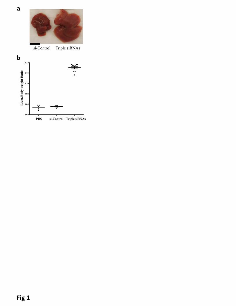
NF2 twice per week for two weeks at the dose of 0.67 mg/kg for each siRNA (hereafter, designated as triple
siRNAs nanoparticle) showed 2.5 folds induction of liver size at Day 15 after the first injections of triple
siRNAs nanoparticle (Fig 1). Similar results were obtained with alternative siRNAs targeting Mst1/Mst2/NF2
(Supplementary table 1, and data not shown).
To characterize this model, different combinations and doses of these siRNAs were examined.
Combinations of si-Mst1 and si-NF2, or si-NF2 alone did not induce growth, while the combination of si-
Mst2 and si-NF2 induced only minor liver growth (Supplementary Fig. 6a). Once we confirmed that all three
siRNAs were necessary for induction of the hepatomegaly, we performed dose titration for each siRNA in
vivo (Supplementary Fig. 6b-d). Based on these results, we optimized the dose for triple siRNA treatment as
0.3 mg/kg for si-Mst1, 0.6 mg/kg for si-Mst2 and 0.2 mg/kg for si-NF2 (Supplementary Fig. 6e). Our
experiments described below used this combined dose.
We further characterized the time course of liver growth induced by siRNA. We found that the growth
started at Day 9 and peaked at Day 15 after the first injection of triple siRNAs nanoparticle (4 injections in
total) (Fig. 2a). After withdrawal of triple siRNAs nanoparticle treatment, the liver shrank back to its original
size at Day 40 (Fig. 2a). In order to examine whether apoptosis is a key mechanism of liver shrinkage, we
performed TUNEL assay and western blots of caspases. We found that in contrast with normal livers and
enlarged livers, the shrinking livers had a significantly higher number of TUNEL-positive cells
(Supplementary Fig. 7a). Furthermore, cleaved caspase 3 and 8 were significantly upregulated in the
shrinking livers (Supplementary Fig. 7b). Our data is consistent with a previous study showing that
conditional inactivation of Yap1 in enlarged mouse liver led to apoptosis of hepatocytes(10).
Remarkably, liver growth induced by the triple siRNAs nanoparticle treatment has an upper limit: prolonged
treatment with siRNAs for an additional one or two weeks (up to 9 injections in total, 2 injections per week,
7
1.1 mg/kg per injection) did not induce further growth of the liver (Fig. 2a), indicating the possibility of
mechanism(s) to restrict Yap1-induced hepatomegaly.
To clarify the targeted cell types in liver, mice were treated with siRNA nanoparticles against triple siRNAs.
Hepatocytes, Kupffer cells, stellate cells and hepatic mononuclear cells were isolated from the livers. We
found that with the formulation (C12-200) and siRNA dose (1.1mg/kg in total) we used, the knockdown
effect is efficient (~90%) in hepatocytes (Supplementary Fig. 8a). In contrast, no significant knockdown was
observed in Kupffer cells, stellate cells and hepatic mononuclear cells. We previously showed that our
siRNA lipid formulation causes deep silencing of integrin β1 in hepatocytes, while no significant knockdown
was found in CD31+, aSMA+, F4/80+ cells(35). Because it is difficult to get a specific commercially
available antibody for the separation of biliary epithelial cells, we performed an alternative approach to
explore whether our lipid nanoparticle could target biliary epithelial cells. We designed and screened a
highly potent siRNA against Cytokeratin 19 (CK19), a specific marker for biliary epithelial cells. We found
that this potent siRNA knockdown of CK19 has an IC50 of 0.07nM in vitro (Supplementary Fig. 8b). We
formulated it into lipid nanoparticles and treated mice. We found it could not induce any silencing of CK19 in
mouse liver (Supplementary Fig. 8c). These data indicate that our lipid nanoparticles cannot induce gene
knockdown in biliary epithelial cells.
Knockdown of Mst1, Mst2 and NF2 reproduces major features of Yap1 hyperactivation
To explore whether triple siRNAs nanoparticle-induced liver growth was due to cell division, we analyzed
the proliferation of hepatocytes using ki-67 staining. We found that proliferation started at Day 7 and peaked
at Day 11, at which point about 25% of total hepatocytes were ki-67-positive (Fig. 2b and c). We
consistently found that expression of related genes to cell cycle was significantly induced (Supplementary
Fig. 9a). Furthermore, we determined whether the proliferation of hepatocytes is due to loss of expression
of components in the Hippo pathway and activation of Yap1. The protein levels of Mst1, Mst2 and NF2 in
livers of triple siRNAs nanoparticle treated animals were examined by immunoblotting. We observed deep
reduction of each protein at Day 11 and 15 (Fig. 2d). In consequence, triple siRNAs nanoparticle treatment
diminished phospho-Yap1 (p-Yap1) by about 95% in total liver lysates (Fig. 2d and Supplementary Figure
10), while the combination of si-Mst1 and si-Mst2 treatment only decreased p-Yap1 level by about 50%
(Supplementary Fig. 10). Consistently, we observed Yap1 accumulation in the nuclei of hepatocytes in liver
sections after treatment with triple siRNAs nanoparticle (Fig. 2e). Interestingly, 12 days after withdrawal of
triple siRNAs nanoparticle treatment (as Day 24), Mst1, Mst2, and NF2, as well as p-Yap1 recovered,
indicating the feasibility of reversible manipulation of Hippo pathway (Fig. 2d). Furthermore, we found the
up-regulation of Yap signature genes including CTGF, Areg, Aurka, Birc5 and AFP (Fig. 2f and
Supplementary Fig. 9b,c). In addition, we observed that the growth of the liver was zone dependent. The
8
periportal area, which contains the highest levels of nutrients and oxygen(36), exhibited the most significant
expansion, whereas the pericentral area had minimal expansion (Supplementary Fig. 11a). Finally, to rule
out the possibility of the involvement of macrophages in liver growth(31), we depleted hepatic macrophages
using Clodronate Liposomes(30). We found that triple siRNAs nanoparticle-induced similar growth of liver in
control and macrophage-depleted mice (Supplementary Fig. 12).
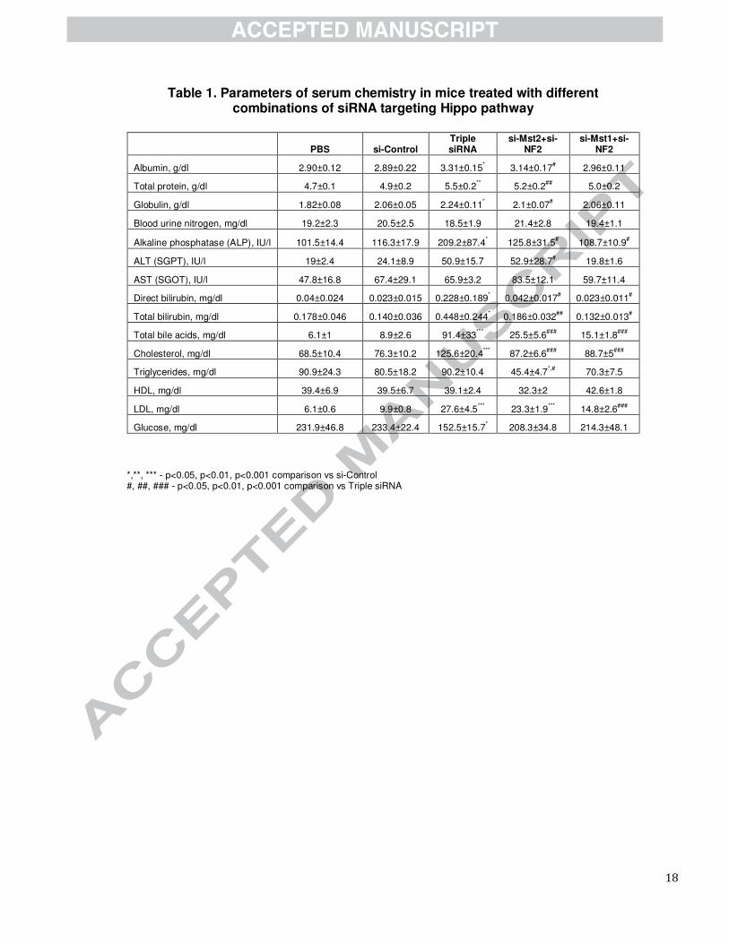
To analyze functions of enlarged livers, we performed analysis of serum biochemistry (Table 1). We found
no significant difference in levels of ALT and AST between PBS, control siRNAs and triple siRNA
nanoparticle treatment, indicating integrity of hepatocytes. Meanwhile, other indicators showed pronounced
hallmarks of cholestasis, including increased levels of alkaline phosphatase, cholesterol, low density
lipoproteins, bilirubin, and bile acids. To further characterize the changes in the liver related to bile flow, we
performed immunohistochemical staining of CK19 to visualize bile ducts and gene expression analysis of
several genes related to bile acid metabolism and transport . The number of CK19 positive cells and the
density of bile ducts were significantly increased after triple siRNAs treatment (Supplementary Fig. 14). This
expansion, together with altered gene expression levels, e.g. NTCP, Abcg5, MDR1, MRP4, and Cyp7
(Supplementary Fig. 9d), indicates that triple siRNA treatment induced remarkable cholestasis.
The triple siRNA induced hepatomegaly is Yap1 dependent.
To validate Yap1-dependent mechanism of the observed phenotype, we depleted Yap1 in triple siRNAs
nanoparticle treated animals. siRNAs against Yap1 (si-Yap1) were designed and screened (Supplementary
Fig. 11). We synthesized 24 siRNAs targeting Yap1. The gene silencing efficiency for individual siRNA was
tested and five efficient siRNAs were identified (Supplementary Fig. 13a). The IC50 for the most potent
siRNA targeting Yap1 was ~0.03nM (Supplementary Fig. 13b). We co-treated animals with LNP-formulated
si-Yap1 with triple siRNAs nanoparticle. We found that si-Yap1 completely blocked the liver growth induced
by triple siRNAs nanoparticle (Fig. 3a), demonstrating that the hepatomegaly induced by triple siRNAs
nanoparticles are Yap1-mediated. Co-treatment of si-Yap1 with triple siRNAs nanoparticles diminished both
pYap1 and Yap1 expression (Fig. 3b), thereby blocking the proliferation of hepatocytes, as indicated by
diminished expression of cell cycle related genes (Fig. 3b and c). Consistently, co-treatment with si-Yap1
also abolished the manifestation of cholestasis associated markers (Supplementary Table 2).
A previous study showed that activation of Yap induces Notch pathway activation with expansion of hepatic
progenitor cells(37). To explore whether activation of Yap induces Notch pathway activation in the triple
siRNA-nanoparticles treated mouse liver, we performed a panel of qPCR analyses for Notch pathway. We
found that Notch pathway signature genes including NOTCH1, NOTCH2, Jag1, SOX9, and HES were all
9
significantly upregulated in triple siRNA treated livers (Fig 3d). Further, such induction was abolished by in
vivo Yap1 siRNA treatment (Fig 3d). Notch pathway has been suggested as an inducer of biliary
specification(38). This is consistent with the expansion of CK19 positive cells in triple siRNA-nanoparticles
treated mouse livers (Supplementary Fig. 14).
p53 controls Yap1-induced liver growth
To further characterize the effects of hepatomegaly, we analyzed gene expression patterns of livers
following long-term triple siRNAs nanoparticle treatment (Fig. 2a). Intriguingly, we observed the up-
regulation of cell cycle checkpoint genes (Supplementary Fig. 7c) and apoptosis related genes
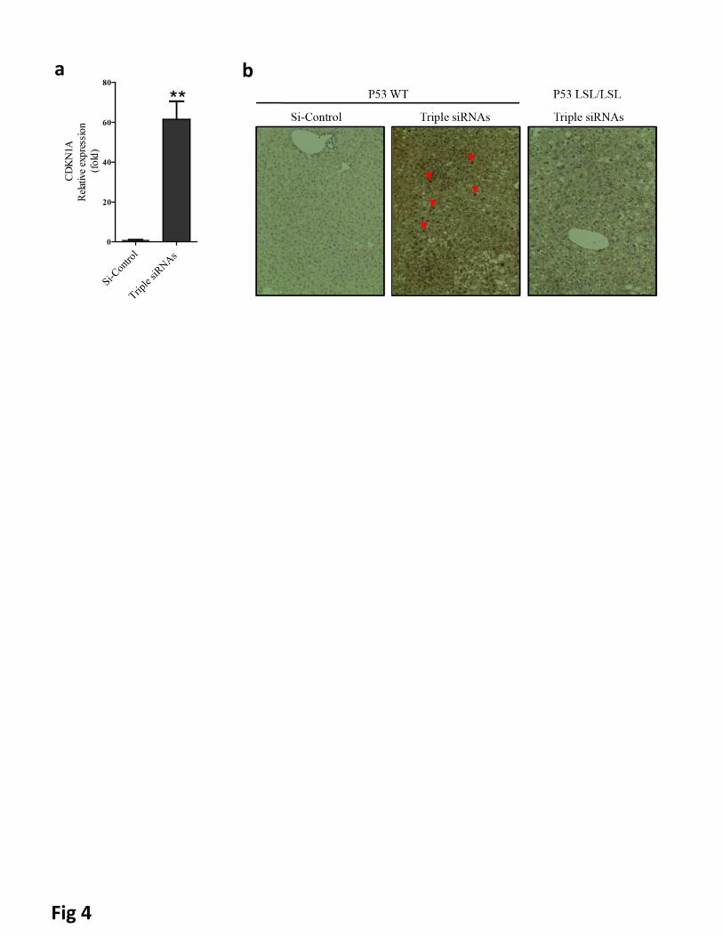
(Supplementary Fig. 7e) with treatment of triple siRNAs nanoparticles. Importantly, we noticed significant
induction of the cyclin-dependent kinase inhibitor 1a (p21) (Fig 4a and Supplementary Figure 15a).
Withdrawal of treatment of triple siRNAs nanoparticle led to diminished p21 expression (Supplementary Fig.
15a). Moreover, co-treatment of si-Yap1 with triple siRNAs nanoparticle abolished p21 induction
(Supplementary Fig. 15b), suggesting that such induction is mediated by Yap1, directly or indirectly. Based
on these results, we hypothesized that the tumor suppressor protein p53, an essential regulator of p21
transcription(39), could be activated as a result of triple siRNAs nanoparticles treatment. Indeed, the
pronounced nuclear staining of p53 indicated its activation with treatment of triple siRNAs nanoparticles
(Fig. 4b). To investigate potential interaction between the Hippo pathway and p53, we compared the
phenotype of triple siRNAs nanoparticles treatment between wildtype mice and p53LSL/LSL mice, which
harbor a Lox-STOP-Lox cassette in the first intron of p53 and are phenotypically equivalent to p53 null
strains(29). In contrast to that liver of wildtype mice, the livers of p53LSL/LSL mice grew much faster than
wildtype mice and exceeded the upper limit of growth (Fig. 5a, b). Remarkably, more than 60% of
hepatocytes were ki-67 positive in p53LSL/LSL mice at Day 14, which was near 3 folds higher than in wildtype
mice (Fig. 5c, d). The H&E staining indicates more severe histological abnormalities in p53LSL/LSL mice than
in wildtype animals with triple siRNAs treatment, such as a higher degree of pleomorphism, extreme
polyploidy, dysplasia of hepatocytes (likely due to swelling with water/fluids), aneuploidy in a portion of
hepatocytes, and intranuclear cytoplasmic inclusions (Fig 5e). To determine wehther a major part of ki-67
positive cells were indeed from hepatocytes, we performed double-staining for keratin 18 (CK18) and ki-67.
We found that a major part of the ki-67 cells in both wildtype p53 and p53 KO were also CK18 positive (Fig
5f), indicating that hepatocytes were under active proliferation. To rule out the possibility of different levels
of knockdown, we measured the levels of Mst1, Mst2, NF2, and pYap1 and found no differences between
wildtype mice and p53LSL/LSL mice (Supplementary Fig. 15c). To further prove that p53 plays a role in
controlling liver growth, we restored p53 by Tamoxifen treatment in p53LSL/LSL; Cre-ER mice, whose stop
cassette in p53 gene can be removed by Cre recombinase. We found that restoration of p53 significantly

10
suppressed liver growth induced by triple siRNAs nanoparticle treatment (Fig. 5e and Supplementary Fig.
15d). Collectively, these results indicate that p53 is activated upon triple siRNAs nanoparticle induced
hepatomegaly, and loss of p53 permits further expansion of liver size.
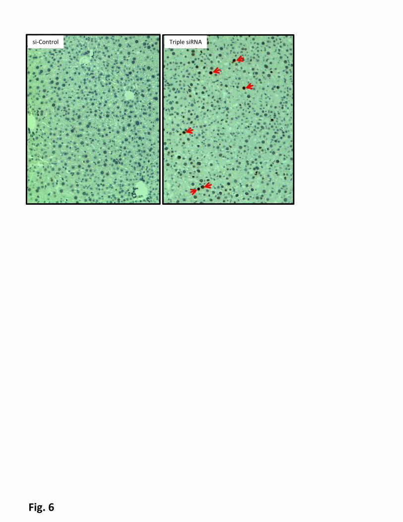
We have hypothesized that p53 may be activated due to DNA damage caused by the elevated levels of bile
acids, a result of cholestasis associated with Yap1-induced hepatomegaly. Using the phosphorylated
histone protein (H2A.X) as an indicator of DNA damage(40) we observed that triple siRNA-induced
hepatomegaly led to DNA damage (Fig. 6). The intensity of the histone phosphorylation correlated to both
the degree of hepatomegaly and p21 levels, i.e. maximal levels of phosphor-H2A.X were observed in the
groups treated with triple siRNA; in contrast, co-treatment with triple siRNAs and si-Yap1 did not upregulate
phospho-H2A.X. It suggests that p53 activation observed in this model is by activation of checkpoint
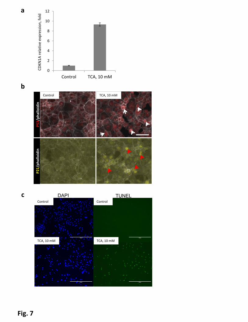
cascades to DNA damage. To explore this mechanism further, we tested the direct effect of bile acids on
p21 activation in murine hepatocyte-derived cell line AML-12. We treated cells with taurocholic acid, a
conjugated primary bile salt which is abundant in mice with cholestasis(41). The taurocholic acid treatment
led to, strong upregulation of p21 mRNA levels and nuclear accumulation of p21 as well as p53 (Fig 7a and
b). To determine whether TCA treatment induced DNA damage in AML12 cells, we treated AML12 cells
with TCA, and performed TUNEL assays. We found TUNEL-positive cells appeared in TCA treated cells but
not control cells (Fig 7c), indicating severe DNA damage in TCA treated cells.
Discussion
Our data demonstrates that it is feasible to manipulate organ size reversibly with siRNA nanoparticles in
vivo. Application of chemically modified synthetic siRNA does not lead to saturation of the RISC complex
and induces minimal immunostimulatory response compared to viral delivery of shRNA(24, 42, 43).
Inhibition at the mRNA level of multiple components of the Hippo pathway is sufficient to activate Yap1 in
vivo, thereby inducing proliferation of hepatocytes and increasing liver size. Contrary to hepatomegaly
induced by complete loss of Mst1/Mst2, one induced by siRNA treatment has an upper limit of growth
(about 2.5 fold). Our data indicates that activation of p53 is a key regulator for limiting liver growth. While
p53 signaling is essential in tumor suppression by coordinating multiple cellular processes, including
activation of DNA repair proteins, arrest of cell cycle, and initiation of cellular apoptosis and senescence
(reviewed in(44)), to our knowledge, our results provide the first demonstration that endogenous p53 plays a
role in size regulation of the liver. Although, we cannot exclude completely direct activation of p53 by loss
of Hippo pathway, e.g. feedback loop through Lats2(45, 46), our results suggest that siRNA mediated
activation of Yap1 can lead to DNA damage response in the liver. Bile acids have been shown to induce
DNA damage in a certain types of cells and cell death in hepatocytes(47-49). Concordant with previous
11
observations, our data show that Yap1 activation leads to disruption of bile duct homeostasis(7) and
provokes a cholestasis-like phenotype.
Crosstalk between Hippo and p53 pathways has been suggested in fly models as well as in mammalian cell
line models(46, 50). Our approach allows investigation of pathway interactions in normal livers. It reveals
indirect interactions between signaling pathways mediated by tissue remodeling and changes in
composition of metabolites. Remarkably, not only is the proliferation rate of hepatocytes in p53LSL/LSL mice
much higher than in the wildtype control, but also the size of hepatocytes in p53LSL/LSL mice is significantly
enlarged (Fig. 5d; Supplementary Fig. 12c, d). It is noteworthy to investigate coordination of p53 and Hippo
pathway during the process of regeneration and under the context of pathological conditions for future
studies. It is known that abnormal bile acids homeostasis can activate p53 in the mouse liver (51). There is
evidence indicating that Yap overactivation can lead to biliary epithelium over-proliferation and irregular
shape(7). To our best knowledge, there is no report showing Yap overactivation provokes a cholestasis-like
phenotype. Dysfunction of bile ducts may be mediated by Yap-dependent dysregulation of gene expression
of bile acid transporters in hepatocytes(52) (Supplementary Fig. 9d) or indirect effects on the bile ducts
function through modulation of paracrine/contact signaling between hepatocytes and cholangioytes (Fig.
3d). Extensive expansion of hepatocytes in the periportal zone may contribute to mechanical distortion of
bile duct function. Our data suggests that Yap activation leads to abnormal bile acid homeostasis, which in
turn triggers p53 activation (Fig 4, 6, 7). Furthermore, our data indicates that loss of p53 led to increased
hepatocyte proliferation when Hippo pathway was suppressed (Fig 5). Previous studies reported that no
obvious abnormal liver histology was found with short-term deletion of p53; in contrast, deletion of p53
induces liver cancer after 10-12 months in mouse model(53). Several studies showed that loss of p53 did
not significantly accelerate phenobarbitone or diethylnitrosamine-induced hepatocellular carcinoma(54-56).
Thus, our finding suggests an important connection between p53 and Hippo pathway, two key pathways for
maintaining quiescence of hepatocytes. Although p21 is an important downstream effector of p53 and its
induction is a recognized marker of p53 activation, the tumour suppressor p53 has a number of targets to
exert its functions(57). The p21-deficient mice have a less obvious phenotype than p53-deficient
animals(58).
We have shown that siRNA nanoparticle-mediated control of organ size is reversible, as indicated by the
shrinking of liver size and restoration of pYap1 after withdrawal of triple siRNAs nanoparticle-treatment (Fig.
2a and d). Livers shrank back to their original size, indicating an intrinsic mechanism for regulating normal
organ size. The technique presented in our manuscript provided a flexible way to induce transient gene
inactivation and subsequent ability to study the mechanism of liver size control in a new manner. A number
12
of questions remain unclear regarding the process of how the liver returns to the exact normal size,
including which signals trigger and stop the apoptosis of hepatocytes, and why only part of cells are
undergoing apoptosis. It is possible that multiple factors are involved in the process in the liver, including
mechanical forces, oxygen levels and surface and nuclear receptors. The method described here allows
dissection of these factors in vivo in a flexible and convenient way. A recent study indicated that a
compensatory network can be activated to buffer against deleterious mutations rather than transcriptional
knockdown(59). Thus, an in vivo genetic knockdown approach may reveal phenotypes which can not be
observed by a knockout approach. As loss of the Hippo pathway is temporary, and the dose of siRNAs can
be titrated to induce temporary proliferation in a portion of hepatocytes without substantial increase of organ
size (data not shown), we believe that modulating liver size in vivo with siRNA-nanoparticles may provide an
efficient tool to explore autonomous and non-autonomous mechanisms for organ size regulation.
Acknowledgements
We thank T. Jacks, A. Sachinidis, K. Meganathan and K. Natarajan for providing experimental materials and
critical review of the manuscript. We thank technical support from Swanson biotechnology Center at Koch
Institute for Integrative Cancer Research. This work was supported in part by the Koch Institute Support
(core) Grant P30-CA14051 from the National Cancer Institute. This work was supported by Alnylam and
NIH RO1-DE016516. H.Y. and R.L.B are supported by 5-U54-CA151884-04 NIH Centers for Cancer
Nanotechnology Excellence and the Harvard-MIT Center of Cancer Nanotechnology Excellence. W.X. is
supported by NIH 5R00CA169512 (to W.X.). V.K. acknowledges support from the Russian scientific fund,
grant number 14-34–00017. The authors acknowledge the service to the MIT community of the late Sean
Collier.
Author Contributions
H.Y., R.L.B., V.K. and D.G.A. directed the project. H.Y. and R.L.B. designed and performed experiments,
and analyzed the data. H.Y. and R.L.B. wrote the manuscript. C.B., S.W, I.Z., V.R., H.N., S.K., L.N., A.A.,
W.X. and M.Z. performed experiments and analyzed data. All authors participated in data discussion and
the manuscript editing.
References
1. Tumaneng K, Russell RC, Guan KL. Organ size control by Hippo and TOR pathways. Curr Biol
2012;22:R368-379.
2. Stanger BZ. The biology of organ size determination. Diabetes Obes Metab 2008;10 Suppl 4:16-
22.

13
3. Shingleton AW. The regulation of organ size in Drosophila: physiology, plasticity, patterning
and physical force. Organogenesis 2010;6:76-87.
4. Pan D. The hippo signaling pathway in development and cancer. Dev Cell 2010;19:491-505.
5. Zhao B, Tumaneng K, Guan KL. The Hippo pathway in organ size control, tissue regeneration
and stem cell self-renewal. Nat Cell Biol 2011;13:877-883.
6. Yu FX, Guan KL. The Hippo pathway: regulators and regulations. Genes Dev 2013;27:355-371.
7. Zhang N, Bai H, David KK, Dong J, Zheng Y, Cai J, Giovannini M, et al. The Merlin/NF2 tumor
suppressor functions through the YAP oncoprotein to regulate tissue homeostasis in mammals. Dev
Cell 2010;19:27-38.
8. Yin F, Yu J, Zheng Y, Chen Q, Zhang N, Pan D. Spatial organization of Hippo signaling at the
plasma membrane mediated by the tumor suppressor Merlin/NF2. Cell 2013;154:1342-1355.
9. Huang J, Wu S, Barrera J, Matthews K, Pan D. The Hippo signaling pathway coordinately
regulates cell proliferation and apoptosis by inactivating Yorkie, the Drosophila Homolog of YAP. Cell
2005;122:421-434.
10. Dong J, Feldmann G, Huang J, Wu S, Zhang N, Comerford SA, Gayyed MF, et al. Elucidation of a
universal size-control mechanism in Drosophila and mammals. Cell 2007;130:1120-1133.
11. Yagi R, Chen LF, Shigesada K, Murakami Y, Ito Y. A WW domain-containing yes-associated
protein (YAP) is a novel transcriptional co-activator. Embo j 1999;18:2551-2562.
12. Vassilev A, Kaneko KJ, Shu H, Zhao Y, DePamphilis ML. TEAD/TEF transcription factors utilize
the activation domain of YAP65, a Src/Yes-associated protein localized in the cytoplasm. Genes Dev
2001;15:1229-1241.
13. Oh S, Lee D, Kim T, Kim TS, Oh HJ, Hwang CY, Kong YY, et al. Crucial role for Mst1 and Mst2
kinases in early embryonic development of the mouse. Mol Cell Biol 2009;29:6309-6320.
14. Song H, Mak KK, Topol L, Yun K, Hu J, Garrett L, Chen Y, et al. Mammalian Mst1 and Mst2
kinases play essential roles in organ size control and tumor suppression. Proc Natl Acad Sci U S A
2010;107:1431-1436.
15. Zhou D, Conrad C, Xia F, Park JS, Payer B, Yin Y, Lauwers GY, et al. Mst1 and Mst2 maintain
hepatocyte quiescence and suppress hepatocellular carcinoma development through inactivation of
the Yap1 oncogene. Cancer Cell 2009;16:425-438.
16. Lu L, Li Y, Kim SM, Bossuyt W, Liu P, Qiu Q, Wang Y, et al. Hippo signaling is a potent in vivo
growth and tumor suppressor pathway in the mammalian liver. Proc Natl Acad Sci U S A
2010;107:1437-1442.
17. Tumaneng K, Schlegelmilch K, Russell RC, Yimlamai D, Basnet H, Mahadevan N, Fitamant J, et al.
YAP mediates crosstalk between the Hippo and PI(3)K-TOR pathways by suppressing PTEN via miR-
29. Nat Cell Biol 2012;14:1322-1329.
18. Varelas X, Miller BW, Sopko R, Song S, Gregorieff A, Fellouse FA, Sakuma R, et al. The Hippo
pathway regulates Wnt/beta-catenin signaling. Dev Cell 2010;18:579-591.
19. Xin M, Kim Y, Sutherland LB, Qi X, McAnally J, Schwartz RJ, Richardson JA, et al. Regulation of
insulin-like growth factor signaling by Yap governs cardiomyocyte proliferation and embryonic heart
size. Sci Signal 2011;4:ra70.
20. Strassburger K, Tiebe M, Pinna F, Breuhahn K, Teleman AA. Insulin/IGF signaling drives cell
proliferation in part via Yorkie/YAP. Dev Biol 2012;367:187-196.
21. Fernandez LA, Northcott PA, Dalton J, Fraga C, Ellison D, Angers S, Taylor MD, et al. YAP1 is
amplified and up-regulated in hedgehog-associated medulloblastomas and mediates Sonic hedgehog-
driven neural precursor proliferation. Genes Dev 2009;23:2729-2741.

14
22. Dong Y, Love KT, Dorkin JR, Sirirungruang S, Zhang Y, Chen D, Bogorad RL, et al. Lipopeptide
nanoparticles for potent and selective siRNA delivery in rodents and nonhuman primates. Proc Natl
Acad Sci U S A 2014.
23. Love KT, Mahon KP, Levins CG, Whitehead KA, Querbes W, Dorkin JR, Qin J, et al. Lipid-like
materials for low-dose, in vivo gene silencing. Proc Natl Acad Sci U S A 2010;107:1864-1869.
24. Zeigerer A, Gilleron J, Bogorad RL, Marsico G, Nonaka H, Seifert S, Epstein-Barash H, et al. Rab5
is necessary for the biogenesis of the endolysosomal system in vivo. Nature 2012;485:465-470.
25. Baughman JM, Perocchi F, Girgis HS, Plovanich M, Belcher-Timme CA, Sancak Y, Bao XR, et al.
Integrative genomics identifies MCU as an essential component of the mitochondrial calcium
uniporter. Nature 2011;476:341-345.
26. Coelho T, Adams D, Silva A, Lozeron P, Hawkins PN, Mant T, Perez J, et al. Safety and efficacy of
RNAi therapy for transthyretin amyloidosis. N Engl J Med 2013;369:819-829.
27. Leuschner F, Dutta P, Gorbatov R, Novobrantseva TI, Donahoe JS, Courties G, Lee KM, et al.
Therapeutic siRNA silencing in inflammatory monocytes in mice. Nat Biotechnol 2011;29:1005-1010.
28. Dahlman JE, Barnes C, Khan OF. In vivo endothelial siRNA delivery using polymeric
nanoparticles with low molecular weight. 2014;9:648-655.
29. Ventura A, Kirsch DG, McLaughlin ME, Tuveson DA, Grimm J, Lintault L, Newman J, et al.
Restoration of p53 function leads to tumour regression in vivo. Nature 2007;445:661-665.
30. Yin H, Cheng L, Holt M, Hail N, Jr., Maclaren R, Ju C. Lactoferrin protects against acetaminophen-
induced liver injury in mice. Hepatology 2010;51:1007-1016.
31. You Q, Holt M, Yin H, Li G, Hu CJ, Ju C. Role of hepatic resident and infiltrating macrophages in
liver repair after acute injury. Biochem Pharmacol 2013;86:836-843.
32. Jeong WI, Park O, Radaeva S, Gao B. STAT1 inhibits liver fibrosis in mice by inhibiting stellate
cell proliferation and stimulating NK cell cytotoxicity. Hepatology 2006;44:1441-1451.
33. Xue W, Meylan E, Oliver TG, Feldser DM, Winslow MM, Bronson R, Jacks T. Response and
resistance to NF-kappaB inhibitors in mouse models of lung adenocarcinoma. Cancer Discov
2011;1:236-247.
34. Soutschek J, Akinc A, Bramlage B, Charisse K, Constien R, Donoghue M, Elbashir S, et al.
Therapeutic silencing of an endogenous gene by systemic administration of modified siRNAs. Nature
2004;432:173-178.
35. Bogorad RL, Yin H, Zeigerer A, Nonaka H, Ruda VM, Zerial M, Anderson DG, et al. Nanoparticle-
formulated siRNA targeting integrins inhibits hepatocellular carcinoma progression in mice. Nat
Commun 2014;5:3869.
36. Cunningham CC, Van Horn CG. Energy availability and alcohol-related liver pathology. Alcohol
Res Health 2003;27:291-299.
37. Yimlamai D, Christodoulou C, Galli GG, Yanger K, Pepe-Mooney B, Gurung B, Shrestha K, et al.
Hippo pathway activity influences liver cell fate. Cell 2014;157:1324-1338.
38. Sparks EE, Huppert KA, Brown MA, Washington MK, Huppert SS. Notch signaling regulates
formation of the three-dimensional architecture of intrahepatic bile ducts in mice. Hepatology
2010;51:1391-1400.
39. el-Deiry WS, Tokino T, Velculescu VE, Levy DB, Parsons R, Trent JM, Lin D, et al. WAF1, a
potential mediator of p53 tumor suppression. Cell 1993;75:817-825.
40. Lu C, Zhu F, Cho YY, Tang F, Zykova T, Ma WY, Bode AM, et al. Cell apoptosis: requirement of
H2AX in DNA ladder formation, but not for the activation of caspase-3. Mol Cell 2006;23:121-132.
41. Zhang Y, Hong JY, Rockwell CE, Copple BL, Jaeschke H, Klaassen CD. Effect of bile duct ligation
on bile acid composition in mouse serum and liver. Liver Int 2012;32:58-69.
15
42. John M, Constien R, Akinc A, Goldberg M, Moon YA, Spranger M, Hadwiger P, et al. Effective
RNAi-mediated gene silencing without interruption of the endogenous microRNA pathway. Nature
2007;449:745-747.
43. Grimm D, Wang L, Lee JS, Schurmann N, Gu S, Borner K, Storm TA, et al. Argonaute proteins are
key determinants of RNAi efficacy, toxicity, and persistence in the adult mouse liver. J Clin Invest
2010;120:3106-3119.
44. Vousden KH, Prives C. Blinded by the Light: The Growing Complexity of p53. Cell
2009;137:413-431.
45. Ganem NJ, Cornils H, Chiu SY, O'Rourke KP, Arnaud J, Yimlamai D, Thery M, et al. Cytokinesis
failure triggers hippo tumor suppressor pathway activation. Cell 2014;158:833-848.
46. Aylon Y, Michael D, Shmueli A, Yabuta N, Nojima H, Oren M. A positive feedback loop between
the p53 and Lats2 tumor suppressors prevents tetraploidization. Genes Dev 2006;20:2687-2700.
47. Woolbright BL, Dorko K, Antoine DJ, Clarke JI, Gholami P, Li F, Kumer SC, et al. Bile acid-induced
necrosis in primary human hepatocytes and in patients with obstructive cholestasis. Toxicol Appl
Pharmacol 2015;283:168-177.
48. Ohtaki Y, Hida T, Hiramatsu K, Kanitani M, Ohshima T, Nomura M, Wakita H, et al. Deoxycholic
acid as an endogenous risk factor for hepatocarcinogenesis and effects of gomisin A, a lignan
component of Schizandra fruits. Anticancer Res 1996;16:751-755.
49. Ajouz H, Mukherji D, Shamseddine A. Secondary bile acids: an underrecognized cause of colon
cancer. World J Surg Oncol 2014;12:164.
50. Zhang W, Cohen SM. The Hippo pathway acts via p53 and microRNAs to control proliferation
and proapoptotic gene expression during tissue growth. Biol Open 2013;2:822-828.
51. Yang H, Li TW, Ko KS, Xia M, Lu SC. Switch from Mnt-Max to Myc-Max induces p53 and cyclin D1
expression and apoptosis during cholestasis in mouse and human hepatocytes. Hepatology
2009;49:860-870.
52. Halilbasic E, Claudel T, Trauner M. Bile acid transporters and regulatory nuclear receptors in
the liver and beyond. J Hepatol 2013;58:155-168.
53. Katz SF, Lechel A, Obenauf AC, Begus-Nahrmann Y, Kraus JM, Hoffmann EM, Duda J, et al.
Disruption of Trp53 in livers of mice induces formation of carcinomas with bilineal differentiation.
Gastroenterology 2012;142:1229-1239.
54. Gould S, Sidaway J, Sansom N, Betton G, Orton T. Phenobarbitone-induced liver response in wild
type and in p53 deficient mice. Toxicol Lett 2001;122:131-140.
55. Martin NC, McGregor AH, Sansom N, Gould S, Harrison DJ. Phenobarbitone-induced ploidy
changes in liver occur independently of p53. Toxicol Lett 2001;119:109-115.
56. Kemp CJ. Hepatocarcinogenesis in p53-deficient mice. Mol Carcinog 1995;12:132-136.
57. Harris SL, Levine AJ. The p53 pathway: positive and negative feedback loops. Oncogene
2005;24:2899-2908.
58. Martin-Caballero J, Flores JM, Garcia-Palencia P, Serrano M. Tumor susceptibility of
p21(Waf1/Cip1)-deficient mice. Cancer Res 2001;61:6234-6238.
59. Rossi A, Kontarakis Z, Gerri C, Nolte H, Holper S, Kruger M, Stainier DY. Genetic compensation
induced by deleterious mutations but not gene knockdowns. Nature 2015;524:230-233.
16
Figure Legends
Figure 1. Treatment of lipid-based nanoparticles (LNP)-formulated siRNAs targeting Mst1, Mst2 and
NF2 (Triple siRNAs) induced hepatomegaly in mice. C57BL/6 female mice were treated with triple
siRNAs, dose matched LNP-siRNA against the luciferase gene (si-Control), or PBS as controls. Livers were
taken 15 days after the first injection. a, Macroscopic views of livers. Scale bar is equal to 1 cm. b, Maximal
induction of liver size in triple siRNA treated mice. Error bars represent standard error (SEM), p<0.01.
Figure 2. In vivo treatment with triple siRNAs induced proliferation of hepatocytes and activation of
Yap1. C57BL/6 female mice were treated with triple siRNAs or dose matched si-Control. Livers were taken
at Day 7, 9, 11, 13, 15, 24, 32 or 40 days after the first injection. a, Liver/body weight ratio at different time
points. Error bars represent SEM, (N≥3). b, Immunohistochemical analysis of hepatocyte proliferation (Day
11). Ki-67 (grey), Phalloidin (yellow), Hoechst (blue). c, Quantification of (b). Error bars represent SEM, **,
p<0.01 (N=3). d, Western blot analysis of Hippo pathway signalling. e, Triple siRNA treatment changed
Yap1 localization in liver (Day 11). Yap1 (yellow), nuclei (blue). f, Expression of Yap1-dependent genes
were induced by triple siRNAs (Day 11). Error bars represent SEM, **, p<0.01 (N=3).
Figure 3. Additional siRNA treatment proved genetic interactions. C57BL/6 female mice were treated
with dose matched si-control, triple siRNAs plus si-Control, or triple siRNAs plus si-Yap1. Livers were taken
11 or 15 days after the first injection. a, weight at Day 15 was recorded. Error bars represent SEM, **,
p<0.01 (N≥5). b, Liver lysate was made and western blot performed. c, Analysis of cell cycle related genes
in liver tissues (Day 11) by qPCR. Error bars represent SEM, **, p<0.01 (N=3). d, YAP activation induces
Notch pathway signaling mRNA expression of Notch pathway genes was determined by RT-qPCR in siLUC,
Triple siRNA, or Triple siRNA+siYAP treatment groups,*p<0.05. N=4
Figure 4. p53 was activated in livers of triple siRNAs treated animal. C57BL/6 mice and p53LSL/LSL were
treated with triple siRNAs or dose matched si-Control. a, The levels of CDKN1A (P21) mRNA in livers of
C57BL/6 mice treated with si-Control or triple siRNAs(Day 14). Error bars represent SEM, **, p<0.01 (N=5).
b, p53 staining in liver sections. Red arrowheads indicate nuclear accumulation of p53.
Figure 5. p53 plays an important role in restraining liver growth. a, Macroscopic views of livers (Day
22). b, Liver/body weight ratio at different time points. Error bars represent SEM, *, p<0.05 (N=4). c,
Hepatocyte proliferation evidenced by ki-67 staining (Day 14). d, Quantification of ki-67+ cells. Error bars
represent SEM, **, p<0.01, *, p<0.05 (N=3). e, H&E staining of the liver sections (Day 22). f,
immunofluorescent staining of ki-67 (red) and keratin 18 (green) of the liver sections (Day 22)., scale bars =
17
100µm. g, p53LSL/LSL; CreER mice treated with tamoxifen (p53 on) or vehicle controls (p53 off). Liver weight
was measured 22 days after the first dose of triple siRNAs. Error bars represent SEM, **, p<0.01 (N≥3).
Figure 6. DNA damage in enlarged liver. Livers were collected at Day 14 after the first injection of triple
siRNAs. DNA damage was evidenced by phospho-H2A.X positive cells (indicated by red arrows).
Figure 7. Bile acids induce activity of p53-pathway in vitro. AML12 cells were treated with 10mM
taurocholic acid (TCA) for 4 hours. Untreated cells serve as a control. a, mRNA levels of CDKN1A(p21) in
AML12 cells. Error bars represent SEM. b, immunofluorescent staining, arrowheads indicate nuclear
accumulation of p53 (white) and p21 (red). Bar indicates 100µm. C Cells were fixed and stained for TUNEL
and counterstained with DAPI, scalebars = 400µm.
18
Table 1. Parameters of serum chemistry in mice treated with different combinations of siRNA targeting Hippo pathway
PBS si-Control Triple siRNA
si-Mst2+si-NF2
si-Mst1+si-NF2
Albumin, g/dl 2.90±0.12 2.89±0.22 3.31±0.15* 3.14±0.17
# 2.96±0.11
Total protein, g/dl 4.7±0.1 4.9±0.2 5.5±0.2** 5.2±0.2
## 5.0±0.2
Globulin, g/dl 1.82±0.08 2.06±0.05 2.24±0.11* 2.1±0.07
# 2.06±0.11
Blood urine nitrogen, mg/dl 19.2±2.3 20.5±2.5 18.5±1.9 21.4±2.8 19.4±1.1
Alkaline phosphatase (ALP), IU/l 101.5±14.4 116.3±17.9 209.2±87.4* 125.8±31.5
# 108.7±10.9
#
ALT (SGPT), IU/l 19±2.4 24.1±8.9 50.9±15.7 52.9±28.7# 19.8±1.6
AST (SGOT), IU/l 47.8±16.8 67.4±29.1 65.9±3.2 83.5±12.1 59.7±11.4
Direct bilirubin, mg/dl 0.04±0.024 0.023±0.015 0.228±0.189* 0.042±0.017
# 0.023±0.011
#
Total bilirubin, mg/dl 0.178±0.046 0.140±0.036 0.448±0.244* 0.186±0.032
## 0.132±0.013
#
Total bile acids, mg/dl 6.1±1 8.9±2.6 91.4±33***
25.5±5.6###
15.1±1.8###
Cholesterol, mg/dl 68.5±10.4 76.3±10.2 125.6±20.4***
87.2±6.6###
88.7±5###
Triglycerides, mg/dl 90.9±24.3 80.5±18.2 90.2±10.4 45.4±4.7*,#
70.3±7.5
HDL, mg/dl 39.4±6.9 39.5±6.7 39.1±2.4 32.3±2 42.6±1.8
LDL, mg/dl 6.1±0.6 9.9±0.8 27.6±4.5***
23.3±1.9***
14.8±2.6###
Glucose, mg/dl 231.9±46.8 233.4±22.4 152.5±15.7* 208.3±34.8 214.3±48.1
*,**, *** - p<0.05, p<0.01, p<0.001 comparison vs si-Control #, ##, ### - p<0.05, p<0.01, p<0.001 comparison vs Triple siRNA
a
b
Fig 1

a
d
b
e
c
f
PBS si‐Control Triple siRNA
0%
5%
10%
15%
20%
25%
30%
9 days 11 days
si‐Control
TriplesiRNAs
PBS
**
**
Ki‐67 positive hepatocytes
Fig 2
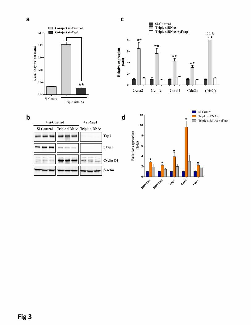
Fig 3
a
b
c
d
NOTCH1
NOTCH2
Jag1
Sox9
Hes1
0
2
4
6
8
10
12si-Control
Triple siRNAs
*
Triple siRNAs +siYap1
*
*
*
*
Rel
ativ
e ex
pres
sion
(fol
d)
a b
Fig 4

a b
c ed
Fig 5
P53 WT P53 LSL/LSLe
f g
si‐Control Triple siRNA
Fig. 6
0
2
4
6
8
10
12
Control TCA, 10 mM
CDKN1A relative expression, fold
Fig. 7
a
b
P53/phalloidin
Control TCA, 10 mM
P21/phalloidin
DAPI TUNELControl Control
TCA, 10 mM TCA, 10 mM
c


