
Clinical variables affecting the marginal degradation of amalgam restorations
Asbjom Jokstad and lvar A. Mjor Department of Anatomy, School of Dentistry, University of Oslo, Oslo, and NIOM, Scandinavian Institute of Dental Materials, Haslum, Norway
Jokstad A, Mjor IA. Clinical variables affecting th~ marginal degradation of amalg'llll
restorations. Acta Odontol Scand 1990;48:379--387. Oslo. ISSN 0001-6357.
The influence of different clinical variables on the marginal degradation of amalgam restorations was studied in a clinical trial of class-II amalgam restorations. Seven Scandinavian
dentists using 5 different alloys placed 468 restorations in 210 patients. The marginal degra
dation was scored on impressions of the restored teeth by using a six-point ordinal rating scale
and was transformed to ridit values. After 5 years 126 patients with 296 restorations remained
in the trial. Ridit analysis and paired comparison tests utilizing the Bonferroni correction
factor at each yearly interval indicated that the extent of degradation of the restoration
margins was influenced by the location in the mouth,. the position on:· the tooth, the ·type of
alloy, and the operator. The results demonstrate that features of the cavity preparation and
the handling of the material by the operator are more important for the degradation of the
restoration margins than other clinical variables. D Amalgam degradation; clinical study;
dental materials; iatrogenic effects; operative dentistry
Asbjomlokstad, Department of Anatomy, Dental Faculty, P. 0. Box 1052 Blindem, University
of Oslo, N-0316 Oslo 3, Norway
Longitudinal clinical trials have demonstrated that the extent of marginal degradation of amalgam restorations is operatordependent (1-9). The mechanism of this dependence is not established, but it has been suggested that the handling properties of the alloy and the condensation techniques of the operators. may play important roles (3, 8). A longitudinal clinical study was therefore initiated with the aim of assessing the operator effect on the performance of class-II amalgam restorations.
Materials and methods
Seven general practitioners residing in Denmark, Sweden, Finland, and Norway agreed to participate in the study. Three were in private practice, two in a public health practice, and two in the school dental service. Their clinical experience varied from 15 to 30 years. Each operator randomly selected the potential participants of the study among their regularly attending patients. The only criteria given for the selection of patients was a preference· for patients requiring at
least three class-II restorations. The need for restorations could be due to primary caries or to failed restorations. The material consisted at base line of 468 2- and 3-surface class-II amalgam restorations in 210 patients. One hundred and forty-seven of these were three-surface restorations. A summary of the assignment of restorations by tooth type is shown in Table 1. One hundred and six patients with 296 .restorations could be followed up throughout the 5-year observatiQn period. The age of the patients varied from 8 to 71 years, with a mean of 28 years. The oral health of each patient was estimated by their DMFS index and caries increment during the trial period. Zero to 0.5 new restorations per year indicated a low caries activity, while more than 2 new restorations per year suggested a high caries activity.
A conventional amalgam alloy and four non-gamma-2 precapsulated alloys were selected for the study (Table 2). Each clinician used three alloys, except two operators, who used two and four alloys each. The alloys were randomly assigned to the teeth to be restored. The operators were informed not to deviate from their clinical routines
380 A. Jokstad & I. A. Mjor ACTA ODONTOL SCAND 48 (1990)
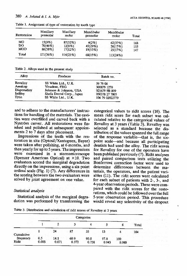
Table 1. Assignment of type of restoration by tooth type
Maxillary Maxillary Mandibular Mandibular Restoration premolar molar premolar molar Total
MO 15(9%) 87(52%) 4(2%) 62(37%) 168 DO 70(46%) 12(8%) 45(29%) 26(17%) 153 MOD 86(58%) 17(12%) 19(13%) 25(17%) 147 Total 171(36%) 116(25%) 68(15%) 113(24%)
Table 2. Alloys used in the present study
Alloy Producer
Revalloy SS White Ltd., U.K. Amal cap Vivadent, FRG Dispersalloy Johnson & Johnson, USA lndiloy Shofu Dental Corp., Japan Tytin SS White Ltd., U.K.
and to adhere to the manufacturers' instructions for handling of the materials. The cavities were overfilled and carved back with a Nystrom carver. All restorations were finished and polished at subsequent appointments 2 to 7 days after placement.
Impressions of the teeth with the restoration in situ (Optosil/Xantopren, Bayer) were taken after polishing, at 6 months, and then yearly for up to 5 years. The impressions were examined in a stereomicroscope (Spencer American Optical) at x 10. Two evaluators scored the marginal degradation directly on the impressions, using a six-point ordinal scale (Fig. 1) (7). Any differences in the scoring between the two evaluators were solved by joint agreement on one value.
Statistical analysis Statistical analysis of the marginal degra
dation was performed by transforming the
Batch no.
59 79 08 300879 1270 021679 9B 809 050378 27 7805 106 79 02022779
categorical values to ridit scores (10). The mean ridit score for each subset was calculated relative to the categorical values of Revalloy at 3 years (Table 3). Revalloy was selected as a standard because the distribution of the values spanned the full range of the response variable--that is, the sixpoint scale--and because all participating dentists had used the alloy. The ridit scores for Revalloy for one of the operators have been published previously (7). Ridit analyses and paired comparison tests utilizing the Bonferroni correction factor were used to determine differences betWeen the materials, the operators, and the patient variables (11). The ridit scores were calculated for each subset of patients with 2-, 3-, and 4·year observation periods. These were compared with the ridit scores for the restorations, which could be followed over the full 5-year observation period. This procedure would reveal any selectivity of the dropout
Table 3. Distribution and calculation of ridit scores of Revalloy at 3 years
Categories
1 2 3 4 5 6 Total
1 24 87 55 13 4 184 Cumulative
frequency 0.5 14.1 60.9 90.8 97.8 100 Ridit 0.003 0.071 0.372 0.758 0.943 0.989

ACTA ODONTOL SCAND 48 (1990)
Table 4. Cumulative loss of restorations over the 5-year observation period
Year
1 2 3 4 5
Observed 449 427 397 340 296 Cumulated loss
Dropout of patients 13 25 42 88 123 Secondary caries 1 6 16 21 24 Bulk fractures 5 9 12 14 18 Tooth fractures 2 3 Extended 1 1 2 2 ·Margin degradation 1 2
patients and the possible influence on the average ridit scores.
Results The loss of restorations was primarily due to patient dropout, especially children who left the school dental service (Table 4). Most of the failed restorations were replaced because of secondary caries on the proximal surfaces and because of bulk fractures (Table 4). There was no correlation between these two criteria for replacement and the ridit values of the replaced restorations. The ridit scores for .each subset of patients with 2-, 3-, 4-, and 5-year observation periods are shown in Fig. 2. No statistical differences between the four subsets could be found. The ridit scores of the subsets were therefore pooled for the subsequent analyses.
Analyses on the basis of intra-oral location of the restorations show that the degradation was slightly higher on the first molars of both the upper and lower jaw than on the other teeth (Fig. 3). In general, the mandibular teeth showed less degradation than the maxillary teeth, whereas the ranking of the
Fig. 1. The scoring system for marginal degradation evaluated on impressions of the teeth with restorations based on the scoring system developed for models (7). The numbers 1 to 6 indicate progressively larger degradation, 1 having marginal relationships equal to or better than the subjacent photograph, 2 having marginal relationships between the two adjacent photographs, and so forth; 6 denotes a marginal degradation equal to or more than on the bottom photograph.
Clinical variables and marginal degradation 381

.. 382 A. Jokstad & J •. A . Mjor
Rldft o.e~~~~~~~~~~~~~~~~~~~~~
O.&
0.4
0.2
Year
ACTA ODONTOL SCAND 48 (1990)
Fig. 2. The ridit means for the restorations with 0-2-years (n = 30), 0-3·years (n = 57), 0-4 years (n = 44), and 0-5 years (n = 296) observation periods, as a function of time. Forty-one restorations were lost during the first 2 years of the study. The number on the illustration at 2 years indicates the critical ratio between the mean ridits of the maximum and minimum values at this point (2-year cohorts).
Each individual paired comparison requires a critical value of 2.6 according to the Bonferroni criterion to be at a significance level of a= 0.05. The ridit score for the whole material is superimposed and marked by triangles.
teeth was similar in both jaws in that the premolars displayed more breakdown than the second molars (Fig. 3). There was no relationship between the ridit scores of the
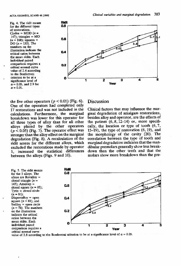
. marginal degradation and the age of the patients. There was, furthermore, no relationship between the degradation and the oral health of the patients. A breakdown of the ridit scores by type of restoration indicates that the marginal degradation was higher on MO restorations than on DO restorations (p < 0.01) (Fig. 4).
Rldft
The kinetics of the marginal degradation of the restorations varied with the type of alloy. Revalloy showed more marginal degradation than any ofthe other alloys from 6 months continuously up to 5 years (Fig. 5). A statistical difference could, however, only be found between Revalloy and Tytin and between Dispersalloy and Indiloy at 4 and 5 years (p < 0.05). A breakdown of the alloy scores by individual operators showed that one of the operators placed restorations with significantly less marginal breakdown than
o.e..--~~~~~~~~~~~~~~~~~~~-,
Fig. 3. The ridit means for the different teeth. Circles = first molars; triangles = second molars; squares = premolars; the ridit scores are for the maxillary teeth (open symbols, n = 101,
O.&
0.4
0.2
Vear
n = 15, n = 17, respectively) and the mandibular teeth (closed symbols, n = 87, n = 26, n = 68, respectively). The number on the illustration indicates the critical ratio between the mean ridits . The individual
paired comparison requires a critical normal curve value of 2.9 according to the Bonferroni criterion to be at a significance level of a= 0.05.
ACTA ODONTOL SCAND 48 (1990) Clinical variables and marginal degradation 383
Aldft Fig. 4. The ridit means for the different types of restorations.
0.8.-----------------------.
O.&
0.4
0.2
Circles = MOD (n = 147); triangles = MO (n = 168); squares = DO (n = 153). The numbers on the illustration indicate the critical ratios between the mean ridits. Each individual paired comparison requires a critical normal curve value of 2.4 according to the Bonferroni criterion to be at a significance level of a= 0.05, and 2.9 for a= 0.01.
0'-----1----2------3------4---~5
Year
the five other operators (p < 0.01) (Fig. 6). One of the operators had completed only 17 restorations and was not included in the calculations. Furthermore, the marginal breakdown was lower for this operator for all three types of alloy than for all other alloys placed by the other operators (p < 0.05) (Fig. 7). The operator effect was stronger than the alloy effect on the marginal degradation (Fig. 8). A recalculation of the ridit scores for the different alloys, which excluded the restorations made by operator 1, increased the statistical differences between the alloys (Figs. 9 and 10).
AldH
Discussion
Clinical factors that may influence the marginal degradation of amalgam restorations, besides alloy and operator, are the effects of the patient (6, 8, 12-14) or, more specifically, the location or type of tooth ( 6, 7, 15-19), the type of restoration (6, 19), and the morphology of the cavity (20). The correlation between the type of tooth and marginal degradation indicates that the mandibular premolars generally show less breakdown than the other teeth and that the molars show more breakdown than the pre-
Fig. 5. The ridit means for the 5 alloys. The alloys are Revalloy = closed triangle (n = 143); Amalcap = closed square (n = 85); Tytin = closed circle
0.8 .----------------------~
(n = 81); Dispersalloy = open square (n = 81); and Indiloy = open circle (n = 78). The numbers on the illustration indicate the critical ratios between the mean ridits. Each individual paired comparison requires a critical normal curve
O.&
Q.4
0.2
Year
value of 2.8 according tci the Bonferroni criterion to be at a significance level of n: = 0.05.

384 A. Jokstad & I. A. Mjor
Rid ft o.e..----~~~~~~~~~~~~~~~~~~-.
0.6
OA
0.2
QL-~~~1~~~-2~~~----,3,----~~~4~~~-:5
Year
ACTA ODONTOL SCAND 48 (1990)
Fig. 6. The ridit means for the six different operators. The ridit means for last operators are not shown because of the low number of submitted restorations. The numbers on the illustration indicate the critical ratios between the mean ridits. Each individual paired comparison requires a critical normal curve value of 2.9 according to the Bonferroni criterion to be at a significance level of oc = 0.05, and 3.4 for oc = 0.01.
molars (6, 15, 19) . . The higher marginal with those in the other teeth (22). The obdegradation specifically in the first molars servation that DO restorations showed less than in the other teeth partially supports marginal degradation than MO or MOD resprevious observations (15) but contrasts with torations is difficult to explain. Some of the the conclusions by other authors (6, 7, 19). differences may be the result of a greater The quality of the occlusal cavity margins frequency of DO restorations in the mandid not differ in the various tooth categories dibular premolars than in the other teeth. with regard to the frequency of external dis- On the other hand, the dimensions of the crepancies (21). However, some of the dif- average DO cavities were larger than the ferences may be attributed to the greater MO restorations, and the frequency of cavity cavity sizes in the first molars and the smaller discrepancies higher (21, 22). To our knowlsizes in the mandibular premolars compared edge there are no previous reports in which
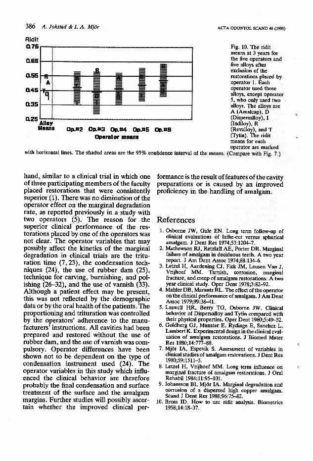
llclt 0.75...-"""!"'"~~~~~~~~~~~~~~~--,
0.45
0.25 Alloy Means Op.#1 Op.#2 Op.#3 Op.#4 Op.#5 Op.#6
Operator means
Fig. 7. The ridit means at 3 years for five alloys and for six of the operators. (One operator was excluded because of the low number of submitted restorations.) Each operator used three alloys, except operator 5, who only used two alloys. The alloys are A (Amalcap), b (Dispersalloy), I (Indiloy), R (Revalloy), and T (Tytin). The ridit means for each
operator are marked by horizontal lines. The shaded areas are the 95% confidence interval of the means.

ACTA ODONTOL SCAND 48 (1990) Clinical variables and marginal degradation 385
Rldlt Fig. 8. The ridit means of Revalloy placed by operator 1 (circles,
0.8 ..--~~~~~~~~~~~~~~~~~~~---.
n = 24), and the ridit means of the best performing amalgam, Indiloy, placed by three other operators (n = 6, n = 25, n = 23). The number on the illustration indicates the critical ratio between the mean ridits. The individual paired comparison requires a critical normal curve value of 2.2 according to the Bonferroni criterion to be at a significance level of IX= 0.05.
O.&
OA
0.2
the degradation of the MO and DO restorations have been compared.
The clinical performance of the different alloys is in accordance with previous reports in that the marginal degradation was less for the non-gamma-2 alloys than for the conventional lathe-cut alloys (1-8, 20). It has been suggested that a superior material would perform better for all dentists ( 4, 6). This hypothesis was strengthened by the present findings. However, the suggestion that the clinical performance of non-gamma-
Rldlt
1 2 3 4 5 Vear
2 alloys is less susceptible to operator variations ( 4) was not supported by the results, since all alloys performed relatively equally for five of the operators. Furthermore, the clinical performance of the restorations placed by one of the operators indicates that any type of amalgam will perform well in the hand of a proficient dentist. That the operator effect can be greater than the alloy effect is not supported by previous reports involving four operators ( 6) and two operators (8). The observation is, on the other
Fig. 9. The ridit means for the 5 alloys excluding operator 1. The alloys are Revalloy = closed triangle (n = 117); Amalcap = closed square (n = 60);
0.8 ..--~~~~~~~~~~~~~~~~~~~---.
Tytin = closed circle (n = 59); Dispersalloy = open square (n = 82), and Indifoy = open circle (n = 80). The numbers on the illustration indicate the critical ratios between the mean ridits . Each individual paired comparison requires a
O.&
OA
0.2
Vear
critical normal curve value of 2.8 according to the Bonferroni criterion to be at a significance level of IX= 0.05. (Compare with Fig. 5.)
386 A. Jokstad & I. A. Mjor
Rid it 0.75
0.65
0.55
0.45
0.35
" A -·q
0.25 Alloy Means
- ._ - -'
--. =----= t:,._ --
Op.•2 Op.•3 Op.a4 Op.as Op.•& Operator means
ACTA ODONTOL SCAND 48 (1990)
Fig. 10. The ridit means at 3 years for the five operators and five alloys after exclusion of the restorations placed by operator 1. Each operator used three alloys, except operator 5, who only used two alloys. The alloys are A (Amalcap), D (Dispersalloy), I (lndiloy), R (Revalloy), and T (Tytin). The ridit means for each operator are marked
with horizontal lines. The shaded areas are the 95% confidence interval of the means. (Compare with Fig. 7.)
hand, similar to a clinical trial in which one of three participating members of the faculty placed restorations that were consistently superior (1). There was no diminution of the operator effect on the marginal degradation rate, as reported previously in a study with two operators (5). The reason for the superior clinical performance of the restorations placed by one of the operators was not clear. The operator variables that may possibly affect the kinetics of the marginal degradation in clinical trials are the trituration time (7, 23), the condensation techniques (24), the use of rubber dam (25), technique for carving, burnishing, and polishing (26-32), and the use of varnish (33). Although a patient effect may be present, this was not reflected by the demographic data or by the oral health of the patients. The proportioning and trituration was controlled by the operators' adherence to the manufacturers' instructions. All cavities had been prepared and restored without the use of rubber dam, and the use of varnish was compulsory. Operator differences have been shown not to be dependent on the type of condensation instrument used (24). The operator variables in this study which influenced the clinical behavior are therefore probably the final condensation and surface treatment of the surface and the amalgam margins. Further studies will possibly ascertain whether the improved clinical per-
formance is the result of features of the cavity preparations or is caused by an improved proficiency in the handling of amalgam.
References 1. Osborne JW, Gale EN. Long term follow·up of
clinical evaluations of lathe-cut versus spherical amalgam. J Dent Res 1974;53:1204-7.
2. Mathewson RJ, Retzlaff AE, Porter DR. Marginal failure of amalgam in deciduous teeth. A two year report. J Am Dent Assoc 1974;88:134-6.
3. Letzel H, Aardening CJ, Fick JM, Leusen Van J, Vrijhoef MM. Tarnish, corrosion, marginal fracture, and creep of amalgam restorations: A two year clinical study. Oper Dent 1978;3:82-92.
4. Mahler DB, Marantz RL. The effect of the operator on the clinical performance of amalgam. J Am Dent Assoc 1979;99:38-41.
5. Laswell HR, Berry TG, Osborne JW. Clinical behavior of Dispersalloy and Tytin compared with their physical properties. Oper Dent 1980;5:49-52.
6. Goldberg GJ, Munster E, Rydinge E, Sanchez L, Lambert K. Experimental design in the clinical evaluation of amalgam restorations. J Biomed Mater Res 1980;14:777-88.
7. Mjor IA, Espevik S. Assessment of variables in clinical studies of amalgam restorations. J Dent Res 1980;59:1511-5.
8. Letze! H, Vrijhoef MM. Long term influence on marginal fracture of amalgam restorations. J Oral Rehabil 1984;11:95-101.
9. Johansson BI, Mjor IA. Marginal degradation and corrosion of a dispersed high copper amalgam. Scand J Dent Res 1988;96:75-82.
10. Bross ID. How to use ridit analysis. Biometrics 1958;14:18-37.
..
o,

a
ACTA ODONTOL SCAND 48 (1990)
11. FleissJL, Chilton NV, Wallenstein S. Ridit analysis in dental clinical studies. J Dent Res 1979;58:7080-4.
12. Letze) H, Vrijhoef MM. The influence of polishing on the marginal integrity of amalgam restorations. J Oral Rehabil 1984;11:89-94.
13. Larson T, Sabott D, Cooley R, Greener EH. A clinical study of marginal integrity and tarnish behaviour of 3 copper rich amalgam systems. J Oral Rehabil 1979;6:61-6.
14. Forsten L, Kallio ML. Marginal fracture of dental amalgams. Scand J Dent Res 1976;84:430-3.
15. Osborne JW, Gale EN. Failure at the margin of amalgams as affected by cavity width, tooth position and alloys selection. J Dent Res 1981;60:681-5.
16. Weiland M, Nossek H, Schulz P. Zur klinischen bewertung der amalgamfiillungstheraphie der kavitiitenklassen 1&11. I. Einfluss von lokalisation, grosse, alter & politur der fiillung sowie patientalter und mundhygiene. Stomatol DDR 1988;38:801-5.
17. Watson PA, Phillips .. RW, Schwartz ML, Gilmore HW. A comparison of zinc-containing and zinc.:free amalgam restorations. J Prosthet Dent 1973;29: 536-41.
18. MacRae PD, Zacherl W, Castaldi CR. A study of defects in class II dental amalgam restorations in deciduous molars. Can Dent Assoc J 1962;28:491-502.
19. Mahler DB, Marantz RL. Marginal fracture of amalgam. Effect of type of tooth and restoration class and size. J Dent Res 1980;59:1497-1500.
20. Leinfelder KF, Mjor IA. Clinical evaluations. In: Mjor IA, ed. Dental materials. Biological properties and clinical evaluations. Boca Raton: CRC Press, 1986:69-91.
21. Jokstad A. The dimensions of everyday class-II cavity preparations for amalgam. Acta Odontol Scand 1989;47:89-99.
Received for publication 12 October 1989
Clinical variables and marginal degradation 387
22. Jokstad A, Mjor IA. The quality of routine class II cavity preparations for amalgam. Acta Odontol Scand 1989;47:53-64.
23. Osborne JW, Gale EN. A two-, t)).ree-, and fouryear follow-up of a clinical study of the effect of trituration on amalgam restorations. J Am Dent Assoc 1974;88:795-7.
24. Letze) H, van't Hof A, Vrijhoef MM. The influence of the condensation instrument on the clinical behaviour of amalgam restorations. J Oral Rehabil 1987;14:133-8.
25. Letze) H, Fick JM, Aardening CH, Van Leusen J, Vrijhoef MM. Influence of rubberdam use on clinical behavior of amalgam restorations. IADR Program and abstracts of papers, 1979:No 349.
26. Leinfelder KF, Strickland WD, Walls IT, Taylor DF. Burnished amalgam restorations. A 2-year clinical evaluation. Oper Dent 1978;3:2-8.
27. May KN Jr, Wilder AD Jr, Leinfelder KF. Burnished amalgam restorations. A two-year clinical evaluation. J Prosthet Dent 1983;49:19~7.
28: Fenton RA, Samples RJ. Immediate polished and as carved Tytin restorations after 12 months. J Dent 1984;12:165-74.
29. Smales RJ, Fenton RA. Immediate polished and as carved Tytin restorations after three years. J Dent 1985;13:79-83.
30. Mayhew RB, Schmeltzer LD, Pierson WP. Effect of polishing on the marginal integrity of high-copper amalgams. Oper Dent 1986;11:8-13.
31. Jeffrey IW, Pitts NB. Finishing of amalgam restorations: to what degree is it necessary? J Dent 1989;17:55-60.
32. Borgmeyer PJ, Advokaat JG, Akerboom HB, Van Reenen GJ. The influence on the use of copalite on the marginal breakdown of amalgam restorations. Results after 2 years. IADR Program and abstracts of papers 1981;60: no 1285.


