
DDISEASE OF THE BLOODISEASE OF THE BLOODDanil Hammoudi.MD

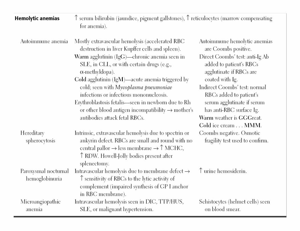
Direct Coombs testDirect Coombs test
The direct Coombs test (also known as the direct antiglobulin test or DAT)is used to detect if antibodies or complement system factors have bound if antibodies or complement system factors have bound to RBC surface antigens to RBC surface antigens in vivoin vivo..
Indirect Coombs testIndirect Coombs test
The indirect Coombs test (also known as the indirect antiglobulin test or IAT) is a used to detect in-vitro antibody-antigen reactions.
Coombs reagent: Coombs reagent is antihuman globulin.
Coombs reagent (also known as Coombs antiglobulin or antihuman globulin)


By numberBy SHAPEBy hemoglobinBy size
Erythrocyte DisordersErythrocyte Disorders
Anisocytosis – various sizesPoikilocytosis – various shapes

•• AnemiaAnemia –• blood has abnormally low
oxygen-carrying capacity– It is a symptom rather than
a disease itself– Blood oxygen levels cannot
support normal metabolism– Signs/symptoms include
fatigue, paleness, shortness of breath, and chills
ERYTHROCYTE DISORDERSERYTHROCYTE DISORDERS
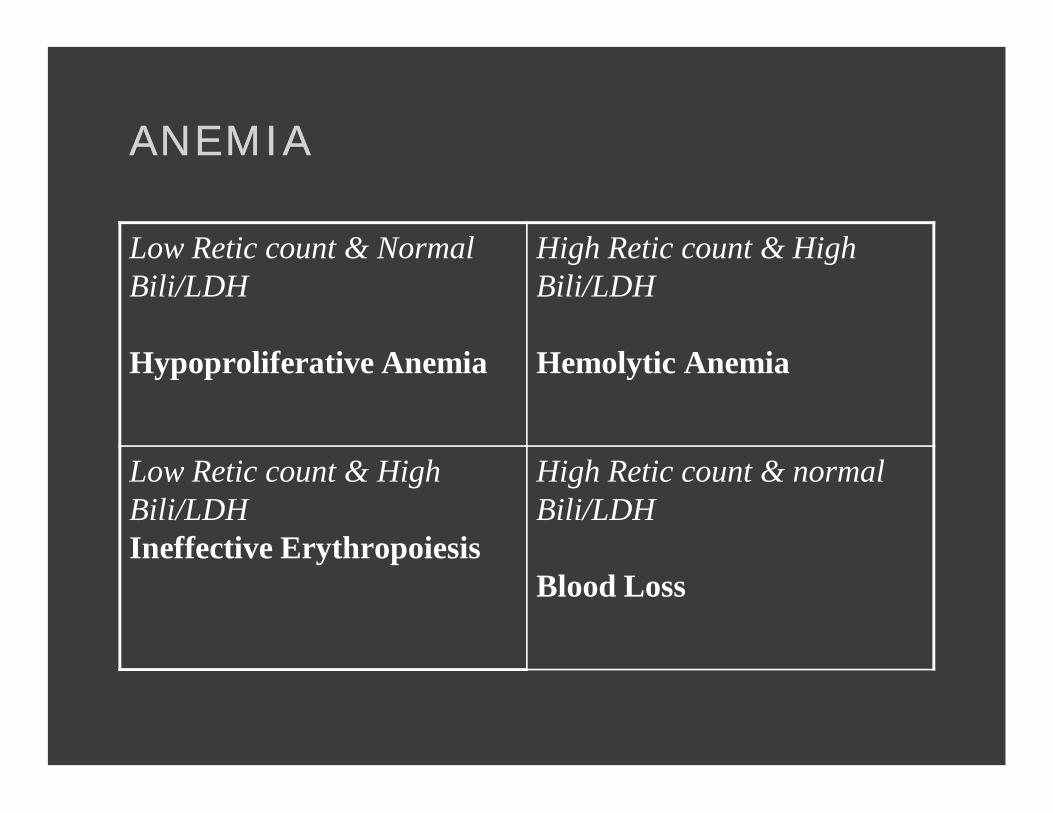
ANEMIAANEMIA
Low Retic count & Normal Bili/LDH
Hypoproliferative Anemia
High Retic count & High Bili/LDH
Hemolytic Anemia
Low Retic count & High Bili/LDHIneffective Erythropoiesis
High Retic count & normal Bili/LDH
Blood Loss
VAGINAL BLEEDING CAUSED BY MENSES
OR OTHER

LAB EVALUATION OF LAB EVALUATION OF HYPOPROLIFERATIVE ANEMIASHYPOPROLIFERATIVE ANEMIAS
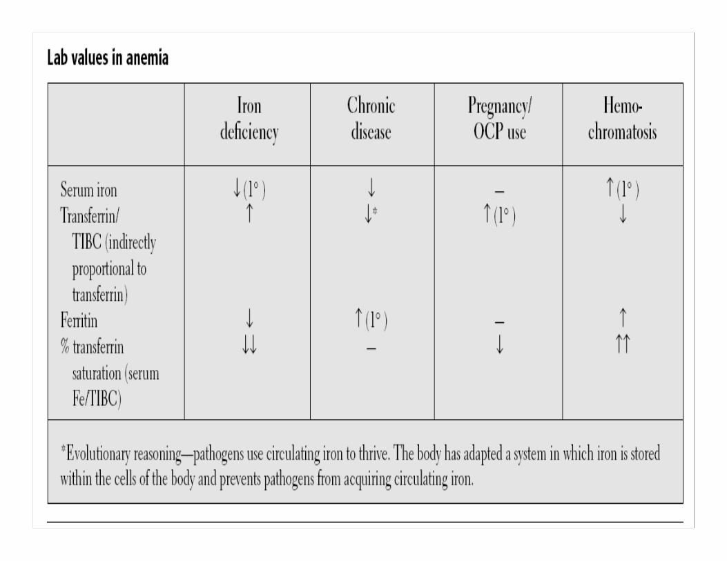
Fe TIBC Ferritin
Fe Deficiency low High(>300) low
Anemia of Chronic Dx
low low Normal to high
Aplastic anemia High Extremely high
Normal to high

EVALUATION OF THE PATIENT EVALUATION OF THE PATIENT
• HISTORY– Is the patient bleeding?
• Actively? In past?– Is there evidence for increased RBC destruction?– Is the bone marrow suppressed?– Is the patient nutritionally deficient? Pica?– PMH including medication review, toxin exposure
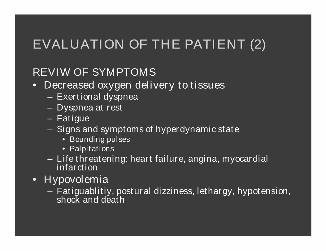
EVALUATION OF THE PATIENT (2)EVALUATION OF THE PATIENT (2)
REVIW OF SYMPTOMS• Decreased oxygen delivery to tissues
– Exertional dyspnea– Dyspnea at rest– Fatigue– Signs and symptoms of hyperdynamic state
• Bounding pulses• Palpitations
– Life threatening: heart failure, angina, myocardial infarction
• Hypovolemia– Fatiguablitiy, postural dizziness, lethargy, hypotension,
shock and death

EVALUATION OF THE PATIENT (3)EVALUATION OF THE PATIENT (3)
PHYSICAL EXAM•Stable or Unstable?
-ABCs-Vitals
•Pallor•Jaundice
-hemolysis•Lymphadenopathy•Hepatosplenomegally•Bony Pain•Petechiae•Rectal-? Occult blood

LABORATORY EVALUATIONLABORATORY EVALUATION
• Initial Testing– CBC w/ differential (includes RBC indices)– Reticulocyte count– Peripheral blood smear

LABORATORY EVALUATION (2)LABORATORY EVALUATION (2)
• Bleeding– Serial HCT or HGB
• Iron Deficiency– Iron Studies
• Hemolysis– Serum LDH, indirect bilirubin, haptoglobin, coombs,
coagulation studies• Bone Marrow Examination• Others-directed by clinical indication
– hemoglobin electrophoresis– B12/folate levels

DIFFERENTIAL DIAGNOSISDIFFERENTIAL DIAGNOSIS
• Classification by Pathophysiology – Blood Loss– Decreased Production– Increased Destruction
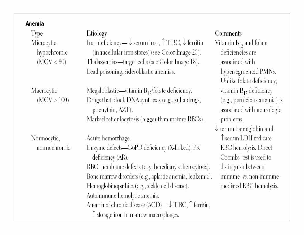
• Classification by Morphology– Normocytic– Microcytic– Macrocytic

•• NormochromicNormochromic, , normocyticnormocytic anemia (normal MCHC, normal MCV). anemia (normal MCHC, normal MCV). These include:
– anemias of chronic disease– hemolytic anemias (those characterized by accelerated
destruction of rbc's)– anemia of acute hemorrhage– aplastic anemias (those characterized by disappearance of
rbc precursors from the marrow)
•• Hypochromic, Hypochromic, microcyticmicrocytic anemia (low MCHC, low MCV). anemia (low MCHC, low MCV). These include: – iron deficiency anemia– thalassemias– anemia of chronic disease (rare cases)
•• NormochromicNormochromic, , macrocyticmacrocytic anemia (normal MCHC, high MCV). anemia (normal MCHC, high MCV). These include: – vitamin B12 deficiency– folate deficiency

Mean corpuscular volume [MCV]Mean corpuscular volume [MCV]
Is a measure of the average red blood cell volume average red blood cell volume (i.e. sizesize) that is reported as part of a standard complete blood count.
In patients with anemia, it is the MCV measurement that allows classification as either a microcytic anemia (MCV below normal range) or macrocytic anemia (MCV above normal range).
Mean corpuscular hemoglobin concentrationMean corpuscular hemoglobin concentrationis a measure of the concentration of hemoglobin in a given volume of packed red blood cell.
It is calculated by dividing the hemoglobin by the hematocrit. A normal value is 32 to 36 g/dl. Hb/ht
It is diminished ("hypochromic") in microcytic anemias, and normal ("normochromic") in macrocytic anemias (due to larger cell size, though the hemoglobin amount or MCH is high, the concentration remains normal). MCHC is elevated in hereditary spherocytosis.

MACROCYTIC ANEMIAMACROCYTIC ANEMIA
• MCV > 100• Megaloblastic:Abnormal
ities in nucleic acid metabolism– B12, Folate
• Non-megaloblastic:AbnormalRBC maturation– Myelodysplasia
• ETOH, liver dz, hypothryroidism, chemotherapy/drugs

MICROCYTIC ANEMIAMICROCYTIC ANEMIA
• MCV <80• Reduced iron
availability• Reduced heme
synthesis• Reduced globin
production


MICROCYTIC ANEMIAMICROCYTIC ANEMIAREDUCED IRON AVAILABILTYREDUCED IRON AVAILABILTY• Iron Deficiency
– Deficient Diet/Absorption– Increased Requirements– Blood Loss– Iron Sequestration
• Anemia of Chronic Disease– Low serum iron, low TIBC, normal serum ferritin– MANY!!
• Chronic infection, inflammation, cancer, liver disease

MICROCYTIC ANEMIAMICROCYTIC ANEMIAREDUCED HEME SYNTHESISREDUCED HEME SYNTHESIS• Lead poisoning• Acquired or
congenital sideroblastic anemia
• Characteristic smear finding: Basophylic stippling

MICROCYTIC ANEMIAMICROCYTIC ANEMIAREDUCED GLOBIN PRODUCTIONREDUCED GLOBIN PRODUCTION• Thalassemias• Smear
Characteristics– Hypochromia– Microcytosis– Target Cells– Tear Drops

LAB TESTS OF IRON DEFICIENCY OF LAB TESTS OF IRON DEFICIENCY OF INCREASED SEVERITYINCREASED SEVERITY
NORMALNORMAL Fe deficiencyFe deficiencyWithout anemiaWithout anemia
Fe deficiency Fe deficiency With mild anemiaWith mild anemia
Fe deficiency Fe deficiency With severe With severe anemiaanemia
Serum IronSerum Iron 6060--150150 6060--150150 <60<60 <40<40
Iron Binding Iron Binding CapacityCapacity
300300--360360 300300--390390 350350--400400 >410>410
SaturationSaturation 2020--5050 3030 <15<15 <10<10
HemoglobinHemoglobin NormalNormal NormalNormal 99--1212 66--77
Serum FerritinSerum Ferritin 4040--200200 <20<20 <10<10 00--1010
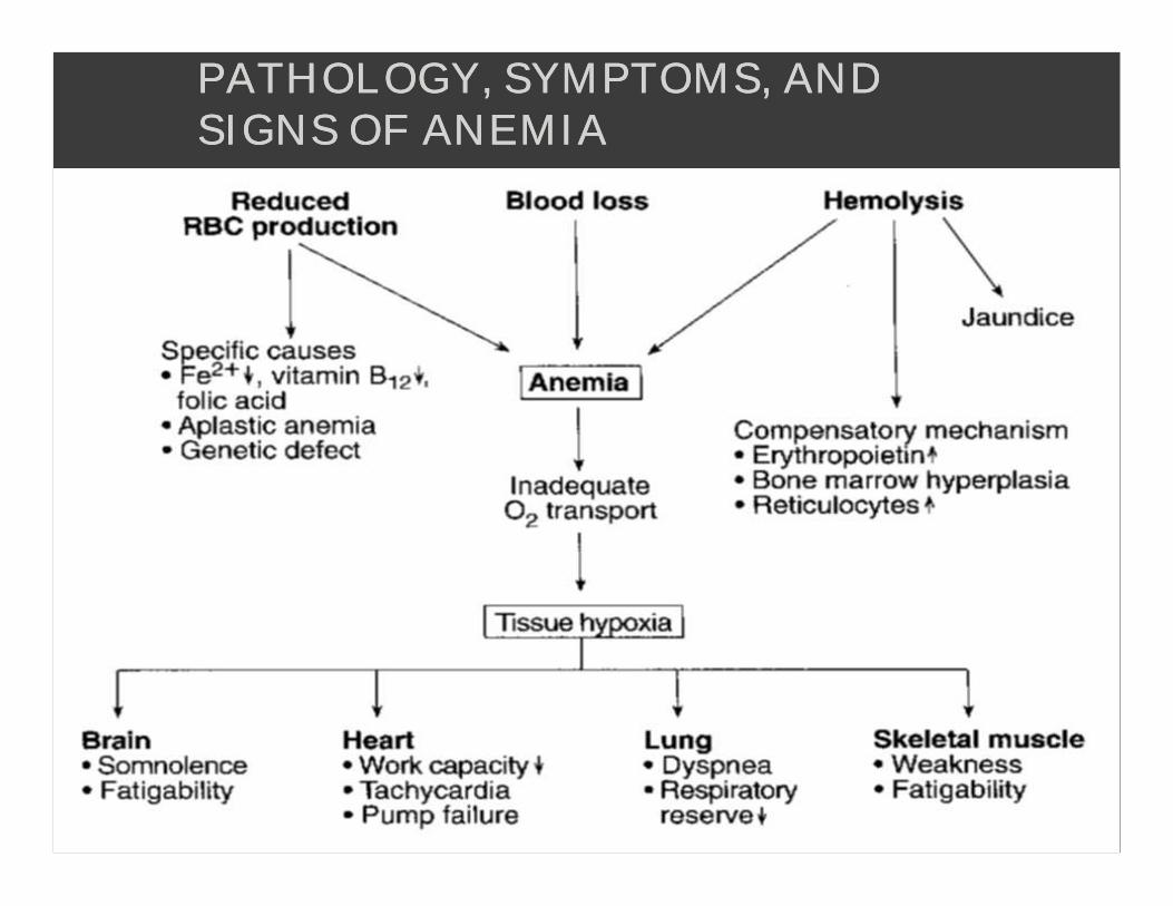
PATHOLOGY, SYMPTOMS, AND PATHOLOGY, SYMPTOMS, AND SIGNS OF ANEMIASIGNS OF ANEMIA
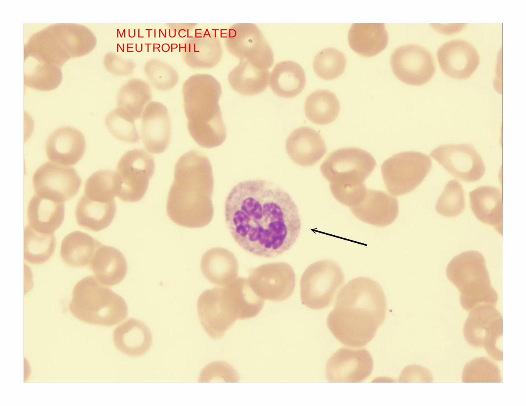
MULTINUCLEATED NEUTROPHIL
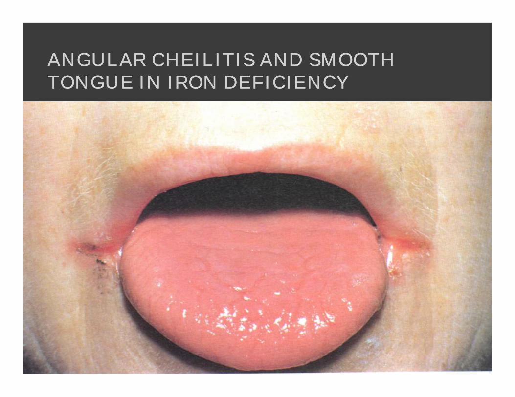
ANGULAR CHEILITIS AND SMOOTH ANGULAR CHEILITIS AND SMOOTH TONGUE IN IRON DEFICIENCYTONGUE IN IRON DEFICIENCY
• The anemia of chronic disease
Iron deficiency anemia
Beta thalassemia minor
Anemia of chronic renal failure
Anemia in cancer patients onchemotherapy
Aplastic anemia
Dysplastic and sideroblastic anemias
Sickle cell anemia
Glucose 6 phosphate dehydrogenasedeficiency and hemolysis
Hereditary spherocytosis
Macrocytic anemia – folate versus vitamin B12 deficiency
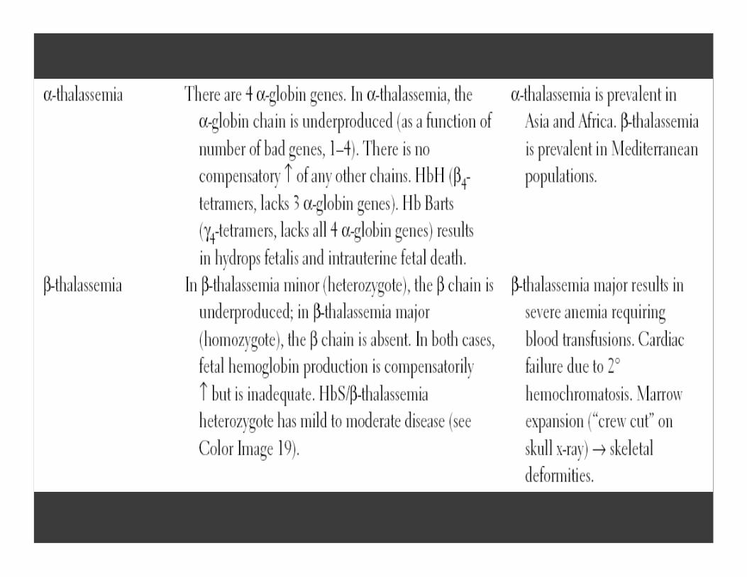
Thalassemia
Anemia in the elderly
Iron loading and hemachromatosis
Differential diagnosis of iron loading

ANEMIAANEMIA: : INSUFFICIENTINSUFFICIENTERYTHROCYTESERYTHROCYTES•• Hemorrhagic anemia Hemorrhagic anemia – result of acute or chronic
loss of blood
•• Hemolytic anemia Hemolytic anemia – prematurely ruptured RBCs
•• AplasticAplastic anemia anemia – destruction or inhibition of red bone marrow

TARGET CELLSTARGET CELLS
Graphic accessed http://diaglab.vet.cornell.edu/clinpath/modules/hemogram/images/target.jpg, 2009.
Target cells (from red blood cells) are associated with
•Hemoglobin C (HbC) disease, •Asplenia, •Liver Disease, Thalassemia•severe Iron deficiency anemia
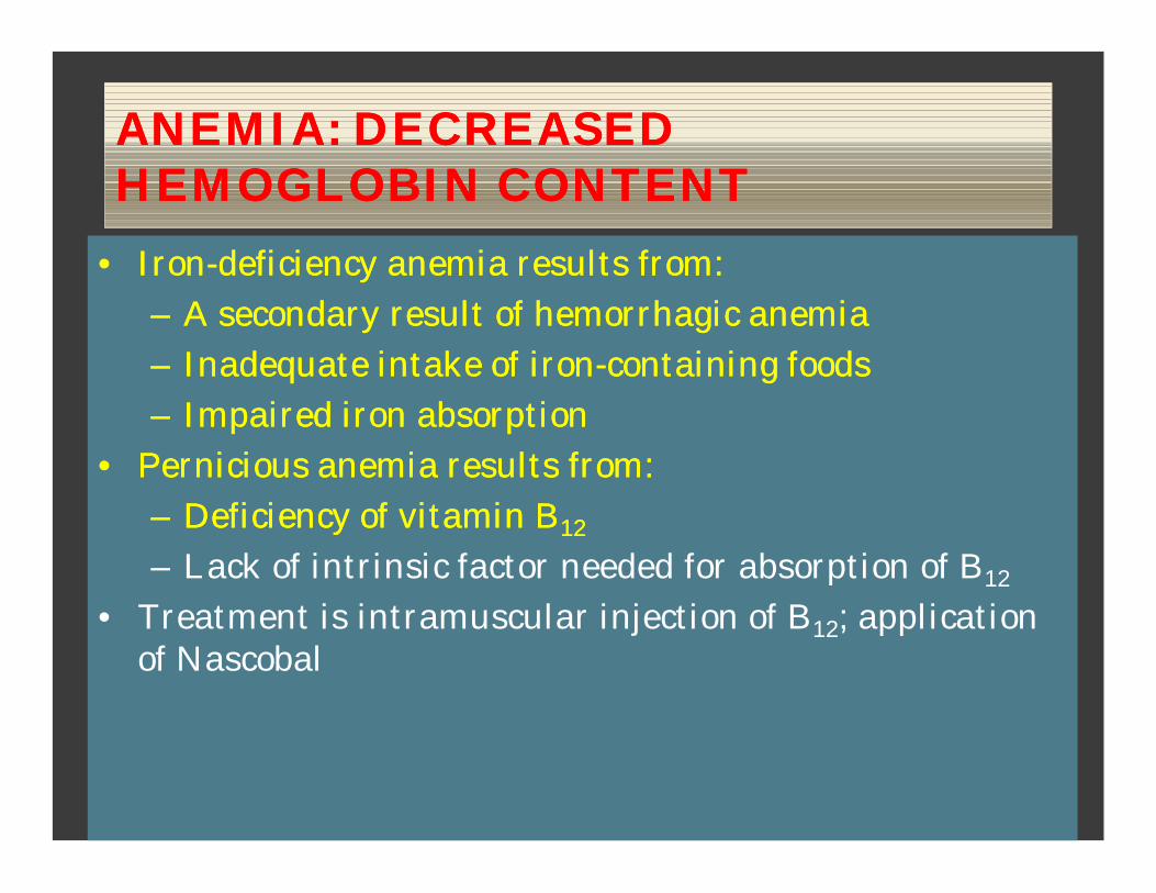
•• IronIron--deficiency anemia results from:deficiency anemia results from:–– A secondary result of hemorrhagic anemiaA secondary result of hemorrhagic anemia–– Inadequate intake of ironInadequate intake of iron--containing foodscontaining foods–– Impaired iron absorptionImpaired iron absorption
•• Pernicious anemia results from:Pernicious anemia results from:–– Deficiency of vitamin BDeficiency of vitamin B1212– Lack of intrinsic factor needed for absorption of B12
• Treatment is intramuscular injection of B12; application of Nascobal
ANEMIA: DECREASED ANEMIA: DECREASED HEMOGLOBIN CONTENTHEMOGLOBIN CONTENT
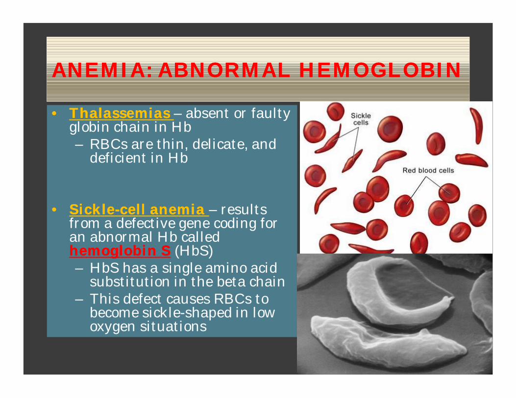
ANEMIA: ABNORMAL HEMOGLOBINANEMIA: ABNORMAL HEMOGLOBIN
•• ThalassemiasThalassemias – absent or faulty globin chain in Hb– RBCs are thin, delicate, and
deficient in Hb
•• SickleSickle--cell anemia cell anemia – results from a defective gene coding for an abnormal Hb called hemoglobin Shemoglobin S (HbS)– HbS has a single amino acid
substitution in the beta chain– This defect causes RBCs to
become sickle-shaped in low oxygen situations

SICKLE CELL ANEMIASICKLE CELL ANEMIA


G6PD DEFICIENCYG6PD DEFICIENCY
Patients with G6PD deficiency are prone to devloping hemolytic anemia in response to sulfonamides such as dapsone and sulfasalazine.
Other precipitating factors are infections, diabetic ketoacidosis, and favismfavism.
POLYCYTHEMIAPOLYCYTHEMIA

POLYCYTHEMIAPOLYCYTHEMIA• Polycythemia – excess RBCs
that increase blood viscosity
• Three main polycythemiasare:–– Polycythemia Polycythemia veravera–– Secondary Secondary
polycythemiapolycythemia–– Blood dopingBlood doping

• When polycythemia occurs and it is not associated with any not associated with any known underlying cause, it is typically referred to as known underlying cause, it is typically referred to as primary polycythemia, polycythemia primary polycythemia, polycythemia veravera, or , or erythremiaerythremia.
• This form of the disease is most common in middle-aged men and people of Jewish descent.
• Primary polycythemia is typically a chronic condition and tends to be progressive.
• In addition to an increase in red blood cells, individuals with the disease generally experience tumorous overgrowth of bone marrow, enlargement of the spleen, and excessive production of platelets and white blood cells.
• There is no known cure for the myeloproliferative disease, but various treatments can help normalize erythrocyte levels and provide symptomatic relief

CAUSESCAUSES POLYCYTHEMIAPOLYCYTHEMIA
• prolonged habitation of high altitudes,
• smoking, • certain types of cancer, • pulmonary disease, • heart disorders,
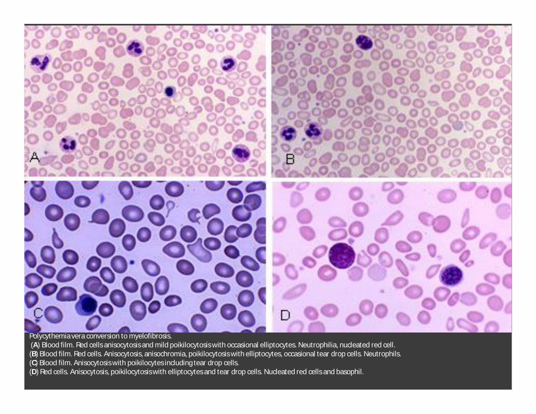
Polycythemia vera conversion to myelofibrosis.(A) Blood film. Red cells anisocytosis and mild poikilocytosis with occasional elliptocytes. Neutrophilia, nucleated red cell. (B) Blood film. Red cells. Anisocytosis, anisochromia, poikilocytosis with elliptocytes, occasional tear drop cells. Neutrophils. (C) Blood film. Anisocytosis with poikilocytes including tear drop cells. (D) Red cells. Anisocytosis, poikilocytosis with elliptocytes and tear drop cells. Nucleated red cells and basophil.

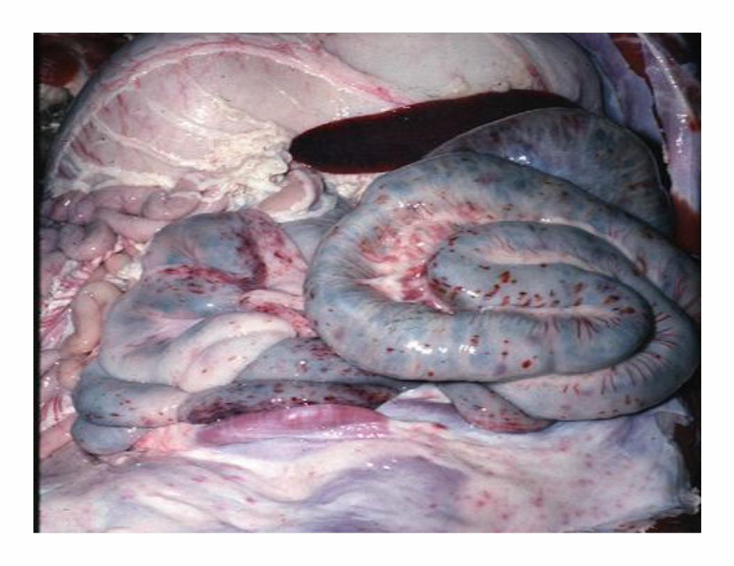
BLEDDING, HEMORRHAGE, BLEDDING, HEMORRHAGE, HEMATOMAS, PURPURA, PETECHIAEHEMATOMAS, PURPURA, PETECHIAE
PUPURA FULMINANS


HEREDITARY HEMORRHAGIC HEREDITARY HEMORRHAGIC TELANGIECTASIATELANGIECTASIA

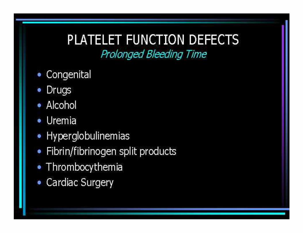
PATHOGENETIC CLASSIFICATION OF PATHOGENETIC CLASSIFICATION OF BLEEDING DISORDERSBLEEDING DISORDERS
HEMOSTASIS ISSUESHEMOSTASIS ISSUES


THROMBOCYTOPENIATHROMBOCYTOPENIA
• Decreased Platelet Production
• • Inadequate number of megakaryocytes
• • Aplastic anemia, marrow injury from
• drugs, radiation, infection, alcohol,
• fibrosis, metastatic tumor• • Ineffective thrombopoiesis
(adequate• megakaryocytes)• • B12/folate deficiency,
myelodysplastic• disorders• • Bone marrow evaluation
will establish the• diagnosis
• Normal platelet count: 140,000 –440,000/mm³
Thrombocytopenia results from:• Decreased marrow production• Increased peripheral destruction• Splenic sequestration• Hemodilution (multiple transfusions)
• Physical examination and bone marrow evaluation distinguish between these possibilities

Increased Platelet DestructionIncreased Platelet Destruction
(Bone marrow evaluation reveals increased number of megakaryocytes)• Immunologic disorders• Antiplatelet antibodies or immune complexes mediate plateledestruction• Idiopathic thrombocytopenic purpura (ITP)
Children: self-limited disorder, usually follows viral Infection
Adults: chronic disorder, immunosuppression usually necessary• Drug-induced (quinine, sulfa)• Sepsis (independent of DIC)• Connective tissue disorders (SLE), lymphoma• Non-immunologic disorders• DIC• Other microangiopathic conditions (TTP, prosthetic cardiac valve)Mechanism of ImmuneThrombocytopenia
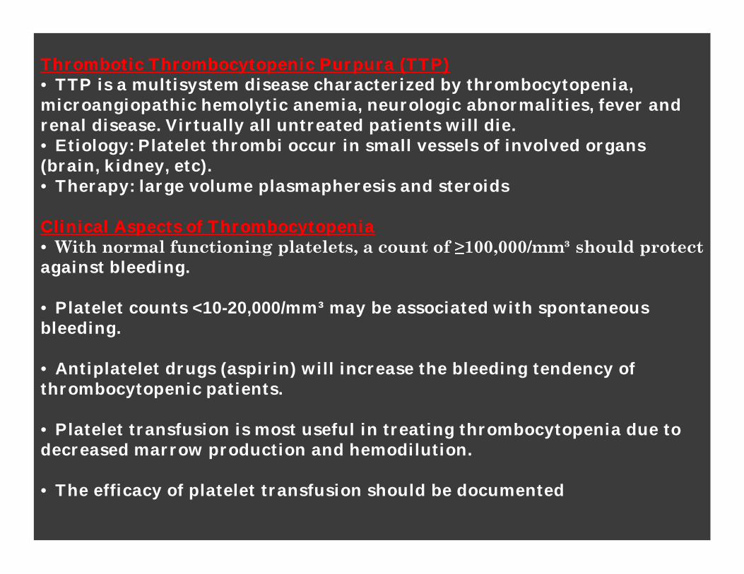
Thrombotic Thrombocytopenic Thrombotic Thrombocytopenic PurpuraPurpura (TTP)(TTP)• TTP is a multisystem disease characterized by thrombocytopenia, microangiopathic hemolytic anemia, neurologic abnormalities, fever and renal disease. Virtually all untreated patients will die.• Etiology: Platelet thrombi occur in small vessels of involved organs (brain, kidney, etc).• Therapy: large volume plasmapheresis and steroids
Clinical Aspects of ThrombocytopeniaClinical Aspects of Thrombocytopenia• With normal functioning platelets, a count of ≥100,000/mm³ should protect against bleeding.
• Platelet counts <10-20,000/mm³ may be associated with spontaneous bleeding.
• Antiplatelet drugs (aspirin) will increase the bleeding tendency of thrombocytopenic patients.
• Platelet transfusion is most useful in treating thrombocytopenia due to decreased marrow production and hemodilution.
• The efficacy of platelet transfusion should be documented


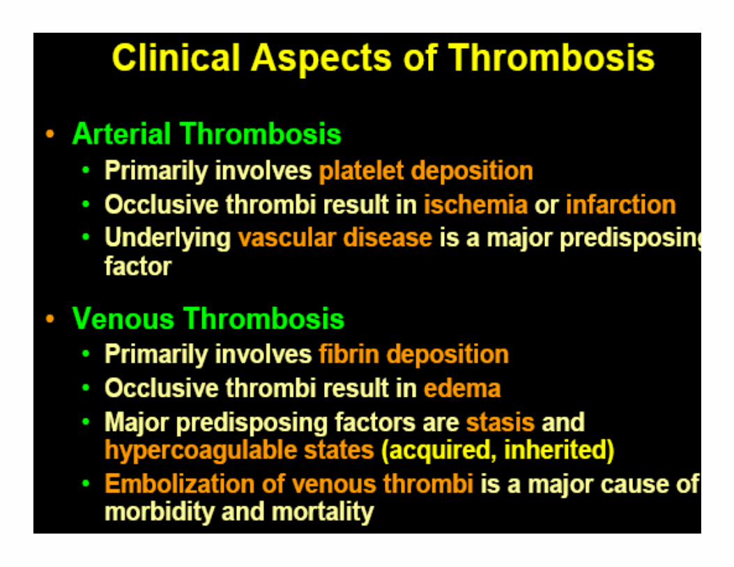
HEMOSTASIS DISORDERS:HEMOSTASIS DISORDERS:THROMBOEMBOLYTICTHROMBOEMBOLYTIC CONDITIONSCONDITIONS
• Thrombus – a clot that develops and persists in an unbroken blood vessel– Thrombi can block circulation, resulting in tissue
death– Coronary thrombosis – thrombus in blood vessel of
the heart
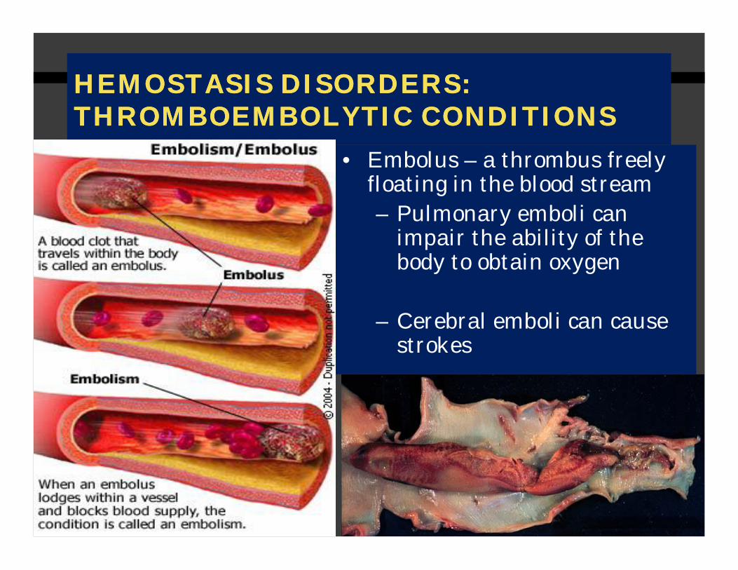
HEMOSTASIS DISORDERS:HEMOSTASIS DISORDERS:THROMBOEMBOLYTICTHROMBOEMBOLYTIC CONDITIONSCONDITIONS
• Embolus – a thrombus freely floating in the blood stream– Pulmonary emboli can
impair the ability of the body to obtain oxygen
– Cerebral emboli can cause strokes

Bone Marrow Megakaryocyte.
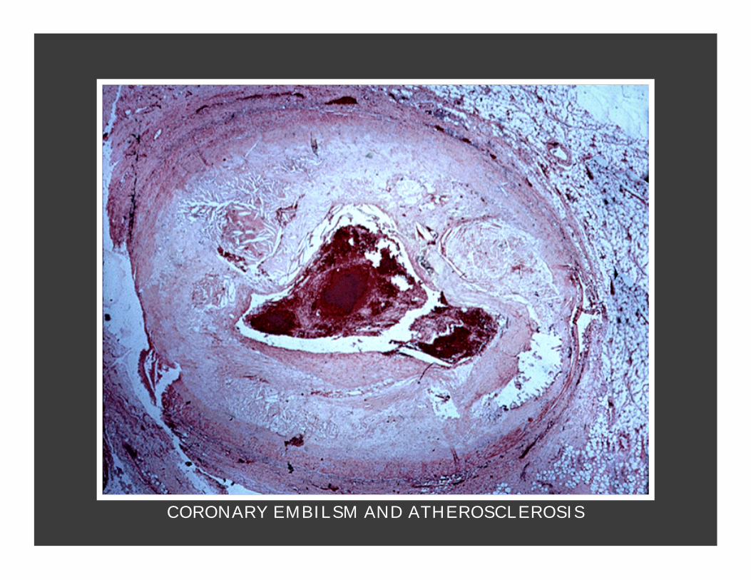
CORONARY EMBILSM AND ATHEROSCLEROSIS

Emboli formation.

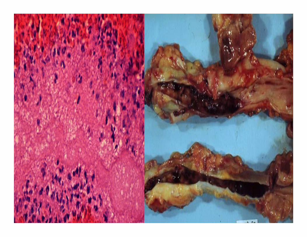

Coronary thromboembol
ism lignezahn.

Embolism .
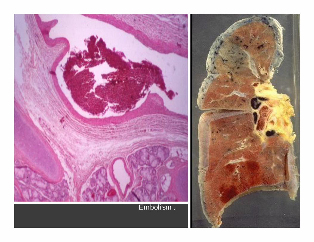
Embolism .

• Substances used to prevent undesirable clots:
–– AspirinAspirin – an antiprostaglandin that inhibits thromboxane A2
–– Heparin Heparin – an anticoagulant used clinically for pre-and postoperative cardiac care
–– WarfarinWarfarin – used for those prone to atrialfibrillation
PREVENTION OF UNDESIRABLE PREVENTION OF UNDESIRABLE CLOTSCLOTS

•• Disseminated Intravascular Coagulation (DIC): Disseminated Intravascular Coagulation (DIC): widespread clotting in intact blood widespread clotting in intact blood vesselsvessels
•• Residual blood cannot Residual blood cannot clotclot
•• Blockage of blood flow and severe bleeding Blockage of blood flow and severe bleeding followsfollows
•• Most common as:Most common as:–– A complication of pregnancyA complication of pregnancy–– A result of septicemia or incompatible blood A result of septicemia or incompatible blood
transfusionstransfusions
HEMOSTASIS DISORDERSHEMOSTASIS DISORDERS


DIC.

•• ThrombocytopeniaThrombocytopenia –condition where the number of circulating platelets is deficient– Patients show petechiaepetechiae due to
spontaneous, widespread hemorrhage
– Caused by suppression or destruction of bone marrow (e.g., malignancy, radiation)
– Platelet counts less than 50,000/mm3 is diagnostic for this condition
– Treated with whole blood transfusions
HEMOSTASIS DISORDERS: BLEEDING HEMOSTASIS DISORDERS: BLEEDING DISORDERSDISORDERS
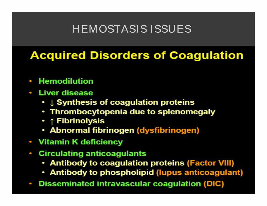
• Inability to synthesize procoagulants by the liver results in severe bleeding disorders
• Causes can range from vitamin K deficiency to hepatitis and cirrhosis
• Inability to absorb fat can lead to vitamin K deficiencies as it is a fat-soluble substance and is absorbed along with fat
• Liver disease can also prevent the liver from producing bile, which is required for fat and vitamin K absorption
HEMOSTASIS DISORDERS: BLEEDING HEMOSTASIS DISORDERS: BLEEDING DISORDERSDISORDERS
•• HemophiliasHemophilias –– hereditary bleeding disorders hereditary bleeding disorders caused by lack of clotting caused by lack of clotting factorsfactors
–– Hemophilia A Hemophilia A –– most common type (83% of all most common type (83% of all cases) due to a cases) due to a deficiency of factor deficiency of factor VIIIVIII
–– Hemophilia B Hemophilia B –– due to a due to a deficiency of factor deficiency of factor IXIX
–– Hemophilia C Hemophilia C –– mild type, due to a mild type, due to a deficiency deficiency of factor XIof factor XI
HEMOSTASIS DISORDERS: HEMOSTASIS DISORDERS: BLEEDING BLEEDING DISORDERSDISORDERS


Czar Nicholas II of Russia and his family, photographed c. 1916, showing his wife Alexandra (who was a carrier of hemophilia), his four daughters, and (in the foreground) his son Alexis, perhaps the most famous European royal with hemophilia. Corbis.

History's most famous carrier of the gene for hemophilia was Victoria (1819-1901), Queen of England and grandmother to most of the royalty in Europe. In 1853, Queen Victoria gave birth to her eighth child, Leopold, Duke of Albany, who had hemophilia and died at the age of 31 from internal bleeding after a fall.
Two of Queen Victoria's four daughters, Alice (b. 1843) and Beatrice (b. 1857), also carried the gene for hemophilia and subsequently transmitted the disease to three of Victoria's grandsons and to six of her great-grandsons.
Alice's daughter Alexandra also was a carrier of hemophilia, and she transmitted the disease to her son Alexis (b. 1904), whose father was Czar Nicholas 11 (1868—1918) of Russia. Alexis is perhaps the most famous of the European royals with hemophilia. Alexis was the heir to his father's throne and his medical condition caused much anxiety in the royal household. Historians are still discussing the role Alexis's condition played in the Russian revolution of 1918.

HEMOSTASIS DISORDERS: BLEEDING HEMOSTASIS DISORDERS: BLEEDING DISORDERSDISORDERS• Symptoms include prolonged bleeding and painful and
disabled joints• Treatment is with blood transfusions and the injection
of missing factors

• hemophilia A (factor VIII deficiency), • hemophilia B (factor IX deficiency, Christmas
disease)

BILIRUBIN ISSUESBILIRUBIN ISSUES


HYPERBILIRUBINEMIAHYPERBILIRUBINEMIA
Increased plasma concentrations of bilirubin (> 3 mg/dL) occurs when there is an imbalance between its production and excretion Recognized clinically as jaundice

Prehepatic (hemolytic) jaundice
• Results from excess production of bilirubin(beyond the livers ability to conjugate it) following hemolysis
• Excess RBC lysis is commonly the result of autoimmune disease; hemolytic disease of the newborn (Rh- or ABO-incompatibility); structurally abnormal RBCs (Sickle cell disease); or breakdown of extravasated blood
• High plasma concentrations of unconjugated bilirubin(normal concentration ~0.5 mg/dL)

Intrahepatic jaundice• Impaired uptake,
conjugation, or secretion of bilirubin
• Reflects a generalized liver (hepatocyte) dysfunction
• In this case, hyperbilirubinemia is usually accompanied by other abnormalities in biochemical markers of liver function

Posthepatic jaundice
• Caused by an obstruction of the biliary tree
• Plasma bilirubin is conjugated, and other biliary metabolites, such as bile acids accumulate in the plasma
• Characterized by pale colored stools (absence of fecal bilirubin or urobilin), and dark urine (increased conjugated bilirubin)
• In a complete obstruction, urobilin is absent from the urine

DIAGNOSES OF JAUNDICE DIAGNOSES OF JAUNDICE
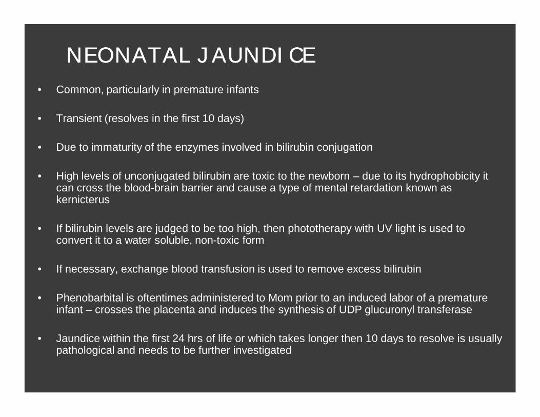
NEONATAL JAUNDICE NEONATAL JAUNDICE • Common, particularly in premature infants
• Transient (resolves in the first 10 days)
• Due to immaturity of the enzymes involved in bilirubin conjugation
• High levels of unconjugated bilirubin are toxic to the newborn – due to its hydrophobicity it can cross the blood-brain barrier and cause a type of mental retardation known as kernicterus
• If bilirubin levels are judged to be too high, then phototherapy with UV light is used to convert it to a water soluble, non-toxic form
• If necessary, exchange blood transfusion is used to remove excess bilirubin
• Phenobarbital is oftentimes administered to Mom prior to an induced labor of a premature infant – crosses the placenta and induces the synthesis of UDP glucuronyl transferase
• Jaundice within the first 24 hrs of life or which takes longer then 10 days to resolve is usually pathological and needs to be further investigated

CAUSES OF CAUSES OF HYPERBILIRUBINEMIAHYPERBILIRUBINEMIA

Benign liver disorder
½ of the affected individuals inherited it
Characterized by mild, fluctuating increases in unconjugatedunconjugatedbilirubinbilirubin caused by decreased ability of the liver to caused by decreased ability of the liver to conjugate conjugate bilirubinbilirubin –– often correlated with fasting or often correlated with fasting or illnessillnessThe source of this hyperbilirubinemia is reduced activity of the enzyme glucuronyltransferase glucuronyltransferase which. conjugates bilirubin and some other lipophilicmolecules.
Males more frequently affected then females
Onset of symptoms in teens, early 20’s or 30’s
Can be treated with small doses of phenobarbitalphenobarbital to stimulate UDP to stimulate UDP glucuronylglucuronyl transferasetransferase activity activity
Gilbert’s SyndromeGilbert’s Syndrome

Alternative, less common names for this disorder include:
• Familial benign unconjugated hyperbilirubinaemia• Constitutional liver dysfunction• Familial non-hemolytic non-obstructive jaundice• Icterus intermittens juvenilis• Low-grade chronic hyperbilirubinemia• Unconjugated benign bilirubinemia• Morbus
GILBERT’S SYNDROMEGILBERT’S SYNDROME
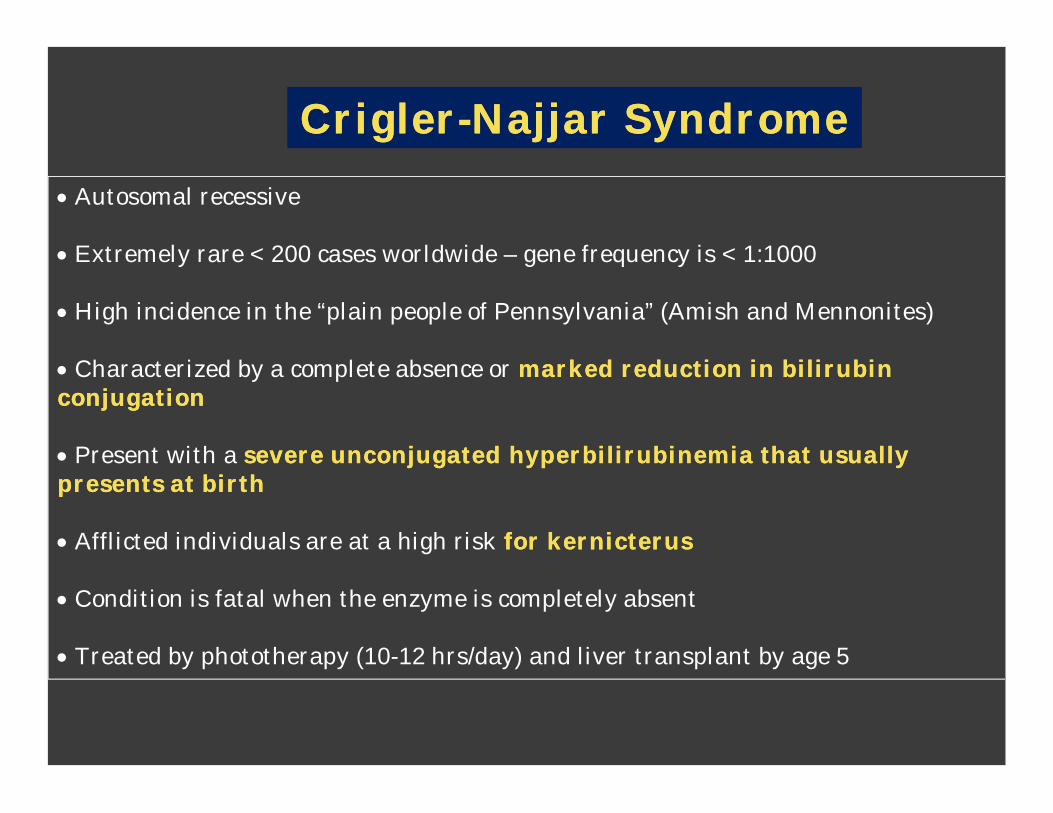
Autosomal recessive
Extremely rare < 200 cases worldwide – gene frequency is < 1:1000
High incidence in the “plain people of Pennsylvania” (Amish and Mennonites)
Characterized by a complete absence or marked reduction in marked reduction in bilirubinbilirubinconjugationconjugation
Present with a severe severe unconjugatedunconjugated hyperbilirubinemiahyperbilirubinemia that usually that usually presents at birthpresents at birth
Afflicted individuals are at a high risk for for kernicteruskernicterus
Condition is fatal when the enzyme is completely absent
Treated by phototherapy (10-12 hrs/day) and liver transplant by age 5
CriglerCrigler--NajjarNajjar SyndromeSyndrome

Characterized by impaired biliarysecretion of conjugated bilirubin
Present with a conjugated hyperbilirubinemia that is usually mild
DubinDubin--Johnson and Rotor’s SyndromesJohnson and Rotor’s Syndromes

LEUKOCYTES DISORDERSLEUKOCYTES DISORDERS

LEUKOCYTES DISORDERS: LEUKOCYTES DISORDERS: LEUKEMIASLEUKEMIAS
• Leukemia refers to cancerous conditions involving WBCs• Acute in kids , chronic in elderly
• Leukemias are named according to the abnormal WBCs involved–– MyelocyticMyelocytic leukemia leukemia – involves myeloblasts [AML.CML]–– Lymphocytic leukemia Lymphocytic leukemia – involves lymphocytes [ALL,CLL
• Acute leukemia involves blastblast--type cells type cells and primarily affects children
• Chronic leukemia is more prevalent in older people


GUM HYPERTROPHY AND HEMORRHAGE IN GUM HYPERTROPHY AND HEMORRHAGE IN ACUTE MONOCYTIC LEUKEMIAACUTE MONOCYTIC LEUKEMIA

MUCOSAL HEMORRHAGE DUE TO MUCOSAL HEMORRHAGE DUE TO SEVERE THROMBOCYTOPENIA IN SEVERE THROMBOCYTOPENIA IN ACUTE LEUKEMIAACUTE LEUKEMIA

ALL.
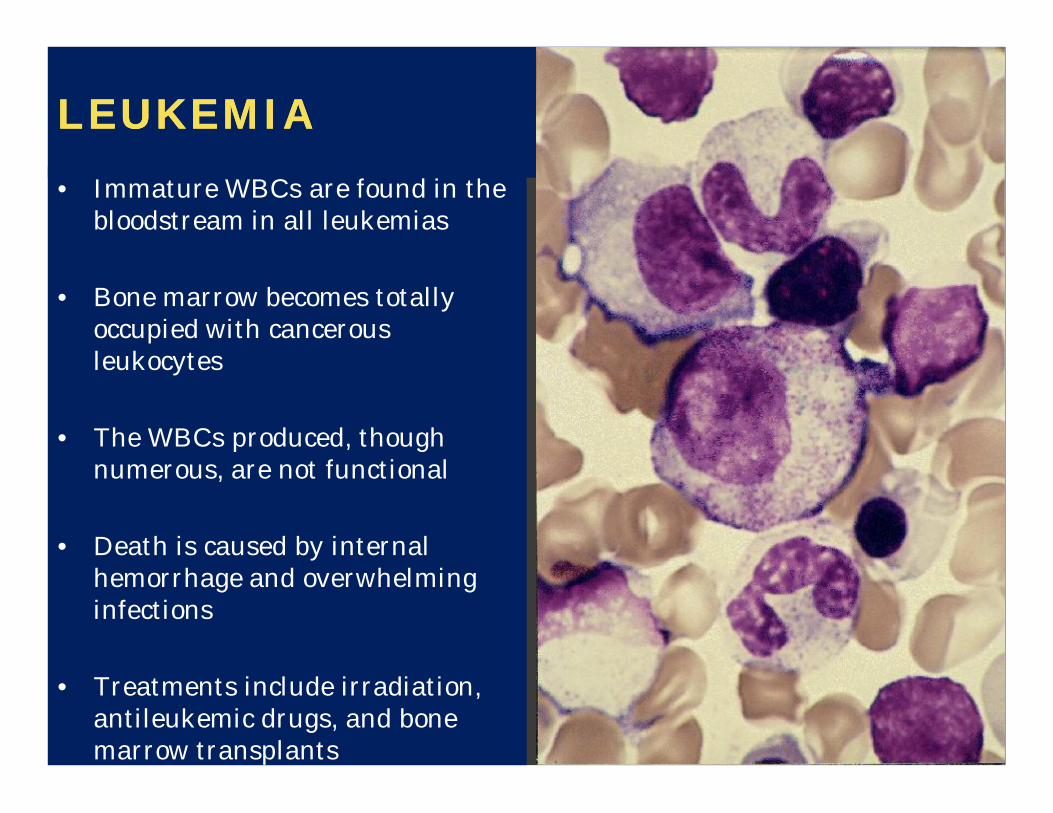
LEUKEMIALEUKEMIA• Immature WBCs are found in the
bloodstream in all leukemias
• Bone marrow becomes totally occupied with cancerous leukocytes
• The WBCs produced, though numerous, are not functional
• Death is caused by internal hemorrhage and overwhelming infections
• Treatments include irradiation, antileukemic drugs, and bone marrow transplants


MULTIPLE MYELOMAMULTIPLE MYELOMA


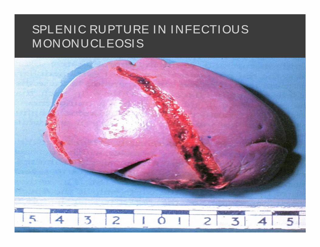
SPLENIC RUPTURE IN INFECTIOUS SPLENIC RUPTURE IN INFECTIOUS MONONUCLEOSISMONONUCLEOSIS

SPLEEN INFILTRATED BY HODGKIN'S SPLEEN INFILTRATED BY HODGKIN'S DISEASEDISEASE
NEUTROPHILSNEUTROPHILS CONDITION CONDITION
LOW LOW NEUTROPHILNEUTROPHIL COUNTS ARE TERMED COUNTS ARE TERMED NEUTROPENIANEUTROPENIA. :. :
--CONGENITAL (GENETIC DISORDER) OR IT CAN DEVELOP LATER, AS IN THCONGENITAL (GENETIC DISORDER) OR IT CAN DEVELOP LATER, AS IN THE CASE OF E CASE OF APLASTICAPLASTIC ANEMIA OR SOME KINDS OF LEUKEMIA. ANEMIA OR SOME KINDS OF LEUKEMIA.
--SIDESIDE--EFFECT OF MEDICATION, MOST PROMINENTLY CHEMOTHERAPY. NEUTREFFECT OF MEDICATION, MOST PROMINENTLY CHEMOTHERAPY. NEUTROPENIA OPENIA MAKES AN INDIVIDUAL HIGHLY SUSCEPTIBLE TO INFECTIONS. NEUTROPENIMAKES AN INDIVIDUAL HIGHLY SUSCEPTIBLE TO INFECTIONS. NEUTROPENIA CAN BE THE A CAN BE THE RESULT OF COLONIZATION BY INTRACELLULAR RESULT OF COLONIZATION BY INTRACELLULAR NEUTROPHILICNEUTROPHILIC PARASITES.PARASITES.
--FUNCTIONAL DISORDERS OF FUNCTIONAL DISORDERS OF NEUTROPHILSNEUTROPHILS ARE OFTEN HEREDITARY. THEY ARE ARE OFTEN HEREDITARY. THEY ARE DISORDERS OF DISORDERS OF PHAGOCYTOSISPHAGOCYTOSIS OR DEFICIENCIES IN THE RESPIRATORY BURST (AS IN OR DEFICIENCIES IN THE RESPIRATORY BURST (AS IN CHRONIC CHRONIC GRANULOMATOUSGRANULOMATOUS DISEASE, A RARE IMMUNE DEFICIENCY, AND DISEASE, A RARE IMMUNE DEFICIENCY, AND MYELOPEROXIDASEMYELOPEROXIDASE DEFICIENCY).DEFICIENCY).
--IN ALPHA 1IN ALPHA 1--ANTITRYPSIN DEFICIENCY, THE IMPORTANT ANTITRYPSIN DEFICIENCY, THE IMPORTANT NEUTROPHILNEUTROPHIL ENZYME ENZYME ELASTASEELASTASEIS NOT ADEQUATELY INHIBITED BY ALPHA 1IS NOT ADEQUATELY INHIBITED BY ALPHA 1--ANTITRYPSIN, LEADING TO EANTITRYPSIN, LEADING TO EXCESSIVE TISSUE XCESSIVE TISSUE DAMAGE IN THE PRESENCE OF INFLAMMATION DAMAGE IN THE PRESENCE OF INFLAMMATION -- MOST PROMINENTLY PULMONMOST PROMINENTLY PULMONARY ARY EMPHYSEMA.EMPHYSEMA.
--IN FAMILIAL MEDITERRANEAN FEVER (FMF), A MUTATION IN THE IN FAMILIAL MEDITERRANEAN FEVER (FMF), A MUTATION IN THE PYRINPYRIN (OR (OR MARENOSTRINMARENOSTRIN) GENE, WHICH IS EXPRESSED MAINLY IN ) GENE, WHICH IS EXPRESSED MAINLY IN NEUTROPHILNEUTROPHIL GRANULOCYTES, GRANULOCYTES, LEADS TO A CONSTITUTIVELY ACTIVE ACUTE PHASE RESPONSE AND CAUSESLEADS TO A CONSTITUTIVELY ACTIVE ACUTE PHASE RESPONSE AND CAUSES ATTACKS OF ATTACKS OF FEVER, FEVER, ARTHRALGIAARTHRALGIA, PERITONITIS, AND , PERITONITIS, AND -- EVENTUALLY EVENTUALLY -- AMYLOIDOSISAMYLOIDOSIS..


