Download - DIVERSITY OF THE HUMAN MEMORY T CELL - ETH Z

Research Collection
Doctoral Thesis
Diversity of the human memory T cell repertoire to pathogensand vaccines
Author(s): Becattini, Simone
Publication Date: 2014
Permanent Link: https://doi.org/10.3929/ethz-a-010271553
Rights / License: In Copyright - Non-Commercial Use Permitted
This page was generated automatically upon download from the ETH Zurich Research Collection. For moreinformation please consult the Terms of use.
ETH Library

DISS. ETH NO 22166
DIVERSITY OF THE HUMAN MEMORY T CELL REPERTOIRE
TO PATHOGENS AND VACCINES
A thesis submitted to attain the degree of
DOCTOR OF SCIENCES of ETH ZURICH
(Dr. sc. ETH Zurich)
presented by
SIMONE BECATTINI
MSc. Medical Biology University of Florence, Italy
Born on 24.06.1983
in Fiesole, Florence, Italy
accepted on the recommendation of
Prof. Antonio Lanzavecchia (examiner) Dr. Federica Sallusto (co-examiner)
Prof. Dr. Salomé LeibundGut-Landmann (co-examiner)
Zurich, 2014

Table of contents
Table of contents
1. Acknowledgments …………………………………………………… 3
2. General Summary
2.1 Summary (English) ……………………………………………… 5
2.2 Riassunto (Italiano) ……………………………………………… 6
3. Table of Abbreviations ……………………………………………… 7
4. Introduction …………………………………………………………… 9
4.1 CD4+ T cells: antigen receptor, subsets, and role in the immune response
4.1.1 Generation of CD4+ T lymphocytes ………………………… 9
4.1.2 αβ T cell receptor: genetically encoded diversity of the immune repertoire …………………………………………………… 13
4.1.3 TCR-mediated signaling and T cell activation by dendritic cells …………………………………………………………… 18
4.1.4 Naïve T cell priming and dynamics of T cell response ……… 21
4.1.5 Generation of effector and memory CD4+ T cells ………… 24
4.1.6 CD4+ T cell subsets: origin, phenotype and function ……… 27
4.1.7 Balancing subsets: when pathology arises from an inappropriate Th response……………………………………………………… 32
4.1.8 Flexibility of CD4+ T cells at the clonal and population level …………………………………………………………… 34
4.1.9 T cell repertoire: facts and open questions ………………… 41
4.2 Pathogens and vaccines object of this study
4.2.1 Candida albicans …………………………………………… 45
4.2.2 Mycobacterium tuberculosis ………………………………… 50
4.2.3 Tetanus toxoid vaccine ……………………………………… 52
5. Aims of the Thesis …………………………………………………… 55

Table of contents
6. Results …………………………………………………………………… 57
6.1 Functional heterogeneity of human memory T cell clones primed by pathogens and vaccines
6.1.1 Functional heterogeneity and clonal composition of C. albicans-specific CD4+ T cells ……………………………………… 57
6.1.2 Unique and shared clonotypes among C. albicans-specific memory T cell subsets ……………………………………………… 62
6.1.3 Extensive clonotype sharing among TT-specific T cell subsets …………………………………………………………… 67
6.1.4 Isolation of sister clones from different memory subsets…… 70
6.1.5 Generation of multiple T cell fates by in vitro priming of naïve T cells ………………………………………………………… 72
6.2 Additional Results
6.2.1 High throughput approach to unravel the relationship between C. albicans antigens and elicited response ……………………… 74
6.2.2 Interrogation of the T cell response in patients with chronic mucocutaneous candidiasis ………………………………… 79
6.2.3 A biochemical approach to identify C. albicans dominant Th17 antigens among cell wall mannoproteins …………………… 84
6.2.4 A biochemical approach to retrieve C. albicans inducing Th2 response ……………………………………………………… 93
6.2.5 In vivo assessment of the non-redundant role of T cells in long-term protection from C. albicans infection ………………… 96
7. Discussion and Outlook ……………………………………………… 101
8. Experimental Procedures ………………………………………… 107
9. References ……………………………………………………………… 116
10. Curriculum Vitae ……………………………………………………… 133
11. List of Publications, Presentations and Awards ……………… 135
1. Acknowledgments
3
1. Acknowledgments
First and foremost I want to thank my mentors, Federica Sallusto and Antonio Lanzavecchia,
for giving me the chance to perform my doctoral studies in their laboratory; I considered this
possibility a great privilege and honor, and tried to capture as much as I could from their
scientific knowledge.
Federica allowed me to work with freedom on very exciting and challenging projects. Her
guidance and feedback were instrumental for me not only to obtain the results presented in
this thesis, but also to acquire the skills I will need to pursue my scientific career.
The many challenges and insights I received from Antonio have been for me a great source of
constructive self-criticisms and learning, deeply impacting on my scientific growth.
A very special thanks goes to my ETH co-supervisor Salomé LeibundGut-Landmann, for her
passionate and enthusiastic support, for her help in critical steps of my doctoral path, and for
representing a beautiful example of genuine fun in science.
I am profoundly obliged to Silvia Monticelli, for making the PhD course an incredible
opportunity to learn critical thinking and get insights on paper reviewing and grant proposal
writing. I profited the most from it, and I will miss it terribly!
I had the fortune to collaborate with several leading scientists, who provided me with
samples, reagent and performed analysis that added great value to my work: in particular, I
would like to thank Anne Puel, Jean-Laurent Casanova, Alex Sette and Ton Schumacher.
Nico Callewaert introduced me with fervor and patience to the fascinating world of
biochemistry, and was kind enough to keep advising me and solve my many doubts
throughout the last years. I really appreciated his help, and hope we will have other chances to
collaborate in the future.
Mathilde Perez did an invaluable work with the handling of the sequences, and she did so
with a kindness and dedication that I truly appreciated.
A unique acknowledgment is for Federico, with whom from the very beginning I have
established way more than just a symbiotic relationship for cell care, experiment set up, and
strenuous fight against Murphy’s law. His presence truly helped me to “keep calm and carry
on”.

1. Acknowledgments
4
Several lab-mates deserve credit for helping me with some of the experiments presented in
this manuscript: in particular, thanks to Daniela, Blanca, Davide, Corinne, Antonino and
Laurent for their patience and commitment. Many others did not directly contributed to this
work, but supported and tolerated me in the lab, and my gratitude goes also to all of them,
especially to Dora, my first guide in Bellinzona.
David always performed impeccable sorting, and him and Marcus provided fantastic support
for daily trouble-shooting; besides, I will keep tight the memories of all the discussions we
had about science, politics, religion in front of a number of (generally emptied) bottles of
wine. That was great, thanks!
Dominik, Gabor, Michele and Silvia have been for me unique pillars to set up experiments,
discuss and criticize data, dissect all possible future and past career scenarios. With them I
also had the chance to travel, climb up to remote mountain hats, cook and play music, and I
really enjoyed all those wonderful moments. I believe they are and will be amazing scientists,
and hope our paths will cross again both inside and outside the lab.
I feel an acknowledgment is due to the Thai Boxing Bellinzona crew: training and fighting in
such a great environment was a terrific medicine to free my mind and motivated me to try
harder.
Stefano “Sammi” is an amazing person that happened to be my friend, introduce me to the
marvels of immunology, and stubbornly believe in me. I owe him more than he thinks.
I want to thank my family and old friends all around the world. First, for supporting me
unconditionally, and for representing Home, at any time, in any place. Secondly, for sharing
their experiences, often reminding me that many things are important in life that we should
appreciate and be grateful for, even when experiments do not work. In particular, thanks to
Ale “Lanze” and Marco “Burro”.
Finally, my deepest gratitude goes to Ilaria, my true Love, for always being by my side, for
making everything possible, for showing and teaching me the wonder that no experiment can
unravel.

2. General Summary
5
2. General Summary
2.1. Summary (English)
Human CD4+ T cells are key players in orchestrating the response to pathogens and vaccines.
Upon first antigen encounter, naïve T cells get activated and clonally expand, meanwhile
acquiring effector functions to control the threat and later becoming memory T cells to assure
appropriate response in case of re-challenge. Different classes of pathogens are known to
induce distinct polarized T cell subsets, each characterized by specific homing and functional
properties, that can mount the most efficient response tailored to the challenging pathogen.
Thus, viruses and intracellular bacteria induce IFN-γ-producing Th1 cells while helmints
induce IL-4-producing Th2 and fungi and extracellular bacteria induce IL-17-producing Th17
cells. Once considered terminally differentiated stages, polarized T cells can show remarkable
flexibility and have been proven to undergo phenotypic switch or divergent differentiation in
several experimental systems. Understanding the generation of diversity in the human T cell
memory compartment would have enormous implications both on a biological and on a
clinical level.
Using cell sorting of human memory Th1, Th2, and Th17 cells followed by CFSE labeling,
antigenic stimulation, and next generation TCR Vβ sequencing, we were able to demonstrate
that memory T cells specific for pathogens such as Candida albicans and Mycobacterium
tuberculosis or tetanus toxoid vaccine could be present in all subsets, albeit at different
frequencies. Interestingly, several clonotypes were present in more than one subset and, in
some cases, even in all subsets, while other clonotypes were restricted to one particular
subset. By cloning antigen-specific T cells from memory subsets we were able to isolate
several T cell clones from Th1, Th2, and Th17 subsets and show that they share the same
TCR but display different transcription factors, cytokine production and chemokine receptor
expression, characteristic of the subset from which they were isolated. Collectively, these
results indicate that the T cell response to pathogens and vaccines can comprise T cell clones
that in spite of identical TCR, display different, sometimes divergent types of effector
functions and provide for the first time demonstration of intraclonal diversification in the
human T cell response.

2. General Summary
6
2.2 Riassunto (Italiano)
I linfociti T CD4+ umani rivestono un ruolo fondamentale nel dirigere la risposta a patogeni e
vaccini. In seguito all’iniziale riconoscimento dell’antigene, le cellule T naive si attivano e
vanno incontro ad espansione clonale, acquisendo allo stesso tempo specifiche proprieta’
funzionali. Queste consentono di controllare il pericolo nell’immediato e, venendo ereditate
da cellule T della memoria, anche di garantire una risposta appropriata in caso di successivo
incontro col medesimo antigene. Classi diverse di patogeni inducono diversi sottotipi di
cellule T, ognuno caratterizzato da specifiche capacita’ migratorie e proprieta’ funzionali,
tramite le quali puo’ rispondere efficacemente al microbo sollecitante. In particolare, virus e
batteri intracellulari promuovono lo sviluppo di cellule Th1 che producono IFN-γ, gli elminti
quello di Th2 che producono IL-4, funghi e batteri extracellulari quello di cellule Th17 che
producono IL-17. Sebbene i subset di linfociti T siano stati per molto tempo considerati
entita’ irreversibilmente differenziate e’ oggi chiaro che queste cellule presentano una
notevole plasticita’, e la loro capacita’ di modificare il proprio fenotipo o di differenziare in
maniera divergente in seguito al riconoscimento dell’antigene e’ stata dimostrata in molti
modelli sperimentali.
Abbiamo utilizzato un protocollo che prevede l’ottenimento di cellule della memoria di tipi
diversi (Th1, Th2, Th17) tramite citofluorimetria a flusso, seguito da marcatura con CFSE,
stimolazione antigenica, e sequenziamento di ultima generazione della regione Vβ del TCR
delle cellule proliferanti. Tramite questo approccio abbiamo potuto dimostrare che le cellule
della memoria specifiche per patogeni quali Candida albicans, Mycobacterium tuberculosis o
il tossoide tetanico (vaccino) sono presenti in tutti i sottotipi di linfociti T, sebbene con
frequenze differenti. Inaspettatamente, abbiamo identificato vari clonotipi presenti in piu’
compartimenti, talvolta addirittura in tutti quelli analizzati, mentre altri sono risultati
selettivamente appartenere ad uno specifico gruppo. Attraverso clonaggio abbiamo isolato
alcune di queste cellule presenti allo stesso tempo nei compartimenti Th1, Th2 e Th17, e
confermato che possiedono un identico TCR, ma differiscono per l’espressione di fattori di
trascrizione, citochine, e recettori chemochinici, in accordo con la classe di appartenenza. In
definitiva, questi risultati dimostrano che l’insieme di cellule T che rispondono ad un
patogeno o ad un vaccino comprende cloni che, nonostante un identico recettore per
l’antigene, mostrano divergenti, talvolta contrapposte, proprieta’ funzionali, e rappresentano
percio’ la prima dimostrazione di differenziamento intra-clonale nella risposta dei linfociti T
nell’uomo.

3. Table of Abbreviations
7
3. Table of Abbreviations
AIRE Autoimmune regulator gene
APC Antigen-presenting cell
BCG Bacillus Calmette-Guerin
BCR B cell receptor
CA Candida albicans
CCR C-C chemokine receptor
CD Cluster of differentiation
CDR Complementary determining region
CKR Chemokine receptor
CLR C-type lectin receptor
CMC Chronic mucocutaneous candidiasis
CMV Cytomegalovirus
cTEC Cortical thymic epithelial cell
CTL Cytotoxic T cell
CWE Cell wall extract
CXCR CXC-chemokine receptor
DC Dendritic cell
DN Double negative
DP Double positive
EC50 Half maximal effective
concentration
GATA3 GATA binding protein 3
GM-CSF Granulocyte macrophage
colony-stimulating factor
HSC Hematopoetic stem cell
ICAM Intracellular adhesion molecule
ICOS Inducible costimulator
IFN Interferon
Ig Immunoglobulin
IL Interleukin
ILC Innate lymphoid cell
iNKT Invariant natural killer T cell
Iono Ionomycin
ITAM Immunoreceptor tyrosine based
activation motif
GC Germinal center
Lm Listeria monocytogenes
LN Lymph node
LPS Lipopolysaccaride
MHC Major histocompatability complex
MoDC Monocyte derived dendritic cell
MTB Mycobacterium tuberculosis
mTEC Medulary thymic epithelial cell
NK Natural killer
NLR Nod-like receptor

3. Table of Abbreviations
8
PAMP Pathogen associated molecular
pattern
PBMC Peripheral blood mononuclear cell
PMA Phorbol 12-myristate 13-acetate
P:MHC Peptide:MHC
PRR Pattern recognition receptor
RAG Recombination-activating gene
SP Single positive
STAT Signal transducer and activator of
transcription
T-bet T-cell-specific T-box
TCR T cell receptor
TCM T central memory cell
TdT Terminal deoxynucleotidyl
transferase
TE T effector cell
TEM T effector memory cell
TFH T follicular helper cell
TGF-β Transforming growth factor beta
Th T helper
TLR Toll-like receptor
TNF Tumor necrosis factor
Treg T regulatory cell
TSLP Thymic stromal lymphopoietin
TT Tetanus toxoid
TSCM T memory stem cell
V(D)J Variable, diversity, joining

4. Introduction
9
4. Introduction
4.1. CD4+ T cells: antigen receptor, subsets, and role in the immune
response
4.1.1. Generation of CD4+ T lymphocytes
T lymphocyte development is a process occurring early in life in the thymus, a mediastinal
organ that enlarges during childhood and undergoes atrophy at puberty. Thymus has a bi-
lobar structure, each lobe being composed by multiple lobules organized in an outer cortical
and an inner medullary region (Abbas et al., 2012; Murphy, 2011). Multipotent lymphoid
progenitors continuously reach the thymus coming from the bone marrow, where they are
generated from hematopoietic stem cell precursors, and undergo a complex sequence of
events that lead to the generation of a large pool of mature T lymphocytes expressing each a
unique T cell receptor (TCR) that can recognize a specific peptide bound to major
histocompatibilty complex (MHC) molecules displayed on the surface of antigen presenting
cells (APCs) (Takahama, 2006). In mature T lymphocytes, the TCR associates with a co-
receptor molecule, either CD4 or CD8, that selectively binds to MHC class II or class I
molecules, respectively. As a result, TCRs associated with CD4 can recognize peptides bound
on MHC class II molecules, which are expressed on the surface of professional APCs, such as
B cells, dendritic cells (DCs), macrophages, and monocytes, while TCRs associated with CD8
can recognize peptides bound to MHC class I molecules, which are expressed by virtually all
nucleated cells in the body. Peptides derived from extracellular antigens are generally
presented on MHC class II molecules, while MHC class I molecules mainly accommodate
peptides derived from antigens from intracellular compartments (Abbas et al., 2012; Murphy,
2011).
The process of T cell development and selection is very restrictive, and only 2-4% of cells
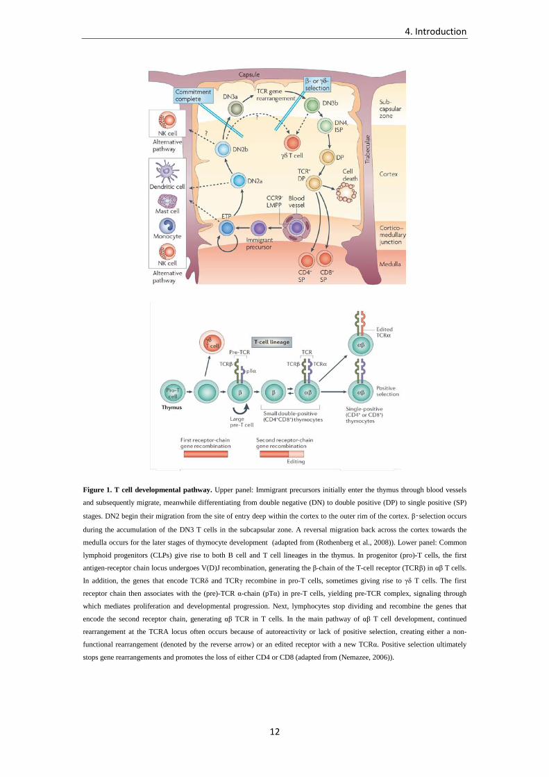
entering the thymus will make egress from the organ. Once in the thymus, the lymphoid
progenitors become CD4–CD8– double negative (DN) thymocytes losing the potential to
differentiate into B or NK cells (Rothenberg et al., 2008) (Figure 1). Based on the expression
of CD44 and CD25, these cells differentiate through 4 distinct stages (DN1 to DN4) in the
outer cortex of the organ (Godfrey et al., 1993). At the stage of DN3, cells can produce a TCR
β chain upon somatic rearrangement of the dedicated locus on chromosome 7, mediated by
the enzymes recombination-activating gene (RAG) 1 and 2 (Figure 1). The TCR β chain pairs
on the cell surface with a pre-TCR α chain (pT-α), transcribed instead from a genetic locus
that does not undergo rearrangement, and this definitely precludes the possibility to develop
4. Introduction
10
along the pathway of γδ T cells (Saint-Ruf et al., 1994, 2009; Takahama, 2006). The pre TCR
αβ can signal downstream thanks to the association with the CD3/ζ complex, allowing further
maturation of the T cell precursor to the DN4 stage, and finally recombination of the α chain
and surface expression of a mature αβ TCR (Pang et al., 2010). Co-receptors start to be
expressed at this stage, generally CD8 first, followed by CD4: the resulting population is
indeed composed by CD4+CD8+ double positive (DP) cells. Most of these thymocytes will die
by neglect, as their TCRs bind too weakly to the endogenous peptides presented in the cortex
by thymyc epithelial cells (TECs), and they do not upregulate TCR-signaling associated pro-
survival factors. Subsequently, T cells lose the expression of one of the two co-receptors and
are instructed to become either CD4+ or CD8+ T lymphocytes that migrate into the medulla
(Germain, 2002; Rothenberg et al., 2008). There is no consensus yet on what signals are
precisely required to make this fate decision. Two models have been proposed: in the first
one, known as the selection model, the choice of which co-receptor has to be maintained is
made randomly, and T cells are then selected based on their capacity to recognize antigens on
the resulting MHC molecule of choice, most of them dying as a consequence of a non
functional TCR-co-receptor coupling. In the second, referred to as the instruction model, the
signals that the T cell receives during thymic antigen presentation drive the selection of the
fittest co-receptor (Germain, 2002). Variations in strength and duration of the TCR signaling
have been proposed to be key factors in the lineage decision process. Notch-Notch ligand
interaction was reported to be crucial in this process, Notch-1 activation being associated with
CD8 SP T cell development (Germain, 2002).
T cells need to be selected by a minimal strength of self recognition, to ensure functionality
and capability to recognize a non-self antigen when presented on MHC molecules. This
process, called positive selection, takes place in the cortex of the thymus, and is mediated by
specialized cortical thymic epithelial cells (cTECs) (Klein et al., 2014). cTECs express high
amounts of MHC class II molecules on their surface and are endowed with a distinguishing
antigen processing machinery. In fact, they possess a unique catalytic subunit of the
proteasome, called β5t, and lysosomal proteases (cathepsin L and thymus-specific serine
protease, TSSP) that determine their unique array of MHC-displayed peptides. Furthermore,
these cells are characterized by constitutive high levels of macroautophagy, a process through
which self proteins from endogenous cell compartments can be presented on MHC class II
molecules (Klein et al., 2014). Positively selected T cells then migrate to the medulla, where
approximately 5% die by apoptosis as a consequence of very strong interaction between the
TCR and self peptide-MHC complexes. This process, known as negative selection, is
essential to purge the T cell repertoire from potentially autoreactive clonotypes. Negative
selection is primarily carried out by medullary thymic epithelial cells (mTECs); through the

4. Introduction
11
action of AIRE gene, mTECs can ectopically express a broad set of tissue restricted antigens
(TRAs), and therefore present self antigens which would not otherwise be available for
screening of auto-reactive T cells (Anderson et al., 2002; Liston et al., 2003). These antigens
can be presented directly by mTECs, or handed over to neighbouring APCs. Recent work has
provided evidence that negative selection starts already in the cortex, where is mediated by
specific subsets of DCs (Daley et al., 2013; Stritesky et al., 2013).
In summary, during thymic development, T cell precursors undergo a process of selection that
is consistent with an affinity model, whereby affinity refers to the strength of interaction
between TCR and self peptide-MHC complexes. Weak but over-threshold interactions are
required to spare thymocytes from death by neglect; strong interactions lead to negative
selection and apoptosis. Cells whose TCRs interact with intermediate strength will develop
either as non-self responding mature naïve T cells or as tolerized natural T regulatory cells
(nTregs) and exit the thymus to relocate in the periphery (Klein et al., 2014).
4. Introduction
12
Figure 1. T cell developmental pathway. Upper panel: Immigrant precursors initially enter the thymus through blood vessels
and subsequently migrate, meanwhile differentiating from double negative (DN) to double positive (DP) to single positive (SP)
stages. DN2 begin their migration from the site of entry deep within the cortex to the outer rim of the cortex. β‑selection occurs
during the accumulation of the DN3 T cells in the subcapsular zone. A reversal migration back across the cortex towards the
medulla occurs for the later stages of thymocyte development (adapted from (Rothenberg et al., 2008)). Lower panel: Common
lymphoid progenitors (CLPs) give rise to both B cell and T cell lineages in the thymus. In progenitor (pro)-T cells, the first
antigen-receptor chain locus undergoes V(D)J recombination, generating the β-chain of the T-cell receptor (TCRβ) in αβ T cells.
In addition, the genes that encode TCRδ and TCRγ recombine in pro-T cells, sometimes giving rise to γδ T cells. The first
receptor chain then associates with the (pre)-TCR α-chain (pTα) in pre-T cells, yielding pre-TCR complex, signaling through
which mediates proliferation and developmental progression. Next, lymphocytes stop dividing and recombine the genes that
encode the second receptor chain, generating αβ TCR in T cells. In the main pathway of αβ T cell development, continued
rearrangement at the TCRA locus often occurs because of autoreactivity or lack of positive selection, creating either a non-
functional rearrangement (denoted by the reverse arrow) or an edited receptor with a new TCRα. Positive selection ultimately
stops gene rearrangements and promotes the loss of either CD4 or CD8 (adapted from (Nemazee, 2006)).

4. Introduction
13
4.1.2. αβ T cell receptor: genetically encoded diversity of the immune
repertoire
T cells must face an enormous diversity of challenging pathogens, providing response to an
hypothetically infinite spectrum of antigens. The amount of genetic information necessary to
encode for a proportional variety of receptors would likely be incompatible with the size of
the genome. T cells, as well as B cells, have therefore developed a very efficient system to
ensure sufficient diversity of their repertoire by undergoing somatic rearrangement on the
genetic loci that encode for their antigen-specific receptor.
Each germline TCR locus comprises different groups of genes called Variability (V),
Diversity (D), Joining (J) and Constant (C) segments, respectively. A specific promoter and a
leader sequence precedes each V segment (Figure 2). The β chain locus, on chromosome 7,
hosts 77 contiguous V genes (of which 48 functional, the rest being pseudogenes whose
function has been impaired by occurred mutations), 2 D, each followed by 7 of 14 J (13
functional) and 1 out of two C genes. The α chain locus, on chromosome 14, is composed by
54 contiguous V (45 functional), 61 contiguous J (50 functional), and 1 C (The International
Immunogenetics Information System, 2014). D segments are absent from this locus; instead,
this region hosts the genetic locus encoding for the δ chain, that is usually co-expressed with a
γ chain to give rise to T cells carrying a different type of TCR called γδ. γδ T cell migration
from the thymus precedes αβ T cell egress (Dunon et al., 1997).
The process through which the TCR genetic segments are joined and generate a functional
chain of the TCR is called V(D)J recombination. Particular sequences, called recombination
signal sequences (RSSs) are located at the 3’ of each V gene segment, 5’ of each J segment,
and on both sides of each D segment. These sequences consist of a conserved stretch of 7
nucleotides (heptamer, 5’CACACTG3’) contiguous to the coding sequence and followed by a
non-conserved spacer of either 12 or 23 non-conserved basis, in turn followed by a second
conserved set of nine nucleotides (nonamer, rich in AT) (Schatz and Spanopoulou, 2005;
Schlissel, 2003). Acting on those particular sequences, a series of enzymes mediate
recombination among the gene segments so to obtain a linear gene containing 1 V, 1 D and 1
J for the β chain, or 1 V and 1 J in the case of the α chain, joined together. In the case of the β
chain, D and J segments are rearranged first, and the V segment is coupled afterwards. The
main enzymes catalyzing this reaction are Rag1 and 2, they are expressed selectively in
lymphocytes and multimerize to form a tetramer (called Rag1-2 recombinase). The reaction
begins with the recognition of spacer sequences and cleavage at the side of each coding
segment; de novo addition of nucleotides at the junctions and end joining between the two
adjacent segments takes place subsequently, both reaction being mediated by other enzymes
4. Introduction
14
(Schatz and Spanopoulou, 2005; Schlissel, 2003). Importantly, Rag1 and Rag2 can only join
segments flanked by RSS of different spacer length (i.e. a gene flanked by a 12 bp spacer RSS
with one adjacent to a 23 bp spacer RSS) (Kavaler et al., 1984; Kurosawa et al., 1981; Sakano
et al., 1981). Due to the spatial organization of RSS within the locus, this makes
recombination possible only between V-D and D-J segments (or V-J in the case of α-chain). It
is believed that the reason for this “12-23 rule” lies on the structure that nucleotidic stretches
of these particular lengths can achieve, most likely 12 nt corresponding to one turn and 23 nt
to two turns of the DNA helix; this would allow proper spatial positioning of the segments to
be recombined. If the joined segments have the same orientation, the process generates
circular products of DNA excision called TRECs (T Cell Receptor Excision Circles),
operatively used to identify recently thymus-emigrated cells (Kong et al., 1999). Terminal
deoxynucleotidyl transferase (TdT) can add de novo nucleotides to the ends of the cleaved
strands before the joining is completed, and this greatly enhances the diversity generated
through somatic recombination (Cabaniols et al., 2001); added nucleotides are classified as P-
or N-, based on the consequent formation of palyndromic sequences or the absence of an
encoding template, respectively. Finally, a series of ubiquitous DNA-modifying enzymes are
recruited and activated that mediate ligation of the ends of the broken DNA strands (Ku70,
Ku80, DNA-PK, Artemis, DNAligaseIV), thus generating a linear sequence of DNA
encoding for the whole TCR chain. While CDR1 and CDR2 are entirely encoded by the V
segment, the region encoded by the VDJ (or VJ) joining corresponds to the CDR3 on the
mature chain (Schatz and Spanopoulou, 2005; Schlissel, 2003).
From a structural point of view each chain of the αβ TCR is composed of two extracellular
immunoglobulin-like domains (one being constant and the other variable in sequence), a short
hinge region, a transmembrane segment and an extremely reduced cytoplasmic tail, that is not
endowed with any signal-transducing domain (Figure 3). The α and β chains are covalently
linked by a disulfide bond at the level of the hinge region. Each variable domain contains
three loops where the aminoacidic variability is concentrated to allow antigen discrimination,
and are therefore called complementary determining regions (CDRs) 1, 2 and 3 (Murphy,
2011) (Figure 3). CDR1s and CDR2s mainly interact with the terminal parts of the peptide
lying on the MHC cleft and with the MHC complex itself respectively, while the two CDR3s
(one belonging to the α and one to the β chain), bearing the highest level of diversity, directly
interact with the peptide from above, allowing fine discrimination of even a single
aminoacidic difference (Garcia and Adams, 2005). It must be noted that despite such fine
specificity, the affinity of TCR for its cognate antigen is low when compared to antibodies
(Kd 10-5/10-7 M vs 10-7/10-11 M) (Huppa and Davis, 2013). This has been postulated to allow
rapid screening of a multitude of peptide:MHC (p:MHC) complexes. The strength of the

4. Introduction
15
signaling upon antigen recognition by TCR is ensured by several mechanisms, such as the
formation of a tight immunological synapse between the T cell and the APC, the presence of
co-stimulatory molecules that amplify the signals, and and the capacity of p:MHC complexes
to serially engage a multitude of TCRs in a reduced amount of time (Valitutti et al., 1995;
Viola and Lanzavecchia, 1996; Viola et al., 1999). A fourth CDR region is present in the β
chain that appears to be selectively engaged by some bacterial molecules capable of triggering
TCRs in an aspecific manner, referred to as superantigens (Murphy, 2011).
As mentioned above, expression of the TCR is a multistep process that takes places in the
thymus and contributes to thymocyte development into mature naïve T cells. Pro T cells are
precursors that lack any form of antigen receptor and rely on IL-7 of stromal origin for
survival. At the stage of DN3, they express a pre-TCR, composed by TCR β chain and an
invariant pT α chain (Saint-Ruf et al., 1994). A productive rearrangemement of the β chain is
necessary to ensure survival of thymocytes. Signals through the pre-TCR are required to
induce allelic exclusion through inactivation of recombining enzymes, thus ensuring that no
further TCR recombination will occur on the remaining chromosome for the second possible
β chain (Pang et al., 2010). Furthermore, this signaling cascade induces strong proliferation in
the thymocyte, now at the stage named DN4. Next, expression of both CD4 and CD8 co-
receptors (DP stage) and α chain locus recombination take place, leading to expression on the
membrane of a complete TCR (Nemazee, 2006; Rothenberg et al., 2008). Nonproductive
rearrangements of both β and α chains can be rescued by subsequent attempts; α-chain in
particular can undergo successive rearrangements, until positive selection or cell death
intervene (Petrie et al., 1993). Notably, unlike for β chain, α locus recombination does not
induce allelic exclusion unless the receptor is positively selected, and as a result roughly up to
1 out of 3 αβ T cells express two functional TCRs on their surface (Padovan et al., 1993). At
this stage, signals received thorugh the TCR, as previously discussed, induce T cells to
differentiate toward a defined lineage, by losing expression of one of the two co-receptors and
becoming single positive (SP) thymocytes. In the meantime the expression of CD3 molecules
is upregulated and the TCR complex appears in its mature form on the cell surface. Naïve T
cells bearing such receptor complex are now fully mature and can migrate into the circulation.
Only roughly one in three developing T lymphocytes will make in frame rearrangements of
their αβ chains and undergo further selection (Klein et al., 2014).

4. Introduction
16
Figure 2. Germline organization of TCR α and β genetic loci and V(D)J recombination. Upper panel: Structure of the α and
β genetic loci on chromosome 14 and 7, respectively. Updated genetic segment numbers are reported in the text. Lower panel:
schematic representation of stages of gene rearrangement in αβ T cells; of note, J segments downstream the rearrangement site
are spliced out from mRNA (lower panel) (from Murphy, 2011).

4. Introduction
17
Figure 3. T cell receptor and the TCR complex. Upper panel: schematic representation of the TCR complex. Each complex is
composed by 1 αβ TCR molecule, 2 CD3 dimers and 1 ζ chain dimer. Hemi-circles represent Ig-domain. Oppositely charged
residues in the transmembrane region (not shown) are responsible for association of the complex. Lower panel: front and side
view (A and B, respectively) of the V domains of a αβ TCR recognizing a peptide (yellow) presented on MHC class I molecule.
CDR loops are represented in color (red for the CDR3) (from Abbas et al., 2012).

4. Introduction
18
4.1.3. TCR-mediated signaling and T cell activation by dendritic cells
When the T cell receptor is triggered, a downstream cascade leads to activation of
transcription factors such as NF-κB and AP-1 and, ultimately, to proliferation and functional
response of the T cell. The TCR intracellular region is not endowed with any signaling
domain, and transduction of the extracellular event into a cytosolic cascade is mediated by
other molecules within the TCR complex. The complex is composed by the αβ TCR, two
CD3 molecules, each consisting of two chains, εδ and εγ respectively, and a homodimer of ζ
chains (Figure 2). These invariant components associate with the TCR through electrostatic
interactions between oppositely charged residues in the transmembrane region (Figure 2)
(Call et al., 2002). All these invariant dimers possess immunoreceptor tyrosine-based
activation motifs (ITAMs) in their intracellular portion, one in each chain of the CD3 chains,
3 in each ζ chain. ITAM domains contain two tyrosine residues that, upon phosphorylation,
provide a site for recruitment of Src-homology 2 (SH2) domain-bearing enzymes (Irving and
Weiss, 1991; Letourneur and Klausner, 1992).
The first event upon antigen recognition and TCR triggering is the activation of Lck, a Src-
family kinase, associated both with the CD3/ζ complex and with the cytoplasmic tail of CD4
or CD8 (Li et al., 2004). Lck phosphorylates ITAM motifs on CD3 and ζ chains, thus
recruiting SH2-domain bearing kinase ZAP70 (ζ-chain Associated 70 KD kinase), which is in
turn phosphorylated by Lck (Chan et al., 1995). Lck also activates other proteins, among
which the adapter protein LAT (Linker for Activation of T cells). This, through the activation
of Phospholypase C-γ (PLC-γ) leads to increase in Ca2+ concentration into the cytosol and
activation of PKCθ. Ca2+ influx promotes activation of transcription factor NFAT through
Calcineurin activity; PKCθ activates NF-κB and AP-1 transcription factors, the latter via
MAPK cascade triggering. As a result, the cell begins to proliferate, produces effector
molecules, such as cytokines, and expresses membrane receptors (Lin and Weiss, 2001;
Smith-Garvin et al., 2009).
When a naïve CD4+ T cell encounters a DC bearing the cognate peptide-MHC class II
complex on its surface, an immunological synapse is formed, where interactions between a set
of integrins on both cellular sides (such as LFA1/ICAM1) contribute to stabilize complexes
between the two cells. p:MHC complexes, TCRs and costimulatory molecules are in fact
constrained toward the center of an integrin ring (Lanzavecchia and Sallusto, 2001).
Importantly, synapse formation increases the sensitivity of TCR for the cognate antigen, thus
lowering of about 100-fold the threshold for activation (Fooksman et al., 2010). During this
interaction, DCs deliver three different types of signals to the T cells: they present the antigen
on their surface MHC molecules (1st signal), provide costimulation (2nd signal) through
4. Introduction
19
surface receptors, and produce cytokines (3rd signal). The strength and type of such signals,
together with other environmental cues, instruct proliferating T cells toward specific effector
and memory fates, acquiring functions that are believed to be optimal for the clearance of the
eliciting pathogen (Lanzavecchia, 1999).
Signal 1 provides the main trigger for T cell activation and induces proliferation; upon TCR
triggering, naïve T cells enter the G1 phase, start producing IL-2 and synthesizing CD25, the
α subunit of the IL-2 receptor, which greatly increases their sensitivity to the cytokine. These
last steps however require to some extent the presence of costimulatory signals (signal 2)
(Acuto and Michel, 2003). Only 10 p:MHC complexes are required to fully activate a CD4+ T
cell, and even less (1-3) to induce effector functions (Irvine et al., 2002). The concept of
progressive threshold for the acquisition of different effector functions was firstly proven on
CD8+ T cells, where cytotoxicity is achieved with low levels of stimulation, while
proliferation and cytokine secretion are induced upon stronger TCR signal (Valitutti et al.,
1996). The fact that very few p:MHC can efficiently activate T cells is due to the very slow
off-rate of dissociation between peptide and MHC, which are almost irreversibly bound, as
well as to the ability of p:MHC complexes to subsequently engage multiple TCRs: it has been
calculated that within 5h a single p:MHC complex can trigger as many as 200 TCRs
(Lanzavecchia et al., 1992; Valitutti et al., 1995). Accordingly, in the presence of
costimulatory molecules, as few as 300-1500 TCRs need to be triggered to activate a T cell
(Viola and Lanzavecchia, 1996; Wei et al., 1999). Very low levels of TCR stimulation is
sufficient to induce naïve T cells to differentiate into effector/memory cells, but fails to
generate a large progeny of memory cells (Zehn et al., 2009).
A second signal is indeed necessary for the functional expansion of naïve T cell clones:
antigen presentation in the absence of costimulatory pathway activation leads either to an
unresponsive state, defined as anergy, in which the T cell is refractory to any further
stimulation, or to switch to a regulatory (tolerogenic) phenotype (Schwartz, 2003). Fully
stimulated T cells proliferate and originate effector and memory progenies; at this stage the
presence of costimulation is not anymore required in case of antigen re-encounter (Schweitzer
and Sharpe, 1998).
The most important and better characterized costimulatory molecules expressed on DCs (and,
in general, on APCs) are CD80 (also known as B7.1) and CD86 (B7.2), that interact with
CD28 which is constitutively expressed on naïve T cells; ICOS ligand (Inducible T cell
Costimulator ligand, ICOSL), whose receptor on T cells is ICOS; CD40 on B cells and DCs
whose receptor on T cells is CD40L. Both ICOS and CD40L are not expressed by naïve T
cells and are upregulated upon TCR stimulation. CD28 triggering by B7.1/B7.2 molecules

4. Introduction
20
increases the production of IL-2 through several mechanisms, acting both at transcriptional
and translational level, and recruits lipid rafts and Lck to the synapse, thus amplifying and
stabilizing the overall amount of TCR complex activation (Acuto and Michel, 2003; Viola et
al., 1999). IL-2 binds to the IL-2 receptor, promoting survival and expansion of T cells
through the induction of anti-apoptotic factors, such as Bcl-xL (Boise et al., 1995). Unlike
CD28, ICOS enhances proliferation and effector functions on T cells in an IL-2-independent
fashion (Greenwald et al., 2005; Hutloff et al., 1999). The CD40-CD40L pair signals in both
directions, making T cells able to enhance the antigen presentation properties of B cells and
DCs (antigen processing, B7 molecule expression, cytokine production) and therefore
indirectly promoting T cell proliferation (Grewal et al., 1997). Through CD40L-CD40
interaction, CD4+ T cells can “license” DCs for priming of CD8+ T cytotoxic responses
(Lanzavecchia, 1998).
CTLA-4 is a molecule expressed on T cells that displays high sequence similarity to CD28;
this molecule, however, has an inhibitory effect on T cell proliferation, possibly through
competition with CD28 for binding to B7 molecules (of note, CTLA-4 affinity for those
receptors is about 20 times higher). This is considered an internal control mechanism whose
aim is to limit proliferation of activated T cells, thus impeding tissue damage (Greenwald et
al., 2005).
The effect of third signal (cytokines) during priming will be analyzed in detail in a subsequent
section. It is just important to point out that different cytokines can imprint expanding T cells
with specific phenotypes which are thought to be optimal to fight the eliciting pathogen and
generate a protective memory pool (Sallusto and Lanzavecchia, 2009).
4. Introduction
21
4.1.4. Naïve T cell priming and dynamics of T cell response
T cells reside in most human tissues, where they can be activated by recognition of foreign
antigens presented by APCs. However, the major site for T cell retention and initiation of the
immune response is the lymph node. Lymph nodes are highly organized organs of reduced
size (ranging from a few mm to 1-2 cm in steady state), bean shaped, and present at very high
and variable numbers in mammals, several hundreds in a human being. They are located
along a system of vessels referred to as lymphatic, through which a complex mixture of
drained interstitial fluids, immune cells, proteins and particulate antigens - called lymph -
flows. Several afferent and one efferent vessels connect lymph nodes throughout the body in a
very articulated net; the role of such apparatus is to constantly filter lymph in order to rapidly
detect any invading pathogen (Abbas et al., 2012; Murphy, 2011).
Lymph nodes are surrounded by a capsule, and are internally structured into a cortical, a para-
cortical and a medullary region, each hosting different cell types. T cells, in particular,
segregate into the paracortical region (also called T cell zone). Here, both naïve T cells -that
have never encountered their cognate antigens- and memory -antigen experienced- T cells
reside and recirculate, randomly screening APCs in search of an activating p:MHC complex.
Particular blood vessel terminations named high endothelial venules, HEVs, reach the
paracortical region and are endowed with a series of unique adhesion molecules that allow
extravasation of blood circulating leukocytes. The paracortical localization of HEVs
facilitates the encounter between lymph borne DCs and T cells and therefore provides an
optimal environment for cellular interaction and initiation of the immune response (Abbas et
al., 2012; Murphy, 2011).
Naïve T cells continuously recirculate through lymphoid organs thanks to the expression of
surface molecules such as CCR7 and CD62L, among others, which mediate extravasation
upon binding of their ligands on HEVs, CCL21 and CD34/GlyCAM-1 respectively. By
continuously moving, every single naïve T cell can screen thousands of antigen presenting
DCs in a few hours, thus greatly increasing the chance to encounter its cognate antigen, an
event whose probability would per se be extremely low. Once inside the lymph node, in fact,
naïve T cells are driven and kept in the T cell zone by a gradient of CCL18-CCL19-CCL21,
produced by stromal cells as well as by DCs. If not activated, T cells leave the lymph node
following upregulation of sphingosine 1-phosphate receptor-1 (S1P1), re-enter the circulation
and resume their screening activity elsewhere (Rot and von Andrian, 2004).
The main APCs involved in the priming process are DCs, cells of myeloid origin that reside
all over organs and tissues, and are endowed with a broad array of innate receptors. DCs reach

4. Introduction
22
very high density at any interface with the outer environment (skin, gut, lungs) and thus with
the invading pathogens, as well as in lymphoid organs. Discovered in 1973 by Steinman and
Cohn, they owe their name to their tree-like morphology characterized by numerous
dendrites, which allow efficient antigen sampling and phagocytosis from the extracellular
space (Steinman and Cohn, 1973). In the peripheral tissue, resident DCs can sense and take up
pathogens as well as soluble antigens, as they can efficiently exert phagocytosis (Reis e Sousa
et al., 1993), fluid phase and receptor mediated pinocytosis and macropinocytosis (Sallusto et
al., 1995; Sallusto and Lanzavecchia, 1994), the latter being active even in steady state at very
high rate. The sensing and uptake are made possible by the impressive variety of phagocytic
and pathogen-sensing receptors these cells are endowed with, such as C-type lectin receptors
(CLRs), toll-like receptors (TLRs), NOD-like receptors (NLR), scavenger receptors (Osorio
and Reis e Sousa, 2011). Following antigen recognition and uptake, DCs undergo a process
known as activation or maturation, that fully enables them for stimulation of naïve T cells, as
they undergo major changes in their biological activities (Reis e Sousa, 2006). First, antigen
uptake is diminished in favor of an improved antigen-presentation capacity (lysosome
acidification, increased proteolysis, augmented MHC class II molecules expression and half
life) (Cella et al., 1997; Reis e Sousa, 2006). Concomitantly, DCs downregulate tissue-
adhesive molecules and enter the lymphatics, reach the subcapsular sinus of a lymph node and
directly migrate to the paracortical T region to interact with T lymphocytes. This migratory
behavior is mediated by the upregulation of the chemokine receptor CCR7, that selectively
drives DCs through a gradient of chemo-attractant factor produced in the T cell area, mainly
CCL19 and CCL21 (Sallusto et al., 1998). Importantly, while migrating, DCs also upregulate
costimulatory molecules such as CD80 and CD86, and initiate production of cytokines. In this
way, tissue resident DCs transport antigen to the draining lymph nodes. Alternatively, the
antigen can be delivered to the lymph node directly via the lymph, where it is taken up by
resident DCs that become activated in situ. Lymph node-resident DCs can also present
antigens handed over by tissue-derived migratory DCs (Allan et al., 2006).
The process through which naive CD4+ T cells are firstly stimulated in the lymph node by
their cognate antigen is known as “priming”. Three main temporal phases have been
identified in priming, that take place subsequently (Mempel et al., 2004). Firstly, DCs and T
cells display high motility, and perform a great number of interactions in search for a high
affinity p:MHC complex recognition. As a second step, when the naïve T cell TCR matches
its cognate p:MHC complex on a DC, both cells arrest and stably interact for 12-24h, during
which the T cell starts producing IL-2. Finally, the T cell dissociates from the DC, the latter
moving away in search of other potential interaction candidates, the former launching an
intensive proliferative activity (Mempel et al., 2004). The time of interaction between DC-T

4. Introduction
23
cell during the priming phase appears to be crucial for the magnitude of the expansion (even
though does not significantly impact on the phenotype of the T cell population); this finding
underlies the short term requirements to imprint a certain fate, at least in the mouse system,
and with differences between CD4+ and CD8+ cells, as CD4+ T cells require longer periods of
antigen exposure to be fully activated (Lees and Farber, 2010).
The clonal expansion that ensue priming peaks around day 7-8 in mice and day 14 in humans,
and can bring a single naïve T cell through more than 15 cell divisions, generating a
population of more than 50,000 daughter cells (Zehn et al., 2012). While proliferating, these
cells differentiate and acquire specific functional properties. This process generates both
effector T cells, that are short lived and readily deal with the invading agent, and memory T
cells, that confer efficient protection in case of pathogen re-attack. The effector phase of the T
cell response is followed by a 1-2 week long contraction phase, during which most of the
clonally expanded cells undergo apoptosis, so that only about 5% of the progeny enter the
memory compartment and remain to confer protection to a second encounter with the same
pathogen (Pepper and Jenkins, 2011).

4. Introduction
24
4.1.5. Generation of effector and memory CD4+ T cells
Following acute phase response and pathogen eradication, the majority (90–95%) of effector
T cells undergo apoptosis, leaving behind a heterogeneous pool of memory cells. These cells
are responsible for systemic immune surveillance and rapid response to re-challenge by the
eliciting threat in virtue of an increased frequency (100-1000 fold) with respect to their naïve
counterpart, decreased activation threshold and high functional capacity (Lees and Farber,
2010; Pepper and Jenkins, 2011).
In 1999, Sallusto et al. identified two distinct subsets of circulating memory T cells on the
basis of effector function, proliferative capacity, and migratory potential (Sallusto et al.,
1999). The authors distinguished, both in the CD4+ and in the CD8+ compartment, central
memory T cells (TCM), expressing CCR7 and CD62L (L-selectin) and therefore endowed with
the capacity to circulate through or reside in lymph nodes, and effector memory T cells (TEM),
lacking those receptors, and expressing receptors for migration to peripheral non-lymphoid
tissues. Upon antigenic stimulation, TCM cells produced interleukin (IL)-2 but very little
effector cytokines, such as IFN-γ or IL-4, and proliferated extensively generating more
differentiated effector T cells (TE) and TEM cells. In contrast, TEM had a lower threshold of
activation compared to TCM, were endowed with effector functions, such as production of
cytokines and perforin, but had limited proliferative capacity (Sallusto et al., 1999). Those
subsets, initially identified in humans, were soon after identified also in the mouse system
(Masopust et al., 2001; Reinhardt et al., 2001). Recently, a third population of memory T
cells, designated as resident memory T cells (TRM), has been described that permanently
resides in peripheral tissues after clearance of infection; however these cells are to date poorly
characterized (Gebhardt et al., 2009; Masopust et al., 2010; Mueller et al., 2013).
All memory T cells, both those residing in lymphoid organs and those sitting in peripheral
tissues, are maintained viable and proliferate at slow rate (homeostatic proliferation) by the
effect of cytokines such as IL-7 and IL-15. Both TCM and TEM cells proliferate in response to
such cytokines, even in the absence of TCR triggering, but TEM fail to expand due to the high
levels of apoptosis, while TCM can self renew and further differentiate (Geginat et al., 2003;
Lees and Farber, 2010). Different requirements for maintenance seem to exist however for
CD8+ and CD4+ cells, the latter showing more stringent need for tonic TCR stimulation by
self antigens and less for IL-7/IL-15 signaling. In particular, tonic signaling was shown to be
necessary for both survival and functionality of memory CD4+ T cells (Kassiotis et al., 2002;
Seddon et al., 2003). CD4+ memory T cells can be relocated to reside in the bone marrow in
niches constituted by IL-7 producing stromal cells (Tokoyoda et al., 2009).

4. Introduction
25
How TE, TCM and TEM are generated following antigen encounter and naïve T cell priming
remain to be fully elucidated. Studies on CD8+ T cells in a mouse model of viral infection
showed that IL-7Rhi memory precursors (memory precursors of effector cells, MPECs) are
already present at the peak of the primary response, while the vast majority of cells expressing
high levels of KLRG1 represent precursors for short-lived effector cells (SLEC) (Kaech et al.,
2003). These finding would predict that memory or effector fates are programmed early at
priming. Importantly, different inflammatory conditions and, more in general, different types
of infection, can induce extremely different ratios between SLEC/MPEC. Such precursors
however have not been identified for CD4+ cells (Lees and Farber, 2010).
Given the irreversible differentiation from TCM to TEM upon antigenic stimulation, a
developmental model was initially proposed according to which TCM - retaining proliferative
capacity - would generate more terminally differentiated TEM. Effector T cells would stand at
the end of this irreversible one-way linear pathway of differentiation (Sallusto et al., 1999). A
few years later, a study challenged this view based on in vivo experiments, proposing that
instead a linear differentiation pathway Tn->TE->TEM->TCM would take place upon infection
(Wherry et al., 2003). However, recent work based on the transfer of single precursors in
naïve hosts and subsequent infection, confirmed the early prediction and provided
mathematical models for the progressive developing pathway. Accordingly, naïve T cells
would generate TCM precursor which in turn would give rise to TEM precursors; in
approximately 10% of cases, naïve T cells would also directly generate TEM precursors. These
precursors would then terminally differentiate into short lived TE. Each precursor in this linear
chain of development would be thus enabled to self-maintain as well as to generate a progeny
of more differentiated and rapidly proliferating cells. In this scenario, TCM would be endowed
with characteristics of stemness in order to maintain and replenish the memory pool
(Buchholz et al., 2013; Gerlach et al., 2010; Stemberger et al., 2007). This is consistent with
the fact that transfer of TCM into naïve hosts confers long-term protection, while TEM have
only limited reconstitution capacity (Gattinoni et al., 2005). The above mentioned findings
have now been confirmed also by single cell transfer experiments on up to three generations
of TCM derived by in vivo primed naïve T cells and appear therefore extremely robust (Graef
et al., 2014). Recently, also a small population with stem cell characteristics was detected
within the T cell memory pool and proposed to account for memory propagation; however
there is no consensus yet on the importance of such subset (Gattinoni et al., 2011).
With respect to CD4+ T cells, potential precursors for TCM and TEM have been identified in a
Th1 model response (Listeria monocytogenes infection) based on the expression of T-bet and
CCR7 (Pepper et al., 2010). It has also been proposed that while CD4+ TEM would derive from
an early precursor giving rise also to TE cells, TCM would be generated from TFH cells or from

4. Introduction
26
a common early precursor in response to ICOSL-mediated signals from B cells (Pepper and
Jenkins, 2011). Even though more careful investigations are required with respect to Th2 and
Th17 subsets regarding the TCM/TEM/TE differentiation, it has been postulated that memory
formation should not be influenced by the functional fate of the cells, as memory T cells can
be generated from cells producing any or none of the lineage specifying cytokines (Lees and
Farber, 2010). In this regard it is worth to note that the TCM pool of humans has been found to
comprise both uncommitted populations, characterized by high self renewing capacity, and T
cells that are already partially committed to differentiation toward defined subsets (Rivino et
al., 2004), strongly suggesting that the model of linear differentiation and pool replenishment
would hold true for any specific CD4+ T cell lineage.
4. Introduction
27
4.1.6. CD4+ T cell subsets: origin, phenotype and function
CD4+ T cells play central roles in shaping the immune response by regulating the function of
many other immune and non-immune cell types (Sallusto and Lanzavecchia, 2009). They help
B cells to produce antibody, regulate the activity of phagocytes and APCs, sustain and
enhance CD8+ T cell response and memory formation, induce a responsive state in peripheral
tissues. Importantly, they also regulate the magnitude and persistence of such responses to
prevent tissue damage. Their role in inducing or enhancing functions in other cells earned
them the title of T helper (Th) cells.
The response to different pathogens has to be accurately tailored, as the effector mechanisms
of clearance differ from one microbe another. To achieve such a multiplicity of functions,
CD4+ T cells are endowed with a surprising capacity of differentiation toward phenotypically
and functionally distinct effector lineages. These lineages, or subsets, are defined as “cell
populations in which a change in cytokine production is promoted by polarizing signals and
stably imprinted by a lineage-specifying transcription factor through epigenetic mechanisms”
(Sallusto and Lanzavecchia, 2009). Importantly, also chemokine receptors, that allow
preferential homing of effector and memory cells to the site of entry of the pathogen eliciting
response, and other surface effector molecules, are coordinately expressed together with
lineage-specifying cytokines in each subset. Thereby this compartment can be described in
terms of functional modules that promote a tailored response (Sallusto and Lanzavecchia,
2009).
Many different factors influence the outcome of the priming process; however, the main
driver of T cell differentiation is considered to be the cytokine milieu produced during
activation, which is mostly determined by DCs. Different pathogens, acting on distinct innate
pathways on DCs, ultimately lead to generation of defined priming microenvironments
(Pulendran, 2005). Cytokines, acting on T cell cytokine receptors, modulate activation of
intracellular cascades that eventually lead to the phosphorylation of a given signaling
transducer and activator of transcription (STAT) protein. As already mentioned, costimulatory
signals and physical parameters of the stimulation, such as strength and duration of the TCR
signaling, influence this process. Once phosphorylated, STAT proteins dimerize and
translocate into the nucleus, where they promote transcription of a series of lineage-specifying
molecules, among which “master” regulator transcription factors. STATs and master
regulators together orchestrate the coordinate expression of a variety of molecules, including
effector cytokines and tissue homing chemokine receptors, that define the identity and thus
the fate of the proliferating T cells. The principal subsets to date identified for CD4+ T cells
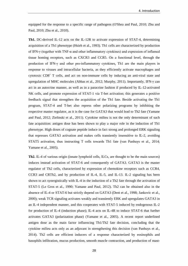
are Th1, Th2, Th17, Th22, Th9, Tfh, and Tregs (Figure 4). Each of these subsets is optimally
4. Introduction
28
equipped for the response to a specific range of pathogens (O'Shea and Paul, 2010; Zhu and
Paul, 2010; Zhu et al., 2010).
Th1. DC-derived IL-12 acts on the IL-12R to activate expression of STAT-4, determining
acquisition of a Th1 phenotype (Hsieh et al., 1993). Th1 cells are characterized by production
of IFN-γ (together with TNF-α and other inflammatory cytokines) and expression of inflamed
tissue homing receptors, such as CXCR3 and CCR5. On a functional level, through the
production of IFN-γ and other pro-inflammatory cytokines, Th1 are the main players in
response to viruses and intracellular bacteria, as they efficiently activate macrophages and
cytotoxic CD8+ T cells, and act on non-immune cells by inducing an anti-viral state and
upregulation of MHC molecules (Abbas et al., 2012; Murphy, 2011). Importantly, IFN-γ can
act in an autocrine manner, as well as in a paracrine fashion if produced by IL-12-activated
NK cells, and promote expression of STAT-1 via T-bet activation; this generates a positive
feedback signal that strengthen the acquisition of the Th1 fate. Beside activating the Th1
program, STAT-4 and T-bet also repress other polarizing programs by inhibiting the
respective master regulator, as it is the case for GATA3 that would lead to Th2 fate (Yamane
and Paul, 2012; Zielinski et al., 2011). Cytokine milieu is not the only determinant of such
fate acquisition: antigen dose has been shown to play a major role in the induction of Th1
phenotype. High doses of cognate peptide induce in fact strong and prolonged ERK signaling
that represses GATA3 activation and makes cells transiently insensitive to IL-2, avoiding
STAT5 activation, thus instructing T cells towards Th1 fate (van Panhuys et al., 2014;
Yamane et al., 2005).
Th2. IL-4 of various origin (innate lymphoid cells, ILCs, are thought to be the main sources)
induces instead activation of STAT-6 and consequently of GATA3. GATA3 is the master
regulator of Th2 cells, characterized by expression of chemokine receptors such as CCR4,
CCR3 and CRTh2, and by production of IL-4, IL-5, and IL-13. IL-2 signaling has been
shown to act synergistically with IL-4 in the induction of a Th2 fate through the activation of
STAT-5 (Le Gros et al., 1990; Yamane and Paul, 2012). Th2 can be obtained also in the
absence of IL-4 or STAT-6 but strictly depend on GATA3 (Dent et al., 1998; Jankovic et al.,
2000); weak TCR signaling activates weakly and transiently ERK and upregulates GATA3 in
an IL-4 independent manner, and this cooperates with STAT-5 induced by endogenous IL-2
for production of IL-4 (induction phase); IL-4 acts on IL-4R to induce STAT-6 that further
activates GATA3 (polarization phase) (Yamane et al., 2005). A recent report underlined
antigen dose as the main factor influencing Th1/Th2 fate decision, concluding that the
cytokine milieu acts only as an adjuvant in strengthening this decision (van Panhuys et al.,
2014). Th2 cells are efficient inducers of a response characterized by eosinophils and
basophils infiltration, mucus production, smooth muscle contraction, and production of mast-

4. Introduction
29
cell-activating IgE; all these effector mechanisms are required to counteract invasion from
multicellular parasites, such as hemints, protect from venom, but also provoke onset of
allergic diseases (Allen and Sutherland, 2014).
Th17. TGF-β, IL-6 and IL-1β (with a few discrepancies observed between the human and the
mouse system) induce STAT-3 (Acosta-Rodriguez et al., 2007a; Bettelli et al., 2006; Chung
et al., 2009; Manel et al., 2008; Mangan et al., 2006; Veldhoen et al., 2006), and ultimately
RORγt (Ivanov et al., 2006), a transcription factor that specifies for the Th17 lineage
(Harrington et al., 2005; Park et al., 2005). IL-21 and IL-23 have been shown to generate an
amplification loop that promotes more efficient Th17 development (Korn et al., 2007;
Nurieva et al., 2007; Volpe et al., 2008; Yang et al., 2008; Zhou et al., 2007). Th17 are
characterized by production of IL17A/IL-17F, IL-22, and GM-CSF and express the mucosal,
CNS, and skin-homing markers CCR6 and CCR4; they have also been reported to express the
NK marker CD161, already at the level of Th17 precursors in the cord blood (Acosta-
Rodriguez et al., 2007a; Acosta-Rodriguez et al., 2007b; Annunziato et al., 2007; Zielinski et
al., 2011). Through the production of IL-17 and IL-22, Th17 promote the recruitment and
activation of neutrophils on the one hand, and act on epithelial cells to induce anti-microbial
peptides, on the other. Th17 are therefore required in case of infection from fungi or
extracellular bacteria (Hernandez-Santos and Gaffen, 2012; LeibundGut-Landmann et al.,
2012).
Th22. A population characterized by the expression of the chemokine receptor CCR6, as well
as skin homing molecules CCR4 and CCR10, and by the capacity to produce IL-22, but not
IL-17, has been recently identified and operatively defined Th22 (Duhen et al., 2009; Trifari
et al., 2009). Polarizing cytokines in this case appear to be IL-6 and TNF, and a fundamental
transcription factor is the xenobiotic receptor aryl-hydrocarbon receptor (AHR). The
functional role of these cells remains to be defined, even though the effect of IL-22 in the
induction of anti-microbial peptides by epithelial cells suggests a role in pathogen control at
the body surface. Interestingly, Th22 have also been shown to contain cells specific for CD1a-
restricted self-antigens (de Jong et al., 2010).
Th9. Cells producing exclusively IL-9, a generally Th2-associated molecule, have also been
identified and defined as Th9. In this case a necessary, though not sufficient, transcription
factor appears to be PU.1, under the control of STAT-6, while defining chemokine receptors
have not been characterized yet (Goswami et al., 2012; Veldhoen et al., 2008). These cells
have been associated to several functions, mainly helminth clearance, and they have been
linked to atopic reactions, tumors, and recently response to skin microbes (Schlapbach et al.,
2014).

4. Introduction
30
TFH. Induction of the transcription factor Bcl-6, probably but not certainly by IL-6 and IL-21,
promotes differentiation of T cells toward TFH fate (Johnston et al., 2009). These cells express
high levels of PD-1, ICOS, and CXCR5, produce IL-21 and are instrumental for mounting an
efficient and high affinity antibody response, as they direct the germinal center reaction
(Crotty, 2011). However, whether this represents a distinct lineage parallel to Th1/2/17, or
rather a functional state of cells belonging to different subsets, remains to be definitively
established (Zhu et al., 2010).
Tregs. Finally, a subset of T cell exist whose role is to dampen the immune response,
protecting from excessive tissue damage during inflammation, as well as from autoimmunity.
These cells have been named regulatory T cells (Tregs), and can be originated through two
distinct pathways. Natural Tregs (nTregs) originate in the thymus, where the driving element
is thought to be the strength of self peptide recognition upon antigen presentation by TECs. In
particular such interaction has been postulated to happen with a strength that is below
threshold for negative selection, but higher than that for non-self specific T lymphocytes. This
would induce tolerization in potentially autoreactive cells and prevent autoimmunity (Klein et
al., 2014). Alternatively, Treg phenotype can be induced in the periphery (iTregs) by antigen
presentation in the absence of costimulatory molecules and inflammatory cytokines (Chen et
al., 2003). Cytokines such as TGF-β and IL-2 have been shown to play an important role in
driving this fate decision by inducing the specifying transcription factors Foxp3 (Fontenot et
al., 2003; Hori et al., 2003). Tregs can dampen the immune response acting through both
release of soluble factors such as TGF-beta, and expression of surface immune-inhibitory
receptors such as CTLA4. It must be acknowledged that to date human in vitro-differentiated
iTregs have failed to show any regulatory activity (Zhu and Paul, 2010).
4. Introduction
31
Figure 4. CD4+ T helper subsets. Upper panel: schematic representation of the T cell priming process: different polarizing cues are delivered by the DC to the naive T cell during antigen presentation, that lead to activation of distinct STAT molecules. These in turn induce expression of lineage-specifying transcription factors (master regulators) and result in acquisition of specific functional properties such as cytokine production. Depicted subsets are widely characterized in the literature (O'Shea and Paul, 2010). Lower panel: updated list containing recently proposed T cell subsets and relative role in immune response as well as in immunopathology (Adapted from (Sallusto and Lanzavecchia, 2009)).

4. Introduction
32
4.1.7. Balancing subsets: when pathology arises from an inappropriate Th
response
Not only the magnitude but also the “flavor” of the response determines the outcome of the
battle between host and pathogen. Striking examples can be found in the literature showing
the importance of an appropriate type of response to reach sterilizing immunity. Most of these
works have proven such concept with respect to the Th1-Th2 dichotomy, as for nearly two
decades these subsets were the only ones known to exist.
A first example is represented by Leishmania major infection. While C57BL/6 mice that are
prone to mount vigorous Th1 response are protected from the infection (through the
production of IFN-γ and the activation of macrophages), BALB/c mice succumb due to their
genetic propensity to generate an IL-4 driven Th2 response (Heinzel et al., 1989).
Importantly, IL-4–/– or IL-4R–/– animals on the same background are largely rescued from
susceptibility to L. major infection, demonstrating that decision on the fate that pathogen-
specific T cells should undertake, regardless of their activation, is of fundamental importance
in terms of response outcome (Noben-Trauth et al., 1999).
On the same line, it was shown that in humans infection with Mycoacterium leprae (the
causative agent of leprosy) is associated with development of the lepromatous form of
disease, which is more aggressive and disseminated, when a Th2 response is predominant. In
contrast, individuals that mount appropriate Th1 response can confine the pathogen and
develop a generally milder form of disease called tuberculoid leprosy (Sieling and Modlin,
1994; Yamamura et al., 1991). Severity of tuberculosis, a diseases provoked by
Mycobacterium tuberculosis, has also been reported to be increased in individuals displaying
cytokine profile skewed toward the production of IL-4/IL-5 rather than IFN-γ (Lindestam
Arlehamn and Sette, 2014).
Early work in different mouse models has shown that susceptibility to Candida albicans can
be associated to a Th2 type of response; IL-4 blockade, as well as pathogen-specific Th1
transfer prior to infection, greatly improves survival of the animals (Romani, 1999). It should
be noted however that these works precede the discovery of the Th17 lineage, and it is thus
likely that all the reported conditions that favored Th1 with respect to Th2 development in
fact provided advantages also to the non-detected Th17 cells, which are now considered to be
the main players in protection against fungi (Hernandez-Santos and Gaffen, 2012).
Human genetic deficiencies offer a unique possibility to study the requirements for protective
immune responses. Seminal work on the importance of IL-17-driven response for C. albicans
clearance or control has been done in the last ten years to understand the immune response of

4. Introduction
33
patients suffering from chronic infection. Patients affected by mutations of different genes can
develop a disease known as chronic mucocutaneous candidiasis (CMC) (Eyerich et al., 2010),
characterized by uncontrolled growth of the fungus on skin, nails and mucosal surfaces. In
particular, loss of function in AIRE (APECED syndrome), IL 17/IL-17R genes, as well as
STAT-3 genes (the latter associated with hyper IgE syndrome, HIES) or gain of function
STAT-1 (STAT-1 GOF), have been implicated in the onset of CMC (de Beaucoudrey et al.,
2008; Liu et al., 2011; Milner et al., 2008; Puel et al., 2011). What all these inborn errors have
in common is that they generate an impairment of the IL-17 axis, eventually allowing C.
albicans overgrowth, and producing a highly invalidating and life-lasting syndrome. It
remains however to be established whether the magnitude of the response is still preserved in
those individuals, and therefore a T cell response is generated having non protective
capabilities (for instance, exaggerated production of IL-4).
The above reported findings underline the importance of regulating not only the magnitude,
but also the type of T cell response that is generated. Conceptually, the activation of an
unwanted type of T cell lineage results in failure of infection control and leads to tissue
pathology.
4. Introduction
34
4.1.8. Flexibility of CD4+ T cells at the clonal and population level
Polarization of effector and memory T cells is considered a remarkably efficient process, as it
consistently leads to the generation of a response that optimally counteracts the inducing
insult. As a matter of fact, indeed, most human beings successfully deal with an enormous
number of potentially harmful microbes every day. However, a growing set of data in the
literature is suggesting a certain degree of flexibility in this system (Ahmadzadeh and Farber,
2002; O'Shea and Paul, 2010; Zielinski et al., 2011).
To understand the magnitude of the heterogeneity possibly achieved by the T cell
compartment, we can analyze the concerning literature by first targeting the single unit of the
immune response, i.e. the clone. Already at this basic level, plenty of possibilities for
generating diversity are associated with T cells. Every single naïve T cell upon stimulation
can undertake several destinies and give rise to phenotypically mixed progenies (“one cell-
multiple fates”), whose averaging accounts for the reproducibility of any pathogen-specific
response. Recent studies have in fact ultimately refuted the “one cell-one fate” model,
according to which each naïve T cell could only give rise to a progeny with homogeneous
characteristics, and the combination of the resulting populations, one for each primed naïve
cells, would account for the overall phenotype of the elicited response (Buchholz et al., 2012).
As already discussed in the previous section, most of the work that has lead to these
conclusions focused on mouse CD8+ lymphocytes, given the narrower range of fates that
associate with this cell type and the availability of dedicated tracking technologies as well as
transgenic models. It must be noted that the main goal of these works was to define
heterogeneity of daughter cells upon priming in terms of effector vs memory development
rather than opposite lineages (Buchholz et al., 2012).
Reiner and colleagues (Chang et al., 2007) showed first that CD8+ T cell asymmetric division
upon Listeria monocytogenes (Lm) infection is involved in the generation of both effector and
memory cells from a single naïve precursor. In particular, being the interaction between DC
and T cell rather prolonged in time, it appeared that, following the first division, one of the
two daughter cells would be proximal to the DC, receive stronger stimulation and be
instructed towards an effector fate. The distal cell, on the contrary, would acquire memory
characteristics. Interestingly, the authors proposed that the mechanism of asymmetric division
could be involved in the generation of different subsets (Th1, Th2, Th17) in the context of a
CD4+ T priming, an hypothesis that to the best of our knowledge has not been investigated in
depth ever since. On the same line, seminal work from Busch and colleagues has followed the
development of congenically marked unexperienced single CD8+ T cells upon transfer into a
naïve host and challenge with Lm (Stemberger et al., 2007). These experiments have shown

4. Introduction
35
that TE, TCM and TEM cells, as defined by the surface expression of CD127 and CD62L, could
be recovered within the progeny of a single CD8+ naïve T cell. Importantly, intra-clonal
heterogeneity was also generated according to IFN-γ, TNF-α, IL-2 production and
degranulation capacity. Shortly after, using a different approach based on genetic labeling (or
barcoding) of individual naïve CD8+ T cells with a retrovirally delivered DNA tag,
Schumacher and colleagues independently confirmed these findings (Gerlach et al., 2010).
A recent work from Lefrançois laboratory identified environmental cues (such as
inflammatory stimuli) as the main trigger for such intra-clonal differentiation, comparing
different infection models, such as VSV and Lm (Plumlee et al., 2013). With respect to CD4+
T cells, single cell transfer approach has been undertaken by Jenkins and colleagues to
determine the intra-clonal diversification patterns upon infection (Tubo et al., 2013). The
conclusions of this study were that different effector fates, such as Th1, TFH and germinal
center (gc) TFH cells (identified on the basis of the expression of transcription factors, surface
markers, cytokine production), could be generated from each naïve precursor. In this
experimental model the relative expansion of each subset was mainly determined by the
nature of the cognate antigen and the resulting bio-physical properties of the TCR-p:MHC
interaction, such as aggregate time. Based on previous work from the same laboratory, as well
as other works showing the importance of signaling strength in the acquisition of TFH
phenotype (Fazilleau et al., 2009; Pepper et al., 2010), the authors proposed that asymmetric
division at the initial stages of stimulation would harbor cellular population with differential
expression of IL-2 receptor. Early signaling through this receptor would induce STAT-5
activation, leading to expression of the transcription factor Blimp-1, that would
simultaneously inhibit Bcl-6 expression, thus preventing TFH fate determination, and induce
expression of IL-12R, leading to STAT-4 signaling and T-bet activation and, therefore, Th1
differentiation. Lack of IL-2 signaling would instead favor ICOS expression and promote TFH
differentiation (Pepper and Jenkins, 2011). However, even if providing valuable insights, this
study did not face the challenge to analyze the generation of completely alternative lineages
during the course of infection, as TFH are currently considered to be rather a differentiation
stage of transient nature, possibly achieved within each subset (Baumjohann et al., 2013;
Yamane and Paul, 2013).
All the above works provide proof of concept that it is possible for a single naïve T cell to
differentiate towards distinct fates. Asymmetric division, environmental cues, strength of
stimulation, number of re-encounters between T cell and DC, have been shown to impact on
the yield of effector vs memory population, and it is therefore likely that such a variety of
factors can modify also the cytokine profile of the primed T cells. Possibly the high efficiency
of fate decision process represents a limiting factor to observe secondary products of the

4. Introduction
36
polarization (i.e., in strongly Th1-favoring priming conditions it will be more difficult to
retrieve any generated Th2 cells). As a matter of fact, there are no reports available in the
literature investigating the possibility that alternative lineages, such as Th1, Th2, or Th17, are
generated from a single cell upon infection.
Interestingly, a recent report identifies through TCR sequencing a certain level of clonotypic
sharing among different humans CD4+ T cell compartments as determined by the expression
of surface chemokine receptors (Wang et al., 2010a). The possibility that heterogeneity takes
place in humans at an intra-clonal level is intriguing; however in the above-mentioned study
the complete lack of information on the antigenic specificity as well as on the phenotype of
the supposedly shared clonotypes do not allow validation for such hypothesis.
Several mechanisms can also be identified that contribute to generate diversity during priming
of response to a pathogen at a multi-cellular level, meaning driving forces that will act
differentially on distinct clonotypes and produce heterogeneous mixes of homogeneous
progenies. First, we identify a primary role for antigen intrinsic properties (sequence,
abundance, etc.) in promoting this process. As already mentioned, stimulation by different
antigens held different ratios among Th1, TFH and gcTFH (Tubo et al., 2013), based on the
aggregate time of TCR stimulation they could promote. This appears to be possible even in
the contest of a single microbe. In fact, Newell and colleagues recently showed that human
CD8+ T cells primed by a virus (CMV, EBV, or Influenza virus) and having the same fine
specificity, as assessed by pMHC-tetramer binding, display enormous differences in terms of
phenotype and cytokine production, creating almost a continuum among all possible
parameter combinations (Newell et al., 2012). In a consecutive paper, the authors described
different phenotype for CD8+ T cells specific to different Rotavirus antigens, that vary in
expression of the trafficking receptors integrin β7 and αE (Newell et al., 2013). Antigen dose
is also a valuable factor in the induction of Th1 vs Th2 fate on T cells (high dose favoring the
first), that was recently shown to act upstream the cytokine signaling, and thus proposed to
occupy a higher hierarchical position in fate determination. This suggests that mixed Th1-Th2
populations could be generated upon priming depending on the availability of each antigen, as
some proteins are produced by the microbe at much higher rate than others (van Panhuys et
al., 2014).
On a second level, it is well established that each pathogen bears a multitude of different
PAMPs, that can potentially drive differential polarizing programs on DCs (Pulendran, 2005).
In the case of C. albicans, β-glucans or α-mannans recognition by Dectin-1 and Dectin-2,
respectively, activate a Th17-polarizing program (LeibundGut-Landmann et al., 2007; Saijo
et al., 2010), TLR4-mediated recognition of O-linked mannans is believed to induce Th1

4. Introduction
37
differentiation through production of type I IFNs or TNF-α (Netea et al., 2008), while TLR2
triggering has been linked to Treg/Th2 induction (Netea et al., 2008). Fungal chitin has also
been proven to promote Th2-skewing conditions (Van Dyken et al., 2014), underlining the
concept that a single microbe can induce multiple polarizing programs in DCs-T cells. The
scenario becomes even more complex if considering that C. albicans, which is a dimorphic
fungus, is differentially recognized by the innate immune system in its yeast vs filamentous
form (Acosta-Rodriguez et al., 2007b; d'Ostiani et al., 2000).
Third, the priming milieu is supposed to play a fundamental role in dictating fate-determining
signals. Pathogens can be encountered at different locations, each constituted by a peculiar
microbiological and immunological environment. The route of infection is indeed well known
to influence T cell phenotype, as shown for intravenous vs intranasal Lm infection, inducing
Th1 and Th17 responses respectively (Pepper et al., 2010). Besides, the site of entry is well
known to strongly influence the migratory capacities of primed cells (Sallusto and
Lanzavecchia, 2009). Fungi such as C. albicans are widely spread on the surfaces and
mucosal tissues of the human body (LeibundGut-Landmann et al., 2012; Underhill and Iliev,
2014); it is presumable that encounter with the microbe in the oral mucosa, in the vagina, in
the gut or in the skin will generate distinct types of response and potentially target different
antigens, as the protein profile of this fungus differs at each anatomical sites, based on
characteristics of the environment, such as microbiota, pH, and osmolarity (Hube, 2004).
On the same line, the origin of the APC has also to be taken in account when considering T
cell fate heterogeneity. Just considering DCs, that are widely recognized as main inducers of
T cell responses, many reports have demonstrated such phenomenon. For instance, splenic
CD8α+ DCs are far better inducer of Th1 responses than CD8α– DCs, due to their increased
production of IL-12 (Pulendran, 2005); CD301b+ DCs are fundamental for priming of Th2
response in vivo (Gao et al., 2013; Kumamoto et al., 2013); human inflammatory DCs and
monocytes have been shown to efficiently induce Th17 response in vitro (Acosta-Rodriguez
et al., 2007a; Segura et al., 2013; Zielinski et al., 2012). The functional status of DCs adds a
further variable to the scenario, as monocyte-derived DCs that efficiently prime Th1
responses shortly after maturation, preferentially induce Th2 polarization overtime when
exhausted (Langenkamp et al., 2000).
The phenotypic flexibility of the T cell memory pool, both within a population and at a clonal
level, is possibly a major factor in generating heterogeneity (Figure 5). This issue becomes
particularly relevant in the human system, due to the life span of human beings and the
resulting high level of exposure to completely different immunostimulatory conditions - such
as infections, allergies, and vaccinations. Plasticity is a feature that has been reported in many

4. Introduction
38
experimental systems, in vitro and in vivo, for CD4+ T cells, at all stages of their life cycle;
yet it is unclear the extent of such phenomenon in the course of a normal response to a
pathogen or a vaccine and what its importance could be in generating protective immunity.
For example, inducing T cells with a broad migratory capacity to different peripheral tissues
could in principle be beneficial to ensure a higher level of protection, and would potentially
represent a key factor to design effective vaccines (Lees and Farber, 2010; Yamane and Paul,
2012). Besides, plasticity represents an important adaptive mechanism for memory cells to
cross-react to pathogens different from those that elicited their priming, given the high
relevance of antigenic sharing between different microbes, and this phenomenon is supposed
to play an important role in elderly individuals, whose naïve repertoire can be dramatically
reduced (Yamane and Paul, 2012).
Cross-reactivity has recently been described as an extremely common feature of T cell, as
underlined by elegant work from Garcia and colleagues (Birnbaum et al., 2014): it is therefore
conceivable that most T clones will be stimulated by different antigens (in a different milieu)
during the course of their lives. Indeed we found that T cell clones specific C. albicans can
react to conidia from up to 8 different Candida species, each generating a peculiar
inflammatory environment (Mele F. et al., unpublished).
Memory T cells can undergo recall response in different conditions upon antigen reencounter,
for instance due to co-infection or different anatomical site of entry of the pathogen, and
therefore modify their phenotypic characteristics. This issue is particularly relevant for those
memory T cells that present a non-polarized phenotype, such as TCM cells (Rivino et al.,
2004). Differences in tonic signaling can induce variability among resting memory T cells, as
some cells may get more frequent or stronger stimulation than others, leading to phenotypic
changes during homeostasis (Lees and Farber, 2010).
Plasticity of effector and memory T cells has been investigated in a variety of experimental
models, trying to interconnect all different T cell lineages. IFN-γ production could be
acquired in vivo by Th2 cells in several mouse models, for instance upon LCMV infection
(Hegazy et al., 2010; Krawczyk et al., 2007). Th1-Th2 plasticity has been shown ex vivo for
human memory cells, upon restimulation under opposite polarizing conditions (Messi et al.,
2003): most instructed T cells retain in fact an epigenetic state of accessibility in genes
associated with other lineages (cytokines, chemokines, surface receptors) (Wei et al., 2009).
Th17 is considered to be an extremely plastic lineage, and Th17 cells easily gained IFN-γ
production in a variety of experimental models. Recent fate mapping or cell transfer studies
showed that in vivo Th17 cells can switch their phenotype to become Th1, or just acquire
production of IFN-γ (Bending et al., 2009; Hirota et al., 2011; Lee et al., 2009). The same has
4. Introduction
39
been reported in ex vivo studies on human cells, demonstrating that IL-12 promotes
conversion of Th17 to Th1 (Annunziato et al., 2007). Interestingly, spectratyping analysis
performed in patients suffering from juvenile idiopathic arthritis (JIA) identified Th17-
derived CCR6+CD161+ Th1 in synovial fluid, thus supporting a model of high plasticity
between these compartments, especially under strong inflammatory conditions (Cosmi et al.,
2011).
Th17-Th2 plasticity has been reported as well, with the identification of an IL-17/IL-4-double
producing T cell population in patients with atopic asthma. The pathogenic potential of these
cells was proven in vivo in a mouse model of asthma; in vitro experiments have suggested that
these cells derive from Th17 cells that were restimulated in allergic environment (i.e. in the
presence of IL-4) (Cosmi et al., 2010; Wang et al., 2010b). The T cell response induced by
vaccination, characterized by highly diverse cytokine production and homing receptor
display, is a striking example of heterogeneity generated in response to a single protein and
under defined conditions (Livingston et al., 2013; Rivino et al., 2004). However, the impact of
this variety on the long-lasting protection resulting from vaccination has never been
considered, most likely due to the fact that protection relies on humoral immunity.
Overall, the analysis of the literature concerning naïve and memory Th fate decision suggests
that the repertoire of T cells specific to a given microbe or vaccine could be highly
heterogeneous, and comprise cells endowed with different homing properties and effector
functions.

4. Introduction
40
Figure 5. An integrated model for CD4+ T cell subset imprinting and flexibility. Shown is a schematic representation of T
cell fate decision upon priming. Dashed arrows represent phenotypic switches that have been reported to occur in experimental
models (from O'Shea and Paul, 2010).

4. Introduction
41
4.1.9. T cell repertoire: facts and open questions
In total, approximately 1011-1012 αβ T cells, of which at least 2.5x10¹¹ CD4+ and 1x10¹¹ CD8+,
are believed to reside in a human being (Bains et al., 2009a; Clark et al., 1999). Circulating αβ
T cell in humans have been estimated to be in the range of 1010, and are generally considered
to represent 2% of the total T cell population throughout the literature. The vast majority of T
cells (estimated as 2.24x10¹¹ in 5 donors (Clark et al., 1999)) have been shown to reside in
lymphoid organs, but most peripheral organs are widely populated by these lymphocytes;
recently, the remarkable amount of 2x10¹⁰ T cells were calculated to be in human skin, which
represents twice as much the amount of T cells in the blood (Clark et al., 2012).
The thymic output of naïve cells decreases overtime, being maximal around 1 year after birth
(2x10⁹ CD4 naive/day), and reaching values of about 3x10⁸/day at the age of 20, 108/day at
the age of 30, and so forth (Bains et al., 2009a; Bains et al., 2009b). Thymic output, however,
is not the only factor contributing to the homeostasis of the naïve pool, as in humans this also
largely depends on peripheral T cell division; competition for IL-7 and availability of self
peptide-MHC providing tonic signals are crucial in this regard (Lees and Farber, 2010). On
this topic the literature shows several discrepancies, mainly attributable to differences in the
adopted experimental approach and mathematical model. McLean and Michie (McLean and
Michie, 1995) suggested that naïve T cells divide on average once every 3.5 years and die
after 20 years, which makes an average of 6 post-thymic divisions for each naïve T cell.
Others have suggested higher proliferation rates: based on telomere length reduction
(estimated in 50-100 bp per cycle), 7-13 division were estimated during the 20 years life of a
naïve T cell (Weng et al., 1995); De Boer and colleagues considered instead only 35-70bp to
be lost during each division, which would result in 10-19 cycles (De Boer and Noest, 1998).
Elsewhere, the rate of cycling naïve T cells was found to approximate 0.8%, which would
suggest 1 division every 125 days, for a total of 60 cycles during a T cell life span
(Sachsenberg et al., 1998). However, most of these works did not take in consideration the
fact, recently reported, that mean interdivision time of naive cells proportionally increases
over time, becoming extremely lengthened in elderly individuals (Bains et al., 2009a).
Surprisingly, according to this work, T cell proliferation in periphery would contribute more
than thymic output to the maintenance of the naïve pool, as the authors calculated that up to
the age of 20 only a maximum of 30% of new naïve appearing in the periphery can be derived
from thymic output.
The number of T cell receptors potentially generated by V(D)J recombination has been
estimated to be between 1016-1020 (Zarnitsyna et al., 2013). Even considering as correct the

4. Introduction
42
lower boundary of such estimate, the actual number of different specificities represented by
the T cell compartment appears to be necessarily several orders of magnitude smaller, as T
cells in the body are 1012 at maximum. Arstila and colleagues estimated that 106 different β
chains would be borne by circulating T cells, of which only 1-2x10⁵ carried by memory T
cells (Arstila, 1999). Each β chain is believed to be paired with approximately 25 different α
chains in the naïve repertoire (given the fact that pre-T cells, after rearrangement of the β
locus, expand massively and generate up to 103 daughter cells that independently rearrange
the α locus, but mostly die afterwards due to neglect), and with only one α chain in the
memory compartment. Given these numbers the authors calculated that the repertoire of
circulating T cells is composed of at least 2.5 x 10⁷ different TCRs.
By generating a novel high throughput approach to in depth sequence αβ TCR, Robins and
collaborators (Robins et al., 2009) calculated that 107 different Vβ chains are borne by
circulating lymphocytes, thus suggesting a repertoire of approximately 2.5 x 10⁸ different
TCRs if the pairing of each β with 25 α chains holds true. The authors further estimated in 1.4
x 106 αβ TCRs the repertoire of memory cells. The level of diversity would then result 4 and
10 times higher than previously thought in the overall and in the memory T cell compartment,
respectively. These values were elegantly extrapolated making use of the “unseen species
problem” approach, which is commonly used in ecology to estimate the number of total
species in large populations from diversity measurements in random finite samples.
Interestingly, up to 400 naïve clones bearing an identical Vβ chain were found in a 5 ml blood
sample; if we assume a ratio 1:1 between CD4+ and CD8+ lymphocytes in the blood, and an
average pairing with 25 different α chains, it can be extrapolated that up to 200/25 = 8 naïve
cells bear an identical TCR within the sample, which would imply a single naïve being
possibly present in up to 800 copies in the average 5 liters of blood of a human being. A
surprisingly similar value can be obtained by analogous extrapolation from Arstila’s work:
every T cell clone would have an average copy number of 1x10³ (0.5x10¹¹ circulating CD4/2
naïve vs memory/2.5x107 αβ TCRs). The fact that several hundreds of naïve T cells bearing
an identical TCR can be present at any time in an individual has important implications for
the generation of a protective memory pool and could, at least partially, contribute to the
production of intra-clonal diversity.
Robins and colleagues further observed that sequences that more closely resemble the
germline had higher frequency. This finding was confirmed in a detailed analysis by Venturi
and colleagues (Venturi et al., 2011), pinpointing that higher frequency clonotypes are more
likely to be shared between naïve and memory pools and among different individuals; the fact
that elevated frequencies are associated with high efficiency of V(D)J recombination indicates

4. Introduction
43
that some TCRs are created multiple times during thymic development and ultimately that
convergent recombination shapes the TCR repertoire.
How many different cells can recognize an antigen? By making use of the T cell library
technique, that allows frequency estimation for T cells specific to naturally processed
antigens, 5-170/106 circulating naïve CD4+ T cells were found to recognize any given antigen
in human beings (Geiger et al., 2009). Memory T cells with the same specificity showed a 10-
1,000-fold increase within individual donors. The frequency of specific-CD4+ memory T cell
was as high as 10,000/106 for complex antigens such as CMV (Geiger et al., 2009). The
frequency of T cells specific to a single antigenic protein, such as TT, ranged from 40/106 in
the naïve compartment to 600-900/106 (up to 4,000/106) in the memory pool, depending on
the donor and the memory compartment considered (Campion et al., 2014; Geiger et al.,
2009). Kwok and collaborators, on the same line, calculated the frequency of human naïve T
cells specific to different epitopes to be of 2-100/105 cells, and increase upon immunization
with the same epitope about 100 folds; importantly, following vaccination, epitope specific
cells that were already over-represented in the naïve repertoire expanded the most, similarly
to what observed by Moon and collaborators in the mouse system (Kwok et al., 2012; Moon
et al., 2007).
In mice 5-8x10⁵ β chains, each pairing with 2.4 α chains on average, have been calculated,
achieving an overall repertoire of 2x10⁶ diverse TCRs (Casrouge et al., 2000). Early estimates
instead postulated the presence of 10 cells/clone/spleen, and, if these numbers held true, the
repertoire would be of 2x10⁷ clonotypes, impressively similar to that of human beings, despite
the remarkable difference in size between the two organisms. In most mice, however, every
single TCR is considered to be found in single copy, and for any given epitope a range of 10-
1000 cells per animal have been calculated to be specific using different sampling approaches.
The most recent and finest strategy, based on pMHC tetramer-enrichment and sorting,
allowed detection of 20-200 epitope-specific CD4+ T cells per mouse, all of which carried a
different TCR (Moon et al., 2007). Shortly after, the same approach was used to calculate the
number of CD8+ T cells specific to different pMHC I complexes, which were found to be in
the range of 80-1200 per mouse, depending on the epitope (Obar et al., 2008). As already
mentioned, the frequency of specific naïve precursors was found to be predictive for the
expansion of the deriving memory pool.
Having revised the basic quantitative aspects of the human TCR repertoire a question arises:
how can <108 TCRs possibly provide effective immunity against an array of >1015 distinct
pMHC complexes? Recent reports that have identified cross-reactivity as one of the major – if
not the main – factors to allow coverage broadening of the T cell repertoire. Wooldridge and

4. Introduction
44
collaborators generated a library of almost 1013 decamer peptides, which were randomly
sampled and tested for recognition by a single CD8+ T cell clone (Wooldridge et al 2012).
Using this approach, the authors could prove that a given TCR can respond to >106 different
peptides, many of which trigger activation even more efficiently (up to 100-fold) than the
natural ligand (Wooldridge et al., 2012). On the same line, a recent work from Garcia lab
(Birnbaum et al., 2014) took advantage of a yeast display library to discover p:MHC ligands
for a given human or mouse TCR. Following identification of hundreds of peptides that
would bind the studied TCR, the authors generated algorithms to predict the sequence of
peptides that would be recognized: production and testing of such ligands proved their
predictions to be accurate (94% of predicted ligands triggered T cell activation). Importantly,
the authors identified naturally occurring sources for peptides with the predicted
characteristics, thus providing compelling evidence that cross-reactivity is not only possible
but also likely to be commonly exploited by T cells during immune responses.

4. Introduction
45
4.2. Pathogens and vaccines object of this study
4.2.1. Candida albicans
C. albicans is a dimorphic fungus that resides as a commensal organism in the gastrointestinal
tract, on the genital mucosa, and on the skin of healthy individuals. Occasionally, overgrowth
at such locations results in tissue damage and invasion; C. albicans is thus considered an
opportunistic pathogen (Conti and Gaffen, 2010).
Infection by C. albicans are generally mild and concern mainly the GI tract and vaginal
mucosa; vulvo-vaginal (VVC) candidiasis, in particular, affects up to 75% of reproductive-
age women at least once, and becomes recurrent (RVVC, with several episodes per year) in 5-
10% of them (Cassone, 2013). Importantly C. albicans represents a major threat for
immunocompromised individuals, such as HIV+ individuals, transplant recipients, and
chemotherapy-treated patients, in which both mucosal and systemic infections are frequent.
Candida genus ranks among the most common microbial causes of nosocomial bloodstream
infections, with a mortality rate of approximately 40%. Although C. albicans is the most
clinically relevant member of the genus, it is in fact worth to acknowledge that other species
diffusely cause pathology, mainly in health care unit patients, in particular C. glabrata, C.
krusei, C. parasilosis and C. tropicalis (Richardson and Rautemaa, 2009). Virulence factors
of the fungus include surface adhesion and biofilm generation, invasion of the tissue through
filamentous growth, secretion of hydrolytic enzymes (Richardson and Rautemaa, 2009).
C. albicans has a complex structure and expresses a broad variety of surface molecules,
mainly proteins and carbohydrates. This determines a very articulated interaction with the
immune system that has been widely characterized and described (LeibundGut-Landmann et
al., 2012; Netea et al., 2008). The cell wall, that resides outside the cellular membrane,
shaping and protecting the fungal particle, represents the most relevant compartment in terms
of immune recognition and activation. It is organized in three main layers: an inner shelf
composed by chitin (β-(1,4)-linked polymer of N-acetylglucosamine (GlcNAc)); a core layer
of β-(1,3) and, in minor amount, β-(1,6) glucans; an outer layer of highly mannosylated GPI
anchored proteins (mannoprotein) (Chaffin et al., 1998; Hernandez-Santos and Gaffen, 2012;
Netea et al., 2008).
Recognition of C. albicans and activation of the immune system proceeds through different,
sometimes synergizing, pathways, and involves all the major known classes of pattern-
recognition receptors, as TOLL-like receptors (TLRs), NOD-like receptors (NLRs), C-type
lectin receptors (CLRs). Acting on these receptors, different PAMPs have been shown to

4. Introduction
46
induce different types of T helper response, some of which deleterious to the clearance of the
fungus (Figure 6).
A variety of TLRs can recognize component of C. albicans cell-wall associated molecules:
TLR4 recognizes O-linked mannan, while TLR2/1 and TLR6/2 heterodimers recognize
phospholipomannans and β-glucans. TLR4 is considered the most important TLR involved in
Candida recognition, and it was shown to induce the production of type I IFNs as well as
TNF-α, eventually inducing Th1 responses (LeibundGut-Landmann et al., 2012; Netea et al.,
2008). At the intracellular level, C. albicans DNA can be recognized by TLR9, which
localizes in the phagosomal membrane. Even though most TLR-deficient mice show in
general an increased susceptibility to C. albicans infection, the fact that patients with MyD88
deficiencies do not display augmented occurrence of fungal diseases, argues against a major
role for TLR-mediated signaling in the response to the fungus. Interestingly, TLR2 pathway
seems to play a detrimental role in the response to Candida, as it has been linked to
production of anti-inflammatory cytokines and induction of regulatory (and possibly Th2)
phenotype in T cells; consistently, TLR2–/– mice showed increased survival in a model of
systemic Candida infection (Netea et al., 2004). This finding well exemplifies the complexity
and variety of polarizing programs that such a pathogen can activate.
NLRs have also been strongly implicated in anti-Candida response, as NLRP3 activation and
association with the adaptor protein ASC (to form the scaffold for the NLRP3 inflammasome)
is crucial for production and release of the mature form of IL-1β, a key cytokine in response
to fungi (Brown, 2011; Zielinski et al., 2012). Accordingly, ASC and NLRP3 knockout
animals showed dramatically increased susceptibility to both systemic and mucosal Candida
infections (Gross et al., 2009; LeibundGut-Landmann et al., 2012).
CLRs are widely considered to be the key receptors in the initiation of the response to C.
albicans, given their ability to induce T cell polarization toward the protective Th1 and Th17
fates. Dectin-1 and Dectin-2 recognize β-glucans and α-mannans, respectively, while α-
mannose is recognized by Mincle, another member of the family (Osorio and Reis e Sousa,
2011). Of note, β-glucans are generally hidden by the mannoprotein layer of the cell wall, and
become exposed only on scars that are left on the mother cell after separation from the bud
(bud scars) or revealed during the process of hyphal transition (Hernandez-Santos and Gaffen,
2012; Netea et al., 2008). Dectin-1 can transduce signals thanks to the presence of ITAM
motifs in its intracellular portion, Dectin-2 and Mincle, instead, activate intracellular cascades
by coupling with the FcγR (Osorio and Reis e Sousa, 2011). All these molecules activate the
downstream kinases Syk and PKCδ and the adaptor protein CARD9, leading to activation of
NFκB, NFAT, and NLRP3 inflammasome; as a result pro-inflammatory cytokines such as IL-

4. Introduction
47
1β, IL-6, IL-23, and TNF-α, are released by DCs, which induce primarily Th17 and Th1
responses (LeibundGut-Landmann et al., 2007; Robinson et al., 2009; Saijo et al., 2010;
Strasser et al., 2012). The crucial role that such receptors exert in the immune response to
Candida is clearly illustrated by studies performed on mouse models, where the non-
redundant downstream molecules on these pathways were knocked out (Gross et al., 2006;
Gross et al., 2009; LeibundGut-Landmann et al., 2007; Strasser et al., 2012), as well as by
epidemiological evidence that patients with mutations in Dectin-1 or CARD9 genes develop
invasive mucocutaneous candidiasis (Ferwerda et al., 2009; Glocker et al., 2009).
Mannose receptor has also been reported to recognize α-mannans and induce IL-1β and TNF-
α production in macrphages, thus leading to Th17 polarization (van de Veerdonk et al., 2009).
Chitin represents a major component of the cell wall of C. albicans and can actively be
recognized by the immune system. Recently, it was reported that chitin induces IL-10
production in APCs via activation of mannose receptor, NOD2 and TLR9 (Wagener et al.,
2014), resulting in dampening of the inflammation. Interestingly, chitin has been linked to
induction of allergy and asthma in a variety of models, due to the potent activation of type 2
ILCs via epithelial production of IL-25, IL-33 and TSLP when delivered to the lungs, which
counteracts the development of a Th17 response (Van Dyken et al., 2014); these findings
further underline the heterogeneity of polarizing signals possibly induced by a complex
pathogen such as C. albicans.
Protective immunity to C. albicans is believed to be mediated mainly by T cell response.
Early studies identified Th1 response as protective and Th2 as deleterious for the clearance of
the pathogen in a variety of models, such as gastric, oropharingeal and systemic infection
(Cenci et al., 1995; Romani, 1999; Romani et al., 1991; Tonnetti et al., 1995). However, it
appeared that mice deficient for IL-12 p40 subunit or IL-12R were more susceptible than
mice deficient for IFN-γ to C. albicans infection. This discrepancy was resolved when IL-23,
a cytokine that has a common sub-unit to IL-12 (p40), as well as a common chain in their
respective receptors (IL-12Rβ1), was identified. IL-23 plays in fact a fundamental role in the
maintenance of Th17 cells (Hernandez-Santos and Gaffen, 2012). Most part of the literature is
in agreement with the concept that Th17 cells have a protective role in candidiasis; this has
been tested in a variety of models of infection (systemic, oral, cutaneous, vaginal)
(Hernandez-Santos and Gaffen, 2012). The fundamental role of the IL-17 axis in Candida
containment is strongly supported by the association between chronic infections and genetic
deficiencies that impair this developmental path in humans. Genetic defects in IL-17 or IL-17
receptor genes, or mutations that impair STAT-1 or STAT-3 function (e.g., STAT-1 GOF),
AIRE mutations leading to APECED syndrome with production of anti-IL17 and anti-IL-22
antibodies, as well as defects in Dectin-1 or CARD9, promote the onset of CMC (Ferwerda et

4. Introduction
48
al., 2009; Glocker et al., 2009; Liu et al., 2011; Milner et al., 2008; Puel et al., 2011; Puel et
al., 2010; van de Veerdonk et al., 2011). Acute and systemic infections tend instead to be
associated with neutropenia or granulocyte-impairing defects (Hernandez-Santos and Gaffen,
2012).
IL-17 acts through several mechanisms to fight fungal infections. Firstly, it promotes
granulopoiesis, neutrophil chemotaxis (by inducing CXCL1-2-5-8 and G-CSF production in
the tissue) and activation (by enhancing myeloperoxidase and NADPH oxidase activity for
ROS production) (Hernandez-Santos and Gaffen, 2012; LeibundGut-Landmann et al., 2012).
Neutrophils are believed to be the most effective cell type in fungal clearance, as they are
endowed with an extremely potent and dedicated enzymatic machinery and can produce
extracellular nets that trap and kill both yeast and hyphal forms of the fungus (Urban et al.,
2006). Secondly, IL-17 together with IL-22 induces production of a plethora of antimicrobial
peptides, such as S100 and β-defensin 2 (Gorr 2009, Liang 2006). A novel mechanism of
action of IL-17 that has been elucidated is the induction of functional competence in NK cells
in a model of systemic candidiasis; only upon exposure to IL-17 these cells become capable
of producing GM-CSF and activating neutrophils, leading to control of the infection (Bar et
al., 2014). It is however not clear what the source of IL-17 is in this and in other experimental
models; in this regard, it has to be acknowledged that a growing amount of reports in the
literature support the role of cell types other than Th17 in the generation of a robust IL-17
axis against C. albicans, such as type 3 ILCs or γδ T cells (Gladiator and LeibundGut-
Landmann, 2013).

4. Introduction
49
Figure 6. An integrated model for C. albicans innate recognition and T cell polarization. Schematic representation of the
innate pathways that can be activated upon recognition of C. albicans associated PAMPs. T cell phenotypes reported to be
generated upon selective triggering of a certain receptor are depicted below. Of note, intracellular TLRs and NLRs are not
indicated in the figure, still were shown to be involved in the process of C. albicans recognition and immune activation (adapted
from (Netea et al., 2008)).
4. Introduction
50
4.2.2. Mycobacterium tuberculosis
The genus Mycobacterium comprises more than 50 species, most of which are non-
pathogenic environmental bacteria closely related to the soil bacteria Streptomyces and
Actinomyces (Cosma et al., 2003). Among those, M. tuberculosis and a few other species are
the etiological agents of tuberculosis (TB), one of the most devastating infectious disease
worldwide, accounting for more than 2 million deaths and 9 million new infections every year
(Andersen and Kaufmann, 2014).
The pathogen enters through the lungs and is generally taken up by macrophages, where it can
survive by preventing fusion of the phagosome with lysosomes. The incapacity to kill the
bacterium leads to a containment strategy that implicates the formation of granulomas,
compact, organized aggregate of mature macrophages. Overtime, macrophages tend to
transform into epithelioid cells, and other cell types, such as neutrophils, DCs, B and T cells,
NK cells, and fibroblasts, are recruited to the granuloma (Ramakrishnan, 2012). Through this
mechanism most infected individuals can control the spread of the bacterium for a life-long
period (a status defined as LTBI, latent TB infection), and only approximately 5-10% fail to
control it and ultimately develop TB disease. Disturbances in the granuloma results in
exaggerated cell death and causes necrosis, thus leading to the formation of a caseous
granuloma that fails to constrain M. tuberculosis and, on the contrary, promotes its growth
and expansion (Andersen and Kaufmann, 2014).
A vaccine, constituted by the Bacille Calmette–Guérin (BCG) strain, is available since
decades and covers nowadays 80% of all infants in the world, making it the overall most
widely used vaccine. Unfortunately, this protects against serious forms of disseminated TB,
but fails to protect against the most prevalent form of this disease, pulmonary TB, and has
therefore a minor impact on TB incidences (Andersen and Doherty, 2005). The BCG is an
attenuated strain of M. bovis, the pathogen responsible for TB in cattle, and was obtained by
continuous in vitro passages for 13 years: this process led to loss of virulence factors, such as
ESAT6 and CFP10. The protection given by BCG vaccination is not life-long, and most
studies have reported that it lasts for 10–20 years (Andersen and Kaufmann, 2014).
As already mentioned the Mycobacterium genus comprises many closely related species,
mostly non-pathogenic soil saprophytes, referred to as environmental mycobacteria. These
bacteria share several features with their pathogenic counterparts, such as a lipid-rich outer
cell wall, genomes with a high GC content, and many similar gene families, including some
of the major immunodominant antigens (such as the Ag85 complex); similarity of antigens
induces frequent cross-reactivity, and it has been postulated that pre-exposure to common
4. Introduction
51
environmental bacteria strongly reduces the efficacy of BCG vaccination and actually
represents a cause for its failure (Andersen and Doherty, 2005).
Immunity against M. tuberculosis is cell-mediated, with T lymphocytes serving as
orchestrators and mononuclear phagocytes as effectors of both protection and pathology. Due
to the intracellular lifestyle of M. tuberculosis, immunity relies on a successful T cell response
against a repertoire of antigenic targets. Th1-type response is believed to be fundamental, as
production of IFN-γ and TNF-α promotes recruitment and activation of anti-mycobacterial
activities in macrophages; consistent with this notion, individuals with genetic defects in the
IFN-γ receptor show an increased susceptibility to mycobacteria (Lindestam Arlehamn et al.,
2013; Lindestam Arlehamn and Sette, 2014). Recently, the response to M. tuberculosis was
shown to be contained in a subset of Th1 cells expressing CCR6 and CXCR3 and producing
high quantity of IFN-γ and modest amounts of IL-17 (Lindestam Arlehamn et al., 2013). IL-
17, TNF-α, GM-CSF, and IL-1β, together with IFN-γ, have also been found to be important
for protection. Instead, several studies indicate that lower IFN-γ/IL-4 or IFN-γ/IL-5 ratios are
found in active TB patients compared to LTBI individuals. T cells other than CD4, namely
CD8, γδ and CD1-restricted T cells, also play an important role in pathogen containment
(Andersen and Kaufmann, 2014; Khader and Cooper, 2008; Lindestam Arlehamn et al., 2013;
Lindestam Arlehamn and Sette, 2014).

4. Introduction
52
4.2.3. Tetanus toxoid vaccine
Clostridium tetani is a gram-positive anaerobic bacterium whose spores are widely distributed
in soil and in the intestines and feces of many animals, such as of horses, sheep, dogs, cats,
and chickens. It usually enters the body through a wound, and in the presence of anaerobic
conditions, the spores germinate (Hatheway, 1990). Toxins, tetanolysin and tetanospasmin,
are produced and diffused systemically through tissue and lymph. Tetanospasmin is
constituted by two proteic subunits linked by a disulfide bond of 100 and 50 KDa,
respectively, and is one of the most potent known toxins, with an estimated minimum human
lethal dose of 2.5 ng per kg of body weight. It acts on the central nervous system, by blocking
the release of inhibitory mediators at the synapse of the spinal cord motoneurons, leading to
unopposed muscle contraction and spasm (Hatheway, 1990) (Centers for Disease Control and
Prevention, 2014). The World Health Organization (WHO) reported 15,516 worldwide cases
of tetanus in 2005, and an estimated 290,000 deaths between 2000 and 2003; most of these
cases occurred in newborn infants in developing countries. Nonetheless, vaccination against
tetanus represents one of the major successes in the history of active immunization (World
Health Organization, 2014).
The vaccine is formulated as tetanus toxoid (TT, the formaldehyde-inactivated preparation of
neurotoxin tetanospasmin) adsorbed on alum salts and delivered intramuscularly. In western
countries, 3-4 doses of the TT vaccine are recommended at infancy (2/4/6/18 months,
generally in combination with the diphtheria/pertussis vaccine), followed by a TT-containing
booster at school-entry age (4-7 years), and one in adolescence (12-15 years). A recall shot
every approximately 10 years is afterwards required to ensure protection; a complete TT
vaccine shot series has a clinical efficacy of virtually 100%. No cases of tetanus were
reported in 2013, neither in Switzerland nor in the US (Centers for Disease Control and
Prevention, 2014; World Health Organization, 2014).
Several mechanisms have been proposed that account for alum potency as an adjuvant, a topic
that has raised many controversies in the literature. A recent work has proven that the most
important mechanism of action is the recruitment of inflammatory monocytes to the site of
injection (i.e. the muscle) following a gradient of CCL2 released by cells of the tissue. Upon
antigen uptake, monocytes migrate to the lymph nodes while differentiating into DCs, and
efficiently prime T cell response (Kool et al., 2008a; Kool et al., 2008b). Furthermore,
enhanced uptake of the particulate antigen and targeting of the antigen to more acidic
organelles are also supposed to enhance antigen presentation. It has also been shown that
intramuscolar injection of antigen in alum salts results in formation of a depot from which the
antigen is slowly released, increasing immunogenicity (Kool et al., 2012).

4. Introduction
53
On a molecular level, the action of alum seems to be dependent mainly on the NLRP3
inflammasome, that is activated both by direct sensing of alum itself, and indirectly, through
release of DAMPs such as ATP and uric acid by the necrotic tissue (uptake of salt crystals
damages lysosomes and induce cell death) (Kool et al., 2012). The induction of a preferential
Th2 response has been linked to production of several factors by APCs as well as to
recruitment of eosinophils, an early source of IL-4, to the site of injection (Jordan et al., 2004;
Kool et al., 2012).
Vaccination-induced protection relies on antibody-mediated neutralization of the toxin. In
fact, protection is estimated using a threshold titer of more than 0.01 IU of anti-tetanus
antibodies per milliliter of blood; anti-tetanus antibodies have an estimated average half-life
of 11 years (Amanna et al., 2007). Notably, beside promoting humoral immunity, the vaccine
induces a very strong T cell response, and TT-specific memory T cells can be isolated from
virtually all immune donors. TT has been used as a model antigen in many in vitro and in vivo
studies. Several reports indicate that the phenotype of such T cells greatly differs in terms of
chemokine receptor expression and cytokine production, even though these features have
never been studied in depth. Most studies detected both Th1 and Th2 responses in healthy
donors upon ex vivo restimulation with the toxoid (Allen et al., 2006; van Riet et al., 2008). In
other studies IFN-γ, TNF-α, IL-2, IL-4, IL-5, IL-13, IL-9, but also IL-17 were detected in
response to boost immunization (Lenarczyk et al., 2000; Livingston et al., 2013). It has been
proposed, though with discrepant conclusions, that the ratio between Th1/Th2/Th17 may
change over time following primary or recall vaccination (Livingston et al., 2013; Rowe et
al., 2000); from these studies it can be extrapolated that Th2 would be favored at first, and
Th1 prevail at later time points. TT-specific cells were found both in TEM and TCM
compartment, in populations enriched for differential expression of the chemokine receptors
CXCR3, CXCR5 and CCR4 (Rivino et al., 2004), implicating induction of extremely
different migratory capacities in T cells primed by the TT-vaccine. Interestingly, also the
hepatitis B virus (HBV) vaccine and the staphylococcal enterotoxin B (SEB) vaccine are
reported to generate a broad cytokine profile in T cells (De Rosa et al., 2004).
All the above mentioned reports indicate that T cell response to tetanus toxoid, and possibly
to any other vaccine antigens, is extremely heterogeneous; this issue needs to be further
addressed as in most cases what determines the efficacy of some vaccines with respect to
others remains unclear, and the shape of the induced T cell response could be of great
relevance.


5. Aims of the Thesis
55
5. Aims of the Thesis
In response to pathogens or vaccines, CD4+ T cells proliferate and differentiate toward
distinct fates (namely Th1, Th2, Th17), each endowed with a peculiar set of homing receptors
and specific cytokine production profile. Th1, Th2 and Th17 are considered alternative fates
of differentiating T cells, which are instructed by signals from the TCR, costimulatory
molecules and innate cytokines. However, T cells can show remarkable flexibility and have
been proven to undergo phenotypic switch or divergent differentiation in several experimental
systems. Understanding the generation of diversity in the human T cell memory compartment
would have enormous implications both on a biological and on a clinical level.
Functional diversity of CD4+ T cells has evolved as a mean to provide the immune system
with the capacity to mount appropriate types of defense against different classes of pathogens.
Th1 cells producing IFN-γ activate macrophage effector function and protect against viruses
and intracellular bacteria, while Th17 cells producing IL-17 and IL-22 activate neutrophils
and protect against fungi and extracellular bacteria and Th2 cells producing IL-4, IL-5 and IL-
13 activate eosinophils and other innate cells to protect against parasites and venoms.
Classical observations in mice and humans with genetic defects in Th1, Th2 and Th17
polarization demonstrate the importance of generating the right type of response against each
type of pathogen.
In this work, we set out to finely dissect the human T cell memory response to microbes and
vaccines (C. albicans, M. tuberculosis and TT), taking advantage of a variety of recently
developed cellular and molecular high throughput screening methods. Our work was in
particular focused on the following questions:
− Do T cells specific to the same pathogen/vaccine display homogeneous properties or
rather belong to distinct subsets?
− Is the antigenic fingerprint of T cell subsets the same or does it differ for each distinct
subset? Can we retrieve antigens that induce specific types of T helper response?
− Can heterogeneity of the T cell response be involved in pathology?
The main result obtained in the course of these studies demonstrate that human memory CD4+
T cells specific for pathogens and vaccines show an unexpected degree of heterogeneity in
terms of polarization, even at the level of single T cell clones.


6. Results
57
6. Results
6.1. Functional heterogeneity of human memory T cell clones primed
by pathogens and vaccines
6.1.1. Functional heterogeneity and clonal composition of C. albicans-
specific CD4+ T cells
To analyze the heterogeneity of human memory CD4+ T cells specific for C. albicans (CA),
we isolated from peripheral blood of healthy donors T helper cell subsets based on chemokine
receptor expression. Th1 cells, isolated as CXCR3+CCR4–CCR6–, produced IFN-γ and
expressed T-bet mRNA, while Th2 cells, isolated as CCR4+CXCR3–CCR6–, produced IL-4
and expressed the highest level of GATA3 mRNA and Th17 cells, isolated as
CCR6+CCR4+CXCR3–, produced IL-17 and expressed ROR-γt (Figure 7). In addition we
isolated a CCR6+CXCR3+CCR4– subset, that we previously defined as Th1*, that produced
IFN-γ and low levels of IL-17 and expressed both T-bet and ROR-γt mRNA (Figure 7).
Cells from the four subsets were labeled with CFSE and stimulated with C. albicans in the
presence of autologous monocytes. In the 20 donors analyzed, CFSElo cells were detected at
higher levels in the Th17 and Th1* subsets, but were also present in Th1 and Th2 subsets
(Figure 8A,B). The pattern of cytokine produced in C. albicans-stimulated cultures was
distinct for each subset. Thus, Th17 cells produced IL-17A, IL-22 and IFN-γ, consistent with
previous reports (Acosta-Rodriguez et al., 2007b; Zielinski et al., 2012), Th1* cells produced
high amounts of IFN-γ and low amounts of IL-17A, Th2 produced IL-4, IL-5 and IL-13, and
classical Th1 cells produced mainly IFN-γ (Figure 8C). The above results indicate that human
memory CD4 T cells primed by C. albicans are functionally heterogeneous in terms of
migratory capacities and cytokine production.
To assess the clonotype composition of C. albicans-specific memory T cells within each Th
subset, we isolated CFSElo cells from C. albicans-stimulated cultures and performed TCRVB
CDR3 sequence analysis on genomic DNA. In the 5 donors analyzed, C. albicans-responsive
T cells were highly polyclonal, with comparable numbers of clonotypes in the 4 subsets
(mean values of 976, 595, 830 and 696 clonotypes in Th1, Th2, Th1* and Th17, respectively,
Figure 8D). Furthermore, the number of clonotypes in each subset varied from donor to donor

6. Results
58
by a factor of approximately 5 fold. For instance, in the Th17 subset there were 290
clonotypes in donor CA-01 and 1,518 clonotypes in donor CA-05 (Table 1). Interestingly, the
frequency of individual clonotypes varied widely, ranging from 20% to 0.01%, with similar
distribution in the 4 subsets (see example in Figure 8E for donor CA-01). In all donors, 3-
4.5% of the clonotypes were present at frequencies >1% and accounted for 34-43% of all
reads (Figure 8F,G).
To address whether the most expanded clonotypes found after in vitro stimulation correspond
to clonotypes that were expanded in vivo, we sequenced, in the same donor, total memory
CD4 T cells directly ex vivo (Figure 8H). From 1.5x10⁶ total memory T cells stimulated in
vitro with C. albicans we isolated from CFSElo cells 5,075 unique clonotypes, which showed
the typical broad frequency distribution. Interestingly, a large proportion of clonotypes and
reads (46% and 84%, respectively) expanded in vitro by stimulation with C. albicans could be
detected in total memory CD4 T cells ex vivo. In addition, there was a highly significant
correlation between in vitro and ex vivo frequencies. These results indicate that C. albicans-
specific T cells undergo different degrees of expansion in vivo, which is reflected by their
frequency found upon in vitro stimulation.

6. Results
59
Figure 7. Sorting strategy to identify functional memory T cell subsets in human peripheral blood. (A) Expression of
chemokine receptors identifies four subsets of human CD4+ memory T cells. Cells expressing CD45RA, CD25, CD14, CD19
were excluded from the gate. (B, C) Cytokine production by the four subsets after stimulation with PMA and Ionomycin (PI) for
4 hours. Dot plots from a representative donor (B). Mean ± s.e.m. of 4 donors (C). (D) TBX21, GATA3, and RORC mRNA levels
in the four subset immediately after sorting. Data are the mean ± s.e.m. of 4 donors.

6. Results
60
Figure 8. Functional heterogeneity and clonal composition of C. albicans-specific CD4 T cells. (A,B) Human memory CD4
T cell subsets were isolated according to the expression of chemokine receptors, labeled with CFSE and stimulated with heat-
killed C. albicans in the presence of autologous monocytes. Shown are CFSE profiles on day 6 and percentage of CFSElo
proliferating cells in a representative donor (A) and median values with 25 and 75 percentiles in 20 donors with whiskers
representing the highest and lowest values in each subset (B). ***P<0.0001, ** P<0.001, * P<0.05 as determined by non
parametric Friedman test. (C) Cytokine production measured on day 6 in the supernatants of the C. albicans-stimulated cultures.
Values represent mean ± s.e.m. (D) Number of TCRVB unique clonotypes detected by sequencing of genomic DNA from C.
albicans-specific T cells isolated form the 4 memory subsets. Each symbol represents a different donor. Lines represent mean
values. (E) Frequency distribution of clonotypes in the four C. albicans-specific T cell subsets from a representative donor.
Dotted line represents the 1% frequency threshold. Indicated are the number of clonotypes with frequencies >1% and their
cumulative frequencies expressed as % of reads (values in parentheses). (F,G) Percent of clonotypes with frequencies >1% (F)
and their cumulative frequencies expressed as % of reads (G) in 5 donors (mean ± s.e.m.). (H) Comparison of clonotype
frequency distribution of C. albicans-specific memory CD4+ T cells (y-axis) and total memory CD4+ T cells sequenced directly
ex vivo after sorting (x-axis). Shown is the Spearman correlation and the significance analyzed by paired t test.

6. Results
61
Table 1
Table 1. Clonotypic composition of the analyzed cell populations. Indicated are, for each donor, the percentage of CFSElo cells obtained from the different subsets in response to the antigen of interest, and the parameters defining their clonotypic composition.

6. Results
62
6.1.2. Unique and shared clonotypes among C. albicans-specific memory T
cell subsets
The above results indicate that memory T cells specific for C. albicans were present in all
subsets analyzed and in each subset there were many C. albicans-specific clones that were
distributed over a wide range of frequencies. It was therefore important to establish whether
this heterogeneous response was due to different clones each polarized to a distinct fate or to
clones that had acquired multiple fates through a process of intraclonal diversification. We
therefore determined whether and to which extent individual clonotypes could be detected in
different subsets. In donor CA-01, out of the 395 clonotypes found in the Th17 subset, 143
were also found in Th1*, while 53 were found in Th2 and only 8 in Th1 (Figure 9A).
Interestingly, the most expanded Th17 clonotypes were all found to be present at high
frequency also in the Th1* compartment. Among the 143 clonotypes shared between Th17
and Th1*, 27 were also found in Th2, and 3 in Th2 and Th1 subsets (Figure 9B). It should be
noted that in all cases, several of the most frequent clonotypes were not found in other
subsets, thus excluding the possibility of a cell contamination and suggesting the presence of
both promiscuous as well as uniquely polarized clonotypes. Similar patterns of clonotype
sharing were found in additional 4 donors analyzed (Figure 10). From the compiled analysis
of the 5 donors, the highest sharing of clonotypes and reads was found between Th17 and
Th1* and the lowest between Th17 and Th1 (Figure 9C). Surprisingly, there was also a
substantial sharing of clonotypes between Th17 and Th2 and between Th1* and Th2.
With regard to the C. albicans-specific response, a sharing of clonotypes between Th17 and
Th1* could have been predicted on the basis of the common expression of T-bet and RORγt
as well as of reported plasticity of Th17 cells. To investigate whether clonotype sharing
between Th1* and Th17 is a general property in these two memory subsets, we analyzed the
TCR repertoire of T cells specific for Mycobacterium tuberculosis. In the two donors
analyzed, M. tuberculosis-reactive cells could be readily identified in Th1* and Th17 subsets,
as well as in a small subset of double positive (DP, CXCR3+CCR4+) cells, which produce
IFN-γ and low amounts of IL-17 (Figure 11A,B). The analysis of M. tuberculosis-reactive,
CFSElo CD4 T cells showed a comparable number of clonotypes in the 3 subsets, with a broad
range of frequency distributions. Interestingly, in contrast to what observed for C. albicans, in
the case of M. tuberculosis-reactive cells only very few clonotypes were shared between Th17
and Th1*, while several, and in particular the most expanded clonotypes, were shared
between the Th1* and DP subsets (Figure 11C,D).
Taken together the above results indicate that there is a substantial sharing of clonotypes
among different subsets, but that the pattern of sharing is dependent on the nature of the

6. Results
63
antigen. In particular, the large sharing of clonotypes between Th17 and Th1* observed in C.
albicans-specific memory T cells contrasts with the distinct repertoires of Th17 and Th1*
memory cells reactive to M. tuberculosis.

6. Results
64
Figure 9. Unique and shared clonotypes among C. albicans-specific memory T cell subsets. (A) Comparison of clonotype
frequency distribution in samples of T cells isolated from C. albicans-stimulated Th1, Th2, Th1* or Th17 subsets from donor
CA-01. Frequencies are shown as % of total reads. The total number of clonotypes in each sample is indicated in parenthesis in
the x- and y-axis. Values in the lower right corner represent the number of shared clonotypes between the two samples. The
Spearman correlation and paired t test value are also shown when significant. (B) Venn diagrams showing the number of unique
and shared clonotypes in the 4 subsets of donor CA-01. (C) Bar histograms showing for 5 donors the percentage of clonotypes
(left) and the percentage of reads (right) that are shared by the indicated subsets.
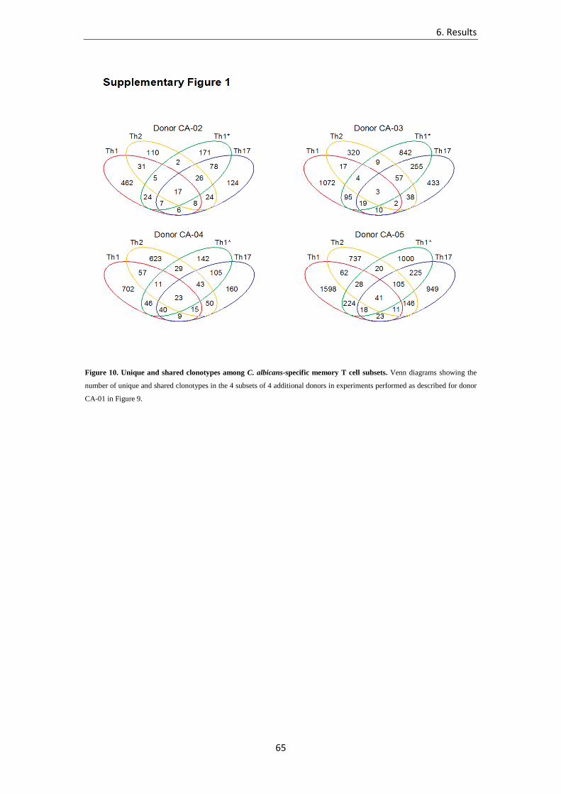
6. Results
65
Figure 10. Unique and shared clonotypes among C. albicans-specific memory T cell subsets. Venn diagrams showing the
number of unique and shared clonotypes in the 4 subsets of 4 additional donors in experiments performed as described for donor
CA-01 in Figure 9.
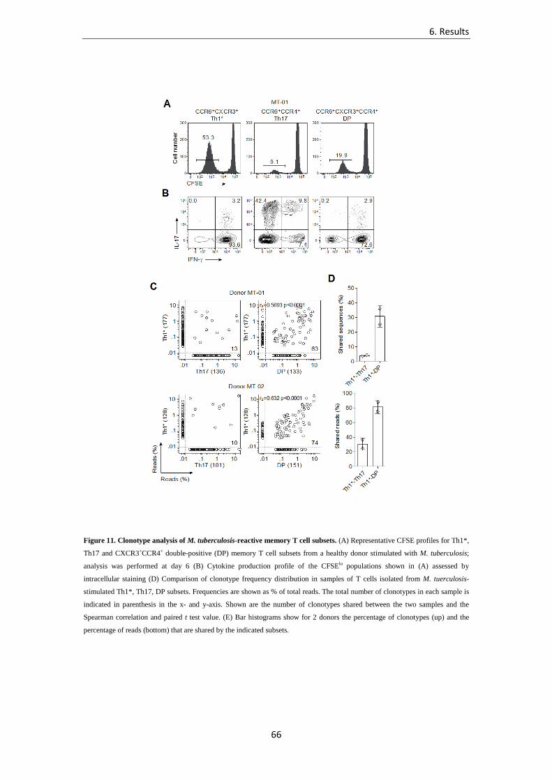
6. Results
66
Figure 11. Clonotype analysis of M. tuberculosis-reactive memory T cell subsets. (A) Representative CFSE profiles for Th1*,
Th17 and CXCR3+CCR4+ double-positive (DP) memory T cell subsets from a healthy donor stimulated with M. tuberculosis;
analysis was performed at day 6 (B) Cytokine production profile of the CFSElo populations shown in (A) assessed by
intracellular staining (D) Comparison of clonotype frequency distribution in samples of T cells isolated from M. tuerculosis-
stimulated Th1*, Th17, DP subsets. Frequencies are shown as % of total reads. The total number of clonotypes in each sample is
indicated in parenthesis in the x- and y-axis. Shown are the number of clonotypes shared between the two samples and the
Spearman correlation and paired t test value. (E) Bar histograms show for 2 donors the percentage of clonotypes (up) and the
percentage of reads (bottom) that are shared by the indicated subsets.
6. Results
67
6.1.3. Extensive clonotype sharing among TT-specific T cell subsets
The heterogeneity of memory T cells observed for C. albicans and M. tuberculosis could be
attributed to the multiplicity of the antigens and microbial products present in the pathogens.
To address whether a single protein antigen would induce a more polarized response, we
studied memory T cells induced by vaccination with Tetanus Toxoid (TT), which is
administered with alum as adjuvant. CFSE-labeled Th1, Th2, Th1* and Th17 cells were
stimulated with TT in the presence of autologous monocytes. Surprisingly, in the four donors
analyzed proliferation to TT was high and comparable in all four subsets (Figure 12A,B). The
pattern of cytokine produced in TT-stimulated cultures was distinct for each subset. The Th1
subset produced IFN-γ, as expected, but also low levels of IL-4, IL-5 and IL-13, while the
Th2 subset produced high levels of IL-4, IL-5 and IL-13, as well as some IFN-γ. The Th1*
and Th17 produced IL-17 and IL-22. In addition, Th1* also produced high levels of IFN-γ
(Figure 12C).
Interestingly, even in the case of a single antigen, the response was highly polyclonal in each
subset with a total of 1,386 and 3,259 individual clonotypes in the two donors analyzed
(Table 1). Furthermore, within each subset the clonal frequency was widely distributed, with
a few dominant clonotypes accounting for a large fraction of total reads (Figure 13).
Surprisingly, there was a high level of clonotype sharing among the 4 subsets, in particular
between Th2 and Th1 (24.4% of clonotypes and 70.4% of reads) and between Th2 and Th17
(17.1% of clonotypes and 62% of reads). Furthermore 83 out of 1,386 clonotypes (in donor
TT-01) and 65 out of 3,259 clonotypes (in donor TT-02) were present in all subsets (Figure
12D and Figure 13). These findings indicate that vaccination with a single protein can prime a
large number of specific T cell clones that acquire different phenotypes and functions.

6. Results
68
Figure 12. Extensive clonotype sharing among TT-specific CD4 T cell subsets. (A,B) CFSE profiles and percentage of
CFSElo proliferating cells in the indicated 4 memory T cell subsets stimulated with TT in a representative donor (A) and median
values with 25 and 75 percentiles in 4 donors, with whiskers representing the highest and lowest values in each subset (B). (C)
Cytokine production measured in the day 6 supernatants of the TT-stimulated cultures. Values represent mean ± s.e.m. (D)
Comparison of clonotype frequency distribution in samples of T cells isolated from TT-stimulated Th1, Th2, Th1* or Th17
subsets from donor TT-01. Frequencies are shown as % of total reads. The total number of clonotypes in each sample is indicated
in parenthesis in the x- and y-axis. Shown are also the number of clonotypes shared between the two samples and the Spearman
correlation and paired t test value. (E) Bar histograms showing for 2 donors the percentage of clonotypes (upper panel) and the
percentage of reads (lower panel) that are shared by the indicated subsets.

6. Results
69
Figure 13. Extensive clonotype sharing among TT-specific CD4 T cell subsets. (A) Frequency distribution of clonotypes in
the 4 TT-specific T cell subsets from donor TT-01 and TT-02. Shown are the 1% frequency threshold, the number of clonotypes
with frequencies >1% and their cumulative frequencies (% of reads) in parentheses. (B,C) Percent of clonotypes with frequencies
>1% (B) and their cumulative frequencies (% of reads) (C) in the 2 donors (mean ± s.e.m.). (D) Comparison of clonotype
frequency distribution in samples of T cells isolated from TT-stimulated Th1, Th2, Th1* or Th17 subsets from donor TT-02.
Frequencies are shown as % of total reads. The total number of clonotypes in each sample is indicated in parenthesis in the x-
and y-axis. Shown are also the number of clonotypes shared between the two samples and the Spearman correlation and paired t
test value. (E) Venn diagrams showing the number of unique and shared clonotypes in the 4 subsets in the two donors.

6. Results
70
6.1.4. Isolation of sister clones from different memory subsets
To provide further evidence that memory T cells expressing the same αβ TCR have different
phenotypes and functional properties we isolated a large number of T cell clones from
samples of C. albicans-specific Th1, Th2, Th1* and Th17 cells that were frozen as a backup.
From donor CA-01 we isolated a total of 242 T cell clones, of which 153 were unique since
they expressed the same TCRVB sequences (Figure 14A). As expected, several clones with
identical TCRVB sequences were found in two subsets (clonotypes 3, 4, 7, 8, 11, 12 13, 17,
18, 64, 75, 77, 82, 206) and in one case in three subsets (clonotype 32). The most frequent
sharing was between Th17 and Th1* and between Th17 and Th2. Sequencing of the TCRVA
chain (done in collaboration with Dr. Ton Schumacher, NKI, The Netherlands) confirmed that
the cells isolated from different subsets carried the same αβ TCR (Figure 14B).
The sister clones isolated form different subsets were functionally characterized for cytokine
production and expression of transcription factors and CCR6. With the exception of IFN-γ,
which was produced by virtually all clones as a consequence of the prolonged in vitro culture,
cytokine production was consistent with the origin of the cells: IL-4 was produced at high
levels by clones isolated from the Th2 subset, while IL-17 and IL-22 were produced by clone
derived from the Th17 and Th1* subsets (Figure 14C). In addition, CCR6 expression and
RORγt mRNA were detected only in clones derived from Th17 or Th1* (Figure 14D-E). As
an example, two sister clones of clonotype 64, isolated from the Th17 or Th2 subset, showed
the characteristic polarized cytokine production and expression of transcription factor (Figure
14F). Another consistent example was provided by two sister clones of clonotype 11 that
were isolated from the Th17 or Th1* subset (Figure 14G). We conclude that T cell with the
same TCR can be found in different subsets and that their phenotype is stably maintained.

6. Results
71
Figure 14. Isolation of sister clones from different memory subsets. T cell clones were isolated from cryopreserved samples
of C. albicans-stimulated CFSElo Th1, Th2, Th1* and Th17 cells from donor CA-01 and TCR αβ sequences were obtained by
Sanger sequencing on amplified cDNA. For each subset the number of individual clonotypes is indicated. (A) The circles
indicate the total number of clones (center), the number of clones unique to that subset (white section) and the number of clones
whose TCR was found also in T cell clones isolated from another subset (color coded sections). (B) List of the clonotypes that
were found in two or more subsets. (C-E) Characterization of sister clones specific for C. albicans isolated from different
memory subsets and expressing one of the shared TCR clonotypes. Shown are cytokine production (C), and expression of
transcription factors (D) and CCR6 (E). (F,G) Characterization of two pairs of sister clones isolated from Th1* and Th17 subsets
(F) and from Th2 and Th17 subsets (G).

6. Results
72
6.1.5. Generation of multiple T cell fates by in vitro priming of naïve T cells
The above data show that the CD4 T cell response to pathogens and vaccines can be highly
heterogeneous, both at the population and at the clonal level. To test whether a single round
of stimulation could imprint these heterogeneous fates, we isolated highly pure naïve CD4 T
cells, which were labeled with CFSE and stimulated with C. albicans in the presence of
autologous monocytes (Zielinski et al., 2011). Proliferating, CSFE-negative T cells recovered
on day 15 produced IFN-γ, IL-17 and IL-22, consistent with previous findings (Figure 15A).
Interestingly, in the same cultures some cells produced IL-4, alone or together with IFN-γ.
These findings demonstrate that multiple polarized cell fates can be generated from naïve T
cells in a single round of stimulation and even in the same culture.
To ask whether the same clonotype could be found in different functional subsets, we sorted
IL-4- and IL-17-secreting cells using the cytokine secretion assay and isolated a number of
clones, which were then characterized phenotypically and molecularly for TCR usage.
Remarkably, the same TCR αβ pair was found in 5 T cell clones isolated from the IL-17+
cells and in 1 clone isolated from IL-4+ cells. Importantly, the clones isolated from IL-17+
cells, produced IL-17 and expressed RORC mRNA, while the clone isolated from IL-4+ cells
produced IL-4 and expressed GATA3, but not RORC mRNA (Figure 15B,D). These findings
demonstrate that intraclonal diversification to exclusive IL-17 and IL-4 production can be
imprinted in a naïve T cells by a single round of in vitro stimulation.

6. Results
73
Figure 15. Generation of multiple fates by in vitro priming of naïve T cells. (A) Proliferation of CFSE-labeled naive CD4 T
cells primed in vitro by C. albicans in the presence of autologous monocytes and intracellular cytokine staining on gated CFSElo
cells on day 15. (B) List of clones isolated by cell sorting of IL-17+ and IL-4+ cells from C. albicans-primed cultures. The T cell
clones with the same TCR are marked with an asterisk. (C,D) Cytokine production (C), and expression of transcription factors
(D) in 3 sister clones isolated from IL-17+ and IL-4+ cells.

6. Results
74
6.2. Additional Results
6.2.1. High throughput approach to unravel the relationship between C.
albicans antigens and elicited response
The genome of C. albicans comprises more than 6,000 ORFs, of which only 1,500 have so
far been characterized (Candida Genome Database, 2014). The work described in Section 6.1
of this thesis provided compelling evidences that multiple types of T helper subsets (namely
Th1, Th2 and Th17) can be generated in response to this complex microbe. Heterogeneity
appears to be represented both at intra-clonal and at population level. The first phenomenon
can possibly be explained with well described mechanisms such as asymmetric division or
differential environmental cues acting on daughter cells during clonal expansion. However,
the vast majority of C. albicans-specific clonotypes identified appeared to be unique to one
cellular compartment. This argues in favor of driving forces that preferentially skew the
polarization of a cell with a given TCR toward a certain fate. Antigen-specific properties,
such as post-translational modification, strength of recognition by TCR, produced amount,
anatomical localization, are likely factors in determining such fate.
We reasoned that if having the possibility to screen distinct T helper repertoires for the
specificity to a wide number of C. albicans proteins we would possibly be able to unravel
some of the underlying relationships between antigens and their induced response. We
therefore selected 80 C. albicans proteins from an in depth analysis of the relevant literature
(Table 2); the criteria utilized encompassed two main constrains, i.e. ultimate characterization
of the protein and reported immunogenic activity. Besides, we took care to select proteins
with different cellular localization (cytosol, cell wall, secreted proteins) and functions
(hydrolysis, adhesion, metabolism, replication, structure maintenance). This would ideally
allow us to link a certain class of proteins with shared features to a consistently induced type
of response.
We proceeded with a bioinformatic analysis to predict and produce immunoprevalent
(dominant within the population) peptides from the selected proteins. This approach presents
the great advantage that only a few peptides must be synthesized, thus avoiding yeast-based
production of the whole protein, with invaluable spare of time and resources. It must be noted
that as C. albicans possesses a slightly modified genetic code, production of those proteins in
bacteria would not result in correct sequence or structure.

6. Results
75
As explained in details in the “Experimental procedures” section, all 15-mer peptides
overlapping by 10 residues spanning each sequence were scored for predicted binding to a
panel of 24 common different HLA class II DR, DP and DQ molecules. A set of 1,273
peptides was selected as top scoring for binding to 12 or more of the 24 HLA class II
molecules probed (in collaboration with Dr. Alessandro Sette, LIAI, CA, USA). These
selection criteria are similar to that adopted in other recent works (Lindestam Arlehamn et al.,
2013; Oseroff et al., 2012). Each protein was represented by a minimum of 4 to a maximum
of 51 peptides (average 16.8). The peptides were synthesized as crude material, individually
resuspended in DMSO and pooled accordingly to the protein of origin.
Using the 80 generated peptide pools, we assessed the level of T cell reactivity in healthy
donors. Total memory T cells (CD4+CD45RO+) were obtained from 5 donors, labeled with
CFSE and co-cultured with autologous monocytes pulsed with each of the 80 peptide pools
(Figure 16a). Some of the peptide pools induced a high proliferative response in 5/5 or 4/5
donors, as detected at day 12 by CFSE dilution; this is consistent with the notion that some
antigens are widely recognized among the human population (immunoprevalent). Some other
peptide pools, instead, induced very strong response on a donor-specific basis, which might
account for more efficient presentation on specific HLA types. Notably, most peptide pools
induced little or undetectable proliferation. Pools representative of metabolic enzymes tended
to be poorly immunogenic in our assay, and we speculate that this might depend on the high
level of sequence homology for such proteins across the eukaryotic domain (not shown).
We decided to focus our attention on the 15 top-ranking peptide pools; the response of 4
different T helper subsets, as defined on the basis of surface expression of chemokine
receptors CCR6, CXCR3, CCR4 (see previous Section 6.1 for details), was analyzed for each
peptide pool, and the relative frequency of antigen-specific T cells determined through T cell
library assay (Geigeret al., 2009) (Figure 16b). As expected, the magnitude of the response
was widely different depending on the analyzed subset. Th17 cells showed high reactivity to
most antigens, while Th1 comprised very rare cells specific to the tested pools. Hyr1 and
MP65, two already described immunodominant antigens of C. albicans cell wall, were
confirmed to be among the most recognized proteins from the fungus. S-aspartyl protease
(Sap) 4, 5, 6, which constitute a separate group of proteases from Sap 1, 2, 3 in terms of
sequence homology, were also identified as novel dominant immunogens inducing exuberant
Th17 responses; interestingly, a deeper analysis of the literature revealed that among the Sap
family, Sap2 was instead the best characterized immunogen, and is currently under evaluation
as a vaccine protein in clinical trials (De Bernardis et al., 2012). The reason for Sap2 being
considered a good vaccine candidate relies on the predominant production of this protein from
in vitro cultured C. albicans, and from the wide presence of Sap2 binding antibodies in
6. Results
76
human sera (Naglik et al., 2003). However, which member of the family is mostly produced
in vivo remains unclear, and antibodies have been reported to be broadly cross-reactive
among members of this family (Naglik et al., 2003). We propose therefore that Sap2 has
erroneously been identified as responsible for an immunogenic effect that has instead to be
attributed to Sap4, Sap5, and Sap6 proteins. The frequency of cells recognizing any of these
three antigens is extraordinary, with an average of roughly 1 cell out of 700-2,000 Th17 being
specific.
Th1* and Th17 showed impressively similar antigenic fingerprints, which would be
consistent with high plasticity between these two compartments, as also suggested by the high
level of clonotype sharing (see Section 6.1). Several differences in terms of fingerprint could
be instead observed among these subsets and the Th2 subset, which seems to preferentially
recognize a distinct set of proteins (for instance, it accounts for the highest frequency of cells
specific to Als6 and Lip5). Interestingly, in the case of Th1, very little response was induced
by any of the tested peptide pools. This finding is in line with the clonotypic analysis we
performed on pathogen-specific Th1 population (see Section 6.1), as such population appears
to be composed mainly by unique clones that are not shared with any other subset. These
results therefore strongly suggest that CCR6– Th1 cells are generated by a distinct set of
proteins of C. albicans that were not included in our assay. It is also possible that Th1
responding to C. albicans are instead cross-reactive T cells that were generated in response
other pathogens, an overall scenario that has been suggested by other groups in a different
context (Birnbaum et al., 2014). In the attempt to consider the relevance of each analyzed
antigen in the generation of a specific type of T cell response, we normalized the obtained
frequencies by the total number of cells within the subset responding to any of the screened
peptide pools. Not surprisingly, we found that the most dominant antigens (Hyr1, MP65,
Sap5) accounted for a very high percentage of the response in the Th1* and Th17
compartments (Figure 16c). Intriguingly though, proteins such as Als6, Lip5 and Plb4 showed
a clear skewing toward a Th1 and Th2 phenotype. The reasons for this preferential association
are still unclear and are currently object of intense investigation. In conclusion, we have
provided evidences for a differential T helper response to antigenic targets within a given
microbe. The identification of C. albicans prototypic “protective” and “deleterious” antigens
(i.e. Th17-inducing MP65, Hyr1, Sap 4, 5, 6 and Th2-inducing Lip5, Als6 and Plb4) raises
the question whether the antigenic fingerprint may be altered in pathological conditions
associated with an overall skewed Th response, such as CMC. We are planning to perform in
vivo experiments using the characterized antigens to verify whether the ex vivo findings
would be confirmed; if so, this approach will allow to dissect the antigen borne signals that
induce polarization of T cells towards a specific fate.

6. Results
77
Table 2. Selected peptide pools for immunogenicity screening. Eighty C. albicans cytosolic, cell-wall associated or secreted proteins were selected (Uniprot accession code is indicated) and predictably immunodominant peptides from each sequence were produced and pooled. Eighty peptide pools were obtained in the end, each composed by a different number of peptides (indicated in last column).

6. Results
78
Figure 16. Antigenic fingerprint of C. albicans-specific cells in different subsets. (a) T cell memory proliferation in response
to 80 C. albicans-derived peptide pools. Peptide pools for 80 C. albicans proteins were obtained (see text, Table 2);
CD4+CD45RO+ memory T cells were obtained by negative selection with bead-based enrichment, CFSE-labeled and co-cultured
with autologous monocytes pulsed with a peptide pool. After 7 days, 50U/ml IL-2 were added to the culture and CFSE dilution
was assessed after additional 5 days. Each pool was tested in duplicate for each donor, 5 donors were tested; bars show average
level of proliferating cells in all 5 donors, color scale indicates the number of donors in which a significant response (defined as
CFSElo % > 5 x background proliferation in the absence of stimulation). (b) T cell libraries were run on Th1, Th2, Th1* and
Th17 cells sorted from 8-10 donors (each plots represents a donor) using the 15 top ranking peptide pool as assessed in a);
average values of frequency are shown as a line. (c) Normalized response is shown for 6 antigens: the values were obtained by
dividing the number of peptide pool-specific cells by the total number of T cells responding to any pool in that subset.

6. Results
79
6.2.2. Interrogation of the T cell response in patients with chronic
mucocutaneous candidiasis
Chronic mucocutaneous candidiasis (CMC) is the collective term for a complex group of
disorders characterized by persistent or recurrent infections of the skin, nails and mucosal
tissues with Candida species, mostly C. albicans (Eyerich et al., 2010). CMC is very
commonly developed by patients with broad and profound T cell deficiencies and can be
provoked by various inborn errors of immunity. Patients with hyper IgE syndrome, caused by
dominant-negative mutations of STAT3, are susceptible principally to CMC and
staphylococcal diseases of the lungs and skin (Milner et al., 2008). These patients have very
low proportions of circulating IL-17A- and IL-22-producing T cells, probably because of
impaired responses to IL-6, IL-21, and IL-23. Individuals with mutations in IL-17F or IL-17
receptor genes have also been shown to develop CMC (Puel et al., 2011). APECED, or
autoimmune polyendocrine syndrome, is caused by AIRE gene mutation and often associates
with CMC; the trigger, in this case, is the production of auto-antibodies against IL-17 and IL-
22 (Puel et al., 2010). Overall, it appears clear that all inborn errors leading to CMC underlie
impairment of the Th17 response.
A further set of mutations that induce gain of function in the STAT-1 transcription factor
(STAT-1 GOF) has been associated with onset of CMC (Liu et al., 2011). Such mutations
occur in the coiled-coil domain of STAT-1 protein and impair the nuclear dephosphorylation
of the activated form, resulting in stronger cellular responses to STAT-1 inducers IFN-α/β,
IFN-γ, and IL-27, and stronger STAT-1 activation in response to the STAT-3-dependent IL-
17-inducers, IL-6 and IL-21. Thus, as highlighted previously, the development of T cells
producing IL-17A, IL-17F, and IL-22 is impaired also in these patients (Liu et al., 2011).
However, the reduced IL-17 production by T cells in these patients was only assessed on the
total CD4+ T cell memory compartment following polyclonal stimulation (PMA+Ionomycin
or α-CD3/α-CD28). This data do not give therefore any insights on how the memory T cell
response to C. albicans is shaped in such patients.
We decided to take advantage of the T cell library technique, recently developed and
extensively used in our laboratory (Campion et al., 2014; Geiger et al., 2009; Lindestam
Arlehamn et al., 2013) to interrogate the repertoire of STAT-1 GOF CMC patients’ memory
T cells in order to obtain quantitative and qualitative information on its composition.
Blood samples were collected from patients as well as age-matched controls at the Institute
Imagine (Paris, France; collaboration with Drs. Anne Puel and Jean Laurent Casanova,
Imagine, France and Rockefeller University NY, USA). CD14+ and CD14– PBMC fractions

6. Results
80
were isolated by magnetic bead enrichment after Ficoll-Plaque gradient, frozen and shipped in
dry ice to our laboratory. CD14– PBMCs were thawed and stained for exclusion markers
(CD8, CD14, CD16, CD19, CD25, CD56), CD4 memory markers (CD4, CD45RO), and
chemokine receptors (CCR6, CXCR3) so to obtain four different memory T cell subsets:
CXCR3+CCR6– classical Th1, CXCR3–CCR6– Th2, CCR6+CXCR3– Th17, and
CCR6+CXCR3+ non-classical Th1* (Figure 17a). This sorting strategy slightly differs from
the one adopted in the work presented Section 6.1 and aimed to maximize the cellular
recovery from the reduced samples in our possess; the main limitation of this strategy would
be represented by an over-representation, and possibly “dilution”, of the Th2 compartment,
which would result enlarged by the inclusion of all CCR6–CCR4–CXCR3– cells generally
excluded from our standard sorting strategy. Still, the obtained four populations of memory T
cells showed clearly distinct cytokine profiles upon ex vivo restimulation, in accordance with
the chemokine-receptor based phenotype prediction (not shown and Figure 17f).
Strikingly, most analyzed samples from patients showed a severely impaired expression of
CCR6, a chemokine receptor whose expression is considered to be controlled by the Th17-
lyneage specifying factor RORγt (Figure 17a,b). Instead, CXCR3 expression, under the
regulation of IFN-γ-induced STAT-1, was found significantly increased. As a result, the
patients showed a dramatically diminished amount of circulating Th17 cells and increased
frequency of classical Th1, as compared to healthy age-matched controls (Figure 17a,b). We
next assessed the expression of CCR6 on non-CD4+ non-CD14+ cells (double negative
fraction) and surprisingly noticed that this population presented an even higher expression of
CCR6 in patients as compared to healthy controls (Figure 17c,d). This finding is extremely
important for two different reasons: first, it represents a valuable internal control for the
validity of the staining, thus confirming a selective lack of CCR6 expression on memory
CD4+ T cells in STAT-1 GOF patients; second, it suggests that CCR6 expression is regulated
by different signals in CD4+ T cells and non-CD4+ T cells (possibly in T vs B cells).
As CCR6+ cells were very rare in STAT-1 GOF patients, we decided to generate clones by
limiting dilution from the two relative subsets (Th1* and Th17) and assess their signature
cytokine profile in order to exclude that functional defects accompanied their reduction in
number. Figure 17e shows that despite the reduced representation in blood, these cells
produced IL-17 and IL-22 in similar amounts when compared to healthy controls. A slight but
not significant increase in IL-17 production by Th1* and, most notably, a strong increase in
IFN-γ production by Th17 was detected in patients (Figure 17e). This suggests that priming in
STAT-1 GOF environment, probably due to contribution of both the APC and naïve T cell,
may tend to skew the proliferating progeny toward an IFN-γ producing phenotype (Th1); in
this scenario Th1* would reasonably represent ex-Th17 (or “missed” Th17) as suggested

6. Results
81
elsewhere (Cosmi et al., 2011), even though clonotypic analysis will be necessary to confirm
this hypothesis.
T cell libraries were prepared and expanded as previously described (Geiger et al., 2009), and
then screened for reactivity to heat-killed C. albicans using autologous monocytes as antigen
presenting cells. 3H-thymidine incorporation was used as readout for proliferation after 3 days
of co-culture, and frequency of C. albicans-specific cells calculated on the basis of the
number of non-responding wells, in accordance with Poisson distribution.
Having targeted with our sorting strategy the whole memory T cell compartment, we were
able to estimate the total number of circulating memory T cells specific to C. albicans just by
summing the frequencies obtained in each subset, normalized to the relative proportion of the
lineage within the whole population. Unexpectedly, the overall frequency of memory T cells
specific to the fungus was unaltered in patients with respect to healthy controls (Figure 17g).
However, the distribution of pathogen-specific cells among subsets differed enormously.
Already on a per subset basis, an average 4-fold increase (p<0.05) was detected in the Th1
compartment of the patients (not shown). When taking in account the relative frequency of
each subset within circulating memory T cells, it appeared striking that response in healthy
controls was Th17-driven (roughly 55% of C. albicans-specific cells belonged to this subset)
and very little Th1-polarized (less than 10%), while patients showed a mirroring distribution,
with 50% of circulating C. albicans-specific cells being Th1 and very few percent Th17
(Figure 17h).
We believe this unbalanced response may be implicated in both susceptibility to C. albicans
infection and on pathogenesis of CMC. First, the lack of circulating T cells expressing the
homing receptor CCR6, considered to be involved in homing to the mucosal sites, might
result in very few protective IL-17 producing cells reaching the site of infection. Secondly, it
is tempting to speculate that classical CCR6– Th1 may instead reach the site of infection due
to expression of CXCR3, homing receptor to inflamed tissues, and worsen the disease by
producing not only IFN-γ but also Th2 types of cytokine (it must be noted that IL-4, IL-5 and
IL-13 are produced by C. albicans-specific classic Th1 in considerable amounts (Becattini et
al., manuscript in preparation). IL-4, IL-5 and IL-13 have long been associated with non-
protective response to C. albicans, in both animal models and humans (Romani, 1999), also
with respect to CMC (Kobrynski et al., 1996). Such cytokines have been shown to dampen
IL-17-mediated neutrophils recruitment in situ, induce diseases such as atopic dermatitis and
render skin more susceptible to infections due to downregulation of β-defensins (Eyerich et
al., 2010). Increased IFN-γ levels at the site of infection could as well play a role in
promoting inflammation and worsening overall tissue homeostasis.

6. Results
82
Our data provide novel important insights on the understanding of C. albicans-specific
memory T cell response in patients suffering from CMC, and set the stage for a whole new
series of experimental questions. Further studies will be required to assess the relative role of
priming vs in situ restimulation of memory cells in generating the observed phenotypic skew,
the cytokine profile and antigenic specificities of skin-resident C. albicans-responding cells as
compared to circulating cells, as well as the clonal relationship between cells residing in
either of these anatomical compartments.

6. Results
83
Figure 17. Phenotype and frequency of C. albicans-specific memory T cells in STAT1-GOF patients suffering from CMC.
(a) Adopted sorting strategy; shown are two representative panels (1 patient, 1 healthy control) for CCR6 and CXCR3 expression
in CD4+ T cells (pre-gated as lin–CD4+CD45RO+, not shown). (b) Quantification of cells sorted as in a) from several donors (n=5
controls, n=7 patients). (c) Representative plot for CCR6 expression in CD4+ T cells and CD14–CD4– cells. (d) Quantification of
CCR6 expression as assessed in c). (e) Representative plot showing the percentage of cytokine producing cells within individual
T cell clones obtained by limiting dilution from healthy controls or patients. (f) Intracellular cytokine staining of T cell library
lines; dots represent average values for each donor. (e) Frequency of circulating C. albicans-specific memory T cells calculated
through T cell library assay; each dot represents one donor. (f) Distribution of C. albicans-specific T cells among different
subsets; values were calculated considering the relative expansion of each circulating subset through surface staining as in a).
**p<0.01, ***p<0.001

6. Results
84
6.2.3. A biochemical approach to identify C. albicans dominant Th17
antigens among cell wall mannoproteins
The cell wall of C. albicans comprises an outer layer of mannans and mannosylated proteins
(mannoproteins) anchored through a GPI molecule to the inner β-glucan layer, that are
considered to be highly immunogenic. The immunogenicity of such proteins depends on
several synergizing factors: first the conjugation with mannan branches, that are recognized
by several innate receptors, such as DC-SIGN, Dectin-2, mannose receptor, Mincle and others
(Osorio and Reis e Sousa, 2011), thus facilitating uptake and efficient activation of DCs
through innate signaling; secondly, the abundance and availability of such proteins, that are
produced in considerable amounts due to their crucial structural or adhesive role, and protrude
from the cell wall, becoming easy targets for uptake by innate cells, and increasing the chance
of being processed and presented (Chaffin et al., 1998; Gomez et al., 1996).
Only a few immunodominant proteins have been identified among the thousands that are
known to occupy this layer; the best studied among those (MP65, Hyr1) are preferentially or
uniquely expressed by the hyphal form of the fungus (Gomez et al., 1996; Luo et al., 2010).
However, mannoproteins are highly represented on the cell wall of conidia (Chaffin et al.,
1998), which are in turn widely recognized by T cells in healthy donors; we therefore
reasoned that abundant immunogenic proteins yet unidentified must be present on the yeast
surface.
As T cells play a key role in protection against fungal infection, it is believed that a vaccine to
protect against C. albicans should elicit strong response of the Th17 subset (Bar et al., 2012).
The search for T cell immunodominant antigens has therefore a remarkable clinical
importance.
We set up a series of biochemical approaches to extract, characterize and identify surface
mannoproteins from log phase-growing C. albicans conidia. The extraction procedure,
described in details in the “Experimental procedures” section, was based on solubilization of
the proteins at high temperature in citrate buffer, precipitation through copper tartrate-
containing Fehling reagent, purification via acidic treatment and dialysis.
Electrophoretic analysis of the extract revealed an accumulation of high molecular weight
proteins at the top of the polyacrylamide gel (Figure 18a, black square). This is consistent
with the large size of the mannose branches, that in yeasts have been estimated to be
composed on average by over 600 mannose units each (Chaffin et al., 1998). Basically very
few proteins were able to enter the gel, either because they had a smaller size or a lower level

6. Results
85
of glycosylation (of note the harsh extraction procedure could likely generate breaks in the
sugar backbone and therefore give rise to products of reduced dimension).
Treatment of the extract with PNGaseF, an enzyme that catalyzes cleavage of the N-linkage
between sugars and Asparagine residues, promoted release of some of the proteins from the
high molecular weight aggregate, thus generating a smear at still high molecular weight
(Figure 18a, blue square) and a clear band at 15-20 KDa (Figure 18a, red square). Following
de-glycosylation we separated the proteic part through acetone based precipitation from the
released sugar moieties remaining in the supernatant.
We first assessed the immunogenic activity of the fraction on total memory CD4+ T cells
upon stimulation in the presence of autologous monocytes. To address also the relative role of
the sugar moiety in this process, as a previous report suggested this would be sufficient to
trigger proliferation and polarization of Th17 (van de Veerdonk et al., 2009), we separately
stimulated T cells with CWE, deglycosylated CWE and recovered soluble sugars. Heat-killed
CA particles were used as positive control. Strikingly, stimulation by CWE induced T cell
activation to a similar extent to whole fungal particles, as assessed by CFSE dilution and
intracellular staining for IFN-γ and IL-17 production; not surprisingly, the effect was not
altered by sugar removal, while mannans alone could not induce substantial T cell
proliferation (Figure 18b). This experiment suggested that cell wall mannoproteins comprises
the most recognized antigens within CA particles, and their aminoacidic backbone is
responsible for recognition by T cells.
We went on trying to determine whether CW mannoproteins would be recognized by
different T helper compartments or rather induce a preferential response in supposedly
protective subsets (CCR6+). A sorting strategy was used based on chemokine receptors
CCR6, CCR4 and CXCR3 that allowed us to sort populations highly enriched for Th1, Th2,
Th17 and non classical CCR6+ Th1*. Ex vivo parallel stimulation of each subset revealed that
CCR6+ cells, Th17 in particular, were preferentially activated by the CWE, as assessed by the
level of T cell CFSE dilution in 4 healthy donors (Figure 18c). CCR6- Th1 and Th2
compartments showed lower response in comparison to that elicited by HK C. albicans
(Figure 18c); this would imply that antigens recognized by Th1 and Th2 may preferentially be
located in the cytosol or be secreted rather than being enclosed in the cell wall. Beside
proliferating to a similar extent, Th1* and Th17 cells produced cytokines in comparable
amount when restimulated with CWE (Figure 18d).
On the same line, the majority of C. albicans-specific T cell clones generated from three
different donors responded upon restimulation with CWE (Figure 18e). We therefore decided
to determine the frequency of T cells specific to such extract within the Th17 subset; heat-

6. Results
86
killed C. albicans and two major antigens proposed or under evaluation for vaccine
development, MP65 and Sap2 (De Bernardis et al., 2012) were selected as positive control.
The extract was recognized by an impressive amount of cells within the Th17 compartment,
with a calculated frequency of CWE-specific T cells nearly as high as that obtained for the
whole pathogen among several tested healthy donors, and consistently higher with respect to
the control immunodominant antigens (Figure 18f, g).
We reasoned that such impressive frequency of CWE-specific cells might be explained either
by the presence of a very potent antigen, or by the synergistic effects of different antigens.
We calculated the EC50 of CWE-specific clones or polyclonal library lines (whose low initial
input, 150 c/w, would reasonably exclude the presence of more than only one responding
cell), and found that the functional affinity for the antigens was extremely high, with EC50
ranging from 0.175 to 0.668 μg/ml (one representative clone shown in Figure 18h). Given the
likely multi-protein composition of the extract, any recognized antigen must act at a lower
concentration with respect to the estimated range, and therefore would represent an interesting
target for immunization strategies.
To rule out the possibility that such antigen would be presented through pathways other than
the classical MHCII, we restimulated CWE-specific T cell clones in the presence of MHCII-
blocking antibodies or using as APCs autologous monocytes fixed with glutharaldeyde prior
to pulse, so to exclude the presence of superantigens that would act independently of
processing. In neither case we could rescue proliferation which was instead clearly detectable
in clones stimulated in standard conditions, thus implying that the CWE-antigen(s) must be
processed and presented on MHCII in a canonical manner (Figure 18i). To definitely
ascertain that sugars were not directly recognized by the TCR we furthermore stimulated
those clones with commercially available protein-free C. albicans mannans, but did not detect
any proliferation (Figure 18j). Taken together these data suggest that a very potent T cell
antigen is comprised in a fraction of cell wall enriched for mannoproteins.
Next, we wondered whether the antigenic potency of CWE would be preserved in the mouse
system. To address this question we performed vaginal or oropharingeal C. albicans infection
of C57BL/6 mice, collected draining lymph nodes after 1 week, isolated cells and
restimulated them in vitro with either heat-killed fungal particles or CWE. As shown in
Figure 19b and c, a very high proportion of Th17 cells were activated by CWE, indicating
that most antigens inducing in vivo priming of naïve T cells would be contained in our extract.
This result has important implications with respect to the debate whether yeast, hyphae or
both growth forms of C. albicans are involved in infection and elicit immune response, and
argues in favor of a strong role for conidia in such contest or, at least, for importance of
6. Results
87
antigens present at both fungal stages. We then asked whether this extract might provide any
protection if used as a vaccine. C57BL/6 mice were immunized with CWE and then
challenged with an intravaginal dose of live C. albicans (Figure 19d). Vaginal lavage was
performed in the three weeks following infection, and proliferating C. albicans particles
quantified through plating and CFU count. A significant decrease in vaginal pathogen burden
could be observeded at all analyzed time points (Figure 19e), showing induction of a
protective response following immunization and suggesting the potential use of the CWE
itself (or of its most immunogenic components) to build a T cell vaccine against C. albicans.
We finally decided to attempt to identify the immunogens within the cell wall preparation by
subjecting CWE to mass spectrometry. First, we tried to narrow down the number of
candidates by fractionating the extract via reverse phase HPLC. Using a C8 column, we
obtained 6 main fractions, and a seventh fraction at very high acetonitrile concentration
containing all remaining (strongly) binding molecules (Figure 20a). When restimulating
CWE-specific T cell library lines, however, we were unable to identify a unique fraction
retaining the immunogenic activity, most likely due to the elution of any targets over different
fractions (as already mentioned, sugar branches can easily break and generate a continuum of
“isoforms” with different molecular weights for any given mannoprotein) (Figure 20b). We
therefore proceeded to mass spectrometric analysis (MS) of the whole extract. A few
candidates were identified with high level of confidence by the run (Figure 20c): Ywp1, in
particular, was highly represented, and turned out to be a heavily mannosylated protein whose
backbone size was compatible with the lower m.w. band detected in gel upon PNGaseF
treatment (Figure 18a, red square). Other potential candidates represented intracellular
enzymatic proteins previously reported to be immunogenic (Candida Genome Database,
2014); a deep search in the literature confirmed that these proteins are commonly found on
the outer cell wall, even though the mechanism through which they get translocated from the
cytosol remains unclear (Chaffin et al., 1998).
The aminoacidic sequences of the identified targets were retrieved from UNIPROT server and
produced as pools of overlapping peptides (15mers) spanning the whole sequence; peptide
pools were generated accordingly. To our surprise, none of the peptide pools induced
significant stimulation neither in libraries from healthy donors (Figure 20d) nor in CWE-
specific clones (Figure 20e). Still, this negative result uncovered that several proteins reported
in the literature to be highly immunogenic (mainly on the basis of specific antibody detection
in blood of immune donors) do not induce a detectable T cell responses.
We reasoned that as the CWE contains proteins, which are highly mannosylated and most
likely tightly aggregated due to the extraction procedure, it was possible that enzymatic

6. Results
88
digestion preceding mass spectrometric analysis would be extremely inefficient, and the most
representative targets remain hidden from our retrieval. We therefore treated the CWE with
PNGaseF in the presence of UREA, a condition that we found to maximize the access of the
hydrolase to the protein backbone, as shown by the appearance of several novel bands upon
silver staining of polyacrylamide gel electrophoresis (Figure 20e). Repetition of MS analysis
identified among the top ranking hits a whole new set of heavily glycosylated, high molecular
weight proteins belonging to the ALS (agglutinin like sequences) family (Figure 20f). These
proteins have been shown to represent important virulence factors in C. albicans infection,
and a member of this family (ALS3) is currently being tested in clinical trials as a bivalent
vaccine against both C. albicans and S. aureus infection (Bar et al., 2012; Ibrahim et al.,
2013).
ALS2 and 4 resulted strongly represented in our sample, ALS1 and 3 possibly also being
present in detectable amounts. As expected, very few peptides could be identified (Figure
19g) and only from one N-terminal extremity of these proteins (most likely the portion
released from sugars upon enzymatic treatment), while none were retrieved from their core,
which resulted to contain an impressive number of potential N- and O-glycosylation sites (not
shown), thus supporting our hypothesis on the causes of missed retrieval by the first MS trial.
We are currently planning to produce these proteins and test them in cellular assays for
immunogenic properties.
In conclusion, we have extracted a mannoprotein-enriched fraction from the cell wall of C.
albicans conidia that induces a strong T cell response, mainly localized in the CCR6+ Th17
and Th1* compartments, both in humans and mice. The responsible antigen(s) is likely to be a
protein and is presented through a canonical MHCII-restricted pathway, acting at very low
concentration. The extract as such induced to some extent a protective response in a mouse
model of vaginal infection. The identification of antigenic targets is made difficult by the very
complex nature and structure of the extract. However, by coupling enzymatic deglycosylation
with mass spectrometry, we believe we have identified the potential immunogens responsible
for its activity. We have planned to produce those antigens in yeast so to preserve
biochemical properties (i.e. glycosylation profile) and test them in ex vivo as well as in vivo
assays. If the identity of these immunogens will be confirmed, this work will have provided
evidences for a preferential localization of Th17-inducing antigens in the cell wall of C.
albicans and identified powerful targets suitable for a vaccine against this pathogen.

6. Results
89
Figure 18. Characterization of immunogenic properties of a mannoprotein-enriched cell wall extract from C. albicans
conidia. (a) Electrophoresis on polyacrylamide gel of the CWE before and after treatment with PNGaseF glycosidase.
Explanation for colored squares are provided in the text. (b) Proliferation and cytokine profile of total CD4+ cells upon
stimulation with CWE: cells were labeled with CFSE and co-cultured for 7 days with autologous monocytes pulsed with the
indicated antigen. Stimulation with PMA + Iono and BFA was performed prior to staining. (c) Proliferation of different CD4+ T
cell compartment in response to CWE. T cells were sorted based on the expression of chemokine receptors and stimulated as in
b). CFSE dilution was assessed by flow cytometry and values normalized to the proliferation induced in Th17. (d) Representative
intracellular cytokine staining plot of Th1* and Th17 cells stimulated as in b) at day 7. (e) Thymidine incorporation of C.

6. Results
90
albicans-specific CD4 clones restimulated in the presence of autologous monocytes and CWE or PBS. Stimulation Index
represents the ratio between CWE/PBS induced proliferation. (f) T cell library response to indicated antigens as measured by 3H-
thymidine incorporation; every dot represent a polyclonal T cell line. Values above 3000 cpm were considered positive. Shown is
one representative experiment. (g) Mean frequency values for T cell specificity to the selected antigens. Values were calculated
from assays as shown in f), each dot represents a different donor. (h) Antigenic titration for CWE-specific T cell clone. T cell
clones or polyclonal lines (see text) were stimulated in the presence of autologous monocytes and decreasing concentrations of
CWE; proliferation was measured through 3H-thymidine incorporation assay and EC50 calculated. Shown is one representative
clone, the calculated range of EC50 was [0.175-0.668] μg/ml among several donors. (i) Assessment of processing pathway
requirements for CWE components. CWE-specific clones were restimulated with CWE in the presence of autologous monocytes
with or without MHC II-blocking antibodies; alternatively, autologous monocytes were fixed in 0.02% glutaraldehyde prior to
pulse with CWE. (j) CWE-specific clones were restimulated with CWE or protein-free mannans in the presence of autologous
monocytes; proliferation was assessed through 3H-thymidine incorporation assay.

6. Results
91
Figure 19. CWE contains antigens that are efficiently recognized in mice and induce protective immune response. (a)
Schematic overview of the experiment: oral/vaginal infection of C57BL/6 mice performed upon 17-beta-estradiol treatment, after
1 week draining lymph nodes were extracted, and T cells restimulated in vitro in the presence of heat-killed C. albicans particles
or CWE. After an o.n. incubation with Golgi-stop cells were stained to assess IL-17 and IFN-γ production. (b) Representative
plot for an intracellular cytokine staining performed as depicted in (a). Cells were pre-gated on CD4+CD44+; the experiments
represent oropharingeal infection, similar results were obtained in the model of vaginal infection. (c) Quantification of IL-17A+
cells from plots as shown in b). Each dot represents a mouse; representative of at least two independent experiments. (d)
Schematic overview of the experimental plan for mouse immunization with CWE. C57BL/6 mice were immunized either with
CWE + MPL or with PBS 56 days prior to infection (CWE inj), then received a booster shot at d-28; 17-beta-estradiol was
injected every week (ESTR) starting two weeks before infection with 106 C. albicans live particles delivered into the vaginal
lumen. Vaginal lavage was performed at day 7, 14 and 21 following infection, and plated on Sabouraud plate; CFU were
determined after o.n. incubation of the plates. (e) Quantification of fungal particles from vaginal lavage as explained in d). Each
dot represents a mouse; data are representative of two independent experiments.

6. Results
92
Figure 20. (a) HPLC fracionation of CWE. CWE was run on a C8 column in acetonitrile gradient and 7 fractions were recovered
(highlighted in blue) and separately resuspended. (b) Proliferation of CWE-specific T cell polyclonal lines with autologous
monocytes and indicated CWE HPLC fraction; cpm values are reported in the table below. (c) CWE components identified by
mass spectrometry on CWE extract and relative location according to the literature. (d) Representative plot of Th17 cell library
screened for specificity to CWE or MS-identified targets produced as overlapping peptides spanning the whole sequences. (e)
CWE-specific T cell clones from 3 different donors were restimulated in the presence of whole CWE or indicated peptide pools.
(f) Electrophoretic run of CWE treated with different deglycosylation protocols; Glycoprofile IV (SIGMA) is an aspecific
chemical deglycosylation kit based on trifluoromethansulfonic acid (TFMS). (g) CWE-retrieved proteins upon mass
spectrometric analysis of deglycosylated extract. Shown are the 12 top ranking targets, ALS family members are highlighted in
red. Note the high score and the low number of identified sequences; explanation is provided in the text.
6. Results
93
6.2.4. A biochemical approach to retrieve C. albicans antigens inducing
Th2 response
Having identified cell wall mannoproteins as a major source of Th17-inducing antigens, we
wondered whether we could retrieve antigens accounting for the Th2 polarization consistently
detected in response to C. albicans in most healthy donors. The identification of such antigens
could have important clinical implications, as a strong correlation between atopy and
sensitivity to Candida infection is reported, and it has been postulated that an allergic type of
inflammation (with in situ recruitment of eosinophiils, and production of IL-4, IL-5, and IL-
13, as well as IgE) is frequent in patients suffering from RVV (Fan et al., 2008; Romani,
1999; Weissenbacher et al., 2004; Witkin et al., 1989).
We reasoned that Th2 induction could associate to production of specific IgE, as Th2-related
cytokines are responsible for the necessary B cell class switch. This is the case for most
allergens, and atopic patients have been reported to develop IgE response to fungi
(Savolainen et al., 1998). We then set up a series of biochemical assays to retrieve Th2-
inducing C. albicans antigens using antigen-specific IgE from human sera as probes.
First, a series of C. albicans lysates were collected, that were either commercially available or
prepared through glass bead-mediated cell disruption or sonication. For the in house
preparation, both methods were used on each form of the fungus (yeast, hyphae), and as the
composition of the 4 obtained lysates appeared to be slightly different, a pool was created that
contained all of them, so to maximize the array of antigens suitable for our search (Figure
21a). The lysates were then used to identify C. albicans-specific IgE containing sera in
ELISA. Assay plates were coated with lysate pool, sera obtained from a frozen bank of more
than 450 samples from healthy donors were used as source of primary antibodies; a secondary
anti-human IgE polyclonal antibody and a tertiary AP-conjugated antibody were used for the
screening. Results are shown in Figure 21b as heat map, relative OD values are reported for
each well. Out of all the screened samples, we could identify only a handful of C. albicans-
specific IgE containing sera (as defined by OD value > 3 x background). This very low
frequency was expected, as the fungus is not reported to induce considerable IgE response in
healthy donors; besides, the presence of high titers of C. albicans-specific IgG (not shown)
most likely overcompeted IgE binding, raising the threshold for detection of the latter.
Presence of C. albicans-specific IgE in these sera was confirmed in subsequent control
assays; titration experiments were performed (Figure 21c), and the relative EC50 was
calculated (Figure 21d).

6. Results
94
Finally, we set out to use the sera to identify antigens in Western blot. Lysates obtained from
yeast or hyphal forms of C. albicans were used as a source of antigen, but Western blot failed
to retrieve any band. However, when repeating the assay after performing lysate
electrophoresis in native (non-denaturing) conditions, in a few cases it was possible to
retrieve a band, at slightly different molecular weight in the two preparations, consistent with
the notion that proteins undergo different post-translational modifications upon switch to
filamentous growth (Chaffin et al., 1998). These results constitute a proof of principle for the
use of IgE to identify C. albicans antigens. Of note, similar approaches have been
successfully used for this purpose elsewhere (Fonseca et al., 2012).
We are now planning to optimize and extensively use this assay to verify whether bands of
the same molecular weight can be consistently found in different donors, and then proceed to
the identification of the recognized antigens through mass spectrometry. The identified
proteins will then be tested with T cell libraries to ascertain the type of response induced in
vivo. We believe this approach could bring particularly relevant results to those clinical
conditions were an increased amount of anti-Candida circulating IgE or in situ allergic-like
reactions have been reported.

6. Results
95
Figure 21. (a) Gel electrophoresis of C. albicans lysates obtained by several procedures. Lysate 5 was obtained from commercial
source (Greer) and presents a different pattern of glycosylation. (b) O.D. values from a 384 plate ELISA; coating was performed
with lysates shown in a), detection with serum derived IgE. (c) Titration of C. albicans-specific IgE contained in a positive
serum; for comparison purposes, 5 negative sera were added as control. (d) EC50 value for C. albicans-specific sera in 7 donors
selected through screening shown in c) and calculated as in d). Each value represents the dilution factor of the serum for which
50% of maximal O.D. is achieved. (e) Native Western blot performed on C. albicans lysates and retrieved with one of the 7 sera
shown in d).
7. Discussion and Outlook
96
6.2.5. In vivo assessment of the non-redundant role of T cells in long-term protection from C. albicans infection
Effective response to different types of infection requires differential involvement of separate
arms of the immune system. For instance, it is well established that systemic candidiasis
occurs in humans when the granulocyte compartment is defective, while T cell deficiencies
lead to chronic or recurrent superficial infections (Hernandez-Santos and Gaffen, 2012).
Th17 have been recognized for several years as the main orchestrator of anti-fungal response,
through the production of cytokines such as IL-17, which induces neutrophil recruitment and
activation, and IL-22, which promotes antimicrobial peptide production by epithelial cells.
However, the recent discovery of ILCs has challenged this view and raised the question
whether these cells might be responsible for first-line defense and instruction of the
forthcoming T cell response. Indeed, ILCs (together with other cell types like NK and γδ T
cells) are characterized by the capacity to become readily activated and react to the presence
of the pathogen as well as to tissue-derived signals within hours. For instance, ILCs have been
recently shown to be required for protection in a model of oropharingeal candidiasis
(Gladiator and LeibundGut-Landmann, 2013). However, IL-17-producing CD4 T cells have
also been shown to confer protection in a similar model, and the differential contribution of
both cell types at different stages of the infection seems a likely explanation (Conti et al.,
2009; Hernandez-Santos et al., 2013).
The vaginal lumen is the most frequent anatomical location for C. albicans infection. Vaginal
infections occur in more than 75% of women during a lifetime and can become recurrent in 5-
10% of cases (Mosci et al., 2013). IL-23-dependent IL-17 but not IL-22 production (attributed
to T cell) has been identified as fundamental for containment of C. albicans infection in the
vagina (Pietrella et al., 2011). However, this work preceded the discovery of ILCs and
therefore the relative eventual division of labor between innate and adaptive compartments at
this location could not be investigated. Recent work from another group identified in fact
ILC-derived IL-22 as the main trigger of a protective anti-Candida response in the vagina (De
Luca et al., 2013).
We set out to assess the relative contribution of T cells in a model of C. albicans vaginal
infection. When considering the previous published studies, we made several observations.
First, the majority of the experiments were conducted within the first weeks of infection, thus
analyzing only the early phases of the host-pathogen interplay, which is generally life-long in
humans. Secondly, the commonly adopted treatment with estrogens, which increases mouse
susceptibility to the infection, is likely to modify the overall tissue homeostasis and the
physiology of the response (Mosci et al., 2013). Third, the use of mouse models that allow

7. Discussion and Outlook
97
discrimination between activation of different cellular compartments (innate vs adaptive, B vs
T) or cytokine pathways has not been adequately exploited by most works to address this
question.
In a first set of experiments (not shown) we could confirm that ILCs are necessary for initial
containment of vaginal infection with C. albicans, as previously shown elsewhere (De Luca et
al., 2013). An early population of CD3-CD90+CD127+ ILCs producing the protective
cytokines IL-17 and IL-22 was found in WT and Rag–/– mice upon infection, both in the
vaginal tissue and in the draining lymph nodes. High levels of mortality in the first weeks post
infection were displayed by Rag-gc–/– mice (that lack B, T, NK, ILCs), by Rag–/– mice treated
with anti-CD127 antibody (lacking B, T cells and CD127+ ILCs but not NK), Rorc–/– (lacking
ILCs and Th17) and Il23p19–/– mice, that were defective in both IL-22 and IL-17 production,
but not by Rag–/– or WT mice. We went on and found that IL-22 was the fundamental player
in protection during this early phase, as Il22–/– mice, that produce normal amount of IL-17,
displayed overlapping susceptibility to the infection as Il23p19–/– mice, thus succumbing to
the infection in the first two weeks (data not shown; Gyuelveszi G. et al, manuscript in
preparation). It thus appears that IL-23, possibly of DC-origin, is necessary to activate a first
wave of response by IL-22 producing ILCs, as reported for other infection models
(Kinnebrew et al., 2012).
We next wondered whether ILCs alone would provide long term resistance to the infection,
and set up kinetics experiment to study the infection course through all its length. As
expected, Rag-gc–/– and Il23p19–/– died shortly after infection; however, if waiting as long as
70-80 days after infection, we could observe a sudden onset of pathology also in Rag–/– mice,
that would eventually lead to death in a few days (Figure 22a). On the contrary, WT mice
would survive even at these later time points. CFU assessment from collected organs revealed
massive Candida presence in the vagina, ovary/uterus, gut and stomach (Figure 22b and not
shown), implicating failure of containment strategies and multi-organ spread of the pathogen.
Notably, pathogen burden was proportionally lower in mice that had resisted longer to the
infection, and were virtually absent in WT mice (Figure 22b), possibly reflecting the
establishment of different types of equilibrium between the host immune system and the
microbe. These data provide evidence that, although not required in the initial phase of the
challenge, cells of the adaptive system, most likely T cells, are required for long-term
containment of the infection.
All these experiments were performed under constant treatment with 17-β-estradiol, which
promotes Candida infection by inducing formation of a squamous epithelium, increasing the
cellular glycogen load, augmenting the pH and inhibiting the local immune response;
however, at later time points this treatment resulted in pathology, provoking liquid

7. Discussion and Outlook
98
accumulation in the ovary and bladder, weight increase and ultimately death of all animals,
including WT (not shown), as previously reported (Mosci et al., 2013).
We therefore decided to assess whether infection would be productive even in the absence of
such treatment. To our surprise, despite doubling of the time required to promote C. albicans
spread, infection was productive in Rag–/– animals (Figure 22c). Following animal sacrifice,
consistent fungal growth could be detected in the vagina, gut and stomach of Rag–/– but not
WT mice (Figure 22d).
Having established a more physiological setup for long term infection study, we asked
whether T or B cell lack would be responsible for Rag–/– mice inability to control Candida
infection at later time points. The fact that T cell- but not B cell-defects strongly correlate
with chronic Candida infection at mucosal surfaces in humans argued in favor of the first
being fundamental for the response. In fact, when performing vaginal infection in the absence
of estrogen treatment, we could clearly observe that, beside Rag-gc–/– animals being highly
susceptible to the infection, Cd3e–/– mice lacking T cells, but not μMT–/– mice lacking B cells,
succumbed to the infection, at even earlier time points when compared to Rag–/– mice (Figure
22f).
Taken together this data show that IL-22-producing ILCs are important to promote protection
during the first phase of vaginal infection with C. albicans. However, such early response
does not achieve pathogen clearance, and a certain fraction of fungal particles survives,
slowly grows and finally takes over the animal in the absence of a productive T cell response.
We thus provide evidences that T cell are necessary to control C. albicans growth and
spreading at later time points during infection, a concept that is compatible with the well
established onset of chronic or recurrent candidiasis in patients suffering from T cell
deficiencies.
Whether and how ILCs instruct or regulate the forthcoming T cell response, and what type of
T cells are required to guarantee protections, are questions that are right now being addressed
in our laboratory.

7. Discussion and Outlook
99
Figure 22. T cells are indispensable to protect the host from C. albicans vaginal infection at late time points. (a) Survival
profile upon C. albicans infection. Mice of the indicated strains were injected with 17-β-estradiol every week starting 7 days
before infection and treated for all the duration of the experiment. At d0 and d1 mice were infected with intra-vaginal delivery of
5x10⁵ particles of live C. albicans (106 conidia in total). Animals were sacrificed according to humane endpoint guidelines. (b)
Pathogen burden in organs from animals sacrificed in the experiment shown in a). (c) Survival curve for mice infected in the
absence of 17-β-estradiol treatment; other than that, experiment was performed as in a). (d) Pathogen burden of mice from
experiment shown in c). (e) Survival curve for mice infected as in c). (f) Pathogen burden of mice from experiment shown in e).


7. Discussion and Outlook
101
7. Discussion and Outlook In response to pathogens or vaccines, CD4+ T cells proliferate and differentiate toward
distinct fates (namely Th1, Th2, Th17), endowed with peculiar phenotypic and functional
properties. Each subset is believed to be optimal for tailoring the response to the eliciting
threat, and provide effective memory in case of further attacks. For a long time, the possibility
that an heterogeneous response could be generated to any encounterd pathogen was not
considered; recent evidences however point towards an overall high plasticity of the immune
system, and of the T cell compartment in particular. Preliminary results from our laboratory
(Zielinski et al., 2011), showing the incomplete homogeneity of cells sharing gross specificity
(i.e. responding to the same pathogen or vaccine) prompted us to investigate this issue in the
human system.
In the present work, we have interrogated the memory T cell response of healthy donors to C.
albicans, M. tuberculosis and the vaccine antigen TT, with a variety of cellular and molecular
high throughput assays. The use of chemokine receptor expression to obtain cells from
differently polarized T helper cell subsets (Sallusto and Lanzavecchia, 2009) allowed us to
simultaneously dissect the repertoires of pathogen or antigen-specific cells in Th1, Th2, Th17
and non-classical CCR6+ Th1* compartments, each characterized by unique homing
properties and effector capacities (cytokine production). We present striking evidences that
the pool of T cells that respond to a given microbe or vaccine is indeed extraordinarily
heterogeneous, challenging a current dogma in cellular immunology (see Graphical abstract
below).
In the case of C. albicans and M. tuberculosis, the response was strongly polarized to one or
few subsets (Th17/Th1* and Th1*, respectively), that are known to mediate protection from
the eliciting pathogen in healthy donors (Acosta-Rodriguez et al., 2007b; Lindestam
Arlehamn et al., 2013). However, other subsets contributed to the response; all the 4 tested
compartments, for instance, contained C. albicans-specific T cells, and we could estimate that
supposedly non-protective Th2 (Romani, 1999) have a only 8-10 times lower frequency than
“protective” Th17 in the blood of healthy individuals. Besides, we determined the antigenic
fingerprint of each analyzed subset and found that different Th compartments preferentially or
uniquely target a peculiar set of C. albicans-derived antigens, possibly reflecting the strength
of the received TCR stimulation (antigen abundance and physical properties) or the presence
of selected antigen-associated PAMPs promoting polarization. Novel Th17-driving
immunodominant antigens could be identified, both via high throughput synthesis-based
approach and biochemical fractionation of the fungal cell wall, where they appear to be

7. Discussion and Outlook
102
concentrated. A biochemical approach was also set to retrieve additional Th2-related antigens
making use of serum IgE. Expanding previous observations from several groups (Lenarczyk
et al., 2000; Livingston et al., 2013; Rivino et al., 2004; Rowe et al., 2000), we also show that
response to TT is characterized by the highest level of heterogeneity, with a virtually even
distribution of responding cells among all tested subsets.
Through deep sequencing-based analysis of clonotypic composition, we could show that
antigen- or pathogen-specific T cell populations contained cells bearing the same Vβ in
different subsets. These cells were isolated, and the identity of the TCR was confirmed on
both the α- and the β-chain. Assessment of the cytokine profile, chemokine receptor and
transcription factor expression, proved the heterogeneity of such clones to hold true. We
therefore postulate that heterogeneity relies on distinct clones receiving different signals
during priming or antigen re-encounter, as well as on intraclonal differentiation (see
Graphical abstract below).
These findings are somehow surprising, as they force us to rethink the in vivo mechanisms of
T cell polarization, as well as the contribution of minor subsets to the generation of an
effective response. The large variety of PAMPs displayed by a complex pathogens that
activate differential polarizing programs in DCs and the stochastic stimulation and
restimulation in different tissues offer likely explanations to our results. Besides, variables
such as antigen dose, strength of TCR triggering, type of involved APCs among others could
possibly contribute to complicate the outcome of a T cell priming.
It may be the case that minor polarization events are just bystander products of a still
remarkably efficient process of fate instruction. However, it is tempting to speculate that
types of response that have been considered detrimental in certain infection models, as it is
the case for Th2 in candidiasis, may instead be helpful if correctly balanced into a wider
context, and that the immune system has positively selected for their occurrence. Broadening
the equipment of pathogen specific cells with different migratory capacities would in
principle ensure patrolling over an increased body surface; the detrimental effect of a
suboptimal type of response would possibly be rapidly overcome by in situ recruitment of the
most numerous protective T cells upon inflammation trigger. A further possibility is that cells
that have been initially primed toward a certain fate, as it is the case for Th17 in C. albicans
infection, undergo phenotypic switch upon restimulation under skewing condition; allergic
inflammation or vaccination with Th2-polarizing adjuvants (such as the broadly employed
alum) could provide such environment. Several reports in the literature furnish proof of
concept for this mechanism (Cosmi et al., 2010; Wang et al., 2010b).

7. Discussion and Outlook
103
The antigenic fingerprint we obtained for C. albicans responding cells of different subsets is a
valuable source of information on the biology of the infection as well as on the dynamics of T
cell response to a microbe. We could indeed identify novel potent proteins that would be
suitable as vaccine targets. T cell response is believed to play a non redundant role in
mediating protection from fungal attack, and the search of immunodominant antigens to elicit
a strong T cell activation started several decades ago (Cassone, 2013). Nonetheless most
reports on C. albicans antigens were based on induction of a humoral response as a read out
for immunogenic properties. Here we determine a broad panel of antigens (at least 15) that
are widely and strongly recognized by T cells in a sizable number of healthy donors. Notably,
these targets were identified through in parallel-screening of as many as 80 antigens, to our
knowledge the broadest analysis of this type ever reported in the literature, thus providing a
series of unbiased internal controls to establish immunogenic hierarchy among C. albicans
proteins. We believe this data will be useful to drive a more rational approach for developing
a prophylactic or therapeutic vaccine against C. albicans.
On a more fundamental level, this analysis allows us to speculate on the generation of the
detected types of response. Antigenic fingerprints of Th17 and Th1* cells appear almost
superimposable; this argues in favor of either a similar antigenic requirements for the
induction of such phenotypes, or a strong relationship between the two compartments,
possibly interconnected by phenotypic switch from one another. This view is supported by
other findings in the literature (Cosmi et al., 2008; Hirota et al., 2011) as well as by our
clonotypic analysis (see below). Th2 cells show a partially overlapping fingerprint with cells
of the CCR6+ compartment, revealing that some antigens preferentially associate with that
kind of response, while some would not; again, clonotypic composition was in line with this
prediction. Notably, CCR6– Th1 cells weakly responded to any of the tested antigens, raising
the question whether these cells are generated following presentation of a different set of
proteins, or may be cross-reactive cells primed in the context of a different microbial
infection. Both scenarios appear to be likely in the light of our additional findings on the TCR
repertoire, as well as of work from other laboratories in a different context (Birnbaum et al.,
2014), as already discussed in the dedicated session (see Graphical abstract below).
This set of data has implications for the understanding of the immune response to infectious
agents, as it suggests that only few antigens can be efficiently targeted by the immune system,
while many others can be exploited as decoys in a previously unseen manner: by inducing a
response of ineffective type. Whole pathogen-based vaccines could be built taking advantage
of this notion and be generated by inducing over-expression of the protective antigens or
forcing down-regulation of the secondary ones.
7. Discussion and Outlook
104
Isolation of heterogeneous clones provides the first strong evidence in support of a “one cell-
multiple fate” model for T cell differentiation in humans. This is in line with many
experimental findings in the mouse system, mostly with respect to the CD8+ compartment and
the development of heterogeneous population of memory T cells (Gerlach et al., 2010; Reiner
et al., 2007; Stemberger et al., 2007). We are aware that homeostatic proliferation generates
sizable populations of unexperienced T cells bearing identical TCR; we cannot rule out the
possibility that the presence of phenotipically different “sister” memory T cells observed in
our system reflects spatially and temporally distinct priming events on “sister” naïve cells.
However, the fact that we could retrieve heterogeneous clonotypes even upon in vitro priming
of a reduced number of naive T cells argues in favor of differentiation happening at a single
precursor level.
We are planning to confirm this finding in the context of single cell priming: we have
estimated that 1 CD4+ T cell out of 5,000 is specific to C. albicans in the naïve repertoire,
therefore by performing in vitro priming of a reduced number of naïve T cells (1,000/well) we
could ensure that only one single precursor can be primed in each culture. Single cell PCR or
secretion assay could be used to ascertain the fate of the generated progeny in proliferating
cultures, and TCR analysis (made possible by our in house-developed RT-PCR-sequencing
approach) will allow confirmation of clonality. The following step will be to dissect the
signals that drive such intraclonal differentiation (asymmetric division, differential APC
interaction with daughter cells, role of microenvironment, etc).
We hypothesized that heterogeneity of T cell response must respect some constrains in terms
of ratio between different yielded subsets in order to provide effective immunity. Indeed, we
found that C. albicans-specific circulating T cells in patients suffering from CMC caused by
inborn STAT-1-GOF mutations were preserved in number, but severely altered in phenotype.
In particular, microbe-specific T cells were mainly found within the IFN-γ/IL-4/IL-5
producing Th1 subset, thus greatly unbalancing the ratio of non-protective vs protective (IL-
17, IL-22) cytokines in these patients.
We have now identified two main follow up studies to this analysis. On the one hand, we
would like to analyze in detail the antigenic fingerprint and fine phenotypic features of C.
albicans-specific T cells in these patients, taking into account different anatomical locations.
Indeed, we wonder whether the information we obtained from the analysis of the circulating
compartment is somehow predictive of the peripheral scenario. By utilizing the antigens we
have shown to preferentially induce Th1, Th2 or Th17 responses we will address the question
whether the antigenic fingerprint remains unaltered in these patients or is rather skewed
toward the recognition of secondary or “non-protective” antigens. Secondly, we will try to
7. Discussion and Outlook
105
characterize the phenotype of T cells in skin biopsies and to compare their TCR repertoire to
that of blood borne T cells; a wide variety of assays could suit to this purpose, both on single
cells (RT PCR, immunohystochemistry, single cell TCR sequencing) or upon restimulation of
tissue-recovered T cells. This should provide insights on the relationship between the
circulating and the peripheral T cell compartment and show which type of T cells that reside
in tissue are recruited from blood and whether in situ-received signals modify their biology.
On the other hand, we are currently expanding our analysis of the T cell memory response to
other cohorts of patients that fail to efficiently control C. albicans growth at mucosal surfaces.
We have obtained samples from women suffering from RVV (in collaboration with Dr. Mihai
Netea, Radboud Medical University, The Netherland), and preliminary results from ex vivo
restimulation and libraries on memory T cells seem to suggest a major involvement of the
Th2 subset in C. albicans recognition (Becattini S., De Gregorio C., et al., unpublished).
Collectively these data point toward a generally unbalanced T cell response as a cause for C.
albicans overgrowth at mucosal surfaces. Of note, this is in line with our findings on the
mouse system, where we could prove T cells to be necessary for Candida containment and
eventually eradication upon vaginal infection. Transfer experiment of in vitro differentiated T
cells are ongoing to discriminate the phenotypic requirements for T cell-mediated protection
in this experimental set up. Based on data in the literature we can predict that Th17 will prove
to be the most effective cells in control of the infection; even in this case, it would be
interesting to assess whether the presence of a certain fraction of cells with different homing
and effector properties would provide a selective advantage and confer broader resistance or
would rather be detrimental.
In conclusion, we believe our work unravels previously unappreciated features of the T cell
response to pathogens and vaccines in physiological and pathological conditions, and paves
the way for the development of new exciting research areas in the field of T cell immunology.

7. Discussion and Outlook
106
Graphical abstract. Heterogeneous T cell responses to pathogens and vaccines. Potential mechanisms for generation of
heterogeneous T cell responses. Left panel: antigens A, B, C generate primarily CCR6+ Th17, but also minor T helper subsets;
Th17 cells tend to switch their phenotype, possibly due to further antigen presentation or exposure to a skewing environment.
Other antigens, instead, depicted as D, E, preferentially generate CCR6– Th1 and Th2 responses. Right panel: in response to
vaccination with TT, possibly due to the presence of alum and the site of injection, uncommitted intermediates are generated that
subsequently undergo stochastic differentiation toward any Th fate. This would explain why the response appears to be evenly
distributed among all different T cell compartments.

8. Experimental Procedures
107
8. Experimental Procedures
Cell Enrichment and Sorting
Blood from healthy donors was obtained from the Swiss Blood Donation Center of Basel and
Lugano, and used in compliance with the Federal Office of Public Health (authorization no.
A000197/2 to F.S). Peripheral Blood Mononuclear Cells (PBMCs) were isolated with Ficoll-
Paque Plus (GE Healthcare). Monocytes and total CD4 T cells were isolated by positive
selection using CD14 and CD4 magnetic microbeads respectively, or by negative selection
using memory CD4 isolation kit (Miltenyi Biotech). Memory T helper cell subsets were
sorted to over 97% purity as follows after gating on CD8–CD14–CD16–CD19–CD25–CD56–
CD45RA– cells: CCR6–CXCR3+CCR4– Th1, CCR6–CXCR3+CCR4– Th2,
CCR6+CXCR3+CCR4– Th1*, CCR6+CXCR3–CCR4+ Th17. Naïve T cells were sorted as
CD8–CD14–CD16–CD19–CD25–CD56–CD45RO–CD45RA+CCR7+CD95–. The following α-
human surface marker antibodies were used for staining: CD45RA FITC (ALB11), CD45RO
PE (UCHL1), CD8 PE-Cy5 (B9.11), CD14 PE-Cy5 (RMO52), CD16 PE-Cy5 (3G8), CD19-
PE-Cy5 (J3-119), CD25 PE-Cy5 (B1.49.9), CD56PE-Cy5 (N901) (all from Beckman
Coulter), CCR6 PE (11A9), CCR4 PE-Cy7 (1G1) (BD Biosciences), CXCR3 AlexaFluor 647
(G025H7), CCR7 BV421 (G043H7), CD95 APC (DX2) (Biolegend). Cells were stained on
ice for 15-20 minutes and sorted with FACSAria III, BD. Viable IFN-γ+, IL-4+ and IL-17+
cells were FACS-sorted using cytokine secretion assay (Miltenyi Biotec).
Ex-Vivo T Cell Stimulation and Intracellular Staining
T cells were cultured in RPMI 1640 supplemented with 2 mM glutamine, 1% nonessential
amino acids, 1% sodium pyruvate, 1% penicillin/streptomycin (all from Life Technologies),
and 5% human serum (Swiss Red Cross). For some experiments up to 500 IU/ml IL-2 was
added to the medium. 50000 sorted T cells were labeled with CFSE and cultured for 6 days
(memory) or 12 days (naive) in the presence of 25000 autologous irradiated monocytes pre-
pulsed for 3-5h with the antigen of interest. Proliferating cells were then sorted based on
CFSE dilution, stained for surface markers or intracellular cytokine production upon
stimulation with PMA/Ionomycin for 5h, BFA being added for the last 2.5 h (all reagents
from SIGMA-ALDRICH). Cells were fixed and permeabilized with Cytofix/Cytoperm (BD
Biosciences) according to the manufacturer’s instructions and then stained with the following
anti-cytokine antibodies: IL-17A eFluor660 (64DEC17), IL-22 PerCP-eFluor710 (22URTI)

8. Experimental Procedures
108
(eBioscience), IFN-γ APC-Cy7 (4S.B3) (Biolegend), IFN-γ FITC (B27), IL-4 PE (MP425D2)
(BD Biosciences). Cytokine concentration in supernatants was assessed using FlowCytomix
assay (eBioscience) according to manufacturer’s instruction.
Microbes/Antigens
C. albicans strain SC5314 was cultured in YPD medium for 16h at 30°C, extensively washed
in PBS and heat-inactivated at 65°C for 30 minutes. Ratio used for stimulation assays was 3
particles per monocyte. Lysate was prepared from mixed cultures of conidia and hyphae.
Briefly, C. albicans was cultured in YPD medium for 16h at 30°C or in YPD supplemented
with 10% human serum for 16h at 37°C. The resulting cultures yielded almost pure
populations of conidia and hyphae respectively. Cells from the two cultures were extensively
washed, resuspended in PBS with complete protease inhibitor cocktail (Roche), mixed and
sonicated on ice for 30 consecutive cycles (45 sec on, 90 sec off, amplitude= 100). The
suspension was then centrifuged at max speed for 15 minutes, and supernatant collected and
sterile filtered through 0.22 μm pore membranes. Presence of a broad array of proteins was
assessed with Imperial Blue staining (Thermo Scientific) of polyacrylamide gel, and
quantification was performed using Bradford reagent (Bio-Rad). C. albicans lysate was used
at a concentration of 2.5 μg/ml in addition to HK conidia, Mycobacterium tuberculosis lysate
(strain H37Rv, bei Resources) and Tetanus Toxoid (provided by G. Galli, Novartis Vaccines,
Siena, Italy) were used at 5 μg/ml.
T Cell Libraries
The method has already been described in greater details elsewhere (Geiger et al., 2009).
Briefly, T cells (500-2,000 cells/well, depending on the subset) were polyclonally expanded
in the presence of 1 μg/ml PHA (Remel, Thermo Scientific), irradiated (45Gy) allogeneic
feeder cells and IL-2 (500 IU/ml). 24-96 wells per subset were generated for each donor.
After 14-21 days, T cells from each well were extensively washed restimulated in the
presence of autologous monocytes pre-incubated with the antigens of interest, and
proliferation was assessed at day 4 after 16 h incubation with 1μCi/ml [3H] Thymidine (GE
Healthcare). Precursor frequencies were calculated based on numbers of wells that scored
negative for proliferation according to the Poisson distribution and expressed per million cells
(Geiger et al., 2009).
8. Experimental Procedures
109
Amplification of TCR-Vβ genes
Individual T cell clone total cDNA was obtained from 1,000-10,000- cells/reaction. Reaction
was carried out using Oligo dT(15) primers (Promega) and SuperscriptIII (Life technologies)
reverse transcriptase, in a reaction mix containing DTT, NP40, dNTPs, RNAsin (Promega).
Reactions were run with the following program: 42°C x 10min, 25°C x 10min, 50°C x 1h,
94°C x 5min. cDNA (3 μl) were added to a PCR mix (final volume 25 μl) containing PfuUltra
II Fusion HS DNA Polymerase (Agilent Genomics). Sequences were amplified using one or
both the designed TCR Vβ-specific forward primer pools (pool fw1, pool fw2, see extended
material) and TBC-rev reverse primer pairing to C1-C2 β chain constant region with the
following program: 95°C x 1min; (95°C x 20sec; 50°C x 20sec; 72°C x 30sec) x 45 cycles;
72°C x 3min. Sequence amplification was assessed through agarose gel electrophoresis;
successfully amplified fragments were sequenced through Sanger method using TBC-Rev
primer.
RNA Extraction and Transcription Factor qRT-PCR:
Total RNA was extracted using TRIzol reagent (Life Technologies) or E.Z.N.A. DNA/RNA
Isolation Kit (OMEGA bio-tek) according to manufacturer’s instructions. qScript cDNA
SuperMix (Quanta Biosciences) was used for cDNA synthesis. Transcripts were quantified by
qRT–PCR on an ABI PRISM 7900HT with predesigned TaqMan Gene Expression Assays:
RORC (Hs01076122_m1), TBX21 (Hs00203436_m1), GATA3 Hs00231122_m1 (Life
Technologies). Expression of target genes was normalized to 18S ribosomal RNA (Applied
Biosystems) and expressed as arbitrary units (A.U.).
TCR Vβ Next-Generation Sequencing
Antigen specific cells were obtained as above. A minimum number of 106 cells was obtained
for all experiments, and each sample was split in two, half of which was frozen as a backup.
In case cells sorted upon stimulation did not reach the required number, they were expanded
for 1-5 days in the presence of 50 IU/ml IL-2. Cells to be analyzed through TCR Next
Generation Sequences were centrifuged and washed in PBS, and genomic DNA was extracted
from the pellet using QUIAamp Micro Kit (QUIAGEN) according to manufacturer’s
instruction. gDNA quantity and purity were assessed through spectrophotometric analysis.
Next generation sequencing (NGS) of CDR3 Vβ regions was performed by Adaptive
Biotechnologies Corp. (Seattle, WA) using the ImmunoSEQ assay (www.immunoseq.com).
8. Experimental Procedures
110
Briefly, following multiplex PCR reaction designed to target any CDR3 Vβ fragment,
amplicons were sequenced using the Illumina HiSeq platform. Raw data consisting of all
retrieved sequences of 87 nucleotides or corresponding amminoacidic sequences and
containing CDR3 region were exported and further processed using in house-developed
software. If not differently stated in the text the assay was performed at Survey level
(detection sensitivity: 1 cell in 40,000); for a few samples a Deep level analysis was used
(detection sensitivity:1 cell in 200,000).
TCR Vβ Sequence Analysis
Data sets of TCR-β sequences were analyzed using algorithms written in Java. DNA
sequences containing frame shift or stop codons were removed prior to analysis. The
experimental noise was determined for each batch of experiments with parallel analysis a pool
antigen specific memory T cells (obtained as depicted above) that was split in two before
DNA extraction and sequencing. Sequences that were present in only split were compared
with the shared sequences. Using the BLASTn algorithm (Camacho et al, 2008), each
sequence that had one or two mismatches with one of the shared sequences, was considered
as a PCR error and removed from the analysis. Finally, all the sequences (shared and not
shared) were plotted together. For each control, we selected the non–shared sequence with the
highest number of reads and set that value as threshold. Sequences with a value of reads
below threshold level were deleted from all experiments. Retrieval of the NGS identified
clones was performed through sequence similarity analysis using BLASTn (Camacho et al,
2008).
Control Experiments on TCR Vβ Sequencing
The probability to find the same T cell clone in two different samples is limited by the size of
the samples, the frequency and responsiveness of a given cell, and the sensitivity of detection.
To take into account these factors in our analysis, we performed two types of controls (Figure
23). In a first set of experiments, we stimulated 1.5x10⁶ Th17 cells with CA, and divided the
CFSElo T cells in two halves that were separately subjected to genomic DNA extraction and
sequencing. In the representative experiment shown in Figure 23A (left panel), 905 and 910
clonotypes were found in the two halves. Of these, 727 (representing 99.4% of total reads)
were present in both samples. The reason why the remaining 0.6% of the reads were detected
only in one of the two halves remains unclear and may be related to the sensitivity of
detection or to the unequal split of very low numbers of expanded cells. In a second series of
8. Experimental Procedures
111
experiments, we sorted 1.5x10⁶ Th cells from the four subsets ex vivo and split the cells in
two halves that were stimulated with CA in the presence of autologous monocytes. TCRVB
CDR3 analysis was then performed on genomic DNA from CFSElo cells that had proliferated
independently in the two cultures from each subset. In one representative experiments, 97%,
94%, 90% and 90% of total reads were found to be shared between the two samples of Th1,
Th2, Th1* and Th17 cells, respectively (Figure 23, right panels). In addition, for most of the
clonotypes, there was a remarkable correlation in the number of reads recorded in the two
samples. We concluded that, due to the in vivo expansion of T cell clones, a sample of
1.5x10⁶ T cells was sufficient to screen more than 90% of the CA-specific repertoire. Based
on these findings, we also introduced a cut-off value equal to the number of reads associated
with the first clonotypes found only in one of the two halves of a sample included as control.
This cut off value excluded clonotypes that accounted for a small percentage of the total
response (Figure 23B,C), for both C. albicans and TT analysis.
Peptide Bioinformatic Anlalyses-Production
80 C. albicans proteins with different cellular location and functions (cytosolic enzymes, cell-
wall associated structural and enzymatic proteins, extracellular hydrolases) were selected
based on a literature screening for reported immunogenic activity. All 15-mer peptides
overlapping by 10 residues spanning each sequence were extracted using bioinformatics tools
(8059 15-mers in total). Next, each peptide was scored for predicted binding to a panel of 24
common different HLA class II DR, DP and DQ molecules for which binding assays are
available. For 23 of the molecules the peptides were scored utilizing the recommended IEDB
consensus algorithm; for one molecule (DRB3*02:02) the IEDB recommended method was
NetMHCIIpan. For each molecule we utilized a consensus score ≥ 20 % to define a binder.
Finally, a set of 1273 peptides was selected corresponding to those peptides scoring in the top
20% for 12 or more of the 24 class II molecules probed. These selection criteria are similar to
what has been recently used in other recent works. (Lindestam Arlehamn et al., 2013; Oseroff
et al., 2012). Each protein was represented by a minimum of 4 to a maximum of 51 peptides
(average 16.8). The peptides were synthesized by A and A (San Diego, CA) as crude material
on a small (1mg) scale, individually resuspended in DMSO and pooled accordingly to the
belonging protein. Individual peptides and pools were divided in aliquots and stored at -80°C.

8. Experimental Procedures
112
Cell Wall Mannoprotein Extraction
C. albicans strain SC5314 was cultured in YPD or LEE’s medium for 16h at 30°C. Harvested
conidia were extensively washed in PBS, pellets weighted and resuspended in 0.5ml of 20mM
Na citrate buffer per g of yeast wet weight. Samples were autoclaved for 1.5h at 121°C and
spun at max speed for 15’. Supernatant, containing highly soluble mannoproteins, was
harvested and froze at -20°C. The following day a mix of equal volumes of Fehling solution I
(7% hydrate copper(II)sulfate in 100ml H2O) and Fehling solution II (35% potassium tartrate
+ 10% NaOH in 100 ml H2O) was prepared and added to the thawed supernatant in a ratio
1:1 for 30’. After centrifugation for 15’ at max speed a pellet was obtained that derived from
precipitation of mannoproteins. Supernatant was removed and pellet dissolved in 70μl of 3N
HCl to promote dissociation of Cu2+ ions from tartrate complex. Proteins were then
precipitated upon addition of a mixture 8:1 MetOH + Acetic acid, incubation for 1h on a
rotating wheal at 4°C and centrifugation. This step was repeated at least twice. Finally, two
steps of wash/dehydration were performed with MetOH and Ether respectively. Pellets were
dried with a vacuum pump and frozen at -80C. Samples were resuspended in water and
quantified using Bradford reagent (BIO-RAD) prior to use.
ELISA of Serum Anti-Candida IgE Detection
C. albicans lysate (Greer) in Carbonate buffer (pH 9.6) was used at 10ug/ml to coat 96 half
area or 384 shallow ELISA plates (Cornig) overnight at room temperature. Plates were
blocked by incubation in BSA 1% (Sigma-Aldrich)for 1h. Different dilutions of sera from
healthy donors were used as source for primary antibodies (IgE) and incubated for 2h.
Secondary antibody polyclonal rabbit anti-human IgE (Dako, 1:1000) was added, and
incubated for 1h at room temperature. Goat-anti rabbit IgG-AP (1:500) was finally added and
incubated for 1h at room temperature. Used substrate was 4-Nitrophenyl phosphate disodium
salt hexahydrate (NPDSH) (Sigma-Aldrich) resuspended in carbonate buffer according to
manufacturer’s instruction. At least 4 washes in PBS Tween 0.05% were performed in
between different incubation steps; all antibodies were diluted in PBS + 1 % BSA. O.D. was
determined by consecutive readings at λ=405. All samples were run in duplicate and controls
comprising all combinations of coating, primary, secondary and tertiary antibodies included
in each assay to determine the level of background.

8. Experimental Procedures
113
Non Denaturing Condition Western Blot
C. albicans lysate samples were run on a native pre-cust 3-12% polyacrylamide gel according
to manufacturer’s instructions (Life Technologies, Native PAGE Novex Bis-Tris Gel
System). After electrophoresis run, samples were transferred on a PVDF membrane using
fast/dry mode. Blocking was performed in milk 5% for 1h. Blotting was carried out in PBS
Tween 0.05%. Membranes were incubated with primary antibody (serum dilutions in PBS +
BSA 1%) overnight at 4°C, followed by 3 washes of 5’ each. Secondary antibody (anti
human IgE-HRP) was added for 30’, followed by 3 washes. Blot was developed with ECL
substrate (Pierce) for 5 min before reading.
Mouse Infection
Vaginal infection: β-estradiol 17-valerate (SIGMA, 200 μg/mice) was solubilized in 150 μl of
ethanol and brought to final volume of 100 μl/mouse in sesam oil. Isofluorane-anesthetized
animals were injected on the back every week for the whole duration of the experiment
starting from d-7, considering d0 the first day of infection. C. albicans strain 3153a, which
has been reported to be optimal for induction of vaginal infection, was cultured in YPD
medium for 16h at 30°C on rotator. Cultured conidia were confirmed to be in log phase
growth by λ 660 nm O.D. measurement, then washed twice with PBS, and delivered into the
vagina of asleep animals in a volume of 10ul. 5x10⁵ particles per mouse were delivered for
two consecutive days (106 particles/mouse in total); particle number was determined through
O.D. measurement, considering that O.D.=1 corresponds to 107 conidia/ml). Mice were
weighted on a weekly up to daily basis depending on the stage of the infection, and
euthanized accordingly to humane endpoint criteria. Vaginal lavage was performed in 100μl
of PBS on asleep animals, and seeded on Sabouraud plates in duplicates. Colony forming
units were assessed upon o.n. culture at 37°C. Organs (vagina, gut, stomach) were removed
from sacrificed animals, smashed and seeded on Sabouraud plates, CFU being assessed as for
lavage. Oropharingeal infection: C57BL/6 mice were set aspleep with isofluorane and
infected by placing a cotton ball saturated with 2 × 10 7 CFU C. albicans (SC5314) sub-
lingually for 75 min, as previously described (Conti et al., 2009). Lymphnodes were collected
at day 8 p.i. and processed as described above. For the immunization experiments mice were
either infected as described above or challenged with sub-cutaneous injection of CWE (100
μg) + monophosphoryl lipid A (MPL, 10 μg, Sigma). Draining lumbar lymph nodes were
collected, smashed, washed with PBS, digested for 75-90 minutes in the presence of
Collagenase D and DNAse (both from Roche), stained for flow cytometric analysis. Utilized

8. Experimental Procedures
114
antibody clones were: CD4 (RM4-5), CD44 (IM7), IL-17A (TC11-18H10), IFN-γ (XMG1.2).
Live-Dead staining (Life Technologies) was always performed on the acquired samples.
Animals were treated in accordance with guidelines of the Swiss Federal Veterinary Office,
and experiments were approved by the Dipartimento della Sanità e Socialità of Canton
Ticino.
Statistical Analysis
Statistical analysis was performed with the Prism software (GraphPad). Data in columns
represent mean ± s.e.m. values, and significance was assessed by non-parametric unpaired
(Friedman) or paired (Kruskal-Wallis) tests. Non-parametric Spearman correlation coefficient
was determined for shared sequences.

8. Experimental Procedures
115
Figure 23. Control experiment on TCR sequencing. (A) Left : Anlaysis of a split sample. C. albicans-specific CFSElo Th17
cells were obtained as described and split in two halves. A pellet was obtained from each half, gDNA was extracted and analyzed
through TCR NGS. The plot shows the relative frequency of each identified sequence in the two samples. The value of frequency
corresponding to the most expanded clone that was possible to retrieve only in one of the two halves was used as a cutoff.
Similar controls were run for each batch of experiments. Right: Parallel stimulation. T cells were sorted as described in the text,
each subset was divided in two halves that were stimulated in parallel with the antigen of interest (shown is TT). Values
represent Spearman correlation coefficient and statistical significance. (B) Cumulative frequency of sequences deleted after
cutoff application. Each dot represents a donor. (C) Average number of reads for deleted sequences (white dots) and validated
sequences (black dots); each dot represents a donor.

9. References
116
9. References Abbas, A.K., Lichtman, A.H., and Pillai, S. (2012). Cellular and Molecular Immunology, 7th
edition. Elsevier. Acosta-Rodriguez, E.V., Napolitani, G., Lanzavecchia, A., and Sallusto, F. (2007a).
Interleukins 1beta and 6 but not transforming growth factor-beta are essential for the differentiation of interleukin 17-producing human T helper cells. Nature immunology 8, 942-949.
Acosta-Rodriguez, E.V., Rivino, L., Geginat, J., Jarrossay, D., Gattorno, M., Lanzavecchia, A., Sallusto, F., and Napolitani, G. (2007b). Surface phenotype and antigenic specificity of human interleukin 17-producing T helper memory cells. Nature immunology 8, 639-646.
Acuto, O., and Michel, F. (2003). CD28-mediated co-stimulation: a quantitative support for TCR signalling. Nature reviews Immunology 3, 939-951.
Ahmadzadeh, M., and Farber, D.L. (2002). Functional plasticity of an antigen-specific memory CD4 T cell population. Proceedings of the National Academy of Sciences of the United States of America 99, 11802-11807.
Allan, R.S., Waithman, J., Bedoui, S., Jones, C.M., Villadangos, J.A., Zhan, Y., Lew, A.M., Shortman, K., Heath, W.R., and Carbone, F.R. (2006). Migratory dendritic cells transfer antigen to a lymph node-resident dendritic cell population for efficient CTL priming. Immunity 25, 153-162.
Allen, J.E., and Sutherland, T.E. (2014). Host protective roles of type 2 immunity: Parasite killing and tissue repair, flip sides of the same coin. Seminars in immunology.
Allen, J.S., Skowera, A., Rubin, G.J., Wessely, S., and Peakman, M. (2006). Long-lasting T cell responses to biological warfare vaccines in human vaccinees. Clinical infectious diseases : an official publication of the Infectious Diseases Society of America 43, 1-7.
Amanna, I.J., Carlson, N.E., and Slifka, M.K. (2007). Duration of humoral immunity to common viral and vaccine antigens. The New England journal of medicine 357, 1903-1915.
Andersen, P., and Doherty, T.M. (2005). The success and failure of BCG - implications for a novel tuberculosis vaccine. Nature reviews Microbiology 3, 656-662.
Andersen, P., and Kaufmann, S.H. (2014). Novel Vaccination Strategies against Tuberculosis. Cold Spring Harbor perspectives in medicine 4.
Anderson, M.S., Venanzi, E.S., Klein, L., Chen, Z., Berzins, S.P., Turley, S.J., von Boehmer, H., Bronson, R., Dierich, A., Benoist, C., et al. (2002). Projection of an immunological self shadow within the thymus by the aire protein. Science 298, 1395-1401.
Annunziato, F., Cosmi, L., Santarlasci, V., Maggi, L., Liotta, F., Mazzinghi, B., Parente, E., Fili, L., Ferri, S., Frosali, F., et al. (2007). Phenotypic and functional features of human Th17 cells. The Journal of experimental medicine 204, 1849-1861.
Arstila, T.P. (1999). A Direct Estimate of the Human T Cell Receptor Diversity. Science 286, 958-961.
Bains, I., Antia, R., Callard, R., and Yates, A.J. (2009a). Quantifying the development of the peripheral naive CD4+ T-cell pool in humans. Blood 113, 5480-5487.
Bains, I., Thiebaut, R., Yates, A.J., and Callard, R. (2009b). Quantifying thymic export: combining models of naive T cell proliferation and TCR excision circle dynamics gives an explicit measure of thymic output. Journal of immunology 183, 4329-4336.

9. References
117
Bar, E., Gladiator, A., Bastidas, S., Roschitzki, B., Acha-Orbea, H., Oxenius, A., and LeibundGut-Landmann, S. (2012). A novel Th cell epitope of Candida albicans mediates protection from fungal infection. Journal of immunology 188, 5636-5643.
Bar, E., Whitney, P.G., Moor, K., Reis e Sousa, C., and LeibundGut-Landmann, S. (2014). IL-17 regulates systemic fungal immunity by controlling the functional competence of NK cells. Immunity 40, 117-127.
Baumjohann, D., Preite, S., Reboldi, A., Ronchi, F., Ansel, K.M., Lanzavecchia, A., and Sallusto, F. (2013). Persistent antigen and germinal center B cells sustain T follicular helper cell responses and phenotype. Immunity 38, 596-605.
Bending, D., De la Pena, H., Veldhoen, M., Phillips, J.M., Uyttenhove, C., Stockinger, B., and Cooke, A. (2009). Highly purified Th17 cells from BDC2.5NOD mice convert into Th1-like cells in NOD/SCID recipient mice. The Journal of clinical investigation 119, 565-572.
Bettelli, E., Carrier, Y., Gao, W., Korn, T., Strom, T.B., Oukka, M., Weiner, H.L., and Kuchroo, V.K. (2006). Reciprocal developmental pathways for the generation of pathogenic effector TH17 and regulatory T cells. Nature 441, 235-238.
Birnbaum, M.E., Mendoza, J.L., Sethi, D.K., Dong, S., Glanville, J., Dobbins, J., Ozkan, E., Davis, M.M., Wucherpfennig, K.W., and Garcia, K.C. (2014). Deconstructing the Peptide-MHC Specificity of T Cell Recognition. Cell 157, 1073-1087.
Boise, L.H., Minn, A.J., Noel, P.J., June, C.H., Accavitti, M.A., Lindsten, T., and Thompson, C.B. (1995). CD28 costimulation can promote T cell survival by enhancing the expression of Bcl-XL. Immunity 3, 87-98.
Brown, G.D. (2011). Innate antifungal immunity: the key role of phagocytes. Annual review of immunology 29, 1-21.
Buchholz, V.R., Flossdorf, M., Hensel, I., Kretschmer, L., Weissbrich, B., Graf, P., Verschoor, A., Schiemann, M., Hofer, T., and Busch, D.H. (2013). Disparate individual fates compose robust CD8+ T cell immunity. Science 340, 630-635.
Buchholz, V.R., Gräf, P., and Busch, D.H. (2012). The origin of diversity: studying the evolution of multi-faceted CD8+ T cell responses. Cellular and Molecular Life Sciences 69, 1585-1595.
Cabaniols, J.P., Fazilleau, N., Casrouge, A., Kourilsky, P., and Kanellopoulos, J.M. (2001). Most alpha/beta T cell receptor diversity is due to terminal deoxynucleotidyl transferase. The Journal of experimental medicine 194, 1385-1390.
Call, M.E., Pyrdol, J., Wiedmann, M., and Wucherpfennig, K.W. (2002). The organizing principle in the formation of the T cell receptor-CD3 complex. Cell 111, 967-979.
Campion, S.L., Brodie, T.M., Fischer, W., Korber, B.T., Rossetti, A., Goonetilleke, N., McMichael, A.J., and Sallusto, F. (2014). Proteome-wide analysis of HIV-specific naive and memory CD4+ T cells in unexposed blood donors. The Journal of experimental medicine 211, 1273-1280.
Candida Genome Database (2014). Candida Genome Database. Casrouge, A., Beaudoing, E., Dalle, S., Pannetier, C., Kanellopoulos, J., and Kourilsky, P.
(2000). Size estimate of the alpha beta TCR repertoire of naive mouse splenocytes. Journal of immunology 164, 5782-5787.
Cassone, A. (2013). Development of vaccines for Candida albicans: fighting a skilled transformer. Nature reviews Microbiology 11, 884-891.
Cella, M., Engering, A., Pinet, V., Pieters, J., and Lanzavecchia, A. (1997). Inflammatory stimuli induce accumulation of MHC class II complexes on dendritic cells. Nature 388, 782-787.

9. References
118
Cenci, E., Mencacci, A., Spaccapelo, R., Tonnetti, L., Mosci, P., Enssle, K.H., Puccetti, P., Romani, L., and Bistoni, F. (1995). T helper cell type 1 (Th1)- and Th2-like responses are present in mice with gastric candidiasis but protective immunity is associated with Th1 development. The Journal of infectious diseases 171, 1279-1288.
Centers for Disease Control and Prevention (2014). Tetanus. Chaffin, W.L., Lopez-Ribot, J.L., Casanova, M., Gozalbo, D., and Martinez, J.P. (1998). Cell
wall and secreted proteins of Candida albicans: identification, function, and expression. Microbiology and molecular biology reviews : MMBR 62, 130-180.
Chan, A.C., Dalton, M., Johnson, R., Kong, G.H., Wang, T., Thoma, R., and Kurosaki, T. (1995). Activation of ZAP-70 kinase activity by phosphorylation of tyrosine 493 is required for lymphocyte antigen receptor function. The EMBO journal 14, 2499-2508.
Chang, J.T., Palanivel, V.R., Kinjyo, I., Schambach, F., Intlekofer, A.M., Banerjee, A., Longworth, S.A., Vinup, K.E., Mrass, P., Oliaro, J., et al. (2007). Asymmetric T lymphocyte division in the initiation of adaptive immune responses. Science 315, 1687-1691.
Chen, W., Jin, W., Hardegen, N., Lei, K.J., Li, L., Marinos, N., McGrady, G., and Wahl, S.M. (2003). Conversion of peripheral CD4+CD25- naive T cells to CD4+CD25+ regulatory T cells by TGF-beta induction of transcription factor Foxp3. The Journal of experimental medicine 198, 1875-1886.
Chung, Y., Chang, S.H., Martinez, G.J., Yang, X.O., Nurieva, R., Kang, H.S., Ma, L., Watowich, S.S., Jetten, A.M., Tian, Q., et al. (2009). Critical regulation of early Th17 cell differentiation by interleukin-1 signaling. Immunity 30, 576-587.
Clark, D.R., de Boer, R.J., Wolthers, K.C., and Miedema, F. (1999). T cell dynamics in HIV-1 infection. Advances in immunology 73, 301-327.
Clark, R.A., Watanabe, R., Teague, J.E., Schlapbach, C., Tawa, M.C., Adams, N., Dorosario, A.A., Chaney, K.S., Cutler, C.S., LeBoeuf, N.R., et al. (2012). Skin Effector Memory T Cells Do Not Recirculate and Provide Immune Protection in Alemtuzumab-Treated CTCL Patients. Science Translational Medicine 4, 117ra117-117ra117.
Conti, H.R., and Gaffen, S.L. (2010). Host responses to Candida albicans: Th17 cells and mucosal candidiasis. Microbes and infection / Institut Pasteur 12, 518-527.
Conti, H.R., Shen, F., Nayyar, N., Stocum, E., Sun, J.N., Lindemann, M.J., Ho, A.W., Hai, J.H., Yu, J.J., Jung, J.W., et al. (2009). Th17 cells and IL-17 receptor signaling are essential for mucosal host defense against oral candidiasis. The Journal of experimental medicine 206, 299-311.
Cosma, C.L., Sherman, D.R., and Ramakrishnan, L. (2003). The secret lives of the pathogenic mycobacteria. Annual review of microbiology 57, 641-676.
Cosmi, L., Cimaz, R., Maggi, L., Santarlasci, V., Capone, M., Borriello, F., Frosali, F., Querci, V., Simonini, G., Barra, G., et al. (2011). Evidence of the transient nature of the Th17 phenotype of CD4+CD161+ T cells in the synovial fluid of patients with juvenile idiopathic arthritis. Arthritis and rheumatism 63, 2504-2515.
Cosmi, L., De Palma, R., Santarlasci, V., Maggi, L., Capone, M., Frosali, F., Rodolico, G., Querci, V., Abbate, G., Angeli, R., et al. (2008). Human interleukin 17-producing cells originate from a CD161+CD4+ T cell precursor. The Journal of experimental medicine 205, 1903-1916.
Cosmi, L., Maggi, L., Santarlasci, V., Capone, M., Cardilicchia, E., Frosali, F., Querci, V., Angeli, R., Matucci, A., Fambrini, M., et al. (2010). Identification of a novel subset of human circulating memory CD4(+) T cells that produce both IL-17A and IL-4. The Journal of allergy and clinical immunology 125, 222-230 e221-224.

9. References
119
Crotty, S. (2011). Follicular helper CD4 T cells (TFH). Annual review of immunology 29, 621-663.
d'Ostiani, C.F., Del Sero, G., Bacci, A., Montagnoli, C., Spreca, A., Mencacci, A., Ricciardi-Castagnoli, P., and Romani, L. (2000). Dendritic cells discriminate between yeasts and hyphae of the fungus Candida albicans. Implications for initiation of T helper cell immunity in vitro and in vivo. The Journal of experimental medicine 191, 1661-1674.
Daley, S.R., Hu, D.Y., and Goodnow, C.C. (2013). Helios marks strongly autoreactive CD4+ T cells in two major waves of thymic deletion distinguished by induction of PD-1 or NF-kappaB. The Journal of experimental medicine 210, 269-285.
de Beaucoudrey, L., Puel, A., Filipe-Santos, O., Cobat, A., Ghandil, P., Chrabieh, M., Feinberg, J., von Bernuth, H., Samarina, A., Janniere, L., et al. (2008). Mutations in STAT3 and IL12RB1 impair the development of human IL-17-producing T cells. Journal of Experimental Medicine 205, 1543-1550.
De Bernardis, F., Amacker, M., Arancia, S., Sandini, S., Gremion, C., Zurbriggen, R., Moser, C., and Cassone, A. (2012). A virosomal vaccine against candidal vaginitis: immunogenicity, efficacy and safety profile in animal models. Vaccine 30, 4490-4498.
De Boer, R.J., and Noest, A.J. (1998). T cell renewal rates, telomerase, and telomere length shortening. Journal of immunology 160, 5832-5837.
de Jong, A., Pena-Cruz, V., Cheng, T.Y., Clark, R.A., Van Rhijn, I., and Moody, D.B. (2010). CD1a-autoreactive T cells are a normal component of the human alphabeta T cell repertoire. Nature immunology 11, 1102-1109.
De Luca, A., Carvalho, A., Cunha, C., Iannitti, R.G., Pitzurra, L., Giovannini, G., Mencacci, A., Bartolommei, L., Moretti, S., Massi-Benedetti, C., et al. (2013). IL-22 and IDO1 affect immunity and tolerance to murine and human vaginal candidiasis. PLoS pathogens 9, e1003486.
De Rosa, S.C., Lu, F.X., Yu, J., Perfetto, S.P., Falloon, J., Moser, S., Evans, T.G., Koup, R., Miller, C.J., and Roederer, M. (2004). Vaccination in Humans Generates Broad T Cell Cytokine Responses. The Journal of Immunology 173, 5372-5380.
Dent, A.L., Hu-Li, J., Paul, W.E., and Staudt, L.M. (1998). T helper type 2 inflammatory disease in the absence of interleukin 4 and transcription factor STAT6. Proceedings of the National Academy of Sciences of the United States of America 95, 13823-13828.
Duhen, T., Geiger, R., Jarrossay, D., Lanzavecchia, A., and Sallusto, F. (2009). Production of interleukin 22 but not interleukin 17 by a subset of human skin-homing memory T cells. Nature immunology 10, 857-863.
Dunon, D., Courtois, D., Vainio, O., Six, A., Chen, C.H., Cooper, M.D., Dangy, J.P., and Imhof, B.A. (1997). Ontogeny of the immune system: gamma/delta and alpha/beta T cells migrate from thymus to the periphery in alternating waves. The Journal of experimental medicine 186, 977-988.
Eyerich, K., Eyerich, S., Hiller, J., Behrendt, H., and Traidl-Hoffmann, C. (2010). Chronic mucocutaneous candidiasis, from bench to bedside. European journal of dermatology : EJD 20, 260-265.
Fan, S.R., Liao, Q.P., Liu, X.P., Liu, Z.H., and Zhang, D. (2008). Vaginal allergic response in women with vulvovaginal candidiasis. International journal of gynaecology and obstetrics: the official organ of the International Federation of Gynaecology and Obstetrics 101, 27-30.
Fazilleau, N., McHeyzer-Williams, L.J., Rosen, H., and McHeyzer-Williams, M.G. (2009). The function of follicular helper T cells is regulated by the strength of T cell antigen receptor binding. Nature immunology 10, 375-384.
9. References
120
Ferwerda, B., Ferwerda, G., Plantinga, T.S., Willment, J.A., van Spriel, A.B., Venselaar, H., Elbers, C.C., Johnson, M.D., Cambi, A., Huysamen, C., et al. (2009). Human dectin-1 deficiency and mucocutaneous fungal infections. The New England journal of medicine 361, 1760-1767.
Fonseca, C., Planchon, S., Renaut, J., Oliveira, M.M., and Batista, R. (2012). Characterization of maize allergens - MON810 vs. its non-transgenic counterpart. Journal of proteomics 75, 2027-2037.
Fontenot, J.D., Gavin, M.A., and Rudensky, A.Y. (2003). Foxp3 programs the development and function of CD4+CD25+ regulatory T cells. Nature immunology 4, 330-336.
Fooksman, D.R., Vardhana, S., Vasiliver-Shamis, G., Liese, J., Blair, D.A., Waite, J., Sacristan, C., Victora, G.D., Zanin-Zhorov, A., and Dustin, M.L. (2010). Functional anatomy of T cell activation and synapse formation. Annual review of immunology 28, 79-105.
Gao, Y., Nish, Simone A., Jiang, R., Hou, L., Licona-Limón, P., Weinstein, Jason S., Zhao, H., and Medzhitov, R. (2013). Control of T Helper 2 Responses by Transcription Factor IRF4-Dependent Dendritic Cells. Immunity 39, 722-732.
Garcia, K.C., and Adams, E.J. (2005). How the T cell receptor sees antigen--a structural view. Cell 122, 333-336.
Gattinoni, L., Klebanoff, C.A., Palmer, D.C., Wrzesinski, C., Kerstann, K., Yu, Z., Finkelstein, S.E., Theoret, M.R., Rosenberg, S.A., and Restifo, N.P. (2005). Acquisition of full effector function in vitro paradoxically impairs the in vivo antitumor efficacy of adoptively transferred CD8+ T cells. The Journal of clinical investigation 115, 1616-1626.
Gattinoni, L., Lugli, E., Ji, Y., Pos, Z., Paulos, C.M., Quigley, M.F., Almeida, J.R., Gostick, E., Yu, Z., Carpenito, C., et al. (2011). A human memory T cell subset with stem cell-like properties. Nature medicine 17, 1290-1297.
Gebhardt, T., Wakim, L.M., Eidsmo, L., Reading, P.C., Heath, W.R., and Carbone, F.R. (2009). Memory T cells in nonlymphoid tissue that provide enhanced local immunity during infection with herpes simplex virus. Nature immunology 10, 524-530.
Geginat, J., Lanzavecchia, A., and Sallusto, F. (2003). Proliferation and differentiation potential of human CD8+ memory T-cell subsets in response to antigen or homeostatic cytokines. Blood 101, 4260-4266.
Geiger, R., Duhen, T., Lanzavecchia, A., and Sallusto, F. (2009). Human naive and memory CD4+ T cell repertoires specific for naturally processed antigens analyzed using libraries of amplified T cells. The Journal of experimental medicine 206, 1525-1534.
Gerlach, C., van Heijst, J.W., Swart, E., Sie, D., Armstrong, N., Kerkhoven, R.M., Zehn, D., Bevan, M.J., Schepers, K., and Schumacher, T.N. (2010). One naive T cell, multiple fates in CD8+ T cell differentiation. The Journal of experimental medicine 207, 1235-1246.
Germain, R.N. (2002). T-cell development and the CD4-CD8 lineage decision. Nature reviews Immunology 2, 309-322.
Gladiator, A., and LeibundGut-Landmann, S. (2013). Innate lymphoid cells: new players in IL-17-mediated antifungal immunity. PLoS pathogens 9, e1003763.
Glocker, E.O., Hennigs, A., Nabavi, M., Schaffer, A.A., Woellner, C., Salzer, U., Pfeifer, D., Veelken, H., Warnatz, K., Tahami, F., et al. (2009). A homozygous CARD9 mutation in a family with susceptibility to fungal infections. The New England journal of medicine 361, 1727-1735.
Godfrey, D.I., Kennedy, J., Suda, T., and Zlotnik, A. (1993). A developmental pathway involving four phenotypically and functionally distinct subsets of CD3-CD4-CD8- triple-

9. References
121
negative adult mouse thymocytes defined by CD44 and CD25 expression. Journal of immunology 150, 4244-4252.
Gomez, M.J., Torosantucci, A., Arancia, S., Maras, B., Parisi, L., and Cassone, A. (1996). Purification and biochemical characterization of a 65-kilodalton mannoprotein (MP65), a main target of anti-Candida cell-mediated immune responses in humans. Infection and immunity 64, 2577-2584.
Goswami, R., Jabeen, R., Yagi, R., Pham, D., Zhu, J., Goenka, S., and Kaplan, M.H. (2012). STAT6-dependent regulation of Th9 development. Journal of immunology 188, 968-975.
Graef, P., Buchholz, V.R., Stemberger, C., Flossdorf, M., Henkel, L., Schiemann, M., Drexler, I., Hofer, T., Riddell, S.R., and Busch, D.H. (2014). Serial transfer of single-cell-derived immunocompetence reveals stemness of CD8(+) central memory T cells. Immunity 41, 116-126.
Greenwald, R.J., Freeman, G.J., and Sharpe, A.H. (2005). The B7 family revisited. Annual review of immunology 23, 515-548.
Grewal, I.S., Borrow, P., Pamer, E.G., Oldstone, M.B., and Flavell, R.A. (1997). The CD40-CD154 system in anti-infective host defense. Current opinion in immunology 9, 491-497.
Gross, O., Gewies, A., Finger, K., Schafer, M., Sparwasser, T., Peschel, C., Forster, I., and Ruland, J. (2006). Card9 controls a non-TLR signalling pathway for innate anti-fungal immunity. Nature 442, 651-656.
Gross, O., Poeck, H., Bscheider, M., Dostert, C., Hannesschlager, N., Endres, S., Hartmann, G., Tardivel, A., Schweighoffer, E., Tybulewicz, V., et al. (2009). Syk kinase signalling couples to the Nlrp3 inflammasome for anti-fungal host defence. Nature 459, 433-436.
Harrington, L.E., Hatton, R.D., Mangan, P.R., Turner, H., Murphy, T.L., Murphy, K.M., and Weaver, C.T. (2005). Interleukin 17-producing CD4+ effector T cells develop via a lineage distinct from the T helper type 1 and 2 lineages. Nature immunology 6, 1123-1132.
Hatheway, C.L. (1990). Toxigenic clostridia. Clinical microbiology reviews 3, 66-98. Hegazy, A.N., Peine, M., Helmstetter, C., Panse, I., Frohlich, A., Bergthaler, A., Flatz, L.,
Pinschewer, D.D., Radbruch, A., and Lohning, M. (2010). Interferons direct Th2 cell reprogramming to generate a stable GATA-3(+)T-bet(+) cell subset with combined Th2 and Th1 cell functions. Immunity 32, 116-128.
Heinzel, F.P., Sadick, M.D., Holaday, B.J., Coffman, R.L., and Locksley, R.M. (1989). Reciprocal expression of interferon gamma or interleukin 4 during the resolution or progression of murine leishmaniasis. Evidence for expansion of distinct helper T cell subsets. The Journal of experimental medicine 169, 59-72.
Hernandez-Santos, N., and Gaffen, S.L. (2012). Th17 cells in immunity to Candida albicans. Cell host & microbe 11, 425-435.
Hernandez-Santos, N., Huppler, A.R., Peterson, A.C., Khader, S.A., McKenna, K.C., and Gaffen, S.L. (2013). Th17 cells confer long-term adaptive immunity to oral mucosal Candida albicans infections. Mucosal immunology 6, 900-910.
Hirota, K., Duarte, J.H., Veldhoen, M., Hornsby, E., Li, Y., Cua, D.J., Ahlfors, H., Wilhelm, C., Tolaini, M., Menzel, U., et al. (2011). Fate mapping of IL-17-producing T cells in inflammatory responses. Nature immunology 12, 255-263.
Hori, S., Nomura, T., and Sakaguchi, S. (2003). Control of regulatory T cell development by the transcription factor Foxp3. Science 299, 1057-1061.

9. References
122
Hsieh, C.S., Macatonia, S.E., Tripp, C.S., Wolf, S.F., O'Garra, A., and Murphy, K.M. (1993). Development of TH1 CD4+ T cells through IL-12 produced by Listeria-induced macrophages. Science 260, 547-549.
Hube, B. (2004). From commensal to pathogen: stage- and tissue-specific gene expression of Candida albicans. Current opinion in microbiology 7, 336-341.
Huppa, J.B., and Davis, M.M. (2013). The interdisciplinary science of T-cell recognition. Advances in immunology 119, 1-50.
Hutloff, A., Dittrich, A.M., Beier, K.C., Eljaschewitsch, B., Kraft, R., Anagnostopoulos, I., and Kroczek, R.A. (1999). ICOS is an inducible T-cell co-stimulator structurally and functionally related to CD28. Nature 397, 263-266.
Ibrahim, A.S., Luo, G., Gebremariam, T., Lee, H., Schmidt, C.S., Hennessey, J.P., Jr., French, S.W., Yeaman, M.R., Filler, S.G., and Edwards, J.E., Jr. (2013). NDV-3 protects mice from vulvovaginal candidiasis through T- and B-cell immune response. Vaccine 31, 5549-5556.
Irvine, D.J., Purbhoo, M.A., Krogsgaard, M., and Davis, M.M. (2002). Direct observation of ligand recognition by T cells. Nature 419, 845-849.
Irving, B.A., and Weiss, A. (1991). The cytoplasmic domain of the T cell receptor zeta chain is sufficient to couple to receptor-associated signal transduction pathways. Cell 64, 891-901.
Ivanov, II, McKenzie, B.S., Zhou, L., Tadokoro, C.E., Lepelley, A., Lafaille, J.J., Cua, D.J., and Littman, D.R. (2006). The orphan nuclear receptor RORgammat directs the differentiation program of proinflammatory IL-17+ T helper cells. Cell 126, 1121-1133.
Jankovic, D., Kullberg, M.C., Noben-Trauth, N., Caspar, P., Paul, W.E., and Sher, A. (2000). Single Cell Analysis Reveals That IL-4 Receptor/Stat6 Signaling Is Not Required for the In Vivo or In Vitro Development of CD4+ Lymphocytes with a Th2 Cytokine Profile. The Journal of Immunology 164, 3047-3055.
Johnston, R.J., Poholek, A.C., DiToro, D., Yusuf, I., Eto, D., Barnett, B., Dent, A.L., Craft, J., and Crotty, S. (2009). Bcl6 and Blimp-1 are reciprocal and antagonistic regulators of T follicular helper cell differentiation. Science 325, 1006-1010.
Jordan, M.B., Mills, D.M., Kappler, J., Marrack, P., and Cambier, J.C. (2004). Promotion of B cell immune responses via an alum-induced myeloid cell population. Science 304, 1808-1810.
Kaech, S.M., Tan, J.T., Wherry, E.J., Konieczny, B.T., Surh, C.D., and Ahmed, R. (2003). Selective expression of the interleukin 7 receptor identifies effector CD8 T cells that give rise to long-lived memory cells. Nature immunology 4, 1191-1198.
Kassiotis, G., Garcia, S., Simpson, E., and Stockinger, B. (2002). Impairment of immunological memory in the absence of MHC despite survival of memory T cells. Nature immunology 3, 244-250.
Kavaler, J., Davis, M.M., and Chien, Y. (1984). Localization of a T-cell receptor diversity-region element. Nature 310, 421-423.
Khader, S.A., and Cooper, A.M. (2008). IL-23 and IL-17 in tuberculosis. Cytokine 41, 79-83. Kinnebrew, M.A., Buffie, C.G., Diehl, G.E., Zenewicz, L.A., Leiner, I., Hohl, T.M., Flavell,
R.A., Littman, D.R., and Pamer, E.G. (2012). Interleukin 23 production by intestinal CD103(+)CD11b(+) dendritic cells in response to bacterial flagellin enhances mucosal innate immune defense. Immunity 36, 276-287.
Klein, L., Kyewski, B., Allen, P.M., and Hogquist, K.A. (2014). Positive and negative selection of the T cell repertoire: what thymocytes see (and don't see). Nature reviews Immunology 14, 377-391.

9. References
123
Kobrynski, L.J., Tanimune, L., Kilpatrick, L., Campbell, D.E., and Douglas, S.D. (1996). Production of T-helper cell subsets and cytokines by lymphocytes from patients with chronic mucocutaneous candidiasis. Clinical and diagnostic laboratory immunology 3, 740-745.
Kong, F.K., Chen, C.L., Six, A., Hockett, R.D., and Cooper, M.D. (1999). T cell receptor gene deletion circles identify recent thymic emigrants in the peripheral T cell pool. Proceedings of the National Academy of Sciences of the United States of America 96, 1536-1540.
Kool, M., Fierens, K., and Lambrecht, B.N. (2012). Alum adjuvant: some of the tricks of the oldest adjuvant. Journal of medical microbiology 61, 927-934.
Kool, M., Petrilli, V., De Smedt, T., Rolaz, A., Hammad, H., van Nimwegen, M., Bergen, I.M., Castillo, R., Lambrecht, B.N., and Tschopp, J. (2008a). Cutting edge: alum adjuvant stimulates inflammatory dendritic cells through activation of the NALP3 inflammasome. Journal of immunology 181, 3755-3759.
Kool, M., Soullie, T., van Nimwegen, M., Willart, M.A., Muskens, F., Jung, S., Hoogsteden, H.C., Hammad, H., and Lambrecht, B.N. (2008b). Alum adjuvant boosts adaptive immunity by inducing uric acid and activating inflammatory dendritic cells. The Journal of experimental medicine 205, 869-882.
Korn, T., Bettelli, E., Gao, W., Awasthi, A., Jager, A., Strom, T.B., Oukka, M., and Kuchroo, V.K. (2007). IL-21 initiates an alternative pathway to induce proinflammatory T(H)17 cells. Nature 448, 484-487.
Krawczyk, C.M., Shen, H., and Pearce, E.J. (2007). Functional Plasticity in Memory T Helper Cell Responses. The Journal of Immunology 178, 4080-4088.
Kumamoto, Y., Linehan, M., Weinstein, Jason S., Laidlaw, Brian J., Craft, Joseph E., and Iwasaki, A. (2013). CD301b+ Dermal Dendritic Cells Drive T Helper 2 Cell-Mediated Immunity. Immunity 39, 733-743.
Kurosawa, Y., von Boehmer, H., Haas, W., Sakano, H., Trauneker, A., and Tonegawa, S. (1981). Identification of D segments of immunoglobulin heavy-chain genes and their rearrangement in T lymphocytes. Nature 290, 565-570.
Kwok, W.W., Tan, V., Gillette, L., Littell, C.T., Soltis, M.A., LaFond, R.B., Yang, J., James, E.A., and DeLong, J.H. (2012). Frequency of epitope-specific naive CD4(+) T cells correlates with immunodominance in the human memory repertoire. Journal of immunology 188, 2537-2544.
Langenkamp, A., Messi, M., Lanzavecchia, A., and Sallusto, F. (2000). Kinetics of dendritic cell activation: impact on priming of TH1, TH2 and nonpolarized T cells. Nature immunology 1, 311-316.
Lanzavecchia, A. (1998). Immunology. Licence to kill. Nature 393, 413-414. Lanzavecchia, A. (1999). Three signals and a master switch in the regulation of T-cell
immunity. Cold Spring Harbor symposia on quantitative biology 64, 253-257. Lanzavecchia, A., Reid, P.A., and Watts, C. (1992). Irreversible association of peptides with
class II MHC molecules in living cells. Nature 357, 249-252. Lanzavecchia, A., and Sallusto, F. (2001). Antigen decoding by T lymphocytes: from
synapses to fate determination. Nature immunology 2, 487-492. Le Gros, G., Ben-Sasson, S.Z., Seder, R., Finkelman, F.D., and Paul, W.E. (1990). Generation
of interleukin 4 (IL-4)-producing cells in vivo and in vitro: IL-2 and IL-4 are required for in vitro generation of IL-4-producing cells. The Journal of experimental medicine 172, 921-929.

9. References
124
Lee, Y.K., Turner, H., Maynard, C.L., Oliver, J.R., Chen, D., Elson, C.O., and Weaver, C.T. (2009). Late developmental plasticity in the T helper 17 lineage. Immunity 30, 92-107.
Lees, J.R., and Farber, D.L. (2010). Generation, persistence and plasticity of CD4 T-cell memories. Immunology 130, 463-470.
LeibundGut-Landmann, S., Gross, O., Robinson, M.J., Osorio, F., Slack, E.C., Tsoni, S.V., Schweighoffer, E., Tybulewicz, V., Brown, G.D., Ruland, J., et al. (2007). Syk- and CARD9-dependent coupling of innate immunity to the induction of T helper cells that produce interleukin 17. Nature immunology 8, 630-638.
LeibundGut-Landmann, S., Wuthrich, M., and Hohl, T.M. (2012). Immunity to fungi. Current opinion in immunology 24, 449-458.
Lenarczyk, A., Helsloot, J., Farmer, K., Peters, L., Sturgess, A., and Kirkham, B. (2000). Antigen-induced IL-17 response in the peripheral blood mononuclear cells (PBMC) of healthy controls. Clinical and experimental immunology 122, 41-48.
Letourneur, F., and Klausner, R.D. (1992). Activation of T cells by a tyrosine kinase activation domain in the cytoplasmic tail of CD3 epsilon. Science 255, 79-82.
Li, Q.J., Dinner, A.R., Qi, S., Irvine, D.J., Huppa, J.B., Davis, M.M., and Chakraborty, A.K. (2004). CD4 enhances T cell sensitivity to antigen by coordinating Lck accumulation at the immunological synapse. Nature immunology 5, 791-799.
Lin, J., and Weiss, A. (2001). T cell receptor signalling. Journal of cell science 114, 243-244. Lindestam Arlehamn, C.S., Gerasimova, A., Mele, F., Henderson, R., Swann, J., Greenbaum,
J.A., Kim, Y., Sidney, J., James, E.A., Taplitz, R., et al. (2013). Memory T cells in latent Mycobacterium tuberculosis infection are directed against three antigenic islands and largely contained in a CXCR3+CCR6+ Th1 subset. PLoS pathogens 9, e1003130.
Lindestam Arlehamn, C.S., and Sette, A. (2014). Definition of CD4 Immunosignatures Associated with MTB. Frontiers in immunology 5, 124.
Liston, A., Lesage, S., Wilson, J., Peltonen, L., and Goodnow, C.C. (2003). Aire regulates negative selection of organ-specific T cells. Nature immunology 4, 350-354.
Liu, L., Okada, S., Kong, X.F., Kreins, A.Y., Cypowyj, S., Abhyankar, A., Toubiana, J., Itan, Y., Audry, M., Nitschke, P., et al. (2011). Gain-of-function human STAT1 mutations impair IL-17 immunity and underlie chronic mucocutaneous candidiasis. The Journal of experimental medicine 208, 1635-1648.
Livingston, K.A., Jiang, X., and Stephensen, C.B. (2013). CD4 T-helper cell cytokine phenotypes and antibody response following tetanus toxoid booster immunization. Journal of immunological methods 390, 18-29.
Luo, G., Ibrahim, A.S., Spellberg, B., Nobile, C.J., Mitchell, A.P., and Fu, Y. (2010). Candida albicans Hyr1p confers resistance to neutrophil killing and is a potential vaccine target. The Journal of infectious diseases 201, 1718-1728.
Manel, N., Unutmaz, D., and Littman, D.R. (2008). The differentiation of human T(H)-17 cells requires transforming growth factor-beta and induction of the nuclear receptor RORgammat. Nature immunology 9, 641-649.
Mangan, P.R., Harrington, L.E., O'Quinn, D.B., Helms, W.S., Bullard, D.C., Elson, C.O., Hatton, R.D., Wahl, S.M., Schoeb, T.R., and Weaver, C.T. (2006). Transforming growth factor-beta induces development of the T(H)17 lineage. Nature 441, 231-234.
Masopust, D., Choo, D., Vezys, V., Wherry, E.J., Duraiswamy, J., Akondy, R., Wang, J., Casey, K.A., Barber, D.L., Kawamura, K.S., et al. (2010). Dynamic T cell migration program provides resident memory within intestinal epithelium. The Journal of experimental medicine 207, 553-564.

9. References
125
Masopust, D., Vezys, V., Marzo, A.L., and Lefrancois, L. (2001). Preferential localization of effector memory cells in nonlymphoid tissue. Science 291, 2413-2417.
McLean, A.R., and Michie, C.A. (1995). In vivo estimates of division and death rates of human T lymphocytes. Proceedings of the National Academy of Sciences of the United States of America 92, 3707-3711.
Mempel, T.R., Henrickson, S.E., and Von Andrian, U.H. (2004). T-cell priming by dendritic cells in lymph nodes occurs in three distinct phases. Nature 427, 154-159.
Messi, M., Giacchetto, I., Nagata, K., Lanzavecchia, A., Natoli, G., and Sallusto, F. (2003). Memory and flexibility of cytokine gene expression as separable properties of human T(H)1 and T(H)2 lymphocytes. Nature immunology 4, 78-86.
Milner, J.D., Brenchley, J.M., Laurence, A., Freeman, A.F., Hill, B.J., Elias, K.M., Kanno, Y., Spalding, C., Elloumi, H.Z., Paulson, M.L., et al. (2008). Impaired T(H)17 cell differentiation in subjects with autosomal dominant hyper-IgE syndrome. Nature 452, 773-776.
Moon, J.J., Chu, H.H., Pepper, M., McSorley, S.J., Jameson, S.C., Kedl, R.M., and Jenkins, M.K. (2007). Naive CD4(+) T cell frequency varies for different epitopes and predicts repertoire diversity and response magnitude. Immunity 27, 203-213.
Mosci, P., Pietrella, D., Ricci, G., Pandey, N., Monari, C., Pericolini, E., Gabrielli, E., Perito, S., Bistoni, F., and Vecchiarelli, A. (2013). Mouse strain-dependent differences in estrogen sensitivity during vaginal candidiasis. Mycopathologia 175, 1-11.
Mueller, S.N., Gebhardt, T., Carbone, F.R., and Heath, W.R. (2013). Memory T cell subsets, migration patterns, and tissue residence. Annual review of immunology 31, 137-161.
Murphy, K.M. (2011). Janeway’s Immunobiology, 8th edition. Garland Science. Naglik, J.R., Challacombe, S.J., and Hube, B. (2003). Candida albicans secreted aspartyl
proteinases in virulence and pathogenesis. Microbiology and molecular biology reviews : MMBR 67, 400-428, table of contents.
Nemazee, D. (2006). Receptor editing in lymphocyte development and central tolerance. Nature reviews Immunology 6, 728-740.
Netea, M.G., Brown, G.D., Kullberg, B.J., and Gow, N.A. (2008). An integrated model of the recognition of Candida albicans by the innate immune system. Nature reviews Microbiology 6, 67-78.
Netea, M.G., Sutmuller, R., Hermann, C., Van der Graaf, C.A., Van der Meer, J.W., van Krieken, J.H., Hartung, T., Adema, G., and Kullberg, B.J. (2004). Toll-like receptor 2 suppresses immunity against Candida albicans through induction of IL-10 and regulatory T cells. Journal of immunology 172, 3712-3718.
Newell, E.W., Sigal, N., Bendall, S.C., Nolan, G.P., and Davis, M.M. (2012). Cytometry by time-of-flight shows combinatorial cytokine expression and virus-specific cell niches within a continuum of CD8+ T cell phenotypes. Immunity 36, 142-152.
Newell, E.W., Sigal, N., Nair, N., Kidd, B.A., Greenberg, H.B., and Davis, M.M. (2013). Combinatorial tetramer staining and mass cytometry analysis facilitate T-cell epitope mapping and characterization. Nature biotechnology 31, 623-629.
Noben-Trauth, N., Paul, W.E., and Sacks, D.L. (1999). IL-4- and IL-4 receptor-deficient BALB/c mice reveal differences in susceptibility to Leishmania major parasite substrains. Journal of immunology 162, 6132-6140.
Nurieva, R., Yang, X.O., Martinez, G., Zhang, Y., Panopoulos, A.D., Ma, L., Schluns, K., Tian, Q., Watowich, S.S., Jetten, A.M., et al. (2007). Essential autocrine regulation by IL-21 in the generation of inflammatory T cells. Nature 448, 480-483.
9. References
126
O'Shea, J.J., and Paul, W.E. (2010). Mechanisms underlying lineage commitment and plasticity of helper CD4+ T cells. Science 327, 1098-1102.
Obar, J.J., Khanna, K.M., and Lefrancois, L. (2008). Endogenous naive CD8+ T cell precursor frequency regulates primary and memory responses to infection. Immunity 28, 859-869.
Oseroff, C., Sidney, J., Vita, R., Tripple, V., McKinney, D.M., Southwood, S., Brodie, T.M., Sallusto, F., Grey, H., Alam, R., et al. (2012). T cell responses to known allergen proteins are differently polarized and account for a variable fraction of total response to allergen extracts. Journal of immunology 189, 1800-1811.
Osorio, F., and Reis e Sousa, C. (2011). Myeloid C-type lectin receptors in pathogen recognition and host defense. Immunity 34, 651-664.
Padovan, E., Casorati, G., Dellabona, P., Meyer, S., Brockhaus, M., and Lanzavecchia, A. (1993). Expression of two T cell receptor alpha chains: dual receptor T cells. Science 262, 422-424.
Pang, S.S., Berry, R., Chen, Z., Kjer-Nielsen, L., Perugini, M.A., King, G.F., Wang, C., Chew, S.H., La Gruta, N.L., Williams, N.K., et al. (2010). The structural basis for autonomous dimerization of the pre-T-cell antigen receptor. Nature 467, 844-848.
Park, H., Li, Z., Yang, X.O., Chang, S.H., Nurieva, R., Wang, Y.H., Wang, Y., Hood, L., Zhu, Z., Tian, Q., et al. (2005). A distinct lineage of CD4 T cells regulates tissue inflammation by producing interleukin 17. Nature immunology 6, 1133-1141.
Pepper, M., and Jenkins, M.K. (2011). Origins of CD4+ effector and central memory T cells. Nature immunology 131, 467-471.
Pepper, M., Linehan, J.L., Pagan, A.J., Zell, T., Dileepan, T., Cleary, P.P., and Jenkins, M.K. (2010). Different routes of bacterial infection induce long-lived TH1 memory cells and short-lived TH17 cells. Nature immunology 11, 83-89.
Petrie, H.T., Livak, F., Schatz, D.G., Strasser, A., Crispe, I.N., and Shortman, K. (1993). Multiple rearrangements in T cell receptor alpha chain genes maximize the production of useful thymocytes. The Journal of experimental medicine 178, 615-622.
Pietrella, D., Rachini, A., Pines, M., Pandey, N., Mosci, P., Bistoni, F., d'Enfert, C., and Vecchiarelli, A. (2011). Th17 cells and IL-17 in protective immunity to vaginal candidiasis. PloS one 6, e22770.
Plumlee, C.R., Sheridan, B.S., Cicek, B.B., and Lefrancois, L. (2013). Environmental cues dictate the fate of individual CD8+ T cells responding to infection. Immunity 39, 347-356.
Puel, A., Cypowyj, S., Bustamante, J., Wright, J.F., Liu, L., Lim, H.K., Migaud, M., Israel, L., Chrabieh, M., Audry, M., et al. (2011). Chronic Mucocutaneous Candidiasis in Humans with Inborn Errors of Interleukin-17 Immunity. Science 332, 65-68.
Puel, A., Doffinger, R., Natividad, A., Chrabieh, M., Barcenas-Morales, G., Picard, C., Cobat, A., Ouachee-Chardin, M., Toulon, A., Bustamante, J., et al. (2010). Autoantibodies against IL-17A, IL-17F, and IL-22 in patients with chronic mucocutaneous candidiasis and autoimmune polyendocrine syndrome type I. The Journal of experimental medicine 207, 291-297.
Pulendran, B. (2005). Variegation of the Immune Response with Dendritic Cells and Pathogen Recognition Receptors. The Journal of Immunology 174, 2457-2465.
Ramakrishnan, L. (2012). Revisiting the role of the granuloma in tuberculosis. Nature reviews Immunology 12, 352-366.
Reiner, S.L., Sallusto, F., and Lanzavecchia, A. (2007). Division of labor with a workforce of one: challenges in specifying effector and memory T cell fate. Science 317, 622-625.

9. References
127
Reinhardt, R.L., Khoruts, A., Merica, R., Zell, T., and Jenkins, M.K. (2001). Visualizing the generation of memory CD4 T cells in the whole body. Nature 410, 101-105.
Reis e Sousa, C. (2006). Dendritic cells in a mature age. Nature reviews Immunology 6, 476-483.
Reis e Sousa, C., Stahl, P.D., and Austyn, J.M. (1993). Phagocytosis of antigens by Langerhans cells in vitro. The Journal of experimental medicine 178, 509-519.
Richardson, M., and Rautemaa, R. (2009). How the host fights against Candida infections. Frontiers in bioscience 1, 246-257.
Rivino, L., Messi, M., Jarrossay, D., Lanzavecchia, A., Sallusto, F., and Geginat, J. (2004). Chemokine receptor expression identifies Pre-T helper (Th)1, Pre-Th2, and nonpolarized cells among human CD4+ central memory T cells. The Journal of experimental medicine 200, 725-735.
Robins, H.S., Campregher, P.V., Srivastava, S.K., Wacher, A., Turtle, C.J., Kahsai, O., Riddell, S.R., Warren, E.H., and Carlson, C.S. (2009). Comprehensive assessment of T-cell receptor beta-chain diversity in alphabeta T cells. Blood 114, 4099-4107.
Robinson, M.J., Osorio, F., Rosas, M., Freitas, R.P., Schweighoffer, E., Gross, O., Verbeek, J.S., Ruland, J., Tybulewicz, V., Brown, G.D., et al. (2009). Dectin-2 is a Syk-coupled pattern recognition receptor crucial for Th17 responses to fungal infection. The Journal of experimental medicine 206, 2037-2051.
Romani, L. (1999). Immunity to Candida albicans: Th1, Th2 cells and beyond. Current opinion in microbiology 2, 363-367.
Romani, L., Mocci, S., Bietta, C., Lanfaloni, L., Puccetti, P., and Bistoni, F. (1991). Th1 and Th2 cytokine secretion patterns in murine candidiasis: association of Th1 responses with acquired resistance. Infection and immunity 59, 4647-4654.
Rot, A., and von Andrian, U.H. (2004). Chemokines in innate and adaptive host defense: basic chemokinese grammar for immune cells. Annual review of immunology 22, 891-928.
Rothenberg, E.V., Moore, J.E., and Yui, M.A. (2008). Launching the T-cell-lineage developmental programme. Nature reviews Immunology 8, 9-21.
Rowe, J., Macaubas, C., Monger, T.M., Holt, B.J., Harvey, J., Poolman, J.T., Sly, P.D., and Holt, P.G. (2000). Antigen-specific responses to diphtheria-tetanus-acellular pertussis vaccine in human infants are initially Th2 polarized. Infection and immunity 68, 3873-3877.
Sachsenberg, N., Perelson, A.S., Yerly, S., Schockmel, G.A., Leduc, D., Hirschel, B., and Perrin, L. (1998). Turnover of CD4+ and CD8+ T lymphocytes in HIV-1 infection as measured by Ki-67 antigen. The Journal of experimental medicine 187, 1295-1303.
Saijo, S., Ikeda, S., Yamabe, K., Kakuta, S., Ishigame, H., Akitsu, A., Fujikado, N., Kusaka, T., Kubo, S., Chung, S.H., et al. (2010). Dectin-2 recognition of alpha-mannans and induction of Th17 cell differentiation is essential for host defense against Candida albicans. Immunity 32, 681-691.
Saint-Ruf, C., Ungewiss, K., Groettrup, M., Bruno, L., Fehling, H.J., and von Boehmer, H. (1994). Analysis and expression of a cloned pre-T cell receptor gene. Science 266, 1208-1212.
Saint-Ruf, C., Ungewiss, K., Groettrup, M., Bruno, L., Fehling, H.J., and von Boehmer, H. (2009). Analysis and expression of a cloned pre-T cell receptor gene. Science. 1994. 266: 1208-1212. Journal of immunology 182, 5165-5169.
9. References
128
Sakano, H., Kurosawa, Y., Weigert, M., and Tonegawa, S. (1981). Identification and nucleotide sequence of a diversity DNA segment (D) of immunoglobulin heavy-chain genes. Nature 290, 562-565.
Sallusto, F., Cella, M., Danieli, C., and Lanzavecchia, A. (1995). Dendritic Cells Use Macropinocytosis and the Mannose Receptor to Concentrate Macromolecules in the Major Histocompatibility Complex Class II Compartment :Downregulation by Cytokines and Bacterial Products. The Journal of experimental medicine 182, 389-400.
Sallusto, F., and Lanzavecchia, A. (1994). Efficient presentation of soluble antigen by cultured human dendritic cells is maintained by granulocyte/macrophage colony-stimulating factor plus interleukin 4 and downregulated by tumor necrosis factor alpha. The Journal of experimental medicine 179, 1109-1118.
Sallusto, F., and Lanzavecchia, A. (2009). Heterogeneity of CD4+ memory T cells: functional modules for tailored immunity. European journal of immunology 39, 2076-2082.
Sallusto, F., Lenig, D., Forster, R., Lipp, M., and Lanzavecchia, A. (1999). Two subsets of memory T lymphocytes with distinct homing potentials and effector functions. Nature 401, 708-712.
Sallusto, F., Schaerli, P., Loetscher, P., Schaniel, C., Lenig, D., Mackay, C.R., Qin, S., and Lanzavecchia, A. (1998). Rapid and coordinated switch in chemokine receptor expression during dendritic cell maturation. European journal of immunology 28, 2760-2769.
Savolainen, J., Kortekangas-Savolainen, O., Nermes, M., Viander, M., Koivikko, A., Kalimo, K., and Terho, E.O. (1998). IgE, IgA, and IgG responses to common yeasts in atopic patients. Allergy 53, 506-512.
Schatz, D.G., and Spanopoulou, E. (2005). Biochemistry of V(D)J recombination. Current topics in microbiology and immunology 290, 49-85.
Schlapbach, C., Gehad, A., Yang, C., Watanabe, R., Guenova, E., Teague, J.E., Campbell, L., Yawalkar, N., Kupper, T.S., and Clark, R.A. (2014). Human TH9 cells are skin-tropic and have autocrine and paracrine proinflammatory capacity. Sci Transl Med 6, 219ra218.
Schlissel, M.S. (2003). Regulating antigen-receptor gene assembly. Nature reviews Immunology 3, 890-899.
Schwartz, R.H. (2003). T cell anergy. Annual review of immunology 21, 305-334. Schweitzer, A.N., and Sharpe, A.H. (1998). Studies using antigen-presenting cells lacking
expression of both B7-1 (CD80) and B7-2 (CD86) show distinct requirements for B7 molecules during priming versus restimulation of Th2 but not Th1 cytokine production. Journal of immunology 161, 2762-2771.
Seddon, B., Tomlinson, P., and Zamoyska, R. (2003). Interleukin 7 and T cell receptor signals regulate homeostasis of CD4 memory cells. Nature immunology 4, 680-686.
Segura, E., Touzot, M., Bohineust, A., Cappuccio, A., Chiocchia, G., Hosmalin, A., Dalod, M., Soumelis, V., and Amigorena, S. (2013). Human inflammatory dendritic cells induce Th17 cell differentiation. Immunity 38, 336-348.
Sieling, P.A., and Modlin, R.L. (1994). Cytokine patterns at the site of mycobacterial infection. Immunobiology 191, 378-387.
Smith-Garvin, J.E., Koretzky, G.A., and Jordan, M.S. (2009). T cell activation. Annual review of immunology 27, 591-619.
Steinman, R.M., and Cohn, Z.A. (1973). Identification of a novel cell type in peripheral lymphoid organs of mice. I. Morphology, quantitation, tissue distribution. The Journal of experimental medicine 137, 1142-1162.

9. References
129
Stemberger, C., Huster, K.M., Koffler, M., Anderl, F., Schiemann, M., Wagner, H., and Busch, D.H. (2007). A single naive CD8+ T cell precursor can develop into diverse effector and memory subsets. Immunity 27, 985-997.
Strasser, D., Neumann, K., Bergmann, H., Marakalala, M.J., Guler, R., Rojowska, A., Hopfner, K.P., Brombacher, F., Urlaub, H., Baier, G., et al. (2012). Syk kinase-coupled C-type lectin receptors engage protein kinase C-sigma to elicit Card9 adaptor-mediated innate immunity. Immunity 36, 32-42.
Stritesky, G.L., Xing, Y., Erickson, J.R., Kalekar, L.A., Wang, X., Mueller, D.L., Jameson, S.C., and Hogquist, K.A. (2013). Murine thymic selection quantified using a unique method to capture deleted T cells. Proceedings of the National Academy of Sciences of the United States of America 110, 4679-4684.
Takahama, Y. (2006). Journey through the thymus: stromal guides for T-cell development and selection. Nature reviews Immunology 6, 127-135.
The International Immunogenetics Information System (2014). IMGT/GENE-DB Query page.
Tokoyoda, K., Zehentmeier, S., Hegazy, A.N., Albrecht, I., Grun, J.R., Lohning, M., and Radbruch, A. (2009). Professional memory CD4+ T lymphocytes preferentially reside and rest in the bone marrow. Immunity 30, 721-730.
Tonnetti, L., Spaccapelo, R., Cenci, E., Mencacci, A., Puccetti, P., Coffman, R.L., Bistoni, F., and Romani, L. (1995). Interleukin-4 and -10 exacerbate candidiasis in mice. European journal of immunology 25, 1559-1565.
Trifari, S., Kaplan, C.D., Tran, E.H., Crellin, N.K., and Spits, H. (2009). Identification of a human helper T cell population that has abundant production of interleukin 22 and is distinct from T(H)-17, T(H)1 and T(H)2 cells. Nature immunology 10, 864-871.
Tubo, N.J., Pagan, A.J., Taylor, J.J., Nelson, R.W., Linehan, J.L., Ertelt, J.M., Huseby, E.S., Way, S.S., and Jenkins, M.K. (2013). Single naive CD4+ T cells from a diverse repertoire produce different effector cell types during infection. Cell 153, 785-796.
Underhill, D.M., and Iliev, I.D. (2014). The mycobiota: interactions between commensal fungi and the host immune system. Nature reviews Immunology 14, 405-416.
Urban, C.F., Reichard, U., Brinkmann, V., and Zychlinsky, A. (2006). Neutrophil extracellular traps capture and kill Candida albicans yeast and hyphal forms. Cellular microbiology 8, 668-676.
Valitutti, S., Muller, S., Cella, M., Padovan, E., and Lanzavecchia, A. (1995). Serial triggering of many T-cell receptors by a few peptide-MHC complexes. Nature 375, 148-151.
Valitutti, S., Muller, S., Dessing, M., and Lanzavecchia, A. (1996). Different responses are elicited in cytotoxic T lymphocytes by different levels of T cell receptor occupancy. The Journal of experimental medicine 183, 1917-1921.
van de Veerdonk, F.L., Marijnissen, R.J., Kullberg, B.J., Koenen, H.J., Cheng, S.C., Joosten, I., van den Berg, W.B., Williams, D.L., van der Meer, J.W., Joosten, L.A., et al. (2009). The macrophage mannose receptor induces IL-17 in response to Candida albicans. Cell host & microbe 5, 329-340.
van de Veerdonk, F.L., Plantinga, T.S., Hoischen, A., Smeekens, S.P., Joosten, L.A., Gilissen, C., Arts, P., Rosentul, D.C., Carmichael, A.J., Smits-van der Graaf, C.A., et al. (2011). STAT1 mutations in autosomal dominant chronic mucocutaneous candidiasis. The New England journal of medicine 365, 54-61.
Van Dyken, S.J., Mohapatra, A., Nussbaum, J.C., Molofsky, A.B., Thornton, E.E., Ziegler, S.F., McKenzie, A.N., Krummel, M.F., Liang, H.E., and Locksley, R.M. (2014). Chitin

9. References
130
activates parallel immune modules that direct distinct inflammatory responses via innate lymphoid type 2 and gammadelta T cells. Immunity 40, 414-424.
van Panhuys, N., Klauschen, F., and Germain, R.N. (2014). T-Cell-Receptor-Dependent Signal Intensity Dominantly Controls CD4 T Cell Polarization In Vivo. Immunity.
van Riet, E., Retra, K., Adegnika, A.A., Jol-van der Zijde, C.M., Uh, H.W., Lell, B., Issifou, S., Kremsner, P.G., Yazdanbakhsh, M., van Tol, M.J., et al. (2008). Cellular and humoral responses to tetanus vaccination in Gabonese children. Vaccine 26, 3690-3695.
Veldhoen, M., Hocking, R.J., Atkins, C.J., Locksley, R.M., and Stockinger, B. (2006). TGFbeta in the context of an inflammatory cytokine milieu supports de novo differentiation of IL-17-producing T cells. Immunity 24, 179-189.
Veldhoen, M., Uyttenhove, C., van Snick, J., Helmby, H., Westendorf, A., Buer, J., Martin, B., Wilhelm, C., and Stockinger, B. (2008). Transforming growth factor-beta 'reprograms' the differentiation of T helper 2 cells and promotes an interleukin 9-producing subset. Nature immunology 9, 1341-1346.
Venturi, V., Quigley, M.F., Greenaway, H.Y., Ng, P.C., Ende, Z.S., McIntosh, T., Asher, T.E., Almeida, J.R., Levy, S., Price, D.A., et al. (2011). A mechanism for TCR sharing between T cell subsets and individuals revealed by pyrosequencing. Journal of immunology 186, 4285-4294.
Viola, A., and Lanzavecchia, A. (1996). T cell activation determined by T cell receptor number and tunable thresholds. Science 273, 104-106.
Viola, A., Schroeder, S., Sakakibara, Y., and Lanzavecchia, A. (1999). T lymphocyte costimulation mediated by reorganization of membrane microdomains. Science 283, 680-682.
Volpe, E., Servant, N., Zollinger, R., Bogiatzi, S.I., Hupe, P., Barillot, E., and Soumelis, V. (2008). A critical function for transforming growth factor-beta, interleukin 23 and proinflammatory cytokines in driving and modulating human T(H)-17 responses. Nature immunology 9, 650-657.
Wagener, J., Malireddi, R.K., Lenardon, M.D., Koberle, M., Vautier, S., MacCallum, D.M., Biedermann, T., Schaller, M., Netea, M.G., Kanneganti, T.D., et al. (2014). Fungal chitin dampens inflammation through IL-10 induction mediated by NOD2 and TLR9 activation. PLoS pathogens 10, e1004050.
Wang, C., Sanders, C.M., Yang, Q., Schroeder, H.W., Jr., Wang, E., Babrzadeh, F., Gharizadeh, B., Myers, R.M., Hudson, J.R., Jr., Davis, R.W., et al. (2010a). High throughput sequencing reveals a complex pattern of dynamic interrelationships among human T cell subsets. Proceedings of the National Academy of Sciences of the United States of America 107, 1518-1523.
Wang, Y.H., Voo, K.S., Liu, B., Chen, C.Y., Uygungil, B., Spoede, W., Bernstein, J.A., Huston, D.P., and Liu, Y.J. (2010b). A novel subset of CD4(+) T(H)2 memory/effector cells that produce inflammatory IL-17 cytokine and promote the exacerbation of chronic allergic asthma. The Journal of experimental medicine 207, 2479-2491.
Wei, G., Wei, L., Zhu, J., Zang, C., Hu-Li, J., Yao, Z., Cui, K., Kanno, Y., Roh, T.Y., Watford, W.T., et al. (2009). Global mapping of H3K4me3 and H3K27me3 reveals specificity and plasticity in lineage fate determination of differentiating CD4+ T cells. Immunity 30, 155-167.
Wei, X., Tromberg, B.J., and Cahalan, M.D. (1999). Mapping the sensitivity of T cells with an optical trap: polarity and minimal number of receptors for Ca(2+) signaling. Proceedings of the National Academy of Sciences of the United States of America 96, 8471-8476.

9. References
131
Weissenbacher, E.R., Weissenbacher, T., and Spitzbart, H. (2004). [The significance of interleukins and of Candida-IgE in chronic recurrent vulvovaginal candidosis]. Mycoses 47 Suppl 1, 37-40.
Weng, N.P., Levine, B.L., June, C.H., and Hodes, R.J. (1995). Human naive and memory T lymphocytes differ in telomeric length and replicative potential. Proceedings of the National Academy of Sciences of the United States of America 92, 11091-11094.
Wherry, E.J., Teichgraber, V., Becker, T.C., Masopust, D., Kaech, S.M., Antia, R., von Andrian, U.H., and Ahmed, R. (2003). Lineage relationship and protective immunity of memory CD8 T cell subsets. Nature immunology 4, 225-234.
Witkin, S.S., Jeremias, J., and Ledger, W.J. (1989). Vaginal eosinophils and IgE antibodies to Candida albicans in women with recurrent vaginitis. Journal of medical and veterinary mycology : bi-monthly publication of the International Society for Human and Animal Mycology 27, 57-58.
Wooldridge, L., Ekeruche-Makinde, J., van den Berg, H.A., Skowera, A., Miles, J.J., Tan, M.P., Dolton, G., Clement, M., Llewellyn-Lacey, S., Price, D.A., et al. (2012). A single autoimmune T cell receptor recognizes more than a million different peptides. The Journal of biological chemistry 287, 1168-1177.
World Health Organization (2014). Tetanus. Yamamura, M., Uyemura, K., Deans, R.J., Weinberg, K., Rea, T.H., Bloom, B.R., and
Modlin, R.L. (1991). Defining protective responses to pathogens: cytokine profiles in leprosy lesions. Science 254, 277-279.
Yamane, H., and Paul, W.E. (2012). Memory CD4+ T cells: fate determination, positive feedback and plasticity. Cellular and molecular life sciences : CMLS 69, 1577-1583.
Yamane, H., and Paul, W.E. (2013). Early signaling events that underlie fate decisions of naive CD4(+) T cells toward distinct T-helper cell subsets. Immunol Rev 252, 12-23.
Yamane, H., Zhu, J., and Paul, W.E. (2005). Independent roles for IL-2 and GATA-3 in stimulating naive CD4+ T cells to generate a Th2-inducing cytokine environment. The Journal of experimental medicine 202, 793-804.
Yang, L., Anderson, D.E., Baecher-Allan, C., Hastings, W.D., Bettelli, E., Oukka, M., Kuchroo, V.K., and Hafler, D.A. (2008). IL-21 and TGF-beta are required for differentiation of human T(H)17 cells. Nature 454, 350-352.
Zarnitsyna, V.I., Evavold, B.D., Schoettle, L.N., Blattman, J.N., and Antia, R. (2013). Estimating the diversity, completeness, and cross-reactivity of the T cell repertoire. Frontiers in immunology 4, 485.
Zehn, D., King, C., Bevan, M.J., and Palmer, E. (2012). TCR signaling requirements for activating T cells and for generating memory. Cellular and molecular life sciences : CMLS 69, 1565-1575.
Zehn, D., Lee, S.Y., and Bevan, M.J. (2009). Complete but curtailed T-cell response to very low-affinity antigen. Nature 458, 211-214.
Zhou, L., Ivanov, II, Spolski, R., Min, R., Shenderov, K., Egawa, T., Levy, D.E., Leonard, W.J., and Littman, D.R. (2007). IL-6 programs T(H)-17 cell differentiation by promoting sequential engagement of the IL-21 and IL-23 pathways. Nature immunology 8, 967-974.
Zhu, J., and Paul, W.E. (2010). Peripheral CD4+ T-cell differentiation regulated by networks of cytokines and transcription factors. Immunol Rev 238, 247-262.
Zhu, J., Yamane, H., and Paul, W.E. (2010). Differentiation of effector CD4 T cell populations. Annual review of immunology 28, 445-489.

9. References
132
Zielinski, C.E., Corti, D., Mele, F., Pinto, D., Lanzavecchia, A., and Sallusto, F. (2011). Dissecting the human immunologic memory for pathogens. Immunol Rev 240, 40-51.
Zielinski, C.E., Mele, F., Aschenbrenner, D., Jarrossay, D., Ronchi, F., Gattorno, M., Monticelli, S., Lanzavecchia, A., and Sallusto, F. (2012). Pathogen-induced human TH17 cells produce IFN-gamma or IL-10 and are regulated by IL-1beta. Nature 484, 514-518.

10. Curriculum vitae
133
10. Curriculum Vitae
Personal data:
Name: Simone Becattini Date and place of birth: June 24th, 1983 – Fiesole, Florence (Italy) Current address: Vicolo Muggiasca 4, 6500 Bellinzona (Switzerland) Phone: +41 76 602 86 96 Skype: simone.becattini E-mail: [email protected] Education
• January 2010 – present: PhD student at the Department of Microbiology, ETH Zurich (Switzerland), Experimental work at the Institute for Research in Biomedicine, Bellinzona (Switzerland)
• January 2009: Board Certification in Biology, University of Florence (Italy) • November 2008: M.Sc. in Medical Biology, summa cum laude, University of Florence
(Italy) • April 2006: B.Sc. in General Biology, summa cum laude, University of Florence (Italy) • July 2002: High School Diploma, Liceo Scientifico "Leonardo da Vinci", Florence
(Italy) Training experiences
• January 2010 – present: PhD training, Institute for Research in Biomedicine, Bellinzona (Switzerland) Supervisors: Dr. Federica Sallusto and Prof. Antonio Lanzavecchia Research project: “Heterogeneity of the human T cell response to microbes and vaccines”
• June 2009 – December 2009: Marie Curie Host Fellowships - Early stage research training (EST), Karolinska Institute, Stockholm (Sweden) Supervisor: Prof. Francesca Chiodi Research project: “Role of Fas/FasL in regulation of inflammation in vaginal tissue during HSV-2 infection”
• February 2008 - November 2008: Master student training, National AIDS Center, Rome (Italy) Supervisor: Dr. Stefano Buttò Research project: “Characterization of variable regions (V1-V5) in gp120 of HIV-1 subtype C in Southern Africa: possible implications to antibody-neutralization escape”
• May 2006 - August 2006: Laboratory of Virology, University of Florence (Italy) Supervisor: Dr. Krystyna Zakrzewska Research project: “Expression of B19V proteins in baculovirus-insect cell systems”
• October 2005-March 2006: Undergraduate student training, Laboratory of Virology, University of Florence (Italy) Supervisor: Dr. Krystyna Zakrzewska Research project: “Set up of molecular methods for B19V genotyping”

10. Curriculum vitae
134
Spoken/Written Languages
Italian: mother tongue English: fluent French: fair References:
Dr. Federica Sallusto (direct doctoral supervisor) Cellular Immunology Laboratory Institute for Research in Biomedicine Via Vincenzo Vela, 6, CH-6500 Bellinzona, Switzerland Phone: +41 91 8200315 E-mail: [email protected] Prof. Antonio Lanzavecchia (official doctoral supervisor) Immune regulation Laboratory Institute for Research in Biomedicine Via Vincenzo Vela, 6, CH-6500 Bellinzona, Switzerland Phone: +41 91 820 0310 E-mail: [email protected] Prof. Dr. Salomé Leibundgut-Landmann (second doctoral supervisor) Institute of Microbiology ETH Zurich HCI G 439, Wolfgang-Pauli-Str. 10, 8093 Zurich, Switzerland Phone: +41 44 632 31 35 E-mail: [email protected] Prof. Dr. Nico Callewaert (collaborator) Unit for Medical Biotechnology Laboratory for Protein Biochemistry and Biomolecular Engineering Department of Biochemistry and Microbiology, UGent VIB Department for Molecular Biomedical Research, UGent Technologiepark 927, 9052 GHENT-ZWIJNAARDE, Belgium Phone: +32 9 331 36 30 E-mail: [email protected]

11. List of publications, presentations and awards
135
11. List of publications, presentations and awards
Publications
• Cenci A., Tavoschi L., D' Avenio G., Narino P., Becattini S., Bernasconi D., Chiappi M., La Torre L., Sukati H., Vardas E., Lo Presti A., Cella E., Ciccozzi M., Picconi O., Monini O., Ensoli B., Buttò S. (2012). “Characterization of variable regions of the gp120 protein from HIV-1 subtype C virus variants obtained from individuals at different disease stages in Sub-Saharan Africa”. J AIDS Clinic Res, S8:006. doi:10.4172/2155-6113.S8-006
• Cenci A., D’Avenio G., Tavoschi L., Chiappi M., Becattini S., Narino P., Picconi O., Bernasconi D., Fanales Belasio E., Vardas E., Sukati H., A. Lo Presti, Ciccozzi M., Monini P., Ensoli B., Grigioni M. and Buttò S. (2014). “Molecular characterization of HIV-1 subtype C gp-120 regions potentially involved in virus adaptive mechanisms”. PLoS ONE 9(4): e95183. doi:10.1371/journal.pone.0095183
• Engen S.A., Rukke H.V., Becattini S., Jarrossay D., Blix I.J, Petersen F.C., Sallusto F., Schenck K. (2014). “The oral commensal Streptococcus mitis shows a mixed memory Th cell signature that is similar to and cross-reactive with Streptococcus pneumonia”. PLoS ONE 9(8):e104306. doi: 10.1371/journal.pone.0104306.
Manuscripts submitted and in preparation
• Mele F., Basso C., Leoni C., Becattini S., Aschenbrenner D., Latorre D., Lanzavecchia A., Sallusto F., Monticelli S. “Low threshold of activation of human memory TH17 cells dependent on miRNA-regulated sustained ERK phosphorylation”. Submitted.
• Becattini S., Latorre D., Mele F., Perez M., De Gregorio C., Cassotta A., Fernandez B., Kelderman S., Schumacher T.N., Corti D., Lanzavecchia A. and Sallusto F. “Functional heterogeneity of human memory T cell clones primed by pathogens or vaccines”. Submitted.
• Kabanova A., Lilleri D., Perez L., Marcandalli J., Agatic G., Becattini S., Fuschillo D., Corti D., Gerna G., Sallusto F. and Lanzavecchia A. “Antibody-Driven Vaccine Design: A Human Cytomegalovirus gHgLpUL128L Subunit Vaccine Selectively Eliciting Potent Neutralizing Antibodies”. Submitted.
• Guylevski G., Becattini S., Basso C., Lanzavecchia A. and Sallusto F. “Innate lymphoid cells and Th17 cells provide early and long-lasting protection against C. albicans”. Manuscript in preparation.
• Teloni R., De Turris V., Chiani P., Becattini S., Sallusto F., Nisini R., Torosantucci A., Gagliardi M.C. “Candida albicans targets a lipid raft/Dectin-1 platform to enter human monocytes and induce antigen specific T cell responses”. Manuscript in preparation.

11. List of publications, presentations and awards
136
Active participation to meetings and schools
Oral presentations:
• May 8, 2014: Zurich Joint Immunology Meeting, Zurich, Switzerland
Presentation title: “Heterogeneity of human T cell response to pathogens and vaccines”
• May 27-June 03, 2013: 8th ENII Summer School on Advanced Immunology, Porto Conte, Italy
Presentation title: “Deciphering the class and specificity of the T cell response to C. albicans”
• April 02-04, 2012: Wolfsberg Meeting, Schloss Wolfsberg, Switzerland
Presentation, title: “Priming of polarized human T cells by microbes”
• March 21-26, 2010: DC Crest 2010, Celerina, Switzerland
Presentation title: “Antibody-driven target discovery: analytic vaccinology”
Poster presentations/others:
• January 12-16, 2014: Keystone Symposium “Tissue Resident Memory T Cells”, Snowbird, Utah, USA. Poster title: Diversity of the human T cell repertoire to pathogens and vaccines”
• August 22-27, 2013: 15th International Congress of Immunology, Milan, Italy. Poster title: “Dissecting the human T cell response to C. albicans: subset-specific patterns of antigen recognition and intraclonal heterogeneity”
• May 27-June 03, 2013: 8th ENII Summer School on Advanced Immunology, Porto Conte, Italy. Poster title: “In search of immunodominant antigens within C. albicans cell wall”
• November 08-09, 2012: SIICA-SSAI Joint workshop “Targeting Dendritic Cells for Immunity and Tolerance”, Ravello, Italy. Poster title: “Identification of C. albicans immunodominant epitopes”
• August 19-21, 2012: Human Immunity Symposium (Cell Symposya), Lisbon, Portugal. Poster title: “Human T cell response to C. albicans”
• February 12-17, 2011: Keystone Symposium “Dendritic Cells and the Initiation of Adaptive Immunity”, Santa Fe, New Mexico, USA. Conference assistant (drafting of a comprehensive summary of all talks)
Honors and awards
• June 2013: Ralph Steinman Memorial Outstanding Poster Award, VIII ENII Summer School on Advanced Immunology, Porto Conte, Sardinia, Italy
• August 2013: Swiss Society for Allergology and Immunology Young Investigator Travel grant to attend the 15th International Congress of Immunology (Milan, Italy)
• May 2013: Institute for Research in Biomedicine 2012-2013 Student Award
• November 2012: Swiss Society for Allergology and Immunology Young Investigator Travel grant to attend SIICA-SSAI Joint workshop (Ravello, Italy)


