EVALUATION OF HYPONATRAEMIA/SIAD
DR ADUAKA PASCHAL

OUTLINEINTRODUCTIONCLASSIFICATIONAETIOLOGYHISTORYEXAMINATIONINVESTIGATIONSCONCLUSION

INTRODUCTIONSodium is a major osmotic solute in
extracellular fluid and an important determinant of extracellular volume status.
Normal daily intake 1 – 2 mmol/kgTotal body sodium in a 70kg man is ~ 3500
mmolThe serum sodium concentration is a measure
of water status rather than total body salt content and a low serum sodium concentration indicates dilute body fluids or an excess of water over sodium

INTRODUCTION (cont’d)Sodium homeostasis is maintained by - thirst- ADH- aldosterone- the kidneys (which can control sodium
reabsorption in the proximal tubule independent of external hormonal input).
Na excretion is not directly related to total body Na content or plasma [Na+]
Normal plasma [Na+] ranges between 135 – 145 mmol/L

INTRODUCTION (cont’d)• Hyponatraemia is plasma[Na+] < 135 mmol/L (severe <
120)• Most common electrolyte disorder. • Frequency is higher in females, the elderly, and in
patients that are hospitalized. • A hospital incidence of 15-20% is common• Patients with hyponatremia have increased morbidity and
mortality compared with patients without hyponatremiaHyponatremia may result from an inappropriate
hypotonic fluid intake, inappropriate fluid retention by excessive ADH, or inadequate renal reabsorption of sodium.

CLASSIFICATION
In clinical practice, it is most useful to consider that cases of abnormal ECF sodium concentration are due to problems with the water control mechanisms
HYPONATRAEMIA
NORMAL OR INCREASED PLASMA
OSMOLALITY
ISOTONIC (PSEUDO)
HYPONATRAEMIA
HYPERTONIC (REDISTRIBUTIV
E)HYPONATRAEMIA
DECREASED PLASMA OSMOLALITY (HYPOTONIC
HYPONATRAEMIA)
HYPOVOLAEMICTotal Body Water Total Body Sodium
EUVOLAEMICTotal Body Water Total Body Sodium
U > 20
HYPERVOLAEMICTotal Body Water Total Body Sodium
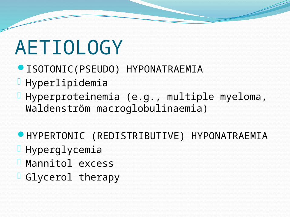
AETIOLOGYISOTONIC(PSEUDO) HYPONATRAEMIA- Hyperlipidemia - Hyperproteinemia (e.g., multiple myeloma,
Waldenström macroglobulinaemia)
HYPERTONIC (REDISTRIBUTIVE) HYPONATRAEMIA
- Hyperglycemia - Mannitol excess - Glycerol therapy

AETIOLOGY (cont’d)HYPOVOLAEMIC HYPONATRAEMIA
Renal Diuretic use Salt-wasting nephropathy (renal tubular acidosis,
chronic renal failure, interstitial nephritis) Mineralocorticoid (aldosterone) deficiency
Extra-renal Volume replacement with hypotonic fluids GI loss (vomiting, diarrhea, fistula, tube suction) Third-space loss (e.g., burns, hemorrhagic
pancreatitis, peritonitis)

AETIOLOGY (cont’d)HYPERVOLAEMIC HYPONATRAEMIA
Urinary [Na+] >20 mEq/L Renal failure (inability to excrete free water)
Urinary [Na+] <20 mEq/L Congestive heart failure Nephrotic syndrome Cirrhosis

AETIOLOGY (cont’d)EUVOLAEMIC HYPONATRAEMIA
Urine [Na+] usually > 20 mEq/L Hypothyroidism (possible increased ADH or deceased glomerular
filtration rate) Pain, stress, nausea, psychosis (stimulates non-osmotic release of
ADH) Drugs: ADH, nicotine, sulfonylureas, morphine, barbiturates, NSAIDs,
acetaminophen, carbamazepine, phenothiazines, tricyclic antidepressants, colchicine, clofibrate, cyclophosphamide, isoproterenol, tolbutamide, vincristine, monoamine oxidase inhibitor
Water intoxication, primary polydipsia Glucocorticoid deficiency Positive pressure ventilation Porphyria Essential (reset osmostat or sick cell syndrome—usually in the elderly)

AETIOLOGY (cont’d)EUVOLAEMIC HYPONATRAEMIA (cont’d)- SIAD: diagnosis of exclusion, the following criteria
must be metDecreased serum osmolality in the absence of diuretic
therapy
Inappropriately elevated urine osmolality (usually >200 mOsm/kg)
Elevated urine [Na+] (typically > 20 mEq/L)
Clinical euvolemia
Normal adrenal, renal, cardiac, hepatic, and thyroid function
Correctable with water restriction

Causes of SIADMalignancy Pulmonary
disordersCNS
DisordersDrugs Others
CarcinomaLungSmall cellMesotheliomaOropharynxGastrointestinal tractStomachDuodenumPancreasGenitourinary tractUreterBladderProstateEndometriumEndocrine thymomaLymphomasSarcomasEwing's sarcoma
InfectionsBacterial pneumoniaViral pneumoniaPulmonary abscessTuberculosisAspergillosisAsthmaCystic fibrosisRespiratory failure associated with positive-pressure breathing
InfectionEncephalitisMeningitisBrain abscessRocky Mountain spotted feverAIDSBleeding and massesSubdural hematomaSubarachnoid hemorrhageCerebrovascular accidentBrain tumorsHead traumaHydrocephalusCavernous sinusthrombosisOtherMultiple sclerosisGuillain-Barré syndromeShy-Drager syndromeDelerium tremensAcute intermittent polyphyria
Drugs that stimulate release of AVP or enhance its actionChlorpropamideSSRIsTricyclic antidepressantsClofibrateCarbamazepineVincristineNicotineNarcoticsAntipsychotic drugsIfosfamideCyclophosphamideNonsteroidal anti-inflammatory drugsMDMA (ecstasy)AVP analoguesDesmopressinOxytocinVasopressin
Hereditary (gain-of-function mutations in the vasopressin V2 receptor)
Idiopathic
Transient
Endurance exercise
General anesthesia
Nausea
Pain
Stress

HISTORYPatients may present to medical attention
with symptoms related to low serum sodium concentrations. However, many patients present due to manifestations of other medical comorbidities, with hyponatremia being recognized only secondarily. For many people, therefore, the recognition is entirely incidental. Patients may develop clinical symptoms due to the cause of hyponatremia or the hyponatremia itself

HISTORY (CONT’D) Symptoms range from nausea, appetite loss, malaise, with mild reduction
in the serum sodium, to lethargy, fatigue, muscle weakness, confusion, restlessness and irritability, spasms, cramps, decreased level of consciousness, headache, and (if severe) seizures and coma
Overt neurologic symptoms most often are due to very low serum sodium levels (usually < 115 mEq/L), resulting in intracerebral osmotic fluid shifts and brain edema. This neurologic symptom complex can lead to tentorial herniation with subsequent brain stem compression and respiratory arrest, resulting in death in the most severe cases
Detailed medication history, including information on OTC drugs the patient has been using, is important because many medications may precipitate hyponatremia
A dietary history with reference to salt, protein, and water intake is useful as well. For patients who are hospitalized, reviewing the records of parenteral fluids administered is crucial.
EXAMINATIONVolume statusDehydration
Oedema

DIFFERENTIAL HISTORY EXAM
Hypotonic replacement of excess fluid loss
history of excessive sweating, vomiting, diarrhea, GI fistulas or drainage tubes, or third spacing of fluids (peritonitis, pancreatitis, burns, small bowel obstruction) and fluid replacement by tap water or hypotonic intravenous fluids
clinical signs of volume depletion: decreased skin turgor, reduced jugular venous pressure, decreased blood pressure; small bowel obstruction: abdominal distension; peritonitis: rebound abdominal tenderness; cutaneous burns
Drug-induced history of use of thiazide diuretics, vasopressin, nonsteroidal anti-inflammatory drugs, nicotine, chlorpropamide, carbamazepine, tricyclic antidepressants, SSRIs, vincristine, thioridazine, cyclophosphamide, clofibrate, mannitol
usually normal
DIFFERENTIAL HISTORY EXAM
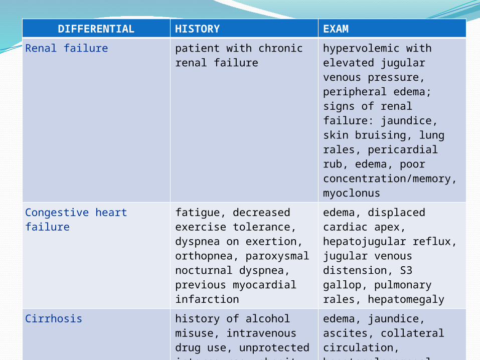
Renal failure patient with chronic renal failure
hypervolemic with elevated jugular venous pressure, peripheral edema; signs of renal failure: jaundice, skin bruising, lung rales, pericardial rub, edema, poor concentration/memory, myoclonus
Congestive heart failure fatigue, decreased exercise tolerance, dyspnea on exertion, orthopnea, paroxysmal nocturnal dyspnea, previous myocardial infarction
edema, displaced cardiac apex, hepatojugular reflux, jugular venous distension, S3 gallop, pulmonary rales, hepatomegaly
Cirrhosis history of alcohol misuse, intravenous drug use, unprotected intercourse, obesity, blood transfusion, known hepatitis infection; fatigue, weakness, weight loss, or pruritus
edema, jaundice, ascites, collateral circulation, hepatosplenomegaly, leukonychia, palmar erythema, spider angiomata, telengiectasia, jaundiced sclera, hepatic fetor, altered mental status

DIFFERENTIAL HISTORY EXAM
Hyperglycemia diabetes: history of diabetes, poor diabetes control, obesity, polyuria, polydipsia, blurred vision; use of causative medications: corticosteroids, niacin, pentamidine, protease inhibitors, some antipsychotics; stress hyperglycemia: recent stroke, myocardial infarction, trauma, infection, inflammation
features of metabolic syndrome in type 2 diabetes
Psychogenic polydipsia
history of schizophrenia or psychotic depression; phenothiazine medications; polydipsia; polyuria
usually normal; weight gain due to high water intake may occur in extreme cases

DIFFERENTIAL HISTORY EXAM
Intracranial surgery history of recent intracranial surgery
signs of volume depletion in cerebral salt-wasting syndrome: decreased skin turgor, reduced jugular venous pressure, decreased blood pressure
Head injury history of head trauma, possible loss of consciousness
may be normal in minor injuries; bruising or lacerations of scalp; evidence of basilar skull fracture: blood in the middle ear cavity (hemotympanum), raccoon eyes (periorbital ecchymosis), postauricular ecchymosis, CSF leakage (rhinorrhea or otorrhea); papilledema, retinal hemorrhage; decreased conscious level; signs of volume depletion in cerebral salt-wasting syndrome: decreased skin turgor, reduced jugular venous pressure, decreased blood pressure

DIFFERENTIAL HISTORY EXAM
Small cell lung cancer (SCLC)
history of cigarette smoking or exposure to tobacco smoke, radon gas, or asbestos; cough, hemoptysis, chest pain, dyspnea, weight loss, fatigue
patient may appear unwell with dyspnea and cachexia, finger clubbing, hypertrophic osteoarthropathy, and dullness to percussion of the lung fields
Irrigation of operative field
irrigation of operative field with hypertonic fluids during transurethral resection of prostate or hysteroscopy, or with hypotonic fluids during endometrial ablation
normal
Artifact in multiple myeloma
positive family history, previous exposure to irradiation or petroleum products, fatigue, pallor, shortness of breath, bone pain usually localized to the back
usually normal

INVESTIGATIONSDiagnosis is by measuring serum [Na+]There are 3 other essential laboratory tests in the
evaluation of patients with hyponatremia that, together with the history and the physical examination, help to establish the primary underlying etiologic mechanism:
1. Serum osmolality readily differentiates between true hyponatremia and pseudohyponatremia secondary to hyperlipidemia, hyperproteinemia, or hypertonic hyponatremia associated with elevated glucose, mannitol, glycine (posturologic or postgynecologic procedure), sucrose, or maltose (contained in IgG formulations).

2. Urinary sodium concentration helps to differentiate between hyponatremia secondary to hypovolemia and SIADH. With SIADH (and salt-wasting syndrome), the urine sodium is greater than 20-40 mEq/L. With hypovolemia, the urine sodium typically measures less than 25 mEq/L. However, if sodium intake in a patient with SIADH (or salt-wasting) happens to be low, then urine sodium may fall below 25 mEq/L.
3. Urine osmolality helps to differentiate between conditions associated with impaired free water excretion and primary polydipsia, in which water excretion should be normal (provided intact kidney function). With primary polydipsia, as with malnutrition (severe decreased solids intake) and reset osmostat, the urine osmolality is maximally dilute, generally less than 100 mOsm/kg. A urine osmolality greater than 100 mOsm/kg indicates impaired ability of the kidneys to dilute the urine. This usually is secondary to elevated vasopressin (ADH) levels, appropriate or inappropriate
Other tests are ancillary


ALGORITHMCorrected [Na+] in Hypertylipid/Hyperprot =
Serum Na x 93 99 – 1.03 (triglyceride
gm/L) – 0.73 (protein gm/L)
Corrected Na in hyperglycaemia = [Na+] + plasma Glc (mmol/L)/4
ORMeasured sodium + 0.016 X
[Serum glucose (mg/dl) – 100]

CONCLUSIONHyponatraemia is associated with many
medical illnesses, usually secondary to the disease process or treatment
Determining the type is essential in order to pinpoint its possible aetiologies
A thorough history, physical examination and baseline and ancillary investigations help to determine the root cause of the hyponatraemic state