Draft
Effect of bovine lactoferrin on Chlamydia trachomatis
infection and inflammation
Journal: Biochemistry and Cell Biology
Manuscript ID bcb-2016-0049.R2
Manuscript Type: Article
Date Submitted by the Author: 14-Sep-2016
Complete List of Authors: Sessa, Rosa; University of Rome, La Sapienza DI PIETRO, MARISA; University of Rome, La Sapienza, Department of Public Health and Infectious Diseases FILARDO, SIMONE; University of Rome, La Sapienza Bressan, Alessia; University of Rome, La Sapienza, rosa, luigi; University of Rome, La Sapienza
Cutone, Antimo; University of Rome, La Sapienza Frioni, Alessandra; SAPIENZA UNIVERSITY OF ROME Berlutti, Francesca; SAPIENZA UNIVERSITY, PUBLIC HEALTH AND INFECTIOUS DISEASES PAESANO, ROSALBA; SAPIENZA UNIVERSITY OF ROME, Department of Gynecological-Obstetric and Urological Sciences Valenti, Piera; University of Rome, La Sapienza, 1Department of Public Health and Infectious Diseases
Keyword: Chlamydia trachomatis, bovine lactoferrin, infection, inflammation, IL-6
https://mc06.manuscriptcentral.com/bcb-pubs
Biochemistry and Cell Biology

Draft
1
Effect of bovine lactoferrin on Chlamydia trachomatis infection and
inflammation
Rosa Sessa1, Marisa Di Pietro
1, Simone Filardo
1, Alessia Bressan
1, Luigi Rosa
1, Antimo
Cutone1, Alessandra Frioni
1, Francesca Berlutti
1, Rosalba Paesano
2 and Piera Valenti
1*
1Department of Public Health and Infectious Diseases, University of Rome, La Sapienza, Rome,
Italy; 2 Department of Gynecological-Obstetric and Urological Sciences, University of Rome, La
Sapienza, Rome, Italy
* Corresponding Author
Piera Valenti
Department of Public Health and Infectious Diseases
University of Rome, La Sapienza
Piazzale A. Moro 5, 00185 Rome (Italy)
Email: [email protected]
Page 1 of 25
https://mc06.manuscriptcentral.com/bcb-pubs
Biochemistry and Cell Biology
Draft
2
Abstract
Chlamydia trachomatis is an obligate, intracellular pathogen responsible for the most common
bacterial sexually transmitted disease worldwide, causing acute and chronic infections. The acute
infection is susceptible to antibiotics, while the chronic one needs prolonged therapies, thus
increasing the risk of developing antibiotic resistance. Novel alternative therapies are needed.
The intracellular development of C. trachomatis requires essential nutrients, including iron. Iron-
chelating drugs inhibit C. trachomatis developmental cycle.
Lactoferrin (Lf), a pleiotropic iron binding glycoprotein, could be a promising candidate against C.
trachomatis infection. Similarly to the efficacy against other intracellular pathogens, bovine Lf
(bLf) could both interfere with C. trachomatis entry into epithelial cells and exert an anti-
inflammatory activity.
In vitro and in vivo effects of bLf against C. trachomatis infectious and inflammatory process has
been investigated.
BLf inhibits C. trachomatis entry into host cells when incubated with cell monolayers before or at
the moment of the infection and down-regulates IL-6/IL-8 synthesized by infected cells.
Six out of seven pregnant women asymptomatically infected by C. trachomatis, after 30 days of
bLf intra-vaginal administration, were negative for C. trachomatis and showed a decrease of
cervical IL-6 levels.
This is the first time that the bLf protective effect against C. trachomatis infection has been
demonstrated.
Key words: Chlamydia trachomatis, bovine lactoferrin, infection, inflammation, IL-6
Page 2 of 25
https://mc06.manuscriptcentral.com/bcb-pubs
Biochemistry and Cell Biology

Draft
3
Introduction
Chlamydia trachomatis, an obligate intracellular pathogen, is the leading cause of bacterial sexually
transmitted infections in the world with an estimated over 131 million new cases per year (Newman
et al. 2015). C. trachomatis genital infection manifests in women as cervicitis, salpingitis and
endometritis, and can progress leading to severe sequelae, such as pelvic inflammatory disease,
ectopic pregnancy and obstructive infertility (Shaw et al. 2011). Importantly, a major concern with
chlamydial genital infections is that approximately 80% of women are asymptomatic, thus resulting
in a reservoir for onwards transmission in the population (Shaw et al. 2011).
C. trachomatis is characterized by a unique biphasic developmental cycle alternating between the
extracellular infectious bodies (Elementary Bodies, EBs), metabolically inactive, and the
intracellular non-infectious bodies (Reticulate Bodies, RBs), metabolically active. The EBs
adhesion and entry into mucosal epithelial cells initiate a signal transduction cascade of the host
cell, leading to the recruitment and reorganization of the actin cytoskeleton at the site of attachment.
Following the fusion of EB-containing endosomes, EBs develop into larger, metabolically active
but non-infectious RBs. Using ATP and nutrients from the host cell, RBs grow and divide by binary
fission within a membrane-bound vacuole, termed inclusion. Subsequently, the RBs asynchronously
transform into EBs, which are released, after approximately 48 hours, from the host cell by lysis
(Wyrick 2010; Bastidas et al. 2013).
In the recent years, it has been demonstrated that C. trachomatis can generate a persistent form
during its developmental cycle as a consequence of several stress-inducing factors (Di Pietro M et
al. 2013; Wyrick 2010). As a result, normal RBs transform into enlarged and morphologically
aberrant RBs, thus stopping the production of infectious EBs (Hogan et al. 2004; Wyrick 2010). In
particular, C. trachomatis enters into the persistence state in the presence of iron-chelating drugs,
which inhibit the developmental cycle and, hence, show its dependence on iron for the achievement
of infectious cycle (Raulston 1997; Thompson and Carabeo 2011). In this regard, iron limitation in
Page 3 of 25
https://mc06.manuscriptcentral.com/bcb-pubs
Biochemistry and Cell Biology

Draft
4
host cells has been shown to be of the utmost importance for the growth and survival of Chlamydia
spp. (Raulston 1997; Al-Younes et al. 2001).
Following C. trachomatis infection, cervical epithelial cells produce several pro-inflammatory
cytokines including TNF-α, IL-1α, IL-6 and IL-8 that augment the cell inflammatory response thus
inducing direct damage to genital tissues. Furthermore, IL-8, in turn, recruits innate immune cells,
which are abundant in the genital mucosa and are able to further worsen chronic inflammation and
tissue-damage of the reproductive system (Redgrove and McLaughlin 2014).
Interestingly, IL-8 recruits, duringthe infection/inflammation, neutrophils that synthesize and
secrete granules containing lactoferrin (Lf) (Masson et al. 1969).
Recently, a great interest in Lf, considered as a prominent component of the first line defense of the
host against infections and inflammation, has been raised.
Lf, an 80 kDa iron-binding glycoprotein, is found in most body fluids including vaginal fluid
(Valore et al. 2002). Lf possesses several biological functions dependent and independent from its
iron binding ability (Valenti and Antonini 2005). Among the biological properties related to its
iron-withholding ability, Lf inhibits bacterial infections, whereas, independently from iron
chelation, its high positive charge favors the binding to microorganisms and/or host cells, thus
hindering the adhesion and entry into epithelial cells (Valenti and Antonini 2005). In addition to
these activities, Lf exerts a potent anti-inflammatory activity, protecting infected host cells from
damages associated to pathological inflammation. In particular, Lf, independently from its iron
binding ability, decreases the synthesis of pro-inflammatory cytokines in infected epithelial cells
(Berlutti et al. 2006; Valenti et al. 2011; Puddu et al. 2011; Frioni et al. 2014).
Given the impact of asymptomatic chlamydial infection on disease outcomes and the
multifunctional features of Lf, the aim of our study was to evaluate the effects of bovine milk-
derivative Lf (bLf) on C. trachomatis infection and on the associated inflammatory state in vitro
and in vivo.
Page 4 of 25
https://mc06.manuscriptcentral.com/bcb-pubs
Biochemistry and Cell Biology
Draft
5
Materials and Methods
Chlamydia trachomatis strain and cell culture
C. trachomatis L2 strain 434/Bu (ATCC VR-902B) was obtained from American Type Culture
Collection.
The human epithelial HeLa-229 cell line from cervix adenocarcinoma (ATCC ® CCL-2.1™) was
cultured at 37°C in Dulbecco’s Modified Eagle Medium (D-MEM, Euroclone, Milan, Italy),
supplemented with 10% fetal calf serum (FCS, Euroclone, Milan, Italy), in humidified atmosphere
with 5% CO2.
Propagation and titration of Chlamydia trachomatis
Elementary body (EB) aliquots of C. trachomatis L2 were stored at −80°C and propagated in HeLa-
229 cells, grown in D-MEM supplemented with 10% FCS, as previously described by
Mastromarino et al. (2014). The infectious titer was assessed by immunofluorescence assay (IFA).
Briefly, HeLa-229 cells grown on glass coverslips in 24-well plates were infected with tenfold
serial dilutions of bacterial stock, incubated for 48 h at 37°C, fixed with methanol and stained with
fluorescein isothiocyanate-conjugated monoclonal (FITC) antibody anti-C. trachomatis
(MicroTrak, Trinity Biotech – USA). The total number of C. trachomatis Inclusion Forming Units
(IFUs) was obtained by counting all fields using a fluorescence microscope (100× magnification).
Lactoferrin
Highly purified bovine milk derivative lactoferrin (bLf) was kindly provided by Morinaga Milk
Industries Co., Ltd. (Tokyo, Japan). The absence of bLf degradation fragments was checked by
SDS-PAGE stained with silver nitrate. Lactoferrin concentration was assessed by UV spectroscopy
on the basis of an extinction coefficient of 15.1 (280 nm, 1% solution). The purity of bLf
corresponded to about 98% as also detected by High Performance Liquid Chromatography (HPLC)
analysis. The bLf iron saturation was about 20% as detected by optical spectroscopy at 468 nm on
the basis of an extinction coefficient of 0.54 (100% iron saturation). LPS contamination of bLf,
estimated by Limulus Amebocyte assay (LAL Pyrochrome kit, PBI International, Milan, Italy), was
Page 5 of 25
https://mc06.manuscriptcentral.com/bcb-pubs
Biochemistry and Cell Biology
Draft
6
0.7±0.06 ng/mg of bLf. Before biological assays, bLf solution was sterilized by filtration using 0.2
µm Millex HV at low protein retention (Millipore Corp., Bedford, Mass.). In all experiments bLf
was used at non-cytotoxic concentration corresponding to 100 µg/ml.
Effects of bovine lactoferrin on infection of HeLa-229 cellswith Chlamydia trachomatis
elementary bodies
i) Pre-incubation of bLf with C. trachomatis EBs
In order to detect the efficacy of bLf against C. trachomatis, 25,000 EBs/ml, corresponding to a
multiplicity of infection (MOI) of 0.05, were pre-incubated in D-MEM with FCS 2% (fresh
medium), in the absence or presence of bLf (100 µg/ml), for 1h or 3hs at 37°C in humidified
atmosphere with 5% CO2. Subsequently, the C. trachomatis EBs suspension was centrifuged at
30,000 × g for 15 min and the supernatant was removed. The pellet containing C. trachomatis EBs
was suspended in fresh medium and used to infect a total of about 105 HeLa-229 cells. Briefly, after
1 hour, the cells were washed with phosphate buffer solution without Ca2+
and Mg2+
(PBS) to
remove the non-internalized C. trachomatis EBs and newly incubated in fresh medium. After 48
hours post infection (h.p.i.) at 37°C in 5% CO2, the total number of C. trachomatis IFU was
determined by IFA.
ii) Pre-incubation of bLf with HeLa-229 cells before the infection with C. trachomatis EBs
HeLa-229 cells were pre-incubated in fresh medium in the absence or presence of bLf (100 µg/ml).
After 1h or 3hs of incubation at 37°C in 5% CO2, bLf was removed by washing the cell monolayers
three times with PBS. Subsequently, HeLa-229 cells were infected with C. trachomatis ata MOI of
0.05 as above described. After 48 h.p.i. at 37°C in 5% CO2, the total number of C. trachomatis IFU
was determined by IFA.
iii) bLf addition to HeLa-229 cells at the moment of infection with C. trachomatis EBs
In this set of experiments, bLf was added to HeLa-229 cells at the moment of infection. Briefly,
HeLa-229 cells were infected with C. trachomatis at a MOI of 0.05 in the absence or presence of
bLf (100 µg/ml). After 1 h at 37°C in 5% CO2, the cells were washed with PBS to remove the non-
Page 6 of 25
https://mc06.manuscriptcentral.com/bcb-pubs
Biochemistry and Cell Biology

Draft
7
internalized C. trachomatis EBs and fresh medium was added. After 48 h.p.i at 37°C and 5% CO2,
the total number of C. trachomatis IFU was determined by IFA.
iv) bLf addition to HeLa-229 cells three hours post C. trachomatis infection
HeLa-229 cells were infected with C. trachomatis at a MOI of 0.05. After 1h of incubation at 37°C
in 5% CO2, the cells were washed with PBS to remove the non-internalized C. trachomatis EBs and
fresh medium was added. After further 3hs of incubation at 37°C in 5% CO2, fresh medium, with or
without bLf (100 µg/ml), was added to the infected cells. After 48 h.p.i. at 37°C and 5% CO2, the
total number of C. trachomatis IFU was determined by IFA.
Detection of cytokines
Preliminary experiments, carried out with C. trachomatis EBs at a MOI of 0.05, showed a very low
cytokine expression by infected HeLa-229 cells. Therefore, HeLa-229 cells were infected with C.
trachomatis EBs at a MOI of 5 in order to reach an higher expression of IL-6 and IL-8 than that
observed at the MOI of 0.05. After 1h of incubation, the cells were washed with PBS to remove the
non-internalized C. trachomatis EBs and supplemented with fresh medium. After further 3hs of
incubation at 37°C in 5% CO2, fresh medium, with or without bLf (100 µg/ml), was added to the
infected cells. The cytokine production was determined in cell monolayer supernatants by ELISA
using Human ELISA Max Deluxe Set (BioLegend, San Diego, CA) after 48 hs of incubation at
37°C in 5% CO2.
Study design
We conducted an open-label cohort study in accordance with the ethical principles of the
Declaration of Helsinki. Approval was granted by the Ethics Committee of Clinica Fabia Mater, Via
Olevano Romano, 25 Rome, Italy (FM MOD 26022010). All pregnant women gave written
informed consent.
One hundred ninety-eight pregnant women from 20 to 40 years without ascertained pathologies,
with normal uterine cavity and intact membranes were enrolled regardless of trimester. Women
Page 7 of 25
https://mc06.manuscriptcentral.com/bcb-pubs
Biochemistry and Cell Biology

Draft
8
were excluded if they had a pathological pregnancy or if during this study were affected by bacterial
vaginal infections unrelated to C. trachomatis.
The exclusion of pregnant women during the clinical trial was also considered on the basis of
voluntary declaration, lack of treatment effectiveness, side effects, protocol infringement, and
missed programmed visits. As a matter of fact, the enrolled pregnant women had a monthly
scheduled visit.
Laboratory tests
At each scheduled visit, in addition to standard assays (haematocrit, glycemia, uricemia, bilirubin,
glutamicoxaloacetic transaminase, glutamic pyruvic transaminase, cholesterol, triglyceride acid and
electrolytes), cervical specimens were collected with polyethylene terephthalate (Dacron) swabs to
detect the presence of C. trachomatis.. In addition, cervical fluids were analyzed to detect IL-6
concentrations.
Chlamydia trachomatis detection
Cervical specimens were analyzed by direct immunofluorescence assay (DFA) using Syva
Microtrack kit (Syva Microtrack, Trinity Biotech, USA) according to the manufacturer’s
instructions. Briefly, the smears were fixed with methanol and stained with fluorescein
isothiocyanate conjugated (FITC) monoclonal antibody against C. trachomatis major outer
membrane protein (MOMP) for 30 minutes at 37°C in a humid chamber. The slides were examined
for the presence of IFUs using fluorescence microscope (100 × magnification).
Treatment against Chlamydia trachomatis infection in pregnant women
Among one hundred ninety-eight pregnant women, seven women, asymptomatically affected by C.
trachomatis, were immediately treated with bLf intravaginal administration. The intravaginal tablet,
containing 100 mg of lyophilized bLf 20% iron saturated, was administered every 8 h for 30 days.
The tablets were administered through a vaginal applicator to obtain a fast and adequate dissolution.
If the treatment with bLf intra-vaginal administration for 30 days was ineffective, the pregnant
women were submitted to antibiotic therapy (Workowski and Bolan 2015).
Page 8 of 25
https://mc06.manuscriptcentral.com/bcb-pubs
Biochemistry and Cell Biology

Draft
9
Maternal side effects
The side effects of bLf intravaginal administration were assessed by monitoring vaginal irritation,
itching and burning.
Fetal and newborn side effects
Fetal vital sign assessments were monitored by ultrasonographic measurements of intrauterine
growth and through the amount of amniotic fluid, expressed as the amniotic fluid index (AFI).
Newborn weight and Apgar score were registered. Apgar score is a practical method of evaluating
the physical condition of a newborn shortly after delivery (Apgar 1953). An Apgar score of 0–3 at
5–10 min of age is predictive of high morbidity and mortality, while an Apgar score of 9–10
means the infant is in the best possible conditions.
Statistical analysis
All values were expressed as mean ± standard deviation (SD) of three replicates from three
independent in vitro experiments. The concentrations of IL-6 in cervical fluid of pregnant women
were expressed as mean values ± SD. Comparison of means was performed by using a two-tailed t-
test for independent samples. A value of P < 0.05 was considered statistically significant.
Page 9 of 25
https://mc06.manuscriptcentral.com/bcb-pubs
Biochemistry and Cell Biology
Draft
10
Results
Effects of bovine lactoferrin on Chlamydia trachomatis infection
We evaluated the effects of bLf, at non-cytotoxic concentration corresponding to 100 µg/ml, on C.
trachomatis infections.
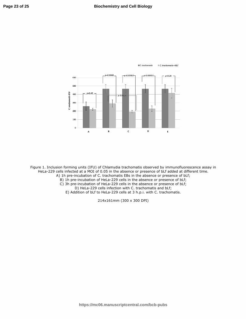
As shown in Figure 1A, no significant reduction in the number of chlamydial IFUs was observed
when chlamydial EBs were pre-incubated with bLf for 1h or 3hs, indicating no direct effect of bLf
on C. trachomatis.
In contrast, bLf was able to inhibit C. trachomatis entry into host cells as evidenced by a significant
reduction of chlamydial IFU observed when HeLa-229 monolayers were pre-incubated with bLf for
1h or 3hs (1h pre-incubation: p=0.0008; 3hs pre-incubation: p=0.00007) (Figure 1 B, C). The
inhibitory effect of bLf on C. trachomatis entry was more pronounced when HeLa-229 cells were
pre-incubated with bLf for 3hs as compared to 1h (p=0.0124) (Figure 1C).
To further confirm the inhibitory effect of bLf on C. trachomatis entry into host cells, bLf was
added at the moment of HeLa-229 monolayer infection with C. trachomatis. The presence of bLf
during the infection phase significantly inhibited C. trachomatis entry into HeLa-229 cells at the
same extent evidenced when bLf was pre-incubated with cell monolayers for 1h or 3hs (Figure 1 B,
C, D). In order to determine whether bLf was also able to inhibit chlamydial replication into host
cell, bLf was added after 3hs of C. trachomatis infection. The addition of bLf under these
experimental conditions resulted in no significant reduction of the number of intracellular
chlamydial IFUs (p=0.28) (Figure 1 E).
Effect of lactoferrin on IL-6 and IL-8 cytokine production by Chlamydia trachomatis-infected
HeLa-229 cells
To investigate the effect of bLf on the inflammatory response, HeLa-229 cells were infected with C.
trachomatis at a MOI of 5 and, after 3hs of infection, bLf was added to the medium. Of note, the
addition of bLf after 3hs post infection did not influence the intracellular number of C. trachomatis
infecting the cell monolayers at the MOI of 0.05 (Figure 1 E) or at the MOI of 5 (data not shown),
Page 10 of 25
https://mc06.manuscriptcentral.com/bcb-pubs
Biochemistry and Cell Biology
Draft
11
thus allowing the detection of the actual synthesis of IL-6 and IL-8 by the same number of
intracellular C. trachomatis. The production of IL-6 and IL-8 was evaluated in the supernatants
(Figures 2 and 3 respectively). The treatment with bLf did not raise the cytokine levels in non-
infected cells as compared to cell monolayers alone. On the contrary, the infection with C.
trachomatis induced a significant increase of both IL-6 and IL-8 levels. The addition of bLf to
infected cells 3hs post infection determined a significant decrease of both IL-6 and IL-8 levels as
compared to bLf-untreated infected cells (P<0.05). In particular, bLf significantly decreased IL-6
and IL-8 concentrations, even if cytokine levels remained higher than those synthesized by non-
infected cell monolayers.
Clinical trial
Among one hundred ninety-eight pregnant women, 16 women, affected by bacterial vaginosis
unrelated to C. trachomatis, and 6 women, for protocol violation, were excluded. One hundred
seventy-six pregnant women completed the study and, among them, seven asymptomatic pregnant
women, positive to C. trachomatis DFA and showing high concentration of IL-6 in cervical fluids,
were treated with the intravaginal administration of bLf (100 mg) every 8hs for 30 days.
After one month, six out of seven cervical specimens were negative to C. trachomatis DFA and the
cervical fluids showed a decrease in IL-6 concentration (from mean values of 250±19 to 50±11
pg/ml). One out of seven pregnant women was positive to C. trachomatis DFA and cervical IL-6
levels did not decrease, ranging between about 270 and 300 pg/ml (Table 1). This patient was
treated with antibiotic therapy.
No maternal and neonatal side effects by bLf intra-vaginal administration were observed.
Page 11 of 25
https://mc06.manuscriptcentral.com/bcb-pubs
Biochemistry and Cell Biology
Draft
12
Discussion
Some mucosal pathogenic bacteria are not only capable of adhering, but also of entering into non-
professional phagocytes, such as epithelial cells. Inside host cells, bacteria are in a protective niche
in which they can replicate and persist, thus avoiding host defences. In addition, antibiotic therapies
are not always effective in the eradication of intracellular pathogens (Armstead and Li 2011).
C. trachomatis is an obligate, intracellular pathogen responsible for the most common bacterial
sexually transmitted disease worldwide, causing acute and chronic infections. Differently from the
acute infection, cured with oral or topical administration of antibiotics, the chronic one is difficult to
eradicate and needs prolonged therapies, thus increasing the risk of developing antibiotic resistance
(Kohlhoff and Hammerschlag 2015) .
Therefore, novel alternative therapies are needed (Sessa et al. 2015). The difficulty to find new
agents anti-C. trachomatis infection resides in the complex life-cycle of this peculiar pathogen. In
fact, C. trachomatis has a unique biphasic developmental cycle alternating between the extracellular
infectious EBs metabolically inactive and the intracellular non-infectious RBs, metabolically active.
Of note, intracellular bacterial pathogens require intracellular nutrients, including iron, for
replication in mammalian cells, and chlamydiae are no exception (Raulston 1997).
Concerning the first phase of C. trachomatis infection, classical anti-bacterial drugs are ineffective
because EBs are metabolically inactive.
Conversely, anti-bacterial drugs could be active against intracellular replicative RBs, since they are
metabolically active. However, antibacterial drugs cannot usually enter inside host cells.
A further key issue is represented by the intracellular re-differentiationof RBs (after intracellular
replication) into EBs, which are released following the lysis of host cells, ready to infect
neighboring epithelial cells and, hence, perpetuate the infectious process (Belland et al. 2003).
Therefore, an ideal drug against C. trachomatis infection should:
- inhibit C. trachomatis EBs adhesion and entry into host cells;
- inhibit C. trachomatis RBs intracellular replication;
Page 12 of 25
https://mc06.manuscriptcentral.com/bcb-pubs
Biochemistry and Cell Biology

Draft
13
- inhibit the re-infection of host cells by EBs, extracellularly released after the re-differentiation of
RBs into EBs.
Lf is thought to play a pivotal role in the prevention of infections. Its ability to sequester iron from
potential pathogens is considered as an important feature in order to contrast infections. Moreover,
its cationic charge is responsible for the binding to bacterial and cell surface components (Valenti
and Antonini 2005). This Lf property has been shown to inhibit the adhesion and entry into
epithelial cells of several facultative intracellular bacteria (Longhi et al. 1993; Ajello et al. 2002; Di
Biase et al. 2004; Willer et al. 2004; Berlutti et al. 2008); however Lf activity against obligate
intracellular bacteria as C. trachomatis has never been observed.
In this study, we utilized a preparation of bLf, iron saturated at 20%, to consent further iron
chelation, an essential nutrient for C. trachomatis developmental cycle (Raulston 1997). In facts, in
the absence of free, available iron, C. trachomatis enters into a persistent state, as evidenced by the
addition of iron-chelating agents, such as deferoxamine mesylate (DFO) or 2,2′-bipyridyl (Bpdl), to
C. trachomatis infected cell monolayers, leading to small-sized inclusions containing enlarged,
aberrant and non-dividing RBs (Thompson and Carabeo 2011), unable to generate infectious
progeny (Wyrick 2010).
Differently from data reported by Thompson and Carabeo (2011), we found that the addition of bLf
to HeLa cell monolayers 3hs post-infection resulted in no significant reduction of the number of
intracellular chlamydial IFU (p=0.28) (Figure 1 E) and of infectious progeny.These conflicting data
could be due to the higher effective concentrations of iron-chelating agents (from 100 to 200 µM)
(Thompson and Carabeo 2011) as compared to 1.25 µM bLf, corresponding to 2.5 µM iron binding
sites, used in this study.
We believe to be very interesting that bLf does not affect both the replication of RBs and the
induction of aberrant RBs, thus avoiding the “silent” reservoir that leads to chronic infection and
inflammation.
Page 13 of 25
https://mc06.manuscriptcentral.com/bcb-pubs
Biochemistry and Cell Biology
Draft
14
In fact, aberrant RBs can contribute to chronic inflammation, even if this aspect is still under
debate. Of note, the recurrent chlamydial disease may also result from the persistence of the
microorganism after unresolved infections (Wyrick 2010).
At the best of our knowledge, we demonstrated, for the first time, that the incubation of cell
monolayers with bLf before the infection or at the moment of the infection inhibited, in a significant
part, C. trachomatis adhesion and entry into epithelial cells. Therefore, the inhibition of C.
trachomatis infectivity by bLf was dependent on its interaction with cell surface. As a matter of
fact, bLf was able to bind to cell surface glycosaminoglycans as well as to heparan sulfate
proteoglycans (Wu et al. 1995; Lang et al. 2011), potential receptors for C. trachomatis adhesion
(Stallmann and Hegemann 2015) .
Conversely, the pre-incubation of bLf with C. trachomatis did not influence its infectivity,
supporting that the specific interaction between bLf and epithelial host cells seems to be the sole
pivotal mechanism responsible for the inhibition of C. trachomatis invasion.
Similarly to the results obtained in epithelial cell monolayers infected with other facultative
intracellular pathogens (Berlutti et al. 2006; Valenti et al. 2011; Frioni et al. 2014), the addition of
bLf significantly decreased the IL-8 and IL-6 levels synthetized by C. trachomatis infected cells. To
avoid that the IL-8 and IL-6 decrease was related to the different number of C. trachomatis IFUs,
these experiments were carried out adding bLf 3 hs post infection. These results demonstrated once
again the ability of bLf to down-regulate pro-inflammatory cytokine synthesis. Although it has been
known for years that exogenous Lf localized to cell nucleus (Ashida et al. 2004; Suzuki et al. 2008;
Valenti et al. 2011), the mechanisms by which bLf could perform its anti-inflammatory activity are
still under debate.
These in vitro results, showing for the first time the protective effects of bLf against C. trachomatis
infection, led us to investigate its efficacy also in asymptomatic pregnant women positive to C.
trachomatis and with high levels of IL-6 in cervical fluids.
Page 14 of 25
https://mc06.manuscriptcentral.com/bcb-pubs
Biochemistry and Cell Biology
Draft
15
Seven Out of one hundred seventy-six pregnant women enrolled in this pilot study, showing
cervical specimens positive to C. trachomatis, were treated with the intravaginal administration of
bLf (100 mg) every 8hs for 30 days.
After one month, six pregnant women were negative to C. trachomatis and showed decreased IL-6
levels in their cervical fluids (from mean values of 250±19 to 50±11 pg/ml).
Similarly to what we observed in the in vitro model, bLf intravaginal administration seems to act by
protecting host cells against the adhesion and entry of chlamydial EBs, extracellularly released after
re-differentiationof RBs to EBs. The simultaneous decrease of IL-6 levels could be a marker for the
lack of re-infection by C. trachomatis EBs in the presence of bLf.
Even if other clinical trials are required, the protective effect of bLf against C. trachomatis,
demonstrated for the first time in this study, suggests a further therapeutic approach based on its
intravaginal administration in addition to that already reported in preventing and curing the preterm
delivery (Paesano et al. 2012).
Acknowledgment
This study was supported by “Sapienza” University to both R S and to P V.
Page 15 of 25
https://mc06.manuscriptcentral.com/bcb-pubs
Biochemistry and Cell Biology
Draft
16
References
Ajello, M., Greco, R., Giansanti, F., Massucci, M.T., Antonini, G., and Valenti, P. 2002. Anti-
invasive activity of bovine lactoferrin towards group A streptococci. Biochem. Cell Biol. 80(1):
119-124.
Al-Younes, H.M., Rudel, T., Brinkmann, V., Szczepek, A.J., and Meyer, T.F. 2001. Low iron
availability modulates the course of Chlamydia pneumoniae infection. Cell. Microbiol. 3(6): 427-
437. doi:10.1046/j.1462-5822.2001.00125.x.
Apgar, V. 1953. A proposal for a new method of evaluation of the newborn infant. Curr. Res.
Anesth. Analg. 32(4): 260-267.
Armstead, A.L., and Li, B. 2011. Nanomedicine as an emerging approach against intracellular
pathogens. Int. J. Nanomedicine 6: 3281-3293.
Ashida, K., Sasaki, H., Suzuki, Y.A., and Lönnerdal, B. 2004. Cellular internalization of lactoferrin
in intestinal epithelial cells. Biometals. 17(3): 311-315.
Bastidas, R.J., Elwell, C.A., Engel, J.N., and Valdivia, R.H. 2013. Chlamydial intracellular survival
strategies. Cold Spring Harb. Perspect. Med. 3(5): a010256. doi:10.1101/cshperspect.a010256.
Belland, R.J., Zhong, G., Crane, D.D., Hogan, D., Sturdevant, D., Sharma, J., Beatty, W.L., and
Caldwell, H.D. 2003. Genomic transcriptional profiling of the developmental cycle of Chlamydia
trachomatis. Proc. Natl. Acad. Sci. U.S.A. 100(14): 8478-8483. doi:10.1073/pnas.1331135100.
Berlutti, F., Schippa, S., Morea, C., Sarli, S., Perfetto, B., Donnarumma, G., and Valenti, P. 2006.
Lactoferrin downregulates pro-inflammatory cytokines upexpressed in intestinal epithelial cells
infected with invasive or noninvasive Escherichia coli strains. Biochem. Cell Biol. 84(3): 351-357.
doi:10.1139/o06-039.
Berlutti, F., Superti, F., Nicoletti, M., Morea, C., Frioni, A., Ammendolia, M.G., Battistoni, A., and
Valenti, P. 2008. Bovine lactoferrin inhibits the efficiency of invasion of respiratory A549 cells of
different iron-regulated morphological forms of Pseudomonas aeruginosa and Burkholderia
cenocepacia. Int. J. Immunopathol. Pharmacol. 21(1): 51-59.
Page 16 of 25
https://mc06.manuscriptcentral.com/bcb-pubs
Biochemistry and Cell Biology
Draft
17
Di Biase, A.M., Tinari, A., Pietrantoni, A., Antonini, G., Valenti, P., Conte, M.P., and Superti, F.
2004. Effect of bovine lactoferricin on enteropathogenic Yersinia adhesion and invasion in HEp-2
cells. J. Med. Microbiol. 53(Pt 5): 407-412. doi:10.1099/jmm.0.05410-0.
Di Pietro, M., Filardo, S., De Santis, F., and Sessa, R. 2013. New insights into Chlamydiae
persistence: an energy metabolism strategy? Int J Immunopathol Pharmacol. 26(2):525-8.
Frioni, A., Conte, M.P., Cutone, A., Longhi, C., Musci, G., di Patti, M.C., Natalizi, T., Marazzato,
M., Lepanto, M.S., Puddu, P., Paesano, R., Valenti, P., and Berlutti, F. 2014. Lactoferrin differently
modulates the inflammatory response in epithelial models mimicking human inflammatory and
infectious diseases. Biometals. 27(5): 843-856. doi:10.1007/s10534-014-9740-9.
Hogan, R.J., Mathews, S.A., Mukhopadhyay, S., Summersgill, J.T., and Timms, P. 2004.
Chlamydial persistence: beyond the biphasic paradigm. Infect. Immun. 72(4): 1843-1855.
doi:10.1128/IAI.72.4.1843-1855.2004.
Kohlhoff, S.A., and Hammerschlag, M.R. 2015. Treatment of Chlamydial infections: 2014 update.
Expert Opin. Pharmacother. 16(2): 205-212. doi:10.1517/14656566.2015.999041.
Lang, J., Yang, N., Deng, J., Liu, K., Yang, P., Zhang, G., and Jiang, C. 2011. Inhibition of SARS
pseudovirus cell entry by lactoferrin binding to heparan sulfate proteoglycans. PLoS ONE. 6(8):
e23710. doi:10.1371/journal.pone.0023710.
Longhi, C., Conte, M.P., Seganti, L., Polidoro, M., Alfsen, A., and Valenti, P. 1993. Influence of
lactoferrin on the entry process of Escherichia coli HB101 (pRI203) in HeLa cells. Med. Microbiol.
Immunol. 182(1): 25-35.
Masson, P.L., Heremans, J.F., and Schonne, E. 1969. Lactoferrin, an iron-binding protein in
neutrophilic leukocytes. J. Exp. Med. 130(3): 643-658.
Mastromarino, P., Di Pietro, M., Schiavoni, G., Nardis, C., Gentile, M., and Sessa, R. 2014. Effects
of vaginal lactobacilli in Chlamydia trachomatis infection. Int. J. Med. Microbiol. 304(5-6): 654-
661. doi:10.1016/j.ijmm.2014.04.006.
Page 17 of 25
https://mc06.manuscriptcentral.com/bcb-pubs
Biochemistry and Cell Biology
Draft
18
Newman, L., Rowley, J., Vander, H.S., Wijesooriya, N.S., Unemo, M., Low, N., Stevens, G.,
Gottlieb, S., Kiarie, J., and Temmerman, M. 2015. Global estimates of the prevalence and incidence
of four curable sexually transmitted infections in 2012 based on systematic review and global
reporting. PLoS ONE. 10(12): e0143304. doi:10.1371/journal.pone.0143304.
Paesano, R., Pietropaoli, M., Berlutti, F., and Valenti, P. 2012. Bovine lactoferrin in preventing
preterm delivery associated with sterile inflammation. Biochem. Cell Biol. 90(3): 468-475.
doi:10.1139/o11-060.
Puddu, P., Latorre, D., Carollo, M., Catizone, A., Ricci, G., Valenti, P., and Gessani, S. 2011.
Bovine lactoferrin counteracts Toll-like receptor mediated activation signals in antigen presenting
cells. PLoS ONE. 6(7): e22504. doi:10.1371/journal.pone.0022504.
Raulston, J.E. 1997. Response of Chlamydia trachomatis serovar E to iron restriction vitro and
evidence for iron-regulated chlamydial proteins. Infect. Immun. 65(11): 4539-4547.
Redgrove, K.A., and McLaughlin, E.A. 2014. The role of the immune response in Chlamydia
trachomatis infection of the male genital tract: a double-edged sword. Front. Immunol. 5: 534.
doi:10.3389/fimmu.2014.00534.
Sessa, R., Di Pietro, M., De Santis, F., Filardo, S., Ragno, R., and Angiolella, L. 2015 Effects of
Mentha suaveolens essential oil on Chlamydia trachomatis. Biomed Res Int. 2015:508071. doi:
10.1155/2015/508071.Shaw, K., Coleman, D., O'Sullivan, M., and Stephens, N. 2011. Public health
policies and management strategies for genital Chlamydia trachomatis infection. Risk Manag.
Healthc. Policy. 4: 57-65. doi:10.2147/RMHP.S12710.
Stallmann, S., Hegemann, J.H. 2015. The Chlamydia trachomatis Ctad1 invasin exploits the human
integrin β1 receptor for host cell entry. Cell. Microbiol. doi:10.1111/cmi.12549.
Suzuki, Y.A., Wong, H., Ashida, K.Y., Schryvers, A.B., and Lönnerdal, B. 2008. The N1 domain of
human lactoferrin is required for internalization by CaCo-2 cells and targeting to the nucleus.
Biochemistry. 47(41): 10915-10920. doi:0.1021/bi8012164.
Page 18 of 25
https://mc06.manuscriptcentral.com/bcb-pubs
Biochemistry and Cell Biology
Draft
19
Thompson, C.C., and Carabeo, R.A. 2011. An optimal method of iron starvation of the obligate
intracellular pathogen, Chlamydia trachomatis. Front. Microbiol. 2: 20.
doi:10.3389/fmicb.2011.00020. eCollection 2011.
Valenti, P., and Antonini, G. 2005. Lactoferrin: an important host defence against microbial and
viral attack. Cell. Mol. Life Sci. 62(22): 2576-2587. doi:10.1007/s00018-005-5372-0.
Valenti, P., Catizone, A., Pantanella, F., Frioni, A., Natalizi, T., Tendini, M., and Berlutti, F. 2011.
Lactoferrin decreases inflammatory response by cystic fibrosis bronchial cells invaded with
Burkholderia cenocepacia iron-modulated biofilm. Int. J. Immunopathol. Pharmacol. 24(4): 1057-
1068.
Valore EV, Park CH, Igreti SL, Ganz T. 2002. Antimicrobial components of vaginal fluid. Am. J.
Obstet. Gynecol. 187(3): 561-568.
Willer, E.daM., Lima, R.deL., and Giugliano, L.G. 2004. In vitro adhesion and invasion inhibition
of Shigella dysenteriae, Shigella flexneri and Shigella sonnei clinical strains by human milk
proteins. BMC Microbiol. 4: 18. doi:10.1186/1471-2180-4-18.
Workowski, K.A., and Bolan, G.A. 2015. Sexually transmitted diseases treatment guidelines, 2015.
MMWR Recomm. Rep. 64(No. RR-3): 1-135.
Wu, H.F., Monroe, D.M., and Church, F.C. 1995. Characterization of the glycosaminoglycan-
binding region of lactoferrin. Arch. Biochem. Biophys. 317(1): 85-92. doi:10.1006/abbi.1995.1139.
Wyrick, P.B. 2010. Chlamydia trachomatis persistence in vitro: an overview. J. Infect. Dis. 201(2):
S88-95. doi:10.1086/652394.
Page 19 of 25
https://mc06.manuscriptcentral.com/bcb-pubs
Biochemistry and Cell Biology

Draft
20
Figure captions
Figure 1. Inclusion forming units (IFU) of Chlamydia trachomatis observed by
immunofluorescence assay in HeLa-229 cells infected at a MOI of 0.05 in the absence or presence
of bLf added at different time.
A) 1h pre-incubation of C. trachomatis EBs in the absence or presence of bLf;
B) 1h pre-incubation of HeLa-229 cells in the absence or presence of bLf;
C) 3h pre-incubation of HeLa-229 cells in the absence or presence of bLf;
D) HeLa-229 cells infection with C. trachomatis and bLf;
E) Addition of bLf to HeLa-229 cells at 3 h.p.i. with C. trachomatis.
Figure 2. IL-6 levels in the supernatants of HeLa-229 cell cultures after 48hs of incubation.
The infection was performed at a MOI of 5 and bLf was added 3 hours post infection. The IL-6
concentrations are expressed as mean values ± standard deviation (SD). A value of P<0.05 was
considered statistically significant.
Figure 3. IL-8 levels in the supernatants of HeLa-229 cell cultures after 48hs of infection. The
infection was performed at a MOI of 5 and bLf was added 3 hours post infection. The IL-8
concentrations are expressed as mean values ± standard deviation (SD). A value of P<0.05 was
considered statistically significant.
Page 20 of 25
https://mc06.manuscriptcentral.com/bcb-pubs
Biochemistry and Cell Biology

Draft
Table 1. General characteristics of pregnant women positive to C. trachomatis before and after the
intravaginal administration of lactoferrin (100 mg).
Before treatment After treatment
n = 6
C. trachomatis DFA Positive Negative
Mean values of cervical IL-6
(pg/ml)
250±19 50±11
Spontaneous delivery (week) 39-40th
Mean values of birth weight (g) 3,727±175
Apgar scores 9/10
n = 1
C. trachomatis DFA Positive Positive
Cervical IL-6 (pg/ml) 263 285
Caesarean section (week) 39th
Birth weight (g) 3,378
Apgar scores 9
Note: DFA, Direct Immunofluorescent Assay.
Page 21 of 25
https://mc06.manuscriptcentral.com/bcb-pubs
Biochemistry and Cell Biology

Draft
Page 22 of 25
https://mc06.manuscriptcentral.com/bcb-pubs
Biochemistry and Cell Biology
Draft
Figure 1. Inclusion forming units (IFU) of Chlamydia trachomatis observed by immunofluorescence assay in HeLa-229 cells infected at a MOI of 0.05 in the absence or presence of bLf added at different time.
A) 1h pre-incubation of C. trachomatis EBs in the absence or presence of bLf;
B) 1h pre-incubation of HeLa-229 cells in the absence or presence of bLf; C) 3h pre-incubation of HeLa-229 cells in the absence or presence of bLf;
D) HeLa-229 cells infection with C. trachomatis and bLf; E) Addition of bLf to HeLa-229 cells at 3 h.p.i. with C. trachomatis.
214x161mm (300 x 300 DPI)
Page 23 of 25
https://mc06.manuscriptcentral.com/bcb-pubs
Biochemistry and Cell Biology

Draft
Figure 2. IL-6 levels in the supernatants of HeLa-229 cell cultures after 48hs of incubation. The infection was performed at a MOI of 5 and bLf was added 3 hours post infection. The IL-6
concentrations are expressed as mean values ± standard deviation (SD). A value of P<0.05 was considered
statistically significant.
214x161mm (300 x 300 DPI)
Page 24 of 25
https://mc06.manuscriptcentral.com/bcb-pubs
Biochemistry and Cell Biology

Draft
Figure 3. IL-8 levels in the supernatants of HeLa-229 cell cultures after 48hs of infection. The infection was performed at a MOI of 5 and bLf was added 3 hours post infection. The IL-8 concentrations are expressed as
mean values ± standard deviation (SD). A value of P<0.05 was considered statistically significant.
214x161mm (300 x 300 DPI)
Page 25 of 25
https://mc06.manuscriptcentral.com/bcb-pubs
Biochemistry and Cell Biology

![Chlamydia trachomatis Genital Infections - Microbial Cellmicrobialcell.com/wordpress/wp-content/uploads/2016/09/2016A-OConnell... · Chlamydia trachomatis [2]. Serovars Ainfections](https://cdn.vdocument.in/doc/165x107/5e1cc35f8cecfe525f4fd6a8/chlamydia-trachomatis-genital-infections-microbial-chlamydia-trachomatis-2.jpg)


