Download - Dysrhythmias and Blocks
Dysrhythmias and Blocks
BKN
1/24/02
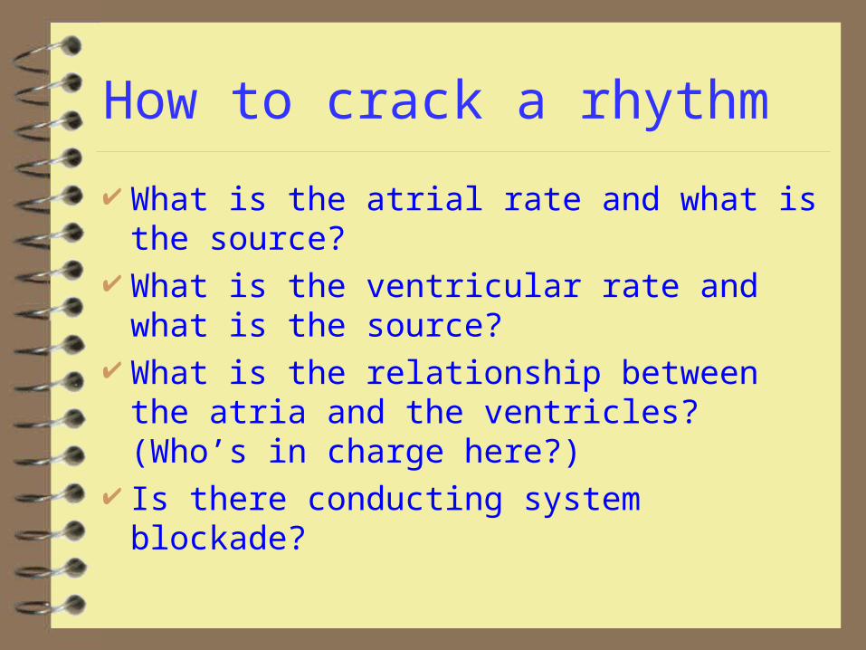
How to crack a rhythm
What is the atrial rate and what is the source?
What is the ventricular rate and what is the source?
What is the relationship between the atria and the ventricles? (Who’s in charge here?)
Is there conducting system blockade?
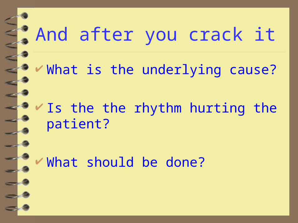
And after you crack it
What is the underlying cause?
Is the the rhythm hurting the patient?
What should be done?
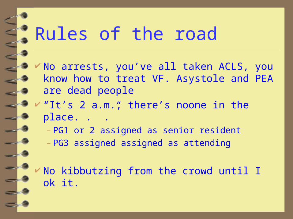
Rules of the road
No arrests, you’ve all taken ACLS, you know how to treat VF. Asystole and PEA are dead people
“It’s 2 a.m., there’s noone in the place. . .”– PG1 or 2 assigned as senior resident– PG3 assigned assigned as attending
No kibbutzing from the crowd until I ok it.
Case 1: Dr. Nelson’s Happy New Year 51 y/o M complaining of palpitations Drunk on homemade wine (ETOH = 225) PE unremarkable Intermittent episodes of rapid pulse and
pallor with BP 95/60 lasting up to 1/2 minute
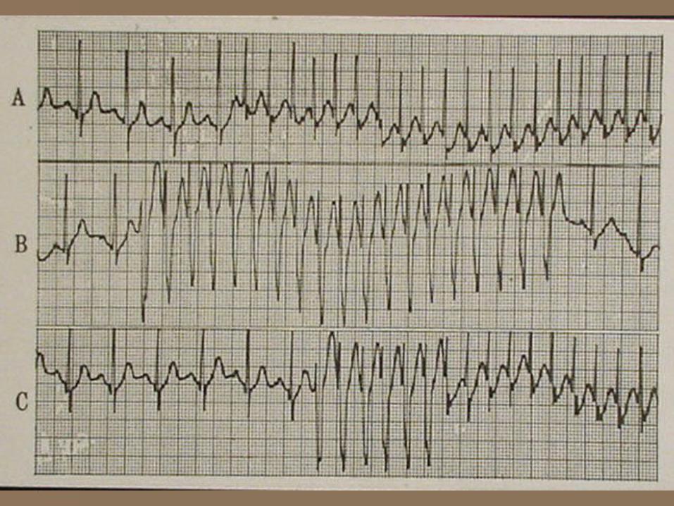
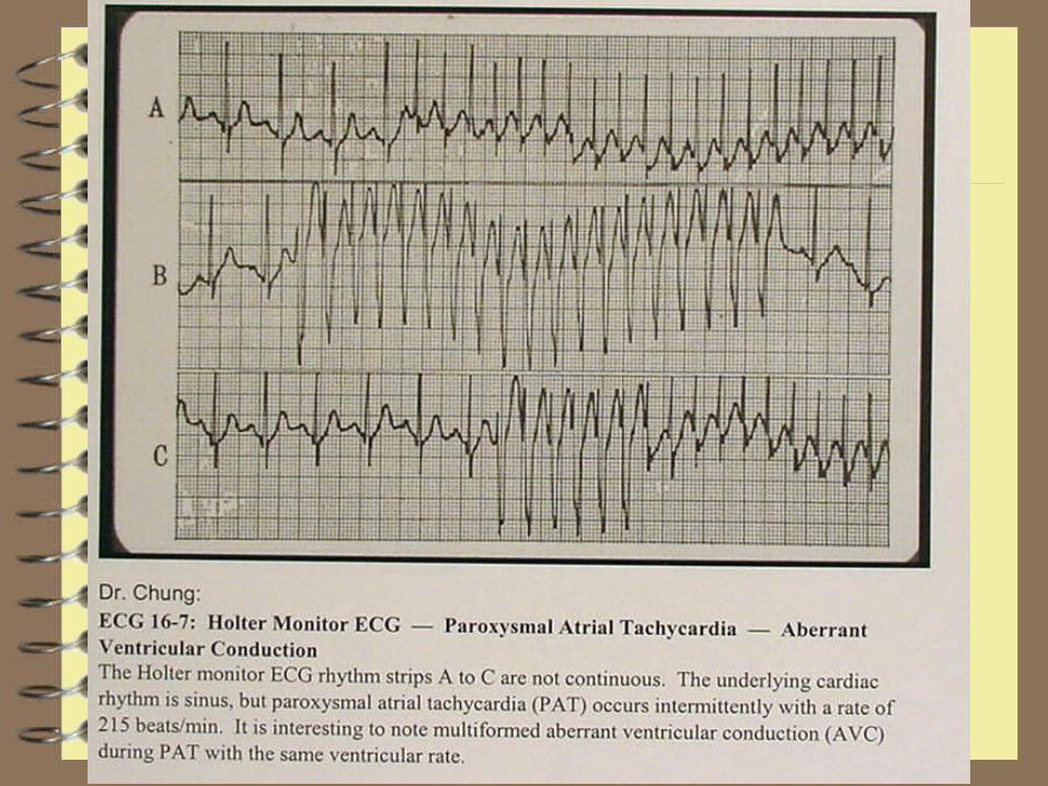
Rhythm strips follow; not continuous
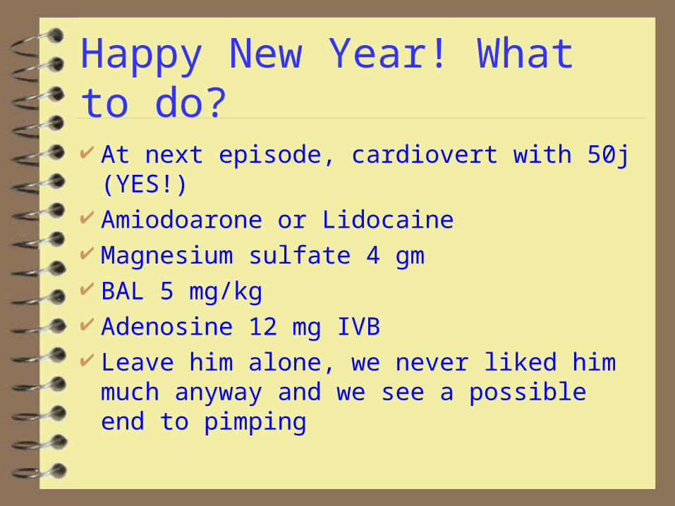
Happy New Year! What to do?
At next episode, cardiovert with 50j (YES!) Amiodoarone or Lidocaine Magnesium sulfate 4 gm BAL 5 mg/kg Adenosine 12 mg IVB Leave him alone, we never liked him much
anyway and we see a possible end to pimping
Happy New Year
What’s the rhythm?
Is it hurting him?
What’s the cause?
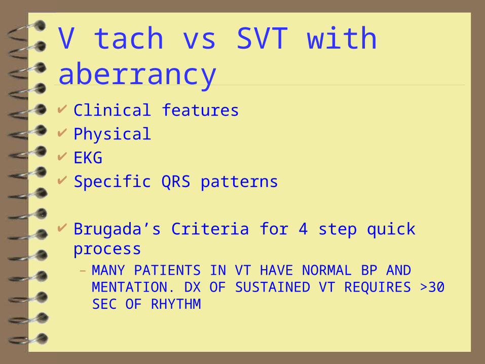
V tach vs SVT with aberrancy
Clinical features Physical EKG Specific QRS patterns
Brugada’s Criteria for 4 step quick process– MANY PATIENTS IN VT HAVE NORMAL BP AND
MENTATION. DX OF SUSTAINED VT REQUIRES >30 SEC OF RHYTHM
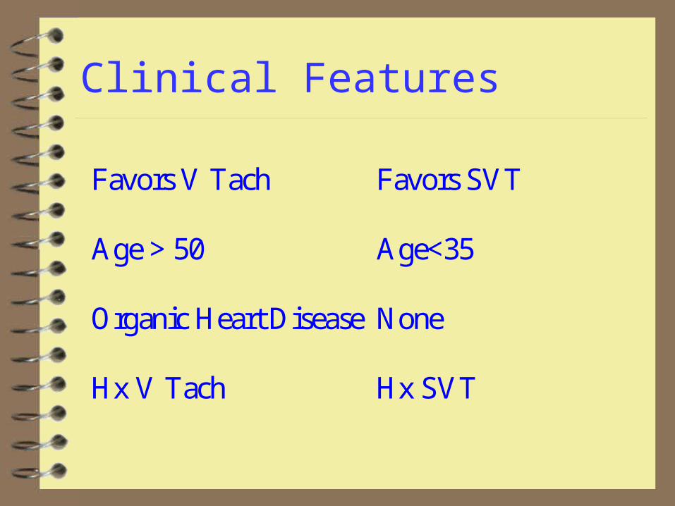
Clinical Features
Favors V Tach Favors SVT
Age > 50 Age<35
Organic Heart Disease None
Hx V Tach Hx SVT
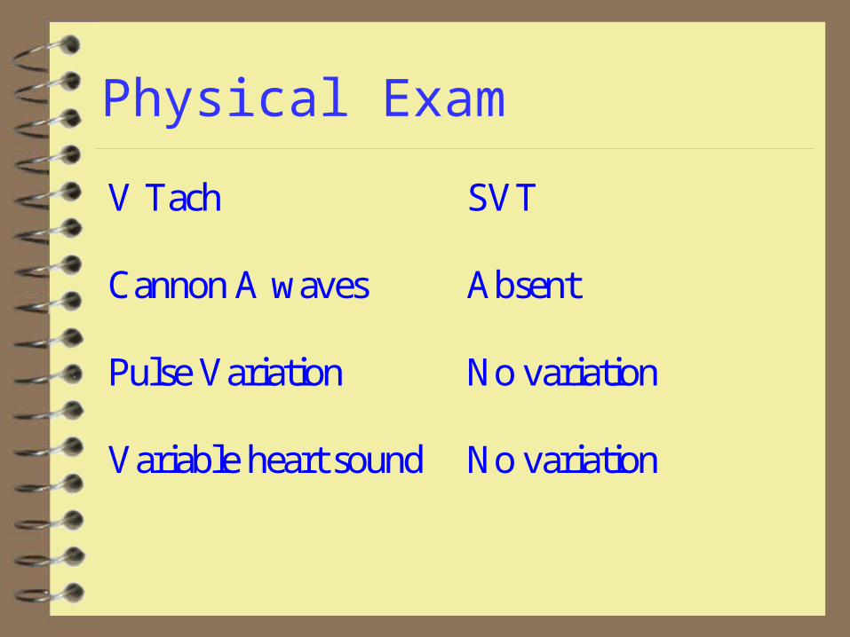
Physical Exam
V Tach SVT
Cannon A waves Absent
Pulse Variation No variation
Variable heart sound No variation
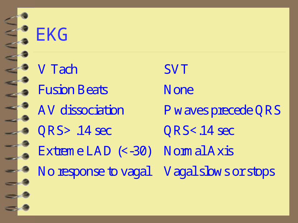
EKG
V Tach SVT
Fusion Beats None
AV dissociation P waves precede QRS
QRS> .14 sec QRS<.14 sec
Extreme LAD (<-30) Normal Axis
No response to vagal Vagal slows or stops
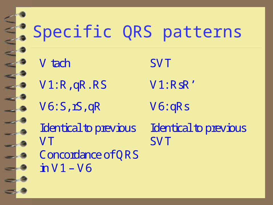
Specific QRS patterns
V tach SVT
V1: R, qR. RS V1: RsR’
V6: S, rS, qR V6: qRs
Identical to previous VT
Identical to previous SVT
Concordance of QRS in V1 – V6
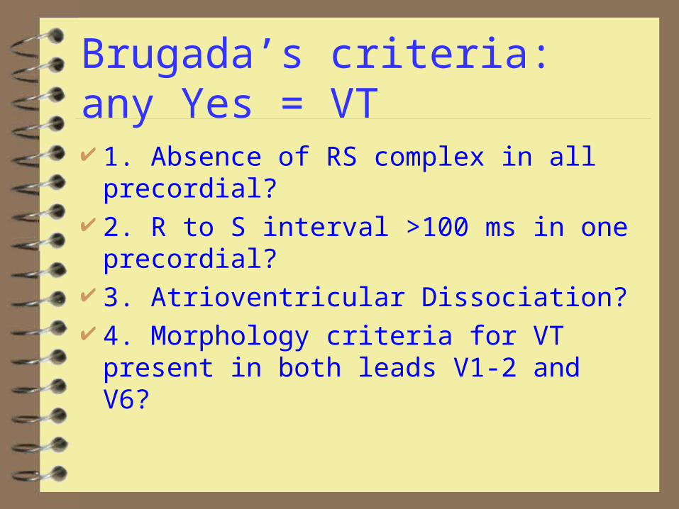
Brugada’s criteria: any Yes = VT
1. Absence of RS complex in all precordial? 2. R to S interval >100 ms in one
precordial? 3. Atrioventricular Dissociation? 4. Morphology criteria for VT present in
both leads V1-2 and V6?
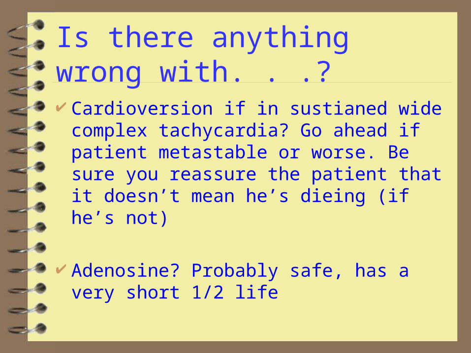
Is there anything wrong with. . .?
Cardioversion if in sustianed wide complex tachycardia? Go ahead if patient metastable or worse. Be sure you reassure the patient that it doesn’t mean he’s dieing (if he’s not)
Adenosine? Probably safe, has a very short 1/2 life
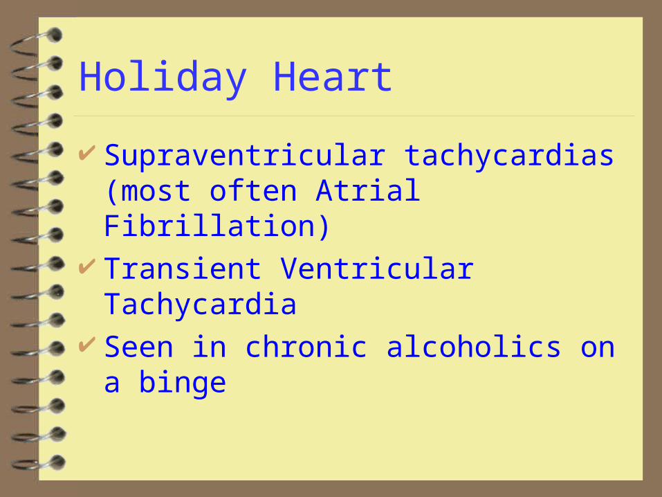
Holiday Heart
Supraventricular tachycardias (most often Atrial Fibrillation)
Transient Ventricular Tachycardia Seen in chronic alcoholics on a binge
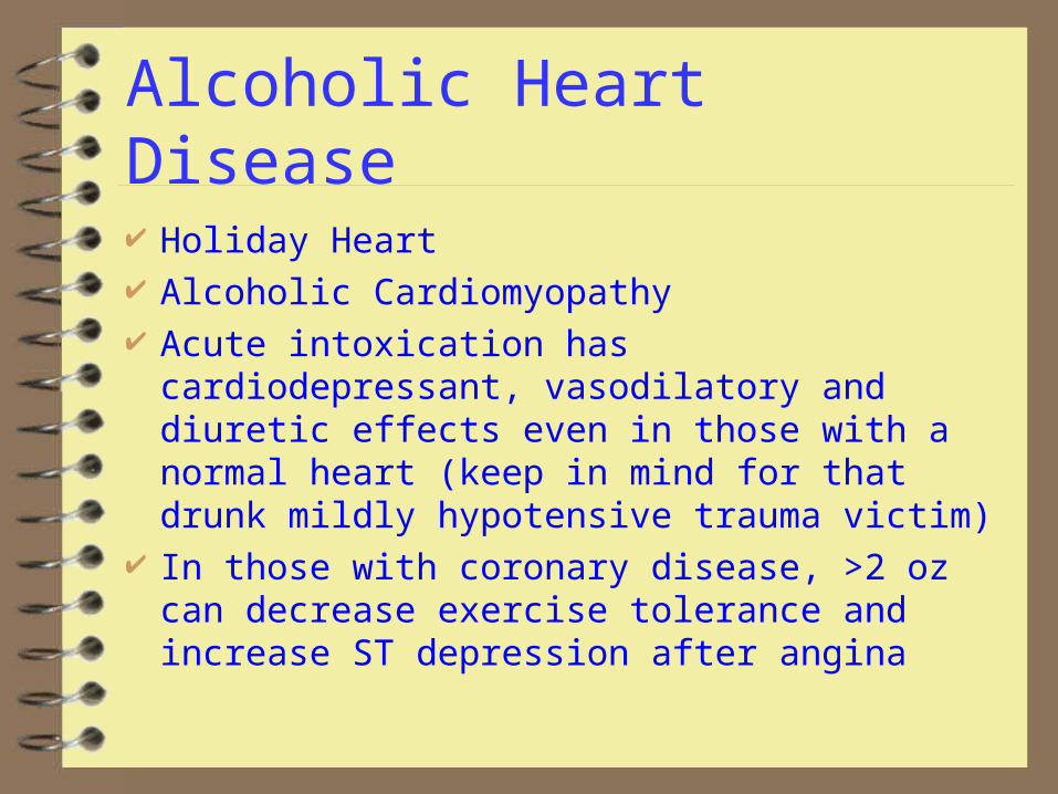
Alcoholic Heart Disease
Holiday Heart Alcoholic Cardiomyopathy Acute intoxication has cardiodepressant,
vasodilatory and diuretic effects even in those with a normal heart (keep in mind for that drunk mildly hypotensive trauma victim)
In those with coronary disease, >2 oz can decrease exercise tolerance and increase ST depression after angina
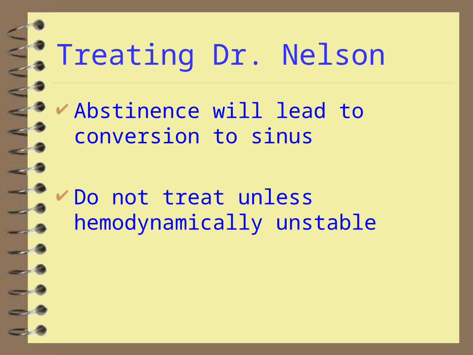
Treating Dr. Nelson
Abstinence will lead to conversion to sinus
Do not treat unless hemodynamically unstable

Case 2: Aunt Jane ain’t right
76 y/o F noted to be increasingly confused by family over last several days
Her Doctor is not available Family doesn’t know details of medical
history Patient complains of being weak
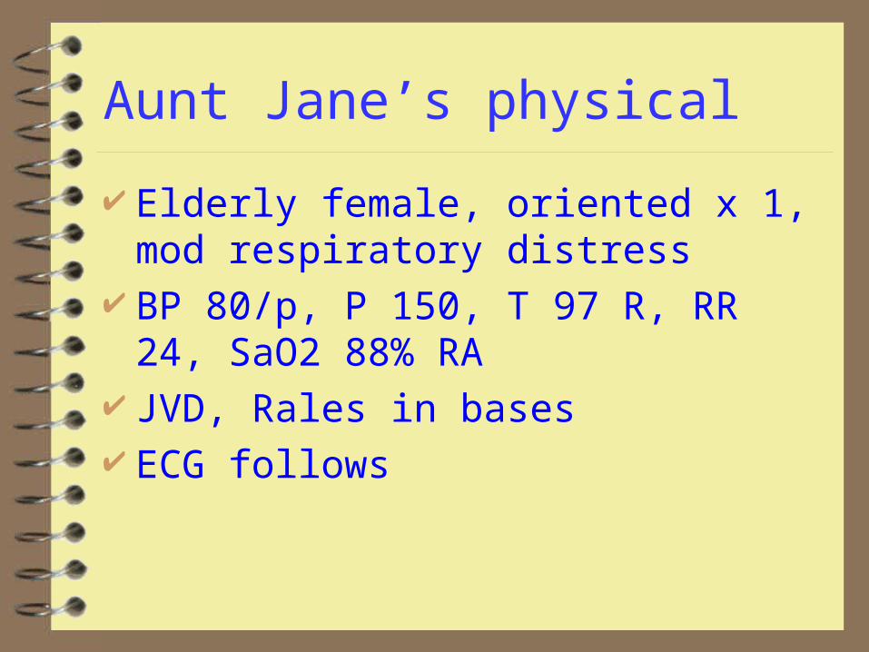
Aunt Jane’s physical
Elderly female, oriented x 1, mod respiratory distress
BP 80/p, P 150, T 97 R, RR 24, SaO2 88% RA
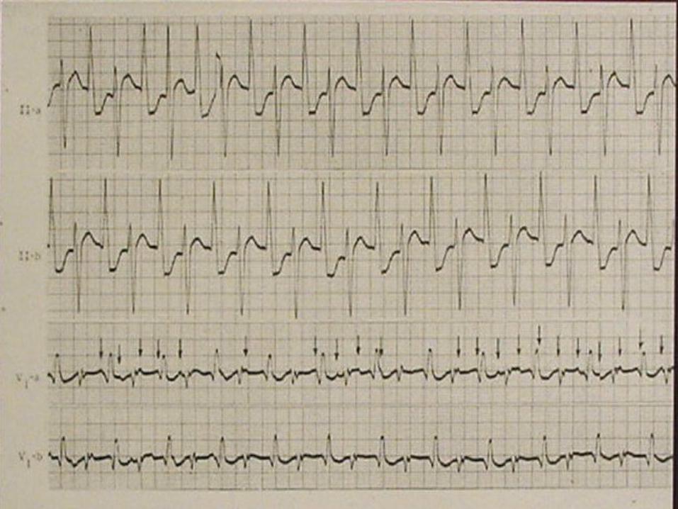
JVD, Rales in bases ECG follows
Case 2: Aunt Jane ain’t right
What’s the rhythm? What’s the underlying cause? Is it hurting her? What to do?
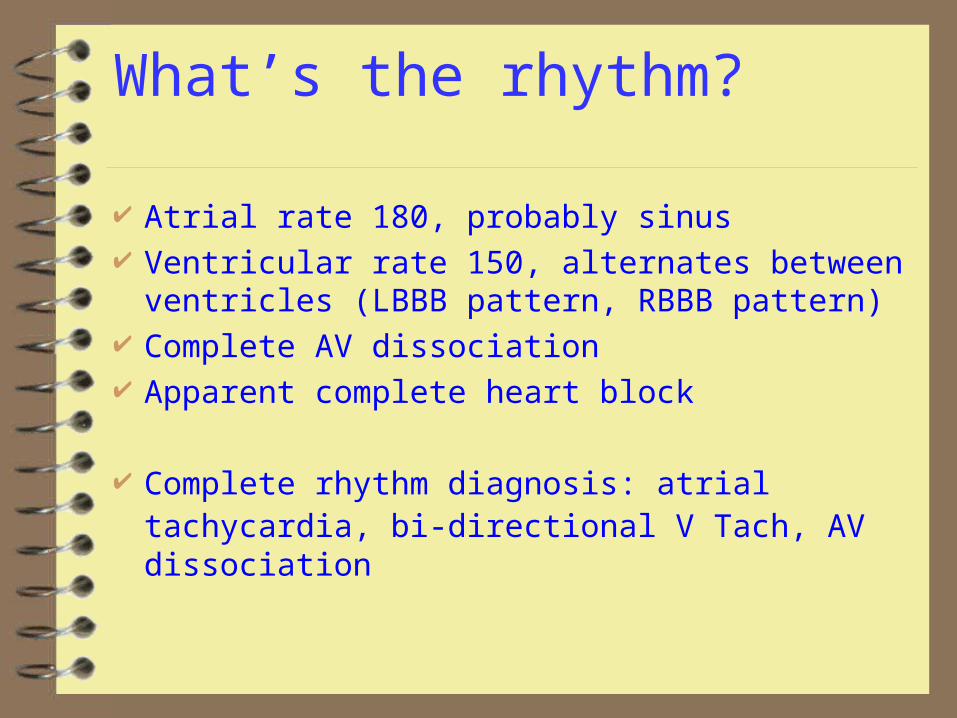
What’s the rhythm?
Atrial rate 180, probably sinus Ventricular rate 150, alternates between ventricles
(LBBB pattern, RBBB pattern) Complete AV dissociation Apparent complete heart block
Complete rhythm diagnosis: atrial tachycardia, bi-directional V Tach, AV dissociation
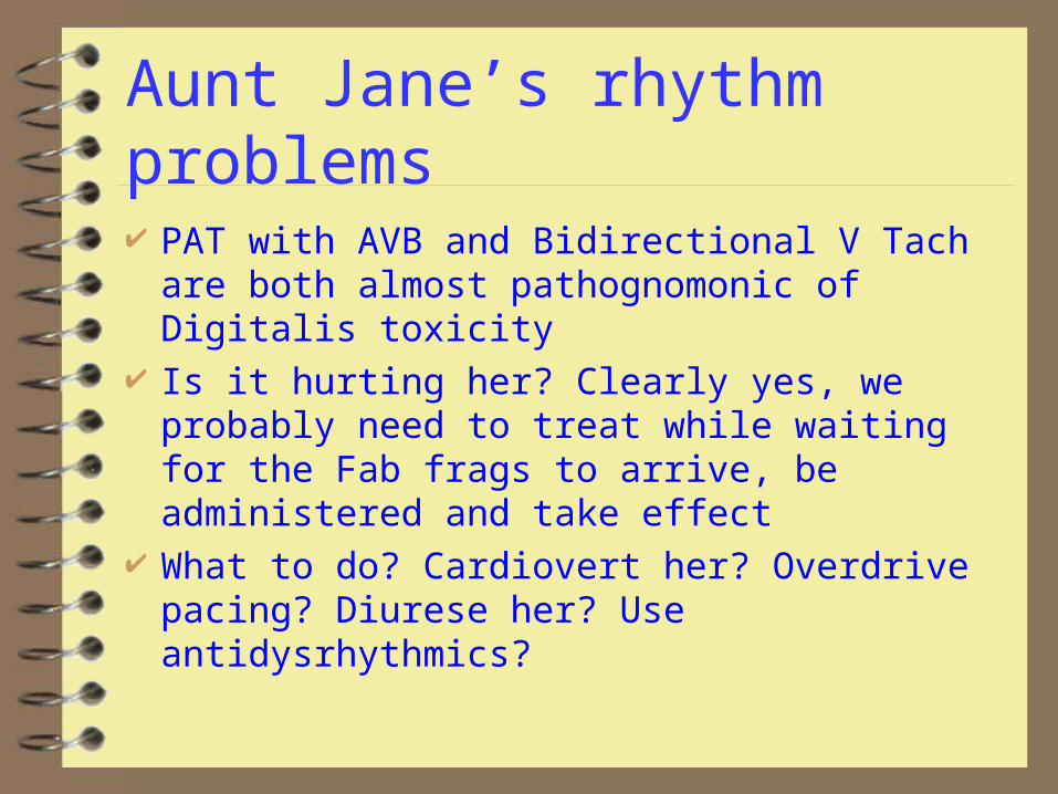
Aunt Jane’s rhythm problems
PAT with AVB and Bidirectional V Tach are both almost pathognomonic of Digitalis toxicity
Is it hurting her? Clearly yes, we probably need to treat while waiting for the Fab frags to arrive, be administered and take effect
What to do? Cardiovert her? Overdrive pacing? Diurese her? Use antidysrhythmics?
Cardiovert?
Cardioversion generally considered contraindicated for Dig toxic rhythms. The heart is irritable due to increased catecholamine sensitivity
Fear cardioversion to refractory VF, VT and asystole Most of that literature is very old and is talking about
cardioverting AF. Applicability to VT unclear. Is Aunt Jane an exception because she’s so sick we
don’t think we’ll make her worse?
Overdrive Pacing?
Transvenous pacing in dig toxicitycarries same dangers as cardioversion (Induces ventricular dysrhymias, mortality 13%)
Transthoracic pacing may be safer, but the pacer would need to be capable of higher rates than the intrinsic rhythm
Diuresis?
Most patients in chronic dig toxicity are already on diuretics and hypokalemic. Hypokalemia worsens Dig toxicity.Must check K and supplement as necessary if going to diurese
Antidysrhythmics
Most antidysrhythmics are contraindicated in Dig toxicity
Magnesium indicated for dig induced tachydysrhythmias. 1-2 gm over 2 minutes then 1-2 gm/hr
Lidocaine or phenytoin are considered safest
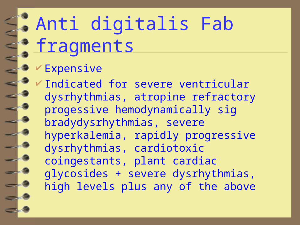
Anti digitalis Fab fragments
Expensive Indicated for severe ventricular
dysrhythmias, atropine refractory progessive hemodynamically sig bradydysrhythmias, severe hyperkalemia, rapidly progressive dysrhythmias, cardiotoxic coingestants, plant cardiac glycosides + severe dysrhythmias, high levels plus any of the above
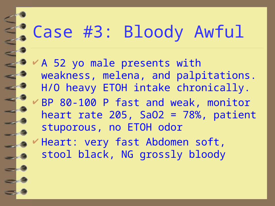
Case #3: Bloody Awful
A 52 yo male presents with weakness, melena, and palpitations. H/O heavy ETOH intake chronically.
BP 80-100 P fast and weak, monitor heart rate 205, SaO2 = 78%, patient stuporous, no ETOH odor
Heart: very fast Abdomen soft, stool black, NG grossly bloody
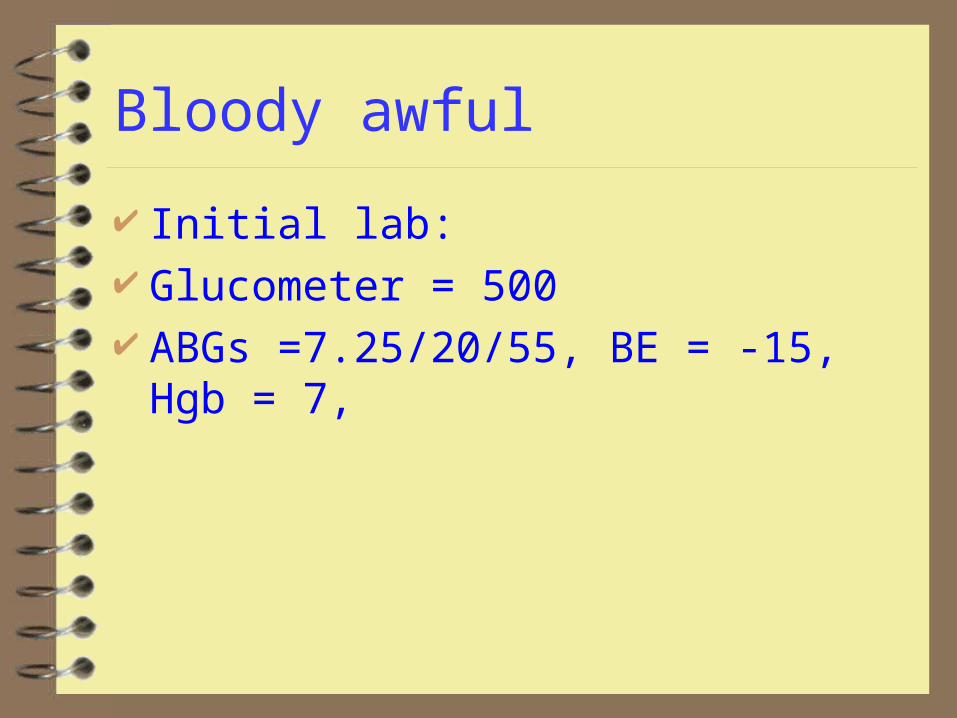
Bloody awful
Initial lab: Glucometer = 500 ABGs =7.25/20/55, BE = -15, Hgb = 7,

Wassup?
What’s the rhythm?
What’s the underlying cause?
Do we need more info?
Is it hurting him?
Wassup?
Rhythm Atrial fibrillation with rapid ventricular response
Underlying causes: hypovolemic shock, alcoholic heart disease, possible DKA
Addl info: CXR borderline cardiomegaly without failure
Is it hurting him? In this clinical scenario, of course. Combination of rate, blood loss, base deficit, anemia suggest that cardiac arrest is imminent

What to do?
Define possible strategies
What to do?
Cardiovert? (electrically or chemically) Slow Vent Response? Correct hypovolemia, anemia, DKA and
hope he gets better from this alone?
Cardioversion
Pro: rapid return to a rate allowing ventricular filling, adequate cardiac output
Con: In presence of chronic AF, atrial thrombus may exist. Cardioversion may lead to arterial embolism (stroke, mesenteric infarct, etc)
Do we think this is acute or chronic AF?
Acute or chronic?
The rate and the clinical scenario suggests acute, but you never really know.
How to do it? Electrical or chemical?
Electrical or Chemical
Electrical is quick and except in the dig toxic is unlikely to have side effects. However, the underlying condition leading to AF is unchanged. The heart may go right back into the rhythm
Chemical conversion is slower, but leads to conditions more likely to allow permanent conversion. However the drugs used (class 1a and 3) can all cause major dysrhythmias and cardiodepression. Digoxin is slower but won’t cause cardiodepression
Summary of recs (most pts hemodynamically stable) For acute AF in patient without failure or
cardiomyopathy: use class 3 agent (Ibutilide has highest success rate and quickest action, but can cause dysrhythmias after single dose)
Chronic AF usually with cardiomyopathy: rate control, anticoagulation for 3 weeks (or TEE) followed by electrical cardioversion

Slow ventricular rate
Calcium channel blockers are preferred for the patient in absence of ventricular dysfunction.
Digoxin for the hemodynamically unstable may take longer to have effect.
Note reading the studies, it appears that the differentiation between “rate-control” and “cardioversion” is probably not real
Correct conditions: always
MI, Ischemia, valvular disease, pericarditis, hyperthyroidism, SSS, contusion, holiday, idiopathic, hypertensive heart, cardiomyopathy, cardiac surgery, catecholamine excess, PE, CHF, WPW
For this patient
It was felt that arrest was imminent and acute AF was most likely, The patient was cardioverted with 200 j. He converted to sinus and within 3 seconds reverted to AF
Class 1a, 3, and Ca blockers were rejected because of patient’s probable cardiomyopathy and hypotension/shock
For this patient
O2 and fluid resuscitation were started Type specific blood was ordered Digoxin 0.5 mg was given IV
Within 5 minutes the patient converted spontaneously to sinus tach. Blood and octriatide started, Pt admitted to ICU, GI consulted.
Case 4: I fainted (4 p.m.)
14 y/o female brought by mother and teacher. Child reportedly was in Biology lab, dissecting a frog which upset her
The biology teacher reports she fell to the ground suddenly, had few jerks of extremities, and awoke, fully concious in about a minute.
Child has no memory of falling and had no prodromal sympotoms
I fainted
Child is awake and alert VS are normal, glucometer is 110, SaO2=98% Complete neuro exam is normal General physical exam is normal
Should we quit? If not, what would you like to do?
I might quit, but
You could easily argue for a b-hcg, bmp, cbc, and a CT/EEG to r/o pregnancy/ectopic, anemia, seizure disorder and subarachnoid bleed
An ECG is harder to justify, but this is a cardiology lecture, so:
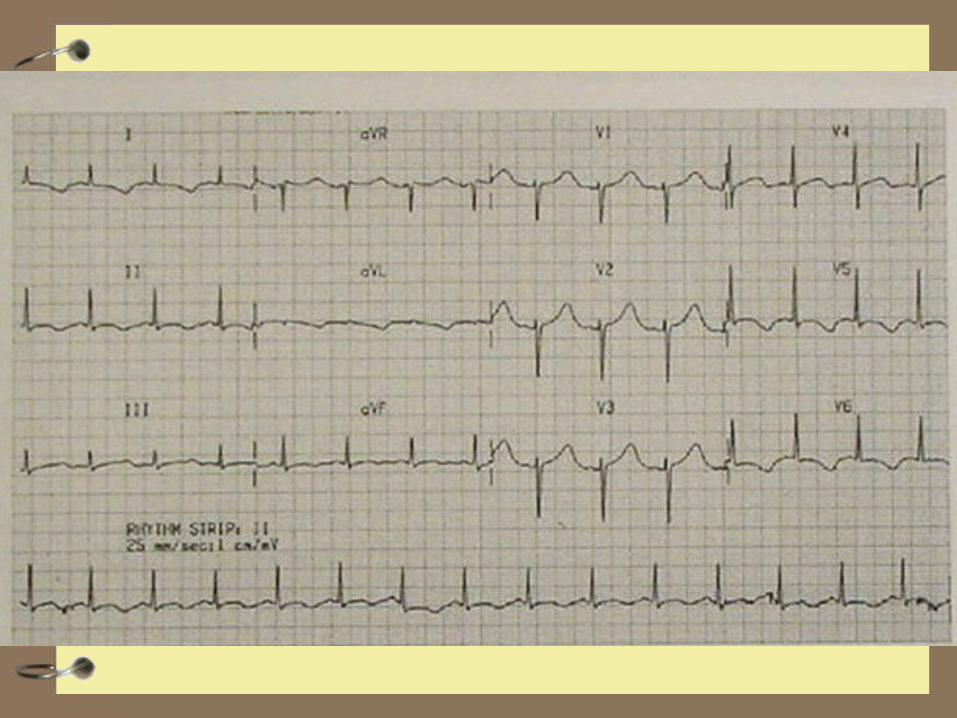

What to do?
Is there a problem?
What will you recommend?
Course
Child is admitted to telemetry, Cardiology consult is planned for the AM
The code team is called at 2 am as the child has arrested
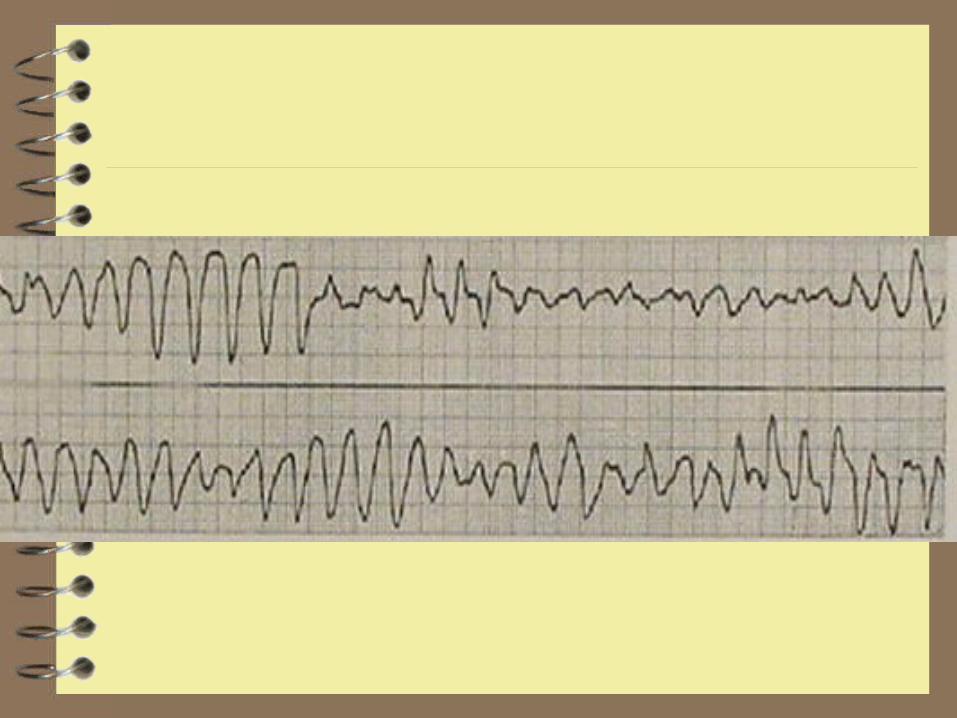
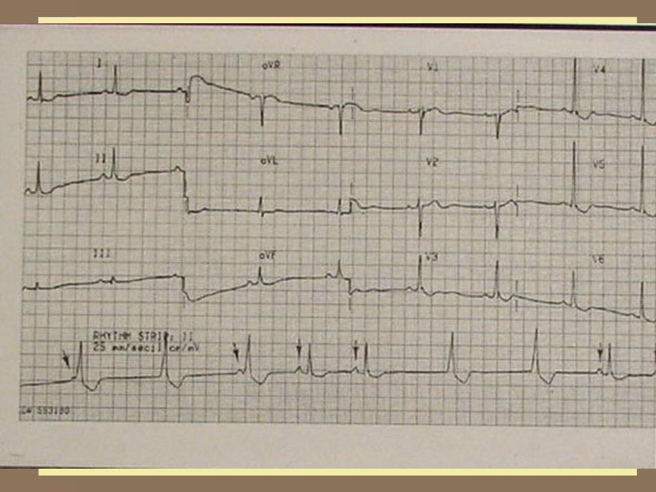
Rhythm strip follows
What to do?
What’s the rhythm?
What’s the immediate treatment? In the short term? In the long term? Would it have been different if the child
had been taking erythromicin?
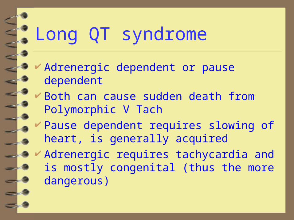
Long QT syndrome
Adrenergic dependent or pause dependent Both can cause sudden death from
Polymorphic V Tach Pause dependent requires slowing of heart, is
generally acquired Adrenergic requires tachycardia and is
mostly congenital (thus the more dangerous)
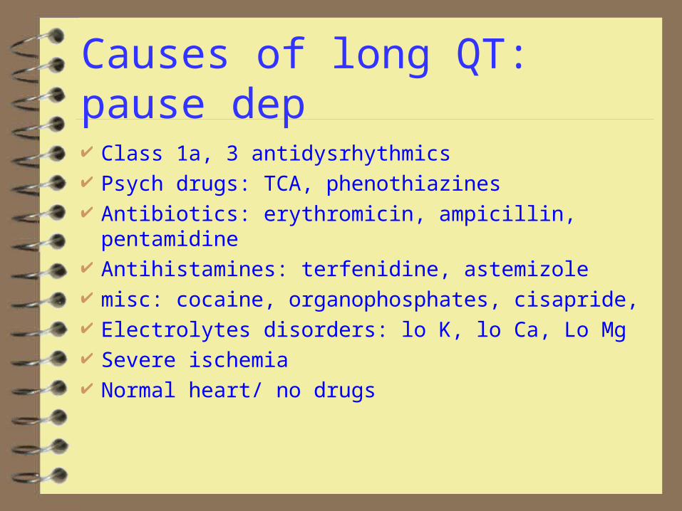
Causes of long QT: pause dep
Class 1a, 3 antidysrhythmics Psych drugs: TCA, phenothiazines Antibiotics: erythromicin, ampicillin, pentamidine Antihistamines: terfenidine, astemizole misc: cocaine, organophosphates, cisapride, Electrolytes disorders: lo K, lo Ca, Lo Mg Severe ischemia Normal heart/ no drugs
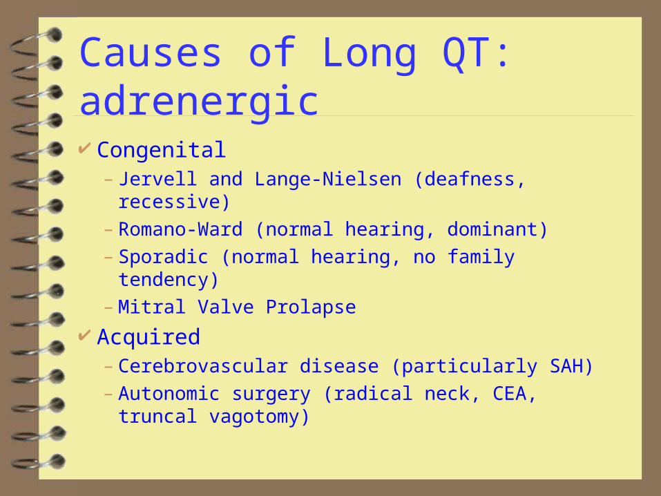
Causes of Long QT: adrenergic
Congenital– Jervell and Lange-Nielsen (deafness, recessive)– Romano-Ward (normal hearing, dominant)– Sporadic (normal hearing, no family tendency)– Mitral Valve Prolapse
Acquired– Cerebrovascular disease (particularly SAH)– Autonomic surgery (radical neck, CEA, truncal
vagotomy)
Immediate treatment for PMVT
Cardioversion in same manner as monomorphic VT
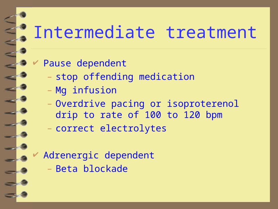
Intermediate treatment
Pause dependent
– stop offending medication
– Mg infusion
– Overdrive pacing or isoproterenol drip to rate of 100 to 120 bpm
– correct electrolytes
Adrenergic dependent
– Beta blockade
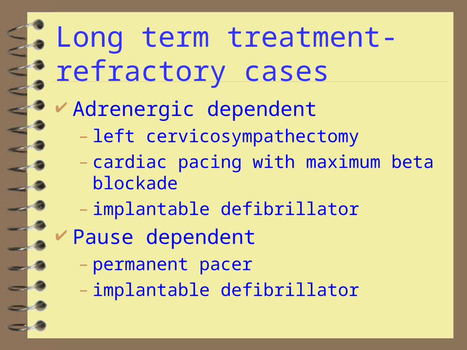
Long term treatment-refractory cases Adrenergic dependent
– left cervicosympathectomy– cardiac pacing with maximum beta blockade– implantable defibrillator
Pause dependent– permanent pacer– implantable defibrillator

Case 5: Weak and Dizzy
70 y/o women presents with occasional episodes of near syncope
PMH: HTN, angina PE: VS nl, CNS nl. CVS: S4, BMP, CBC nl ECG follows
What to do
Diagnosis
treatment
Weak and dizzy
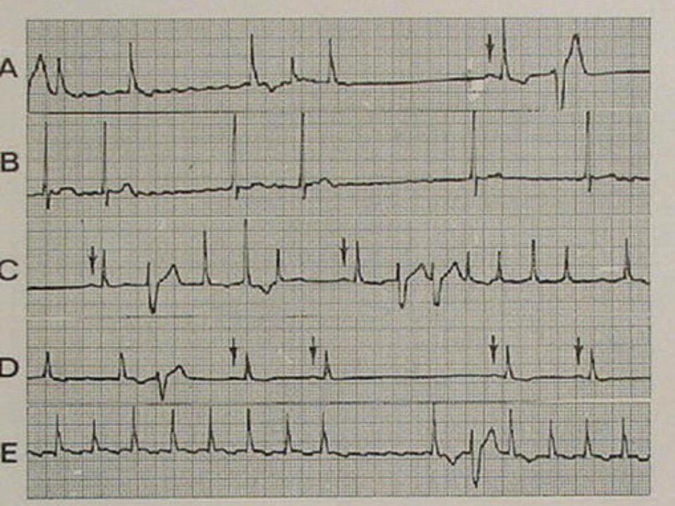
Sinus arrest, intermittent ventricular escape rhythm, incomplete AV dissociation
Treatment: trans thoracic pacing, EPS referral
While hooking up the pacer, the following strips are collected
Weak and Dizzy
What additional diagnoses added?
Any additional therapy?

Weak and dizzy
Brady-tachycardia syndrome, an advanced manifestation of Sick Sinus syndrome
May have to use antidysrhythmics to slow rate. This increases block and necessitates a permanent pacer
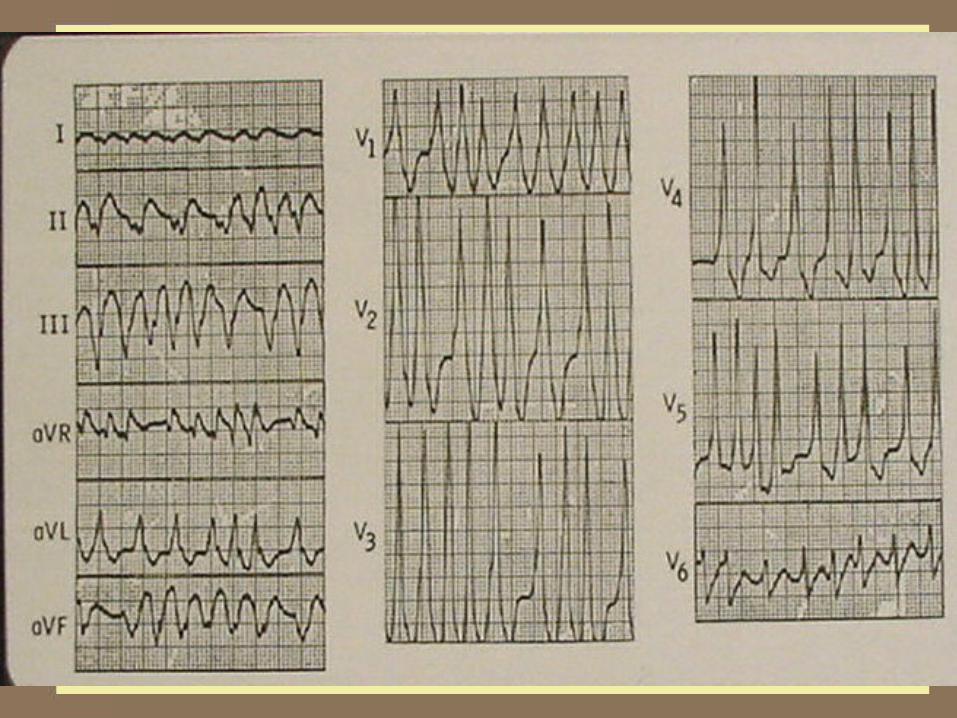
Case 6: My heart is running away
30 y/o m presents with rapid heart beat, dyspnea and weakness. Has had similar episodes in past but no treatment
PE: Pulse rapid, weak. BP 60/p, monitor heart rate 220
ECG follows
What’s next?
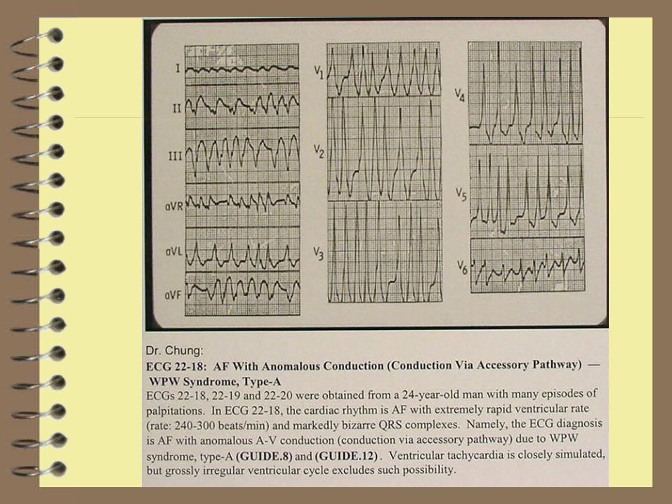
Rhythym is Wolfe-Parkinson-White syndrome with Atrial Fibrillation
What’s next
Cardioversion gives following rhythm change. Now what?
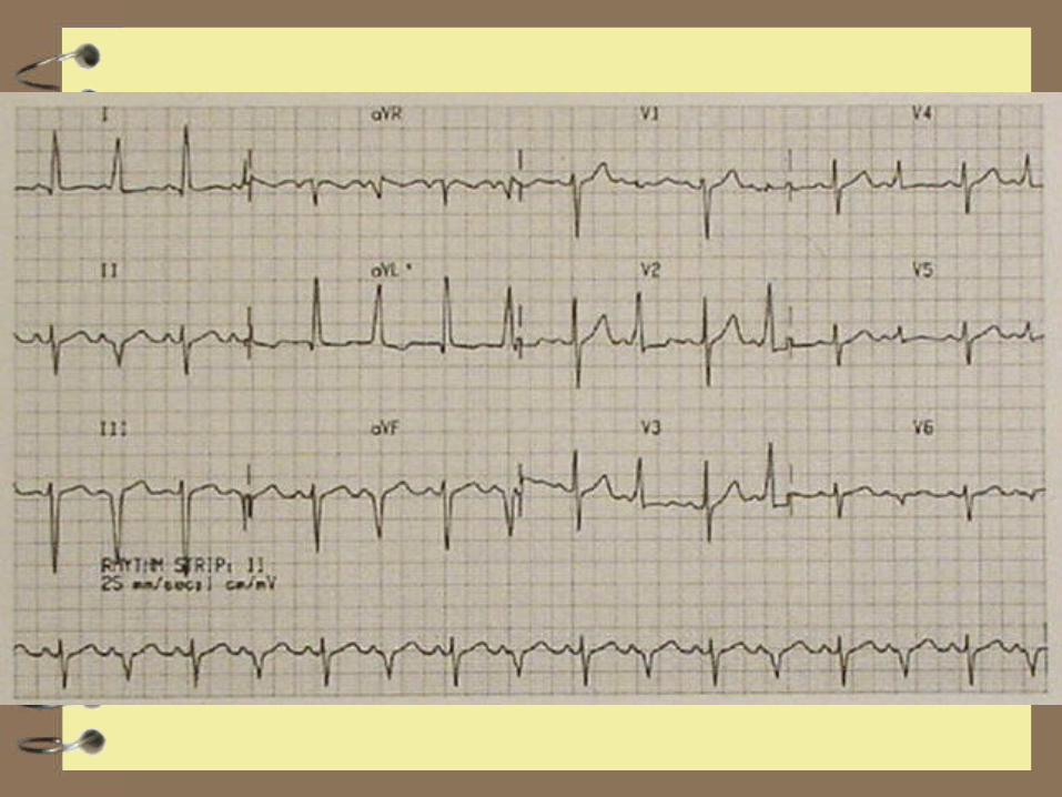
WPW with alternating conduction Orthodromic tachycardia when conduction
down AV node and reciprocates up Bundle of Kent
Antidromic tachycardia when conduction down Bundle of Kent and back up AV node
WPW with tachydysrhythmias
Antidromic regular tachycardia or any irregular tachycardia, regardless of QRS duration at high risk for V Fib
All drugs that block the AV node are contraindicated as they may increase rate of conduction through the accessory pathway, accelerating rate of dysrhythmia leading to V Fib
No-Nos
No Digitalis, Ca channel Blockers, Beta-blockers, or adenosine
Regimen of choice: cardioversion if unstable, procainamide, referral for catheter radioablation of pathway
Second line drugs: other 1a and 1c
Case 7: I need a doctor for Med Control
56 y/o had a syncopal episode, feels well now, wants to refuse therapy
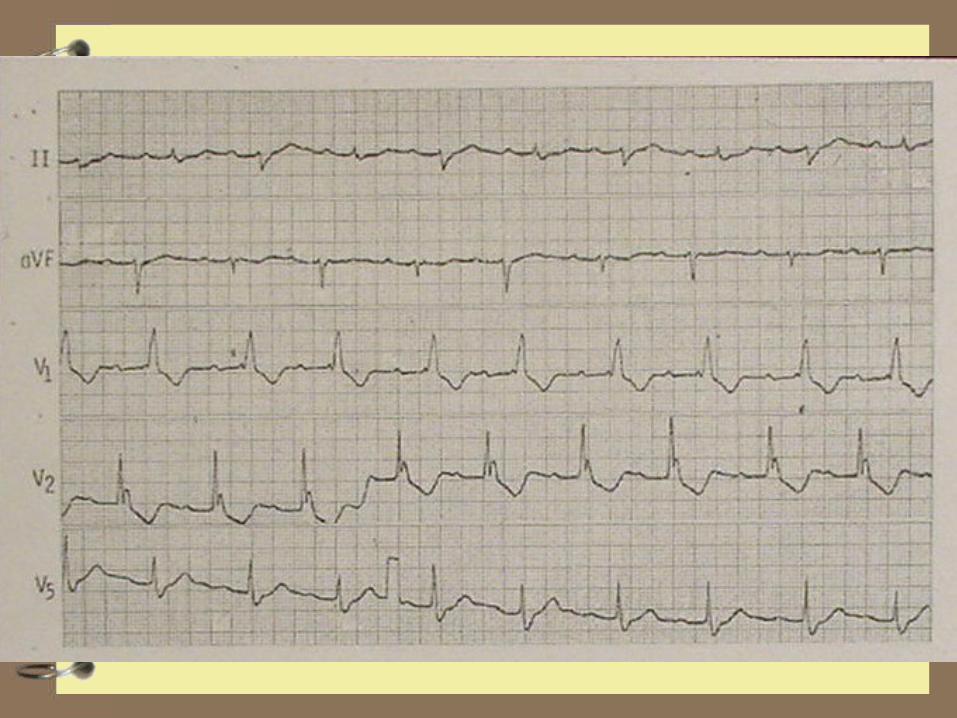
The paramedics fax this ECG, what do you think?
Not so fast buddy
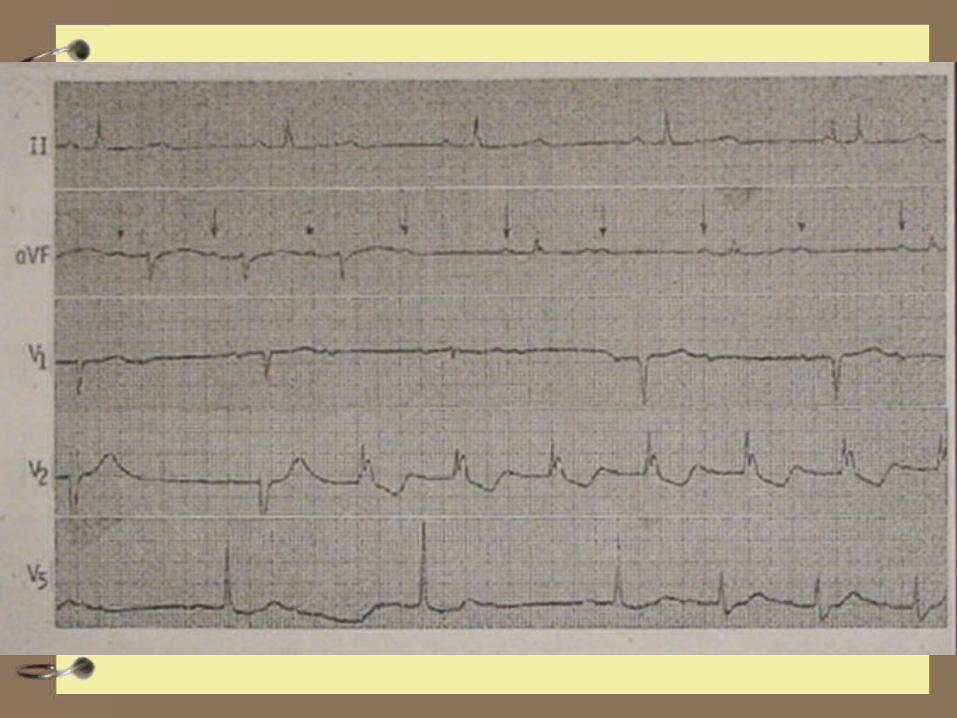
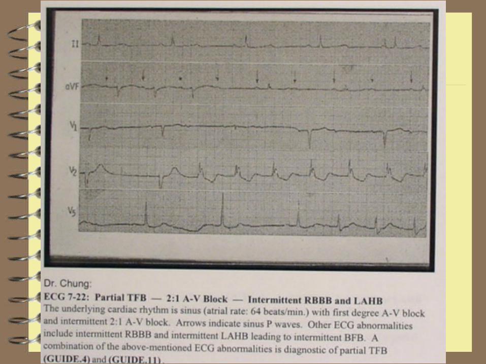
Patient has First degree AV block, RBBB and intermittent LAHB
Consider this a “Partial Trifasicular Blcok”
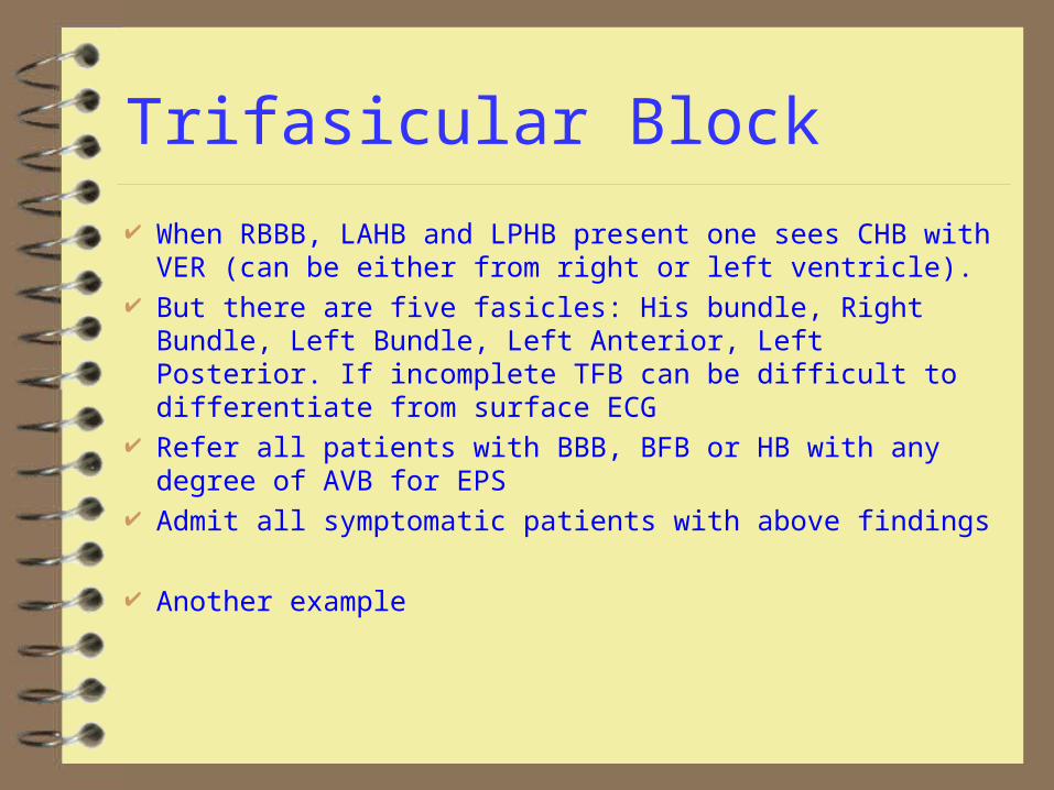
Trifasicular Block
When RBBB, LAHB and LPHB present one sees CHB with VER (can be either from right or left ventricle).
But there are five fasicles: His bundle, Right Bundle, Left Bundle, Left Anterior, Left Posterior. If incomplete TFB can be difficult to differentiate from surface ECG
Refer all patients with BBB, BFB or HB with any degree of AVB for EPS
Admit all symptomatic patients with above findings
Another example
Quickie no 1
83 yo bedridden male found to be less responsive than usual
FMS finds in V tach with pulse, under med control gives lido with conversion, during transport BVM ventilated, aspirates.
On hospital arrival RSI performed, SaO2 improves from 60% to 95%. % minute later V tach recurs, pt cardioverted. Now what meds?
Quickie #1
Pt given lidocaine boluses 100 and 50 while procainamide loading dose prepared
While being loaded, qrs changes, here’s the rhythm strip, what’s going on?
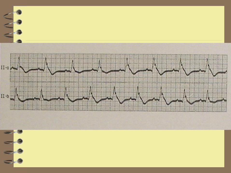
Quickie #1
Sinus mechanism with wide complex, suggestion of j point elevation. Given the clinical situation (elderly, bedridden). Patient may be hypothermic.
Check a temp, If temp below 94 no more carediodepressant antidysrhythmics.
Warm the patient (passive or active internal, not active external)
Quickie #2
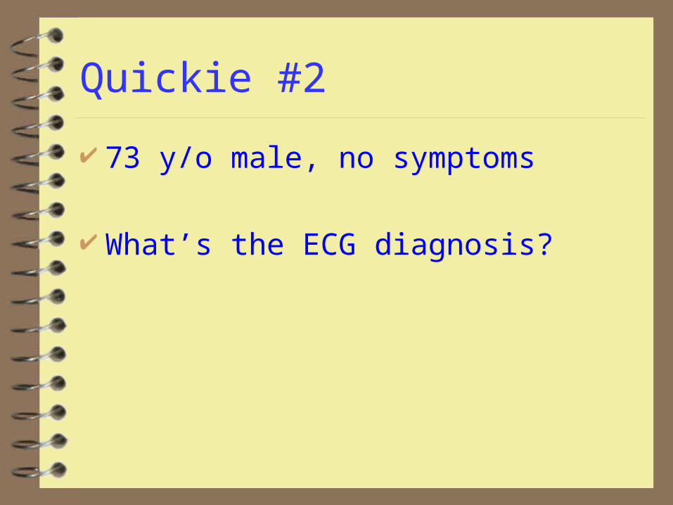
73 y/o male, no symptoms
What’s the ECG diagnosis?
Quickic #2
Alternating RBBB and LBBB. What looks like a second P on half the beats is really the initial deflection of a RBBB
Dig toxic rhythm? Needs EPS?
Quickie #3
58 y/o female feels weak No PMH, doesn’t like Doctors Glucometer 450 ECG follows: findings? Probable cause?
RX?
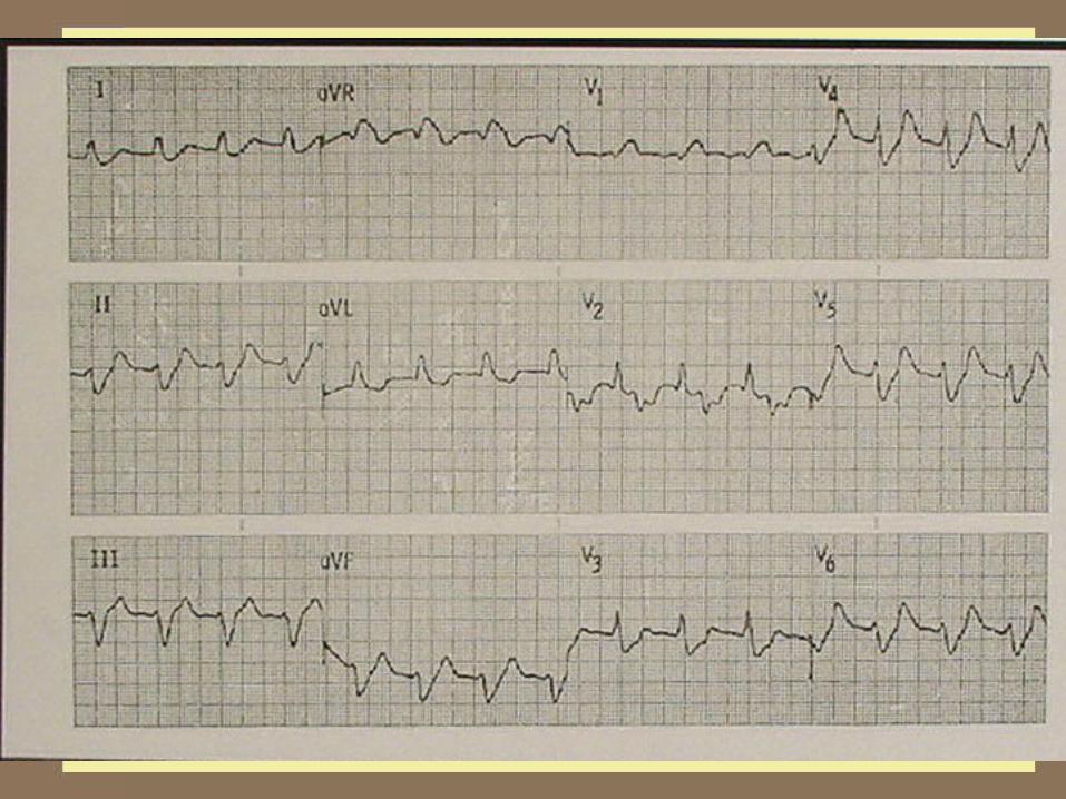
Quickie #3
ECG findings: Diffuse Intraventricular block (RBBB with LAHB?), flattened p waves. Beginning to look like a sine wave
Probable cause: Hyperkalemia sec to renal failure
Immediate Rx: CaCl2 10 ml slow push, Insulin 10 units (don’t need D50 since hyperglycemic), Kayexalate
Quickie #4
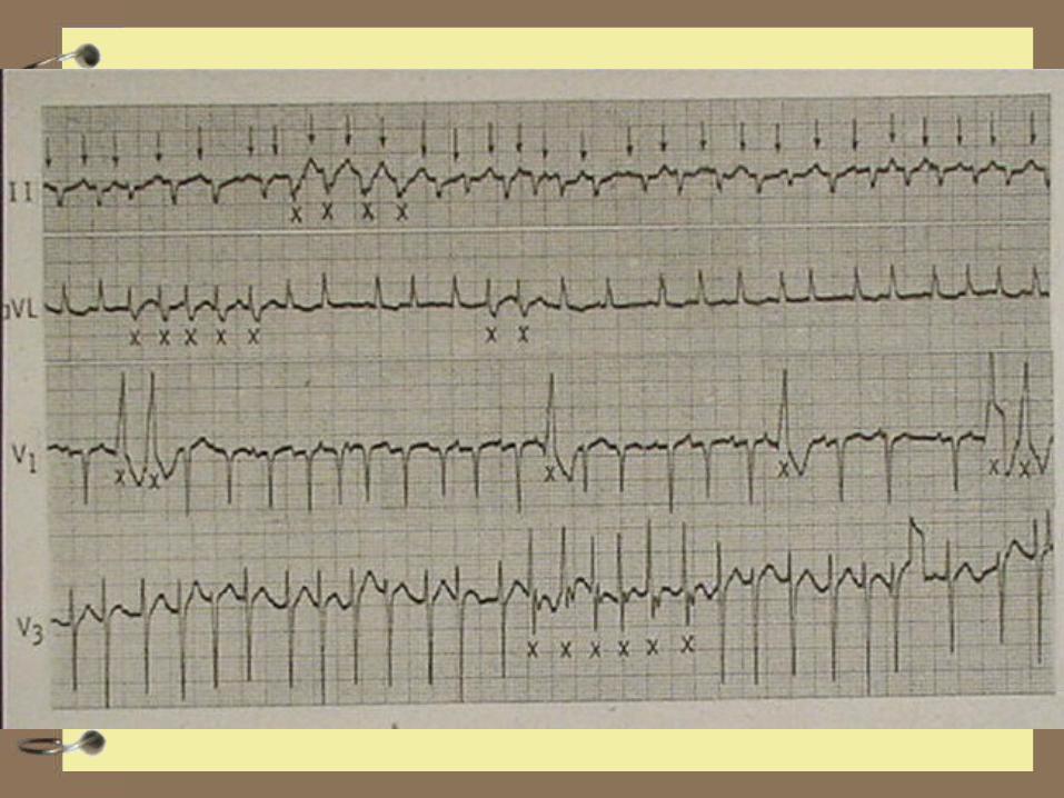
58 y/o in 1 vehicle accident on Saturday Night, smells of ETOH, BP 80/p, patient comatose: PE, FAST, Xrays, Head CT
What are the ECG findings? What should you do to confirm the probable
diagnosis? If confirmed, what should be done now?

Quickie #4
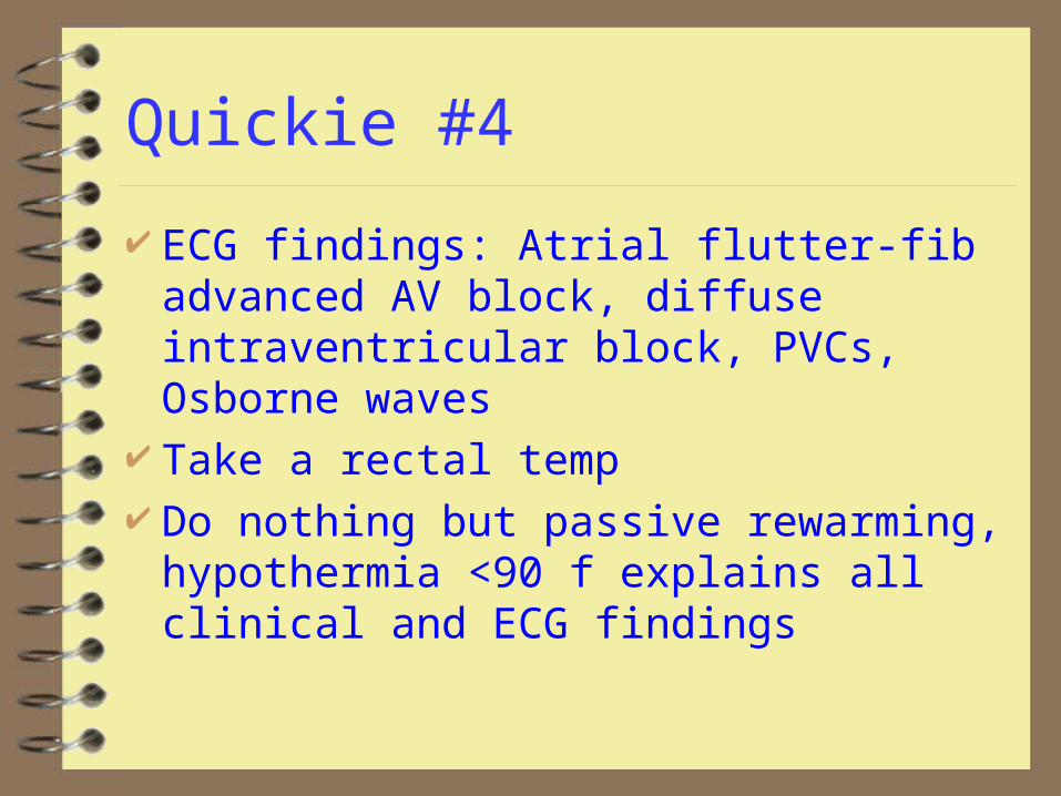
ECG findings: Atrial flutter-fib advanced AV block, diffuse intraventricular block, PVCs, Osborne waves
Take a rectal temp Do nothing but passive rewarming,
hypothermia <90 f explains all clinical and ECG findings
Causes of Diffuse Intraventricular Block
MI Severe Hypertensive Heart Disease Cardiomyopathy Hyperkalemia Elderly (senile fibroelastosis?) Hypothermia

Quickie #5
48 y/o male feels dizzy and weak. BP 90/p ECG findings? Probable cause? Rx?
Quickie #5
ECG findings: Multifocal atrial tachycardia, aberrant conduction
Cor pulmonale with hypoxia, prescribed beta agonists or methylxanthines may be contributing, may be dig toxic
Correct hypoxia, withdraw offending agents, electrical cardioveriosn unlikely to be successful until underlying condition treated
Rate control, if needed by Ca channel blockers, Mg second line
Dig toxicity treated in usual manner
Quickie #6
47 y/o male with idiopathic cardiomyopathy What’s the rhythm?
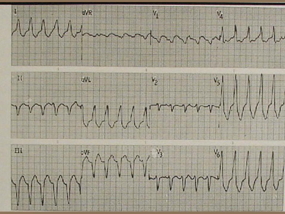
Quickie #6
Wide complex tachycardia, confirmed as V tach by AV dissociation by p waves before the 4th and 13th QRS and after the 5th
Quickie #7
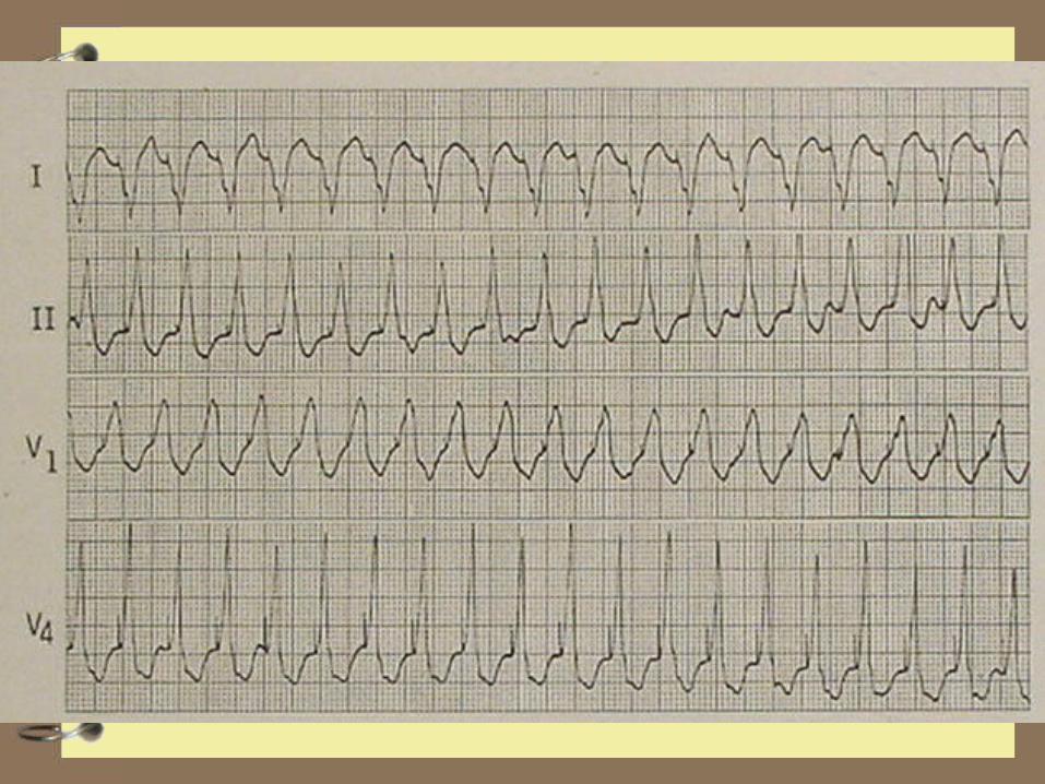
80 y/o F, confused, BP 80/p, pulse 167 ECG diagnosis? What can you do?

Quickie #7
Dx: Pacemaker mediated tachycardia and malpositioned electrode (RBBB pattern suggests anode in Left Ventricle
Try to convert to fixed rate output 70 bpm with magnet
Overdrive it with transthoracic and try to slow it Open the pocket and disconnect the wires or cut
them Find out where the tip is: Left ventricle?
Pulmonary outflow track? Pericardial sac?
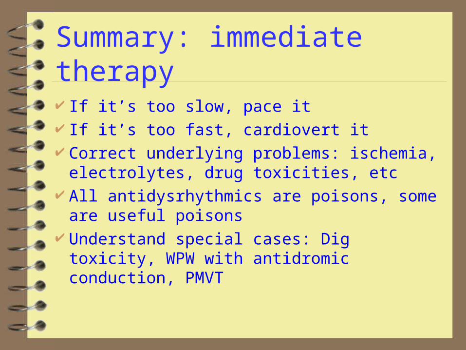
Summary: immediate therapy
If it’s too slow, pace it If it’s too fast, cardiovert it Correct underlying problems: ischemia,
electrolytes, drug toxicities, etc All antidysrhythmics are poisons, some are
useful poisons Understand special cases: Dig toxicity, WPW
with antidromic conduction, PMVT
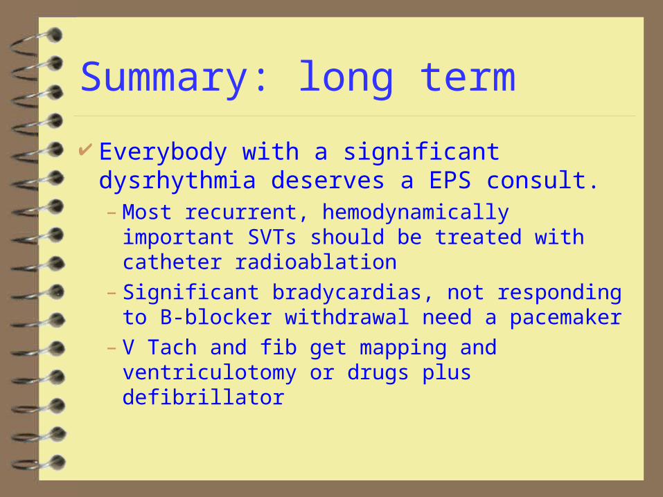
Summary: long term
Everybody with a significant dysrhythmia deserves a EPS consult.– Most recurrent, hemodynamically important
SVTs should be treated with catheter radioablation
– Significant bradycardias, not responding to B-blocker withdrawal need a pacemaker
– V Tach and fib get mapping and ventriculotomy or drugs plus defibrillator


