Ectopic Eruption of the Maxillary First Permanent Molar:
Rate and Predictive Factors of Self-Correction
and
Survey of Specialists' Attitudes Regarding Intervention
by
Basma Dabbagh
A thesis submitted in conformity with the requirements
for the degree of Master in Science of Pediatric Dentistry
Graduate Department of Dentistry
University of Toronto
© Copyright by Basma Dabbagh 2013
ii
Ectopic Eruption of the Maxillary First Permanent Molar:
Rate and Predictive Factors of Self-Correction and
Survey of Specialists' Attitudes Regarding Intervention
Basma Dabbagh
Master of Science in Pediatric Dentistry
Graduate Department of Dentistry
University of Toronto
2013
Abstract
Purpose: To retrospectively assess the incidence and predictive factors for self-correction of
ectopic eruption of maxillary permanent first molars (EE) and the prevailing attitudes amongst
surveyed specialists regarding intervention in cases of EE.
Methods: Charts of patients diagnosed with EE were assessed for predictive clinical and
radiographic factors. An online survey was sent to pediatric dentists and orthodontists.
Results: The rate of self-correction was 71%. One third of self-corrections occurred after age 9.
Increased amount of impaction (r(43)=0.59, p<.001) and degree of resorption (r(57)=0.41,
p=.001) were positively correlated with irreversibility. Orthodontists estimated the spontaneous
self-correction rate to be lower (t(1178)=19.2, p<.001) than pediatric dentists.
Conclusions: One third of self-corrections occurred after 9 years of age and delaying treatment
of EE may be a viable option when uncertain of the outcome. Reliable predictive factors of
irreversibility of EE were identified. Differences exist between pediatric dentists and
orthodontists regarding management of EE.
iii
Acknowledgments
Dr Andrews, thank you for your support throughout this project. You were always available to
help me and answer my questions with a smile. It was a pleasure to have you as my supervisor.
Dr Sigal, thank you for your wise and helpful input. You always made time for me and I greatly
appreciate it.
Dr Titley and Dr Tompson, thank you for your time, your helpful advice and suggestions to
make this work better.
Dr Nainar, thank you for analyzing my radiographs and for your insight and advice.
Darsi, Ed and Trang, thank you for being my family for the last three years. I couldn’t have done
it without you and I couldn’t have wished for better classmates.
Mom, Dad, Nada, André and Sophia, from near and far, you have always been there to support
and encourage me. I am very grateful. Sophia you bring so much joy to our lives.
Teta Laura, you waited for me to come back. Je t’aime. Tu seras toujours dans mon coeur. I will
miss you.

iv
Table of Contents
Table of Contents ........................................................................................................................... iv
List of Tables ............................................................................................................................... viii
List of Figures ................................................................................................................................ ix
List of Appendices ......................................................................................................................... xi
Chapter 1 Ectopic Eruption of the First Permanent Molar ............................................................. 1
1 Literature review ........................................................................................................................ 1
1.1 Definition ............................................................................................................................ 1
1.2 Prevalence ........................................................................................................................... 1
1.3 Etiology ............................................................................................................................... 2
1.4 Diagnosis ............................................................................................................................. 3
1.5 Types of ectopic eruption and their incidence .................................................................... 4
1.6 Consequences of ectopic eruption ...................................................................................... 5
1.7 Indication for intervention .................................................................................................. 7
1.7.1 Potential predictive factors ..................................................................................... 7
1.7.1.1 Rotation of the permanent first molar ....................................................... 7
1.7.1.2 Age and observation period ...................................................................... 8
1.7.1.3 Severity of lock ......................................................................................... 8
1.7.1.4 Presence of an enamel ledge ..................................................................... 9
1.7.1.5 Resorption ............................................................................................... 11
1.7.1.6 Amount of impaction of the permanent tooth under the distal contour .. 12
1.7.1.7 Angulation of the permanent tooth ......................................................... 13
1.7.1.8 Partial eruption in the mouth .................................................................. 14
1.7.1.9 Bilateral ectopic eruption ........................................................................ 14

v
1.7.2 Conclusion ............................................................................................................ 14
1.8 Treatment .......................................................................................................................... 14
1.8.1 Minimal intervention ............................................................................................ 15
1.8.1.1 Elastic separator ...................................................................................... 15
1.8.1.2 Brass wire technique ............................................................................... 16
1.8.1.3 Helical spring .......................................................................................... 16
1.8.1.4 De-Impactor spring ................................................................................. 17
1.8.1.5 Surgical exposure of the permanent tooth .............................................. 17
1.8.1.6 Disking of the distal surface of the second primary molar ..................... 17
1.8.2 Appliance therapy with retention of the second primary molar ........................... 18
1.8.2.1 Humphrey-type appliance ....................................................................... 18
1.8.2.2 Halterman appliance ............................................................................... 19
1.8.2.3 Multi loop unilateral bonded appliance .................................................. 19
1.8.3 Extraction of the second primary molars and space regaining ............................. 20
1.8.3.1 Removable appliances ............................................................................ 20
1.8.3.2 Cervical Headgear .................................................................................. 20
1.8.3.3 Space maintenance ................................................................................. 22
1.8.4 Management of ectopically erupting mandibular molars ..................................... 22
1.9 Associated anomalies ........................................................................................................ 22
1.10 Survey .............................................................................................................................. 23
1.10.1 Advantages of online surveys ............................................................................... 23
1.10.2 Disadvantages of online surveys ........................................................................... 24
1.10.3 Response rate ........................................................................................................ 25
1.11 Rationale………………………………………………………………………………...25
2 Retrospective Chart Review ..................................................................................................... 27
vi
2.1 Objectives ......................................................................................................................... 27
2.2 Materials and Methods ...................................................................................................... 28
2.2.1 Inclusion and exclusion criteria ............................................................................ 28
2.2.2 Outcome ................................................................................................................ 28
2.2.3 Data collection and analysis .................................................................................. 29
2.2.4 Sample size ........................................................................................................... 30
2.2.5 Informed consent process ..................................................................................... 31
2.3 Results. .............................................................................................................................. 32
2.3.1 Demographics ....................................................................................................... 32
2.3.2 Outcome ................................................................................................................ 32
2.3.3 Observation time ................................................................................................... 33
2.3.4 Inter-rater and intra-rater correlation of radiographic factors ............................... 33
2.3.5 Predictive factors of irreversible outcome ............................................................ 34
2.3.6 Adverse events ...................................................................................................... 35
2.3.7 Associated anomalies ............................................................................................ 36
2.4 Discussion ......................................................................................................................... 36
2.5 Conclusions ....................................................................................................................... 41
3 Online survey of pediatric dentists and orthodontists .............................................................. 42
3.1 Objective ........................................................................................................................... 42
3.2 Materials and methods ...................................................................................................... 42
3.2.1 Inclusion criteria ................................................................................................... 42
3.2.2 Sample size ........................................................................................................... 42
3.2.3 Survey content ...................................................................................................... 43
3.2.4 Confidentiality and informed consent ................................................................... 44
3.2.5 Data analysis ......................................................................................................... 44
3.3 Results. .............................................................................................................................. 44
vii
3.3.1 Demographics ....................................................................................................... 44
3.3.2 Ectopic eruption cases ........................................................................................... 51
3.4 Discussion ......................................................................................................................... 58
3.5 Conclusions ....................................................................................................................... 63
References ..................................................................................................................................... 64
Appendices .................................................................................................................................... 73
viii
List of Tables
Table 1. Reported prevalence of ectopic eruption of the first permanent molar ............................ 2
Table 2. Published rate of self-correction of ectopically erupting first permanent molars ............. 5
Table 3. Age at diagnosis and outcome of ectopic eruption ......................................................... 33
Table 4. Inter-rater and intra-rater correlation of radiographic parameters assessed .................... 34
Table 5. Multiple regression analysis of all predictive factors for irreversible outcome ............. 35
Table 6. Multiple regression analysis of select reliable predictive factors for irreversible ouctome
....................................................................................................................................................... 35
ix
List of Figures
Figure 1: Presentation of minimal and severe lock of ectopically erupting permanent first molars
......................................................................................................................................................... 9
Figure 2: UCLA flowchart for the management of ectopically erupting first permanent molars 10
Figure 3: Grading of degree of resorption of ectopically erupting first permanent molars ......... 12
Figure 4: Representation of the measurement of the amount of impaction of the first permanent
molar ............................................................................................................................................. 13
Figure 5: Measurement of angulation of the first permanent molar ............................................. 13
Figure 6: Helical spring ................................................................................................................ 17
Figure 7: Distribution of location of ectopic eruption of the maxillary first permanent molar .... 32
Figure 8. Gender distribution of respondents ............................................................................... 45
Figure 9. Gender distribution of active members of the AAPD and the AAO ............................. 45
Figure 10. Age distribution of respondents ................................................................................... 46
Figure 11. Age distribution of active members of the AAPD and the AAO ................................ 46
Figure 12. Number of years that respondents have been in practice ............................................ 47
Figure 13. Type of training of respondents ................................................................................... 47
Figure 14. Type of institution where respondents received their training .................................... 48
Figure 15. Primary activity of respondents ................................................................................... 49
Figure 16. Geographical distribution of respondents .................................................................... 50
Figure 17. Representation of geographical districts of the AAPD ............................................... 50
x
Figure 18. Survey case 1 ............................................................................................................... 51
Figure 19. Respondents’ classification of the ectopic eruption in case 1 ..................................... 51
Figure 20. Specialists decision to intervene in case 1 ................................................................... 52
Figure 21. Choice of treatment modality of respondents who initiated treatment in case 1 ......... 52
Figure 22. Survey case 2 ............................................................................................................... 53
Figure 23. Specialist’s classification of ectopic eruption in case 2 .............................................. 53
Figure 24. Specialists’ choice of treatment modality in case 2 ..................................................... 54
Figure 25. Survey case 3 ............................................................................................................... 55
Figure 26. Specialists’ classification of the ectopic eruption in case 3A ...................................... 55
Figure 27. Specialists’ classification of the ectopic eruption in case 3B ...................................... 56
Figure 28. Specialist’s choice of treatment modality in case 3B .................................................. 57
Figure 29. Estimated percentage of self-correction by survey respondents ................................. 58
xi
List of Appendices
Appendix A. Information letter to parents
Appendix B. Survey introduction letter
Appendix C. Pediatric dentist survey
Appendix D. Orthodontist survey
Appendix E. Manuscript submitted to NuSmile Graduate Research Student Award (AAPD)
1
Ectopic Eruption of the First Permanent Molar
1 Literature review
1.1 Definition
For the purposes of this investigation, ectopic eruption of the maxillary first permanent molar (EE)
is defined as a local disturbance characterized by a mesial path of eruption causing the permanent
tooth to be locked under the distal undercut of the second primary molar. Other paths of ectopic
eruption can be buccal or lingual. This phenomenon causes various degrees of resorption of the
roots of the primary tooth. Ectopic eruption of the first permanent molar may occur unilaterally or
bilaterally in the maxilla or in the mandible (Young, 1957).
1.2 Prevalence
Ectopic eruption of the maxillary first permanent molar has a prevalence that ranges between 0.75 to
4.3% (Table 1). It is increased by four-fold in persons with cleft lip and palate (Carr & Mink, 1965).
A higher prevalence, 19.8%, has been reported in siblings (Kurol & Bjerklin, 1982a). Some authors
report a higher incidence in males (Bjerklin & Kurol, 1981; Young, 1957), while others found no
statistically significant difference between sexes (Chintakanon & Boonpinon, 1998; Kimmel, Gellin,
Bohannan, & Kaplan, 1982; Pulver, 1968).
2
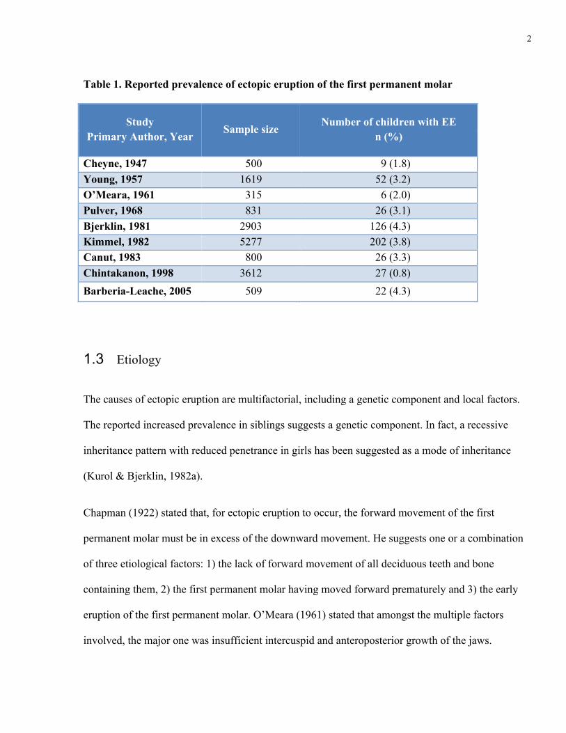
Table 1. Reported prevalence of ectopic eruption of the first permanent molar
Study Primary Author, Year
Sample size
Number of children with EE n (%)
Cheyne, 1947 500 9 (1.8) Young, 1957 1619 52 (3.2) O’Meara, 1961 315 6 (2.0) Pulver, 1968 831 26 (3.1) Bjerklin, 1981 2903 126 (4.3) Kimmel, 1982 5277 202 (3.8) Canut, 1983 800 26 (3.3) Chintakanon, 1998 3612 27 (0.8) Barberia-Leache, 2005 509 22 (4.3)
1.3 Etiology
The causes of ectopic eruption are multifactorial, including a genetic component and local factors.
The reported increased prevalence in siblings suggests a genetic component. In fact, a recessive
inheritance pattern with reduced penetrance in girls has been suggested as a mode of inheritance
(Kurol & Bjerklin, 1982a).
Chapman (1922) stated that, for ectopic eruption to occur, the forward movement of the first
permanent molar must be in excess of the downward movement. He suggests one or a combination
of three etiological factors: 1) the lack of forward movement of all deciduous teeth and bone
containing them, 2) the first permanent molar having moved forward prematurely and 3) the early
eruption of the first permanent molar. O’Meara (1961) stated that amongst the multiple factors
involved, the major one was insufficient intercuspid and anteroposterior growth of the jaws.
3
Other proposed factors for ectopic eruptions are: lack of bony growth in the angle of the tuberosity
region at the right time (Cheyne & Wessels, 1947); larger than normal mean sizes of all maxillary
permanent and primary teeth, larger affected first permanent molars and second primary molars,
smaller maxilla, posterior position of the maxilla in relation to the cranial base, delayed calcification
of some affected first permanent molars and abnormal angulation of eruption, which was more
pronounced in the irreversible type (Pulver, 1968). Another study of 129 children, found only two of
the previous factors associated with irreversible ectopic eruption of the maxillary first permanent
molar: significantly larger permanent molars and a more pronounced angle of eruption. A tendency
for a shorter arch length is mentioned as a factor for irreversibility but was not statistically
significant (Bjerklin & Kurol, 1983). Chintakanon and Boonpinon reported that important
etiological factors were the eruption path of the permanent molars and the size of the mandibular
second primary molars. The authors investigated whether the presence of high interproximal carious
lesion reduces the prevalence of ectopic eruption and found no correlation (Chintakanon &
Boonpinon, 1998). Harrison & Michal (1984) reported that inadequate placement of a stainless steel
crown on the second primary molar is an iatrogenic factor of ectopic eruption of first permanent
molars. Once the crown is replaced with a properly adapted one, the situation usually self-corrects.
1.4 Diagnosis
Early diagnosis of ectopic eruption can be made in children between five and seven years old on a
periapical or bitewing radiograph when the first permanent molar is positioned more superiorly and
mesially. Later in the eruption process, signs of resorption of the second primary molar roots are
evident on radiographs. The first clinical sign of ectopic eruption is the inclination of the occlusal
plane of the second primary molar. In most cases, the distal aspect will be canted occlusally which
may result in an anterior open bite (Carr & Mink, 1965; Salzmann, 1957). Frequently, there will be
4
delayed eruption of the permanent tooth (Harrison & Michal, 1984). In some cases, as the
permanent tooth erupts, the distal cusps will appear first through the gingiva (Young, 1957).
1.5 Types of ectopic eruption and their incidence
Two types of ectopic eruption were first described in a study by Young in 1957: 1) the jump type,
where the ectopic molar ends up releasing its hold and erupting into occlusion; 2) the hold type
where the permanent molar remains locked under the distal contour of the primary second molar
until the latter exfoliates prematurely or treatment is provided. These types are also known
respectively as self-correcting or reversible and irreversible or impacted. For the purpose of
consistency in this text, the terms self-correction and irreversibility will be used.
In Young’s study sample of 1619 children, 52 children presented with 78 ectopic eruptions of which
47 (60%) were self-correction cases and 31 (40%) were of the irreversible type. However, the author
states that ‘66% of all ectopically erupting molars jump the hurdle and erupt normally’, and is often
cited for this number (Young, 1957). Two other studies report rates of self-correction consistent
with Young’s results: 59% of 186 ectopically erupted teeth (Bjerklin & Kurol, 1981), and 69.4% of
36 teeth (Barberia-Leache, Suarez-Clùa, & Saavedra-Ontiveros, 2005).
On the other hand, other studies report different numbers for the incidence of self-correction. In fact,
one study reported that 32 of 35 (91%) ectopic eruptions found in 26 individuals were self-
correcting, a much greater incidence than the above mentioned reports (Pulver, 1968). Another
study of 3612 Thai students showed that only 2 (6.25%) out of 32 ectopic molars were self-
correcting. However, in this study, examination was done at a later age and only clinical diagnosis
was used when the molars were erupted. This study design would cause a large number of self-
corrected teeth to be missed and result in the very low incidence of self-correction (Chintakanon &

5
Boonpinon, 1998). A lower degree of self-correction, 22%, is reported for persons with cleft lip and
palate (Carr & Mink, 1965).
In conclusion, one should note that the rate of self-correction of ectopically erupting first permanent
molars has been reported with some variability (Table 2). This inconsistency may be due to a bias
caused by different study designs, diagnostic methods or early implementation of treatment on self-
correcting ectopic eruptions.
Table 2. Published rate of self-correction of ectopically erupting first permanent molars
Study
Primary Author,
Year
Number of
children with
EE
Number of
ectopic
molars
Type
Reversible Irreversible
Young, 1957 52 78 47 (60%) 31 (40%)
Pulver, 1968 26 35 32 (92%) 3 (8%)
Bjerklin, 1981 126 186 110 (59%) 76 (41%)
Chintakanon, 1998 23 32 2 (6%) 30 (94%) Barberia-Leache, 2005 22 36 25 (69%) 11 (31%)
Mooney, 2007 28 NA (50%) (50%)
NA: not indicated
1.6 Consequences of ectopic eruption
Reversible ectopic eruption can result in various degrees of resorption of the primary second
primary molar which is usually maintained until timely exfoliation. Rarely, the deciduous tooth may
exfoliate early in which case a space maintainer might be indicated to preserve the space until the
second bicuspid erupts. In a study of 92 self-correcting ectopic molars, adverse events occurred in
only one of the resorbed primary teeth, which needed to be extracted because of an infection.

6
Another primary tooth exfoliated prematurely, soon after the eruption of the permanent molar. The
other 90 resorbed primary teeth were maintained until normal exfoliation and served as excellent
maintainers of space and function (Kurol & Bjerklin, 1982b).
Irreversible ectopic eruption results in early loss of the primary second molar before the complete
eruption of the permanent first molar. If no treatment is initiated, this will lead to the mesial eruption
of the first permanent molar resulting in space loss, crowding of the corresponding posterior
segment and possible impaction of second premolar. Early loss of the second primary molar may
occur 4 to 5 years prior to the normal exfoliation date. Malocclusion is further complicated by the
overeruption of the opposing permanent molar (Yuen, Chan, & Tay, 1985). Future corrective
treatment may be complicated, lengthy and costly including distalizing and uprighting of the
permanent molar by use of a fixed or removable appliance and subsequent long term space
maintenance.
Ectopic eruption can rarely lead to undetected caries on the partially erupted permanent first molar.
Occasionally, pain can be associated with the resorbing primary tooth. Abscess formation has been
reported as a consequence of ectopic eruption (Kupietzky, 2000), but it is believed to be caused by
carious pulp exposure of the resorbed teeth or some other condition, for example infection of the
periodontal pocket produced around the area, rather than the extensive resorption into the pulp. In
fact, tertiary dentin being laid down and obliterating the pulpal exposure has been observed in these
resorbed teeth (Young, 1957). Histologically, the primary second molars roots show signs of active
resorption creating lacunae containing dentinoclasts, as well as areas of hard tissue deposition. This
repair tissue has been interpreted as bone (Kurol & Bjerklin, 1982b).

7
1.7 Indication for intervention
Untreated irreversible ectopic eruption of first permanent molars may cause premature loss of the
primary second molar and result in unfavorable occlusion and space deficiency for the second
premolar (Harrison & Michal, 1984; Kennedy & Turley, 1987). Less frequently abscess formation
and pain may occur (Kupietzky, 2000). On the other hand, if the molar is self-correcting, treatment
is unnecessary. Delivering treatment when not indicated may be detrimental, cause bacterial
infiltration, increase the risk of infection and accelerate the loss of the primary tooth. If an
unnecessary treatment is provided, cost and time of the patient and the practitioner are exhausted
(Kurol & Bjerklin, 1982b). Therefore, proper diagnosis of the type of eruption is crucial for the
delivery of appropriate treatment. Unfortunately, this is a challenging task, as no definitive criteria
have been established to accurately predict the outcome. However, a few authors have presented
guidelines and recommendations to aid in determining when to intervene.
1.7.1 Potential predictive factors
1.7.1.1 Rotation of the permanent first molar
Two pathways of eruption have been described by Young (1957). In the first, the permanent molar
erupts broadside, meaning that the whole of its mesial surface brushes along the distobuccal root
and distal surface of the primary. These cases were most likely self-correcting and delaying
treatment is recommended. In the second direction of eruption, the permanent molar rotates so that
the mesio-buccal cusp becomes locked and the distal cusps continue to erupt. This creates a greater
eruption angulation. Space loss is greater in this type and intervention was recommended (Young,
1957). Bjerklin (1983) also found that molars presenting with irreversible ectopic eruption showed a
tendency to have rotated mesiopalatally.
8
1.7.1.2 Age and observation period
In a study conducted on 126 cases of ectopic eruption, Bjerklin and Kurol (1981) observed that in
approximately 90 % of cases, the type of ectopic eruption could be assessed during the child’s 7th
year of life. The remaining 10% were assessed between 8 and 9 years of age. In case of doubt, the
authors recommend postponing treatment for a few months (Kurol & Bjerklin, 1982b). Young stated
that self-correction can occur between 6 months to 2 years after diagnosis of ectopic eruption
(Young, 1957). Most authors recommend an observation period of 3 to 6 months from diagnosis
before intervening (Harrison & Michal, 1984; Kennedy & Turley, 1987). On the other hand,
initiating treatment early may afford a better chance for proper alignment and positioning of the
permanent tooth. Far more potential harm to the primary and permanent molars is risked if clinical
treatment is postponed (Harrison & Michal, 1984).
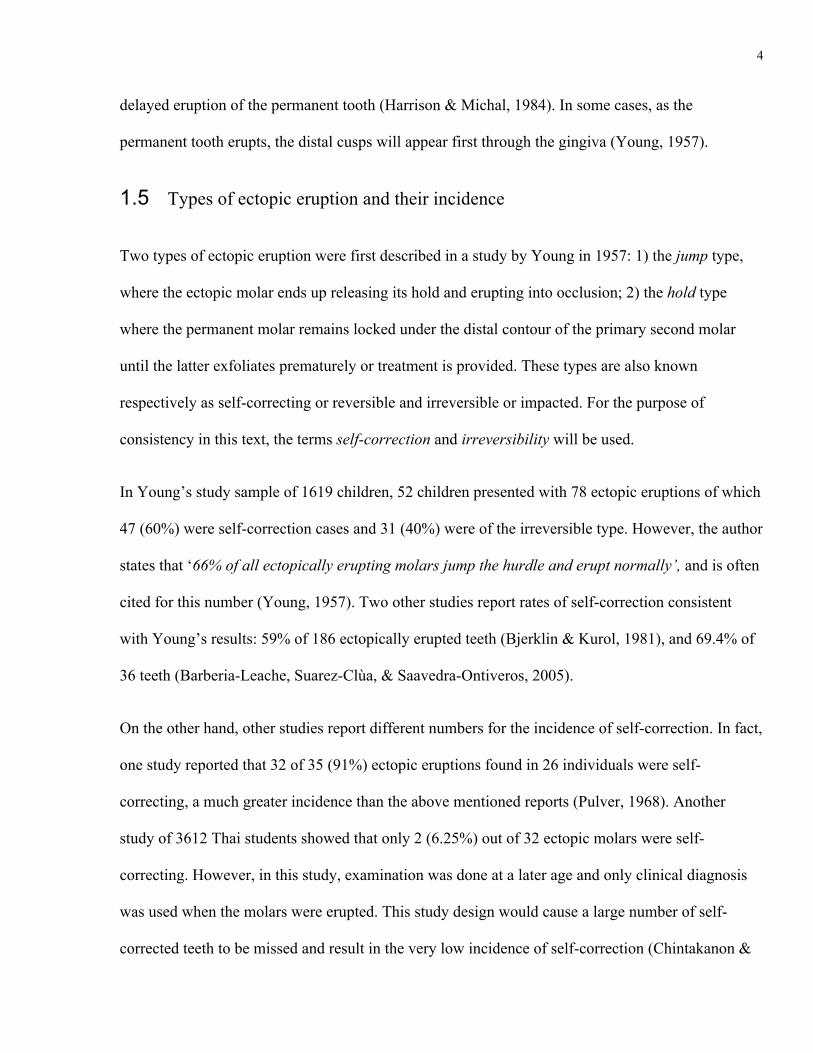
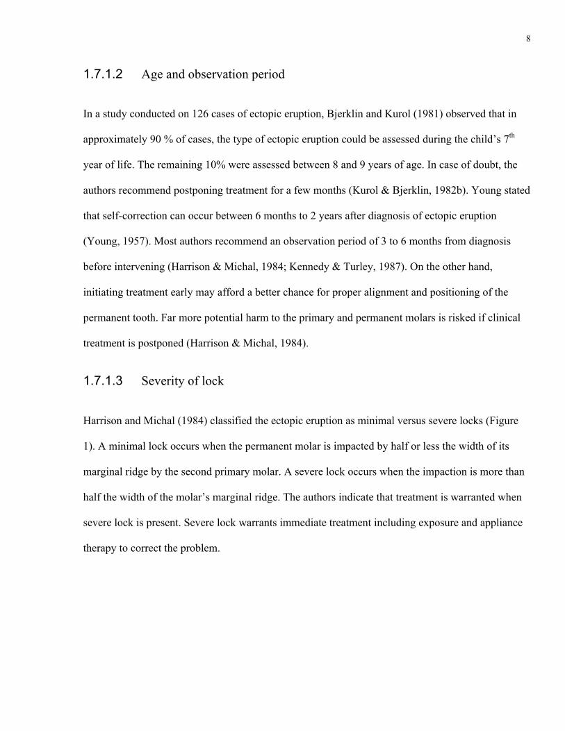
1.7.1.3 Severity of lock
Harrison and Michal (1984) classified the ectopic eruption as minimal versus severe locks (Figure
1). A minimal lock occurs when the permanent molar is impacted by half or less the width of its
marginal ridge by the second primary molar. A severe lock occurs when the impaction is more than
half the width of the molar’s marginal ridge. The authors indicate that treatment is warranted when
severe lock is present. Severe lock warrants immediate treatment including exposure and appliance
therapy to correct the problem.
9
Minimal lock: the ectopic permanent molar is impacted by one
half or less the width of its marginal ridge
Severe lock: the permanent molar is impacted by more than
one half the width of its marginal ridge
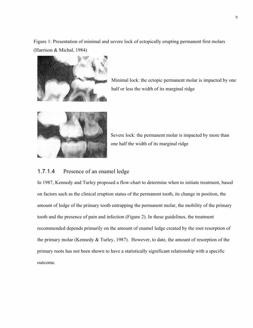
1.7.1.4 Presence of an enamel ledge
In 1987, Kennedy and Turley proposed a flow-chart to determine when to initiate treatment, based
on factors such as the clinical eruption status of the permanent tooth, its change in position, the
amount of ledge of the primary tooth entrapping the permanent molar, the mobility of the primary
tooth and the presence of pain and infection (Figure 2). In these guidelines, the treatment
recommended depends primarily on the amount of enamel ledge created by the root resorption of
the primary molar (Kennedy & Turley, 1987). However, to date, the amount of resorption of the
primary roots has not been shown to have a statistically significant relationship with a specific
outcome.
Figure 1: Presentation of minimal and severe lock of ectopically erupting permanent first molars
(Harrison & Michal, 1984)

10
Figure 2: UCLA flowchart for the management of ectopically erupting first permanent molars
(Kennedy & Turley, 1987)
11
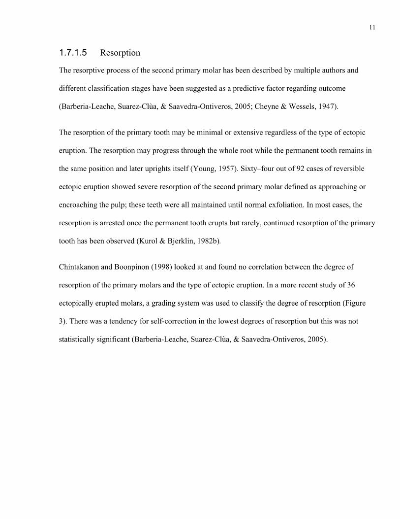
1.7.1.5 Resorption
The resorptive process of the second primary molar has been described by multiple authors and
different classification stages have been suggested as a predictive factor regarding outcome
(Barberia-Leache, Suarez-Clùa, & Saavedra-Ontiveros, 2005; Cheyne & Wessels, 1947).
The resorption of the primary tooth may be minimal or extensive regardless of the type of ectopic
eruption. The resorption may progress through the whole root while the permanent tooth remains in
the same position and later uprights itself (Young, 1957). Sixty–four out of 92 cases of reversible
ectopic eruption showed severe resorption of the second primary molar defined as approaching or
encroaching the pulp; these teeth were all maintained until normal exfoliation. In most cases, the
resorption is arrested once the permanent tooth erupts but rarely, continued resorption of the primary
tooth has been observed (Kurol & Bjerklin, 1982b).
Chintakanon and Boonpinon (1998) looked at and found no correlation between the degree of
resorption of the primary molars and the type of ectopic eruption. In a more recent study of 36
ectopically erupted molars, a grading system was used to classify the degree of resorption (Figure
3). There was a tendency for self-correction in the lowest degrees of resorption but this was not
statistically significant (Barberia-Leache, Suarez-Clùa, & Saavedra-Ontiveros, 2005).

12
I. Mild: resorption limited to cementum or with minimum dentin penetration
II. Moderate: resorption of the dentin without pulp exposure
III. Severe: resorption of distal root leading to pulp exposure
IV. Very severe: resorption that affects the mesial root of the primary tooth
(Barberia-‐Leache, Suarez-‐Clùa, & Saavedra-‐Ontiveros, 2005)
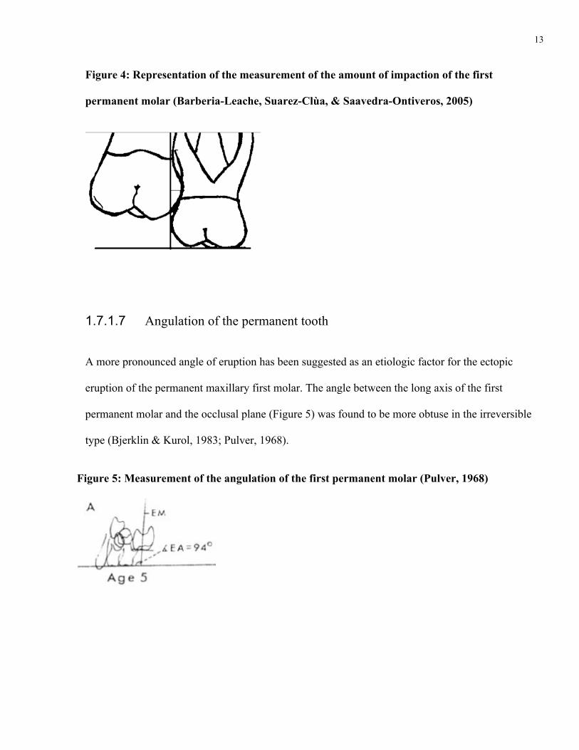
1.7.1.6 Amount of impaction of the permanent tooth under the distal contour
The amount of impaction of the first permanent molar was described by Barberia-Leache (2005) as
the distance from the area of maximum convexity of the mesial contour of the permanent tooth to a
tangential plane to the distal surface of the primary tooth (perpendicular to the occlusal surface)
(Figure 4). No statistical correlation between this measurement and the degree of resorption of the
primary molar was found. The correlation between the amount of impaction and the outcome of the
ectopic eruption was not assessed.
Figure 3: Grading of degree of resorption of ectopically erutping first permanent molars
(Barberia-Leache, Suarez-Clùa, & Saavedra-Ontiveros, 2005)
13
Figure 4: Representation of the measurement of the amount of impaction of the first
permanent molar (Barberia-Leache, Suarez-Clùa, & Saavedra-Ontiveros, 2005)
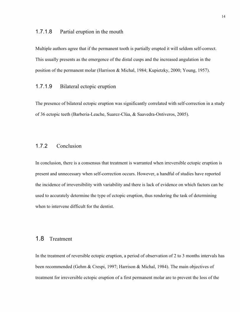
1.7.1.7 Angulation of the permanent tooth
A more pronounced angle of eruption has been suggested as an etiologic factor for the ectopic
eruption of the permanent maxillary first molar. The angle between the long axis of the first
permanent molar and the occlusal plane (Figure 5) was found to be more obtuse in the irreversible
type (Bjerklin & Kurol, 1983; Pulver, 1968).
Figure 5: Measurement of the angulation of the first permanent molar (Pulver, 1968)
14
1.7.1.8 Partial eruption in the mouth
Multiple authors agree that if the permanent tooth is partially erupted it will seldom self-correct.
This usually presents as the emergence of the distal cusps and the increased angulation in the
position of the permanent molar (Harrison & Michal, 1984; Kupietzky, 2000; Young, 1957).
1.7.1.9 Bilateral ectopic eruption
The presence of bilateral ectopic eruption was significantly correlated with self-correction in a study
of 36 ectopic teeth (Barberia-Leache, Suarez-Clùa, & Saavedra-Ontiveros, 2005).
1.7.2 Conclusion
In conclusion, there is a consensus that treatment is warranted when irreversible ectopic eruption is
present and unnecessary when self-correction occurs. However, a handful of studies have reported
the incidence of irreversibility with variability and there is lack of evidence on which factors can be
used to accurately determine the type of ectopic eruption, thus rendering the task of determining
when to intervene difficult for the dentist.
1.8 Treatment
In the treatment of reversible ectopic eruption, a period of observation of 2 to 3 months intervals has
been recommended (Gehm & Crespi, 1997; Harrison & Michal, 1984). The main objectives of
treatment for irreversible ectopic eruption of a first permanent molar are to prevent the loss of the

15
second primary molar and to regain lost arch length by repositioning the first permanent molar
distally. The retention of the second primary molar will allow proper space maintenance and normal
eruption of the second premolar (Kennedy & Turley, 1987). Treatment varies according to the
severity of the impaction as well as the maintainability of the primary second molar.
Treatment may be classified into three main categories: minimal intervention, appliance therapy
with retention of the primary second molar and appliance therapy with extraction of the second
primary molar (Gungor & Altay, 1998).
1.8.1 Minimal intervention
Interproximal wedging consists of creating a separation between the mesial surface of the permanent
first molar and the distal surface of the second primary molar to allow the permanent molar to free
itself from the undercut of the primary tooth. This can be done with an elastic separator, a brass wire
or a spring and is indicated as an initial treatment or when the impaction of the permanent molar
does not appear severe. These wedging techniques have many advantages: they generally minimize
chair time, they do not require impressions or laboratory procedures and they do not damage the
permanent teeth (Gehm & Crespi, 1997). Other minimal interventions include gingivectomy to
expose of the permanent tooth and disking of the distal surface of the primary second molar.
1.8.1.1 Elastic separator
The simplest treatment consists of placing an orthodontic separating elastic between the primary
second molar and the permanent first molar. Topical anesthetic may permit more comfortable
placement of the elastic (Glenn, 1978). Placement with waxed floss instead of pliers may prevent

16
damage to the gingiva. The elastic must be replaced every 7 to 14 days until there is overcorrection
of the impaction. Spontaneous loss of the separator indicates correction (Kennedy & Turley, 1987).
Evidently, the tooth must have emerged somewhat from the gingiva to allow for proper positioning
of the elastic.
1.8.1.2 Brass wire technique
If the permanent molar is not erupted, the brass wire technique may be used. This technique requires
local anesthesia. A brass wire is threaded through the interproximal area and looped around the
marginal ridge of the permanent molar. Both ends of the wire are twisted until snug and tucked into
the interproximal area to avoid discomfort. The wire must encircle the contact area. A bitewing
radiograph is taken to confirm the correct placement of the wire. The patient is seen at regular
intervals to tighten the wire. The wire may be removed once it slips through the contact during
tightening (Yaseen, Naik, & Uloopi, 2011). This technique may result in infection or early loss of
the primary molar, therefore careful supervision is recommended (Kupietzky, 2000).
1.8.1.3 Helical spring
A triangular helical spring has been described as an adjunct to the elastic separator technique. It is
suggested that the space created by the brass wire or the elastic separator is limited and therefore not
effective. A spring composed of three helical loops in a triangular shape is thus fabricated and
inserted between the permanent first molar and the second primary molar. The wedging spring
should be reactivated or replaced every 3 weeks (Y. H. Kim & Park, 2005).

17
(Y. H. Kim & Park, 2005)
1.8.1.4 De-Impactor spring
This prefabricated looped wire (Arkansas Dental Products) is wedged into the interproximal area
and the occlusal loop is twisted to created a separation between the primary and permanent teeth
(Venn, 1985).
1.8.1.5 Surgical exposure of the permanent tooth
If the tooth is not erupted clinically, simple excision of the overlying tissue may be the treatment of
choice. This can be easily done with a scalpel or electrosurgery. Harrison and Michal (1984)
observed that in minimal lock cases, self-correction occurred more frequently and more quickly
after surgical exposure. They also recommend surgical exposure of the first permanent molar if it is
positioned very high to avoid the use of any appliance. Self-correction should be observed within 3
to 4 months, if the condition has not improved, appliances are used (Harrison & Michal, 1984).
1.8.1.6 Disking of the distal surface of the second primary molar
Distal disking of the primary molar is suggested in cases where there is severe crowding warranting
future premolar extraction or when the premolars are absent. This will reduce the undercut created
by the distal contour of the primary tooth and allow the permanent tooth to erupt slightly mesially
Figure 6: Helical spring

18
while retaining the second primary molar as a space maintainer until future orthodontic treatment is
initiated (Kennedy & Turley, 1987).
1.8.2 Appliance therapy with retention of the second primary molar
Multiple appliances have been described using anchorage on the second primary molar with a
distalizing or tipping force on the permanent molar. These are indicated in cases of irreversible
ectopic eruption where the tooth is partially erupted. If the tooth has not emerged, a gingivectomy is
indicated prior to placement of the appliance. These appliances may increase the amount of
resorption on the second primary molar because of the stress placed on the molar. Often the second
primary molar is mobile and temporarily slightly extruded during treatment. It usually self-corrects
as soon as the appliance is removed.
1.8.2.1 Humphrey-type appliance
The Humphrey appliance consists of an orthodontic band fitted on the second primary molar with a
distal extending free arm engaging the occlusal pit of the first permanent molar to create a distal
movement. The original appliance often necessitated a small cavity preparation in the occlusal pit of
the permanent tooth to prevent the arm from slipping (Humphrey, 1962). In view of better bonding
techniques nowadays, it is preferred to activate the distalizing arm against a bonded button or resin
stop on the occlusal surface of the permanent tooth (Gehm & Crespi, 1997). Activation of this
appliance is done every 3 to 4 weeks. The appliance may be designed in such a way that the spring
assembly can be removed for adjustments without removing the band. The advantage of this type of
appliance is that it is fast acting and can be easily removed once the EE is corrected. A disadvantage
is that because of lack of anchorage, the first and second molars may move mesially and arch length
may be lost (Kurol & Bjerklin, 1986). Also, this appliance allows only anterio-posterior movement.
19
As the permanent molar moves distally, the primary tooth may cant occlusally (Gungor & Altay,
1998). To avoid relapse, it is important to make sure the permanent tooth has erupted sufficiently to
clear the resorbed area before removing this appliance.
Many modifications can be made to this appliance. Helical loops may be incorporated to allow for
vertical as well as buccal and lingual movement (Kennedy & Turley, 1987). The helical wires may
be doubled as in the Gropers appliance (Groper, 1992).
1.8.2.2 Halterman appliance
This appliance consists of a band placed on the second primary molar with a large diameter soft
wire with a distal hook placed 2 mm distal to the clinical crown of the permanent tooth. A tight
loop chained elastic is placed between the distal hook and a button bonded to the permanent molar.
The elastic chain creates a distal force on the permanent tooth. Follow-up is recommended every 3
weeks. If more correction is needed, the tension on the elastic may be increased or the appliance
may be removed and the wire repositioned more distally with a three-prong plier. The appliance
may be removed once the impaction of the first permanent molar is corrected (Halterman, 1982).
1.8.2.3 Multi loop unilateral bonded appliance
This modified bilateral Halterman appliance can be used to treat bilateral ectopic eruption of the
first permanent molar. The bands are placed on the first primary molars, and joined by a transpalatal
bar with an acrylic button for stabilization. Bilateral distal extensions with hooks are fabricated on
which chain elastics are placed on buttons bonded to the permanent teeth and to the distal hooks. An
advantage of this appliance is that it does not apply any force on the primary second molars, which

20
may have severe root resorption. This appliance may also be used to regain space if the second
primary molar is lost prematurely (Weinberger, 1992).
1.8.3 Extraction of the second primary molars and space regaining
In cases where the second primary molars have been lost prematurely a space regainer is then
necessary to distalize the first permanent molar once it is erupted. Extraction and space regaining are
also recommended in cases of irreversible ectopic eruption where there is severe resorption,
mobility, pain or infection (Chapman, 1923).
1.8.3.1 Removable appliances
A removable appliance with adams clasps for retention and fingersprings to distalize the first
permanent molars can be used. The advantage of this appliance is that it permits better oral hygiene.
As with all removable appliances, the main issue is that patient compliance is necessary. The use of
this appliance is recommended in unilateral cases only because in bilateral cases the reciprocal
forces from the activated springs may dislodge it (Kurol & Bjerklin, 1986). This issue can be
remedied by the addition of Adams clasps on the central incisors or a Hawley bow to increase
anchorage (Kennedy & Turley, 1987). An anterior bite plane may be required in cases where there is
severe space loss and tipping of the first permanent molar.
1.8.3.2 Cervical Headgear
Extra-oral traction is an effective way to distalize molars, especially in bilateral situations. Cervical
headgear has been suggested as a treatment for space regaining and to promote posterior growth of
the maxilla (Kurol & Bjerklin, 1984). It has been proposed that ectopic eruption may result from a
21
smaller maxilla. Therefore further development and growth of the posterior segments is of
importance, and may affect the longer term treatment results.
In a study of 46 children, cervical traction applied for an average of 9 months in children with
irreversible ectopic eruption resulted in uprighting of the maxillary first permanent molar to good
occlusion. This treatment was successful in 70% of the children, where sufficient space was created
for the second premolar. Poor cooperation was the main reason for failure in the remainder of the
sample. The cervical traction also led to decreased sagittal maxillary growth and to proclination of
the maxillary incisors. Cervical traction gave the best results in older children, whose second
premolars were near eruption or were erupting at the end of treatment (Kurol & Bjerklin, 1984). A
proper cephalometric analysis is recommended prior to the treatment as it may inhibit growth of the
maxilla. Cervical headgear should be avoided in patients who show a tendency for mandibular
prognathism (Kurol & Bjerklin, 1984).
Cervical traction has been a controversial treatment option. In fact, patients presenting with ectopic
eruption of the first permanent molar have been shown to have a more retrusive maxilla and a
tendency to dolicocephaly and shorter anterior cranial base (Pulver, 1968). These morphogenetic
characteristics are usually a contraindication to use extra-oral forces to move the affected molars
distally. Advocates of this theory recommend the use of removable appliances or plates with
horizontal reciprocal action (Canut & Raga, 1983; Pulver, 1968). However, a long-term follow-up
of 45 children treated with extra-oral cervical traction was done and a discriminant analysis showed
that all possible negative effects (increased proclination of the incisors, distal tipping of the
normally erupting first permanent molars in unilateral cases and reduction of maxillary growth) had
been eliminated. Hence, cervical traction treatment may be an adequate treatment if the patient has
no other malocclusion (Bjerklin, Gleerup, & Kurol, 1995).
22
1.8.3.3 Space maintenance
Space maintenance is necessary after any of the above mentioned active treatments until the
eruption of the second premolar. A band and loop appliance or a transpalatal arch are appropriate
space maintainers for this purpose (Kennedy & Turley, 1987).
1.8.4 Management of ectopically erupting mandibular molars
Ectopic eruption of the first permanent molars is less frequent in the mandible than in the maxillary
arch (Young, 1957). Distal movement of the permanent first molar is more challenging because of
the reduced anchorage available from mandibular anterior teeth and the presence of denser bone in
the mandible. A major challenge in the treatment of this condition is to avoid proclination of the
lower incisors and intercanine width expansion (Kennedy, 2008).
Only a few case reports have described treatment of mandibular ectopic eruption of the first
permanent molars in the mixed dentition. The same appliances described for the maxilla may be
adapted for use in the mandibular arch. Treatment duration may be longer due to the denser bone
(Kennedy, 2008; Yaseen, Naik, & Uloopi, 2011).
1.9 Associated anomalies
Children with EE are more likely to have one associated dental anomaly (Mooney, Morgan, Rodd,
& North, 2007). Significantly more frequent anomalies associated with EE were the infraocclusion
of primary molars (Baccetti, 1998; Bjerklin, Kurol, & Valentin, 1992) and cleft lip and palate
(Mooney, Morgan, Rodd, & North, 2007). EE has also been associated with agenesis of the second

23
premolars (Baccetti, 1998; Pulver, 1968), reduced size of maxillary lateral incisors, enamel
hypoplasia (Baccetti, 1998) and supernumerary teeth (Pulver, 1968). Additionally, ectopic eruption
of the permanent canines has been associated with the ectopic eruption of the first permanent molars
(Bjerklin, Kurol, & Valentin, 1992).
1.10 Survey
Studying healthcare professionals attitudes is essential as they play a key role in the rapidly
changing public health system (Kellerman & Herold, 2001). This was traditionally done through
postal surveys. In the past two decades, web-based health-related studies, including online survey
studies, have increased exponentially (Cantrell & Lupinacci, 2007).
1.10.1 Advantages of online surveys
Survey research through online data collection is advantageous for researchers and participants.
Researchers can benefit from online data collection because it is less expensive, a larger pool of
participants may be reached without geographical limitations, data collection time is decreased,
methodological control and efficiency of data entry and analysis are increased and it is possible to
follow-up with participants (Ahern, 2005). Advantages for study participants include increased
anonymity, ability to provide information at their own pace, increased sense of control, increased
willingness to participate because it is a novel approach, convenience and ease of use (Ahern, 2005;
Schleyer & Forrest, 2000). Additionally, the possibility of integrating better quality images, for
example radiographs, and sound can produce more intuitive and rich context for research
opportunities (Duffy, 2002; Schleyer & Forrest, 2000). It can also prevent erroneous answers by
prohibiting multiple or unanswered responses (Schleyer & Forrest, 2000).
24
1.10.2 Disadvantages of online surveys
Despite the many advantages of online surveys, they may pose unique methodological problems.
The lack of control of the testing environment by the researcher may result in extraneous variables
that might bias the response in studies that require accurate timing or involve interpersonal
interactions (Ahern, 2005; Eaton & Struthers, 2002). Also, differences in the respondents’ computer
equipment can affect the appearance of the questionnaire or the ease of using it; these variations
include the configuration of the user’s screen resolution, internet connection speed, memory
resources and software applications (Leece et al., 2004). The main challenge with internet survey’s
validity is participant selection bias. In fact, non-response bias is essential to evaluate the
representativeness of a survey and may reduce its validity if there are significant differences
between participants and non-participants. Typically, for an online survey, the sample pool will
include people who are internet literate and who have access to a computer. This may result in
some age and gender discrepancy. A high response rate is therefore ideal to reduce participant
selection bias. Additionally, determining the number of survey recipients is difficult due to the
possibility of faulty email addresses, the email not being received or read by the recipient. This
creates an inherent problem in accurately determining the actual response rate of a web-based
survey. This inaccuracy may be reduced by a higher number of responses. In some studies
comparing internet to traditional paper surveys, relatively lower response rates were reported in
web-based surveys (Leece et al., 2004); although numerous researchers across disciplines found
there were no differences in data collected from internet research compared to paper and pencil data
(Ahern, 2005; Barry, 2001; Parashos, Morgan, & Messer, 2005; Schleyer & Forrest, 2000).

25
1.10.3 Response rate
A response rate can be generally defined as the proportion of individuals selected into a sample who
are eligible and ultimately participate in the survey. A high response rate from any sample is
essential for the data to be representative of the entire population (Tambor et al., 1993), as it can
reduce the effects of nonresponse bias caused by socio-demographic and behavioral differences
between responders and non-responders (Parashos, Morgan, & Messer, 2005). There is no
scientifically proven minimally acceptable response rate. A response rate of 60% has been used as a
threshold of acceptability, but it is just a “rule of thumb” (Johnson & Wislar, 2012). Past surveys of
health professions reported response rates ranging from nine to 94%.
Recently, empirical assessments have concluded that the response rate of a survey may not be as
strongly correlated with the quality or the representativeness of the survey as has been previously
thought (Keeter, Miller, Kohut, Groves, & Presser, 2000). In fact, if a survey has a high response
rate but its non-respondents are very different from the respondents, this might produce more biased
results than a survey with lower response rate where there are little differences between respondents
and non-respondents. On the other hand, substantial differences between respondents and non-
respondents have been described even with a moderately high response rate (60-70%) (Johnson &
Wislar, 2012). One can argue that a healthcare professional survey may differ from a general
population survey by the fact that the group surveyed may have similar demographics (Tambor et
al., 1993). Increasing response rates may not reduce non-respondent bias if the additional
respondents are more similar to the early respondents than the remaining non-respondents. In a
study comparing early late and non-respondent to a physician’s survey, little difference in
demographic factors was found. This may be explained by the homogeneity of the group surveyed.
26
The existing variations among physicians may not be related to the willingness to respond to a
survey (Kellerman & Herold, 2001).
Effective strategies to increase response rates include shorter questionnaires, ease of access and pre-
paid monetary incentives (Kellerman & Herold, 2001; Tambor et al., 1993). Sending follow-up
reminders substantially increases response rates (Braithwaite, Emery, De Lusignan, & Sutton,
2003). Ease of use of the survey is very important not to deter participants away. For example,
having to enter an identification number and a pin to access the questionnaire is an additional step
that can discourage respondents from completing the survey. Theoretically, surveys to healthcare
professionals should elicit higher response rates than those of less educated respondents. In contrast,
some professionals may resist surveys that pose questions that stereotype or generalize issues or are
restrictive; do not make sense to them and take too much time out of an already overburdened
schedule (Kellerman & Herold, 2001). Therefore, a well-designed survey is very important.
1.11 Rationale
The rate of self-correction of ectopic eruption has been reported with a high degree of variability.
The literature agrees that while self-correcting ectopic eruptions do not require any treatment,
intervention is warranted in irreversible cases and may prevent early loss of the deciduous molar
and concomitant space loss. Multiple guidelines and predictive factors have been reported in the
literature to predict the type of ectopic eruption. However, very few of these factors have been
systematically assessed. The lack of evidence-based guidelines in the diagnosis of ectopically
erupting first permanent molars makes it difficult for the clinician to know when to intervene. It may
also create discrepancies in treatment between clinicians thus affecting the quality of care that is
provided to patients.
27
Chapter 2
PART I
2 Retrospective Chart Review
2.1 Objectives
• To determine the incidence of self-correction in a sample population presenting with ectopic
eruption of the maxillary first permanent molar (EE) where no interceptive treatment was
initiated.
• To determine the age range at which self-correction of ectopic eruption of the maxillary first
permanent molar occurs.
• To assess which clinical and radiographic factors are predictive of irreversible versus self-
correction of ectopic eruption of the maxillary first permanent molar.
• To assess the occurrence of adverse events in self-correcting and irreversible ectopic eruption
of the first permanent molar in a population where no corrective treatment was initiated.
28
2.2 Materials and Methods
Cases of ectopic eruption of the first permanent molar identified between 2000 and 2012 were
reviewed. All cases were obtained from a single private practice where no interceptive treatment
was initiated.
2.2.1 Inclusion and exclusion criteria
The inclusion criteria were:
• Healthy children (ASA I, II)
• Diagnosis of ectopic eruption of a permanent first molar with radiograph available for
diagnosis
• Minimum follow-up of two years or until the permanent tooth has reached its normal
position in the occlusal plane
Cases with craniofacial anomalies, cases where no radiographs were available or cases with
unknown outcome were excluded from this study.
2.2.2 Outcome
The outcomes assessed were defined as:
• Self-Correcting (SC): the ectopic maxillary first permanent molar erupted into occlusion and
the primary second molar was retained with various degrees of resorption;
• Irreversible (IRR): the maxillary first permanent molar remained locked under the distal
contour of the primary second molar until the latter exfoliated prematurely or extraction was
performed due to signs and symptoms.

29
2.2.3 Data collection and analysis
Clinical predictive factors assessed were the gender, the age at diagnosis and outcome of the ectopic
eruption of the maxillary first permanent molar, bilateral versus unilateral occurrence and the
features of the primary occlusion (primary molar occlusion, primary canine occlusion and Baume
type).
Bite-wing radiographs obtained at the time of diagnosis were scanned and printed to scale.
Radiographs were analyzed by a single investigator (B.D.) for the following factors:
1) The thickness of the enamel ledge on the primary second molar (<1mm, ≥1mm) (Kennedy &
Turley, 1987) ;
2) The severity of lock (mild : <1/2 of the marginal ridge of the permanent molar, severe :≥1/2
of marginal ridge) (Harrison & Michal, 1984) ;
3) The angulation of the permanent first molar with the occlusal plane (<90°, = 90°, >90°)
(Pulver, 1968);
4) The degree of resorption on the roots of the primary teeth (Barberia-‐Leache, Suarez-‐Clùa, &
Saavedra-‐Ontiveros, 2005):
• Mild: resorption limited to cementum or with minimum dentin penetration
• Moderate: resorption of the dentin without pulp exposure
• Severe: resorption of distal root leading to pulp exposure
• Very severe: resorption that affects the mesial root of the primary tooth
5) The amount of impaction of the first permanent molar (Barberia-‐Leache, Suarez-‐Clùa, &
Saavedra-‐Ontiveros, 2005)
• The distance from the area of maximum convexity of the mesial contour of the

30
permanent tooth to a tangential plane to the distal surface of the primary tooth
(0-0.9mm, 1-1.9mm, 2-2.9mm, >3mm).
Ten radiographs were randomly chosen and assessed by three calibrated raters for inter-rater
reliability to determine the reproducibility of these measurements between clinicians. Additionally,
the principal investigator was assessed for reliability by assessing ten radiographs at three separate
times. Cronbach's Alpha coefficient was used to determine inter-rater and intra-rater reliability.
A binomal logistic regression analysis with a probit link accounting for dichotomous dependent
variables was conducted to assess the correlations between each factor and the outcome. A full
regression model using a logit link was used to assess the relationship between all factors,
controlling for other predictive factors, and irreversible outcome. Data was analyzed using R
v.2.15.3 (R Foundation for Statistical Computing, Vienna, Austria).
2.2.4 Sample size
A sample size was calculated to assess the number of cases necessary to detect a different rate of
self-correction than the accepted rate in the literature (66%) (American Academy of Pediatric
Using G*Power (v. 3.1):
Test: Exact - Proportion: Difference from constant (binomial test, one sample case)
Analysis: A priori: Compute required sample size
Input: Tail(s) = One
Effect size g = 0.2399998 [Calculated using an odds ratio of 4.636 from proportions
P1 = 0.66 and P2 = .90, where P1 represents the accepted incidence of self-correction
in the literature (66%) and P2 represents the expected incidence (90% )]
α err prob = 0.05
Desired Power (1-β err prob) = 0.9
Constant proportion = 0.66
Output:
Total sample size = 25
31
Dentistry, 2009; Young, 1957). The expected rate of self-correction in this sample population where
no early intervention was initiated was 90%. Sample size calculation was done using G*Power (v.
3.1) in order to detect a difference from the accepted literature in rate of self-correction of ectopic
molars at 90% power and at 5% level of statistical significance.
A sample size of 25 was required for statistical significance, but to increase the power of the
analysis all the cases available were included in the study.
2.2.5 Informed consent process
An information letter describing the purpose of the research, the anonymous data collection and
instructions on how to withdraw from the study was mailed to parents of children who met the
inclusion criteria (Appendix I). Data collection was started 2 months after the letters were sent to
allow parents to opt out of study. Ethics approval was obtained from the University of Toronto
Health Sciences Research Ethics Board.

32
2.3 Results
2.3.1 Demographics
Two cases were not included in the study. One patient had Down syndrome and the second
presented with lower bilateral ectopic eruption of the first permanent molars.
A total of 66 cases of ectopic eruption of maxillary first permanent molars in 45 patients (20 males;
25 females) were identified. The occurrence of the ectopic eruption was bilateral in 21 and unilateral
in 24 patients (16 right; 8 left). Right-sided unilateral ectopic eruption was more common than left-
sided unilateral ectopic eruption but this was not statistically significant (χ²(2, N=24)= 5.733; p
=.057). There was no significant relationship between gender and the position of the ectopic molar.
Figure 7: Distribution of location of ectopic eruption of the maxillary first permanent molar
2.3.2 Outcome
One case was further excluded from the study because of unknown outcome. Sixty–five teeth were
33
included in the study. The rate of self-correction of ectopic eruption (SC) of maxillary first
permanent molars in this sample population where no intervention was initiated was 71% (46/65).
The rate of irreversible ectopic eruption (IRR) was 29%.
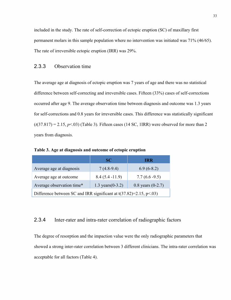
2.3.3 Observation time
The average age at diagnosis of ectopic eruption was 7 years of age and there was no statistical
difference between self-correcting and irreversible cases. Fifteen (33%) cases of self-corrections
occurred after age 9. The average observation time between diagnosis and outcome was 1.3 years
for self-corrections and 0.8 years for irreversible cases. This difference was statistically significant
(t(37.817) = 2.15, p<.03) (Table 3). Fifteen cases (14 SC, 1IRR) were observed for more than 2
years from diagnosis.
Table 3. Age at diagnosis and outcome of ectopic eruption
SC IRR
Average age at diagnosis 7 (4.8-9.4) 6.9 (6-8.2)
Average age at outcome 8.4 (5.4 -11.9) 7.7 (6.6 -9.5)
Average observation time* 1.3 years(0-3.2) 0.8 years (0-2.7)
Difference between SC and IRR significant at t(37.82)=2.15, p<.03)
2.3.4 Inter-rater and intra-rater correlation of radiographic factors
The degree of resorption and the impaction value were the only radiographic parameters that
showed a strong inter-rater correlation between 3 different clinicians. The intra-rater correlation was
acceptable for all factors (Table 4).
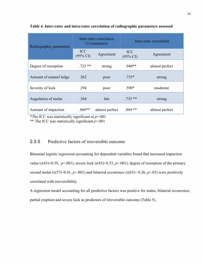
34
Table 4. Inter-rater and intra-rater correlation of radiographic parameters assessed
Radiographic parameters
Inter-rater correlation (3 examiners) Intra-rater correlation
ICC (95% CI) Agreement ICC
(95% CI) Agreement
Degree of resorption .723 ** strong .940** almost perfect
Amount of enamel ledge .262 poor .735* strong
Severity of lock .294 poor .590* moderate
Angulation of molar .364 fair .735 ** strong
Amount of impaction .869** almost perfect .869 ** almost perfect
*The ICC was statistically significant at p=.001 ** The ICC was statistically significant p<.001
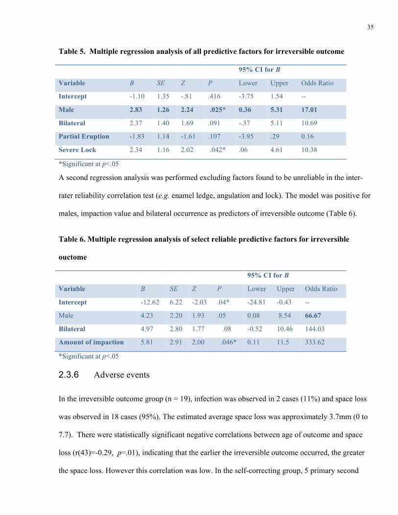
2.3.5 Predictive factors of irreversible outcome
Binomial logistic regression accounting for dependent variables found that increased impaction
value (r(43)=0.59, p<.001), severe lock (r(43)=0.53, p=.001), degree of resorption of the primary
second molar (r(57)=0.41, p=.001) and bilateral occurrence (r(63)= 0.26, p=.03) were positively
correlated with irreversibility.
A regression model accounting for all predictive factors was positive for males, bilateral occurrence,
partial eruption and severe lock as predictors of irreversible outcome (Table 5).
35
Table 5. Multiple regression analysis of all predictive factors for irreversible outcome
95% CI for B
Variable B SE Z P Lower Upper Odds Ratio
Intercept -1.10 1.35 -.81 .416 -3.75 1.54 --
Male 2.83 1.26 2.24 .025* 0.36 5.31 17.01
Bilateral 2.37 1.40 1.69 .091 -.37 5.11 10.69
Partial Eruption -1.83 1.14 -1.61 .107 -3.95 .29 0.16
Severe Lock 2.34 1.16 2.02 .042* .06 4.61 10.38
*Significant at p<.05
A second regression analysis was performed excluding factors found to be unreliable in the inter-
rater reliability correlation test (e.g. enamel ledge, angulation and lock). The model was positive for
males, impaction value and bilateral occurrence as predictors of irreversible outcome (Table 6).
Table 6. Multiple regression analysis of select reliable predictive factors for irreversible
ouctome
95% CI for B
Variable B SE Z P Lower Upper Odds Ratio
Intercept -12.62 6.22 -2.03 .04* -24.81 -0.43 --
Male 4.23 2.20 1.93 .05 0.08 8.54 66.67
Bilateral 4.97 2.80 1.77 .08 -0.52 10.46 144.03
Amount of impaction 5.81 2.91 2.00 .046* 0.11 11.5 333.62
*Significant at p<.05
2.3.6 Adverse events
In the irreversible outcome group (n = 19), infection was observed in 2 cases (11%) and space loss
was observed in 18 cases (95%). The estimated average space loss was approximately 3.7mm (0 to
7.7). There were statistically significant negative correlations between age of outcome and space
loss (r(43)=-0.29, p=.01), indicating that the earlier the irreversible outcome occurred, the greater
the space loss. However this correlation was low. In the self-correcting group, 5 primary second
36
molars (11%) were extracted after the eruption of the permanent first molar. None of the self-
correcting cases required distalization due to space loss.
In the irreversible outcome group, 9 primary teeth necessitated extraction. Seven space maintainers
were placed and 4 space regainers. Three cases required no treatment, as the space loss was minimal
or the permanent successor was not present and space closure was intended. Three cases were
referred to an orthodontist because of other anomalies or malocclusion.
2.3.7 Associated anomalies
Five cases of EE presented with agenesis of one or multiple second premolars.
2.4 Discussion
This retrospective chart review was done in a single private office where treatment of ectopic
eruption of the first permanent molar was initiated only when the patient presented with signs and
symptoms, including pain, infection or mobility of the primary tooth.
Ectopic eruption of the first permanent molar was identified in 45 patients. There was no difference
in occurrence between males and females. This is in agreement with multiple reports (Chintakanon
& Boonpinon, 1998; Kimmel, Gellin, Bohannan, & Kaplan, 1982; Pulver, 1968). An increase in
right-sided unilateral occurrence has been reported previously (Barberia-Leache, Suarez-Clùa, &
Saavedra-Ontiveros, 2005). In this sample, a higher number of right-side ectopic eruptions was
noted but the difference was not statistically significant. The unilateral to bilateral occurrence ratio
was similar in this population which is in accord with one study (Bjerklin & Kurol, 1981), while
another report found a significant higher bilateral occurrence (Barberia-Leache, Suarez-Clùa, &
Saavedra-Ontiveros, 2005).
37
The 71% rate of self-correction in this study was higher than previously reported in the majority of
the published literature (Table 2) (Barberia-Leache, Suarez-Clùa, & Saavedra-Ontiveros, 2005;
Bjerklin & Kurol, 1981; Young, 1957).
In this sample population, treatment was not initiated until the primary second molar presented with
very severe resorption resulting in mobility, pain or infection or the primary tooth was lost
prematurely. In some cases, the observation period was longer than 2 years. The majority of the
literature recommends that treatment be initiated after an observation period of 3 to 6 months if self-
correction has not occurred (Harrison & Michal, 1984; Kennedy & Turley, 1987). It has also been
recommended that the type of ectopic eruption be determined during the child’s seventh year of life
(Kurol & Bjerklin, 1982a). In this sample population, one third of the self-corrections occurred after
a 2 year observation period and after 9 years of age. Had treatment been initiated earlier, the rate of
irreversible ectopic eruption would have been higher. Delaying intervention in these cases was
beneficial as it avoided unnecessary treatments. Self-corrections occurring after the age of 9 may be
due to the exfoliation of the first primary molar resulting in the mesial drift of the second primary
molar and the freeing of the permanent first molar. This phenomenon may lead to loss of arch
length, although, in this sample population, none of the self-correcting cases showed clinically
significant space loss. This hypothesis could be looked at in a future prospective study. The
pediatric dental practice in which this study was performed differs from the average pediatric
practice as it provides orthodontic care for a large number of patients. Dentists that do not provide
orthodontic services may not feel as comfortable delaying interceptive treatment.
In the self-correcting group, 5 second primary molars (11%) were lost after the eruption of the first
permanent molar but prior to the time of normal exfoliation. In a study of 92 resorbed primary
molars due to self-correcting ectopic eruption, only 2 (2%) were lost prematurely. It may be

38
hypothesized that the higher incidence in this sample may have been due to the prolonged presence
of the permanent tooth against the roots of the primary teeth which increased the resorption.
Nonetheless, the treatment for these teeth was minimal and consisted of space maintenance.
Intervening early in the irreversible cases may have prevented the early loss of some primary teeth
and space loss in the corresponding arch. Space loss was in fact the most common adverse event
occurring in all but one of irreversible cases. At the same time, the cases that presented with an
increased space loss occurred earlier. In fact, increased space loss was significantly correlated with
earlier age at diagnosis, shorter observation period and earlier age at outcome. This signifies that
irreversible ectopic eruption may not have been prevented as it is more aggressive and occurs
earlier, within the first 3 to 6 months.
Of the 19 cases of irreversible ectopic eruption, 4 required space regainers, 6 required space
maintainers only and 6 required no treatment as the space loss was minimal or space closure was
intended due to agenesis of the second premolar. An additional 3 cases were referred to an
orthodontist due other anomalies or malocclusions. The lack of a control group renders the task of
determining the advantages and disadvantages of delaying treatment versus intervening early in
these cases difficult.
In summary, delaying treatment in this sample population did not cause major adverse events. In
fact, only 4 cases required active distalization of the first permanent molar. This indicates that
delaying intervention when unsure of the type of ectopic eruption can be a viable option and may
prevent unnecessary treatment and cost.
The present study findings indicate that the higher grades of resorption are correlated with
irreversible outcome. Previous studies found a non-statistically significant tendency for self-

39
correction in the lowest grades of resorption this may be due to a smaller sample size (Barberia-
Leache, Suarez-Clùa, & Saavedra-Ontiveros, 2005; Chintakanon & Boonpinon, 1998).
The correlation of the amount of impaction with the outcome had not been assessed previously and
had the highest correlation with irreversible outcome. This radiographic parameter was also found to
be a reliable measure between different clinicians, therefore the amount of impaction seems to be a
good reliable measure to aid in determining irreversibility of ectopic eruption of the maxillary first
permanent molar.
Predictive measurements previously reported in the literature were found to have poor statistical
reliability. In fact the poor inter-rater reliability results for the enamel ledge measurement, the
permanent tooth angulation and the severity of lock indicates clinicians’ inability to consistently
reproduce these measurements. This may be due to the poor description of these measurements in
the original articles, as well as the lack of standardization of radiographs. Some radiographic
measurements were therefore excluded from the second regression model.
The first regression model was positive for males, partial eruption, bilateral occurrence and severe
lock. Due to the assumptions of logistic regression, whereas dependent variables are eliminated,
some variables were not included in this model. Additionally, severe lock showed poor inter-rater
reliability. Therefore, a second regression model was done excluding the variables that showed poor
inter-rater reliability and including those that showed good reliability between clinicians. This
model was found to be more accurate and applicable clinically and indicated that males presenting
with bilateral ectopic eruption of the maxillary first permanent molars and increased amount of
impaction were more likely to have an irreversible outcome. Looking at the second multiple
regression model, only the impaction value is significant on its own. Although males and bilateral
are not significant, they do have a substantial impact on the fit of the model overall and were

40
retained by a stepwise selection algorithm. Hence, they are still contributing to the model. The
multiple regression models yielded very high odds ratios. This is due to the small sample size as
well as the uneven distribution of the data. Future studies with a larger sample may determine more
accurate odds ratio.
The regression analyses were done assessing each tooth as an independent case. This allowed
inclusion of bilateral cases where the outcome was different for each side. A higher number of cases
thus increased the power of the analyses. Unfortunately, this method may have created an inherent
bias towards factors that are not independent, mainly gender and bilateral cases. On the other side,
the relationship between gender, unilateral or bilateral occurrence and outcome was assessed and
was not found to be significant. Future analysis may involve a multilevel model approach.
The retrospective chart review was limited by the data available in the charts as well as the quality
and availability of the radiographs. A future prospective cohort study looking at the amount of
impaction with standardized radiographs may help in developing evidence based guidelines for the
decision to initiate treatment of ectopic eruption of maxillary first permanent molars.
41
2.5 Conclusions
1) Self-correction of ectopic eruption of maxillary first permanent molars may occur after the age
of 9 and delaying intervention can be a viable treatment option when unsure of the type of
ectopic eruption.
2) Increased amount of impaction and degree of resorption were identified as reliable predictors of
irreversible outcome of ectopic eruption of first permanent molars.
3) Ectopic eruption presenting bilaterally in males with an increased amount of impaction are more
likely to be of the irreversible type and require earlier intervention.
42
Chapter 3
PART II
3 Online Survey of Pediatric Dentists and Orthodontists
3.1 Objective
To evaluate the prevailing attitudes of pediatric dentists and orthodontists towards intervention in
cases of ectopic eruption of maxillary first permanent molars.
3.2 Materials and methods
An online survey was developed using the SurveyMonkey online tool (SurveyMonkey, Palo Alto
Calif). The survey was sent to all active members of the American Academy of Pediatric Dentistry,
the Canadian Academy of Pediatric Dentistry and the American Association of Orthodontists. One
reminder email was sent one month after the initial email.
3.2.1 Inclusion criteria
All active members of the Canadian Academy of Pediatric Dentistry (CAPD), the American
Academy of Pediatric Dentistry (AAPD) and the American Association of Orthodontists (AAO)
with a valid email address were asked to participate to the survey.
3.2.2 Sample size
The sample size needed to determine if predictor variables influence the decisions of practitioners to
initiate treatment of an ectopically erupting permanent molar in different clinical scenarios was
43
determined following the rule: N=104 + k; where k is the number of prediction variables, assuming
a medium-size relationship between the independent variables (predictors) and dependent variables
(decision of treatment or observation for different cases), where α = .05 and power set at 80%.
The prediction variables assessed were:
1. Gender of practitioner
2. Age of practitioner
3. Years of practice
4. Type of training
5. Setting of training
6. Primary setting of current practice
7. Specialty
The minimum sample size required was 111 practitioners for regression analysis. More practitioners
were surveyed to have a better representativeness of the North American population.
3.2.3 Survey content
The survey included questions on demographics and 3 cases of ectopic eruption for dentists to
review and decide on whether to intervene or observe. The first case was characterized by a bilateral
occurrence of the ectopic eruption of the maxillary first permanent molar; the second case was
marked by the age of the patient at diagnosis, 9 years old; and the third case presented an ectopic
eruption causing minimal resorption and impaction that increased over the course of a year. The
survey also comprised of general questions regarding factors used to determine if intervention is
warranted in a case of ectopic eruption of the first permanent molar. Pearson’s Chi-squared test was
44
conducted to compare responses between orthodontists and pediatric dentists.
3.2.4 Confidentiality and informed consent
The IP addresses of all respondents were blocked to ensure confidentiality. The survey did not
contain any identification cookies. A letter of introduction (Appendix III) including the study’s
purpose, the survey’s description and assurance of confidentiality, prefaced the survey.
3.2.5 Data analysis
The data set was compiled by the SurveyMonkey websites and exported to a Microsoft Excel
spreadsheet and was analyzed using R v.2.15.3 (R Foundation for Statistical Computing, Vienna,
Austria). Descriptive statistics and frequency distributions were used to determine if there is a
consensus among practitioners toward management of ectopic molars. Multiple regression analysis
was used to determine if any factors predict the decision of a practitioner to treat or observe an
ectopic eruption.
3.3 Results
The total number of respondents was 1591, specifically 950 pediatric dentists and 641
orthodontists. The respective response rate was 22% and 7%. The rate of response from pediatric
dentists was significantly higher than orthodontists (χ²(1, N=1591)= 801.7; p <.001).
3.3.1 Demographics
There was a significant difference in the gender distribution of pediatric dentists and orthodontists
(χ²(1, N=1591)= 109; p <.001). (Figure 8). This difference is similar to the AAPD and AAO gender

45
distribution (Figure 9). The CAO data was not available.
(source: AAPD member statistics, March 2013; personal correspondence with AAO)
There were significantly more pediatric dentists in the 30-39 age group and significantly more
orthodontists in the >60 years old group (χ²(4, N=1591)= 109; p <.001) (Figure 10). Again, there are
78%
22%
AAO
78%
22%
AAO
78%
22%
AAO
Figure 9. Gender distribution of active members of the AAPD and the AAO
Figure 8. Gender distribution of respondents

46
some similarities with the AAPD and AAO age group distribution (Figure 11).
Figure 10. Age distribution of respondents
Figure 11. Age distribution of active members of the AAPD and the AAO
(source: AAPD member statistics, March 2013; personal correspondence with AAO)
11%
35%
20% 21%
13%
0.20%
22% 25% 24%
29%
0%
5%
10%
15%
20%
25%
30%
35%
40%
< 30 30-‐39 40-‐49 50-‐65 > 65
AAPD AAO
2.2%
36.1%
23.8% 22.1%
15.8%
2.0%
17.0% 20.3%
26.8%
33.9%
0.0%
5.0%
10.0%
15.0%
20.0%
25.0%
30.0%
35.0%
40.0%
20-‐29 30-‐39 40-‐49 50-‐59 >60
Pediatric Dentists Orthodontists
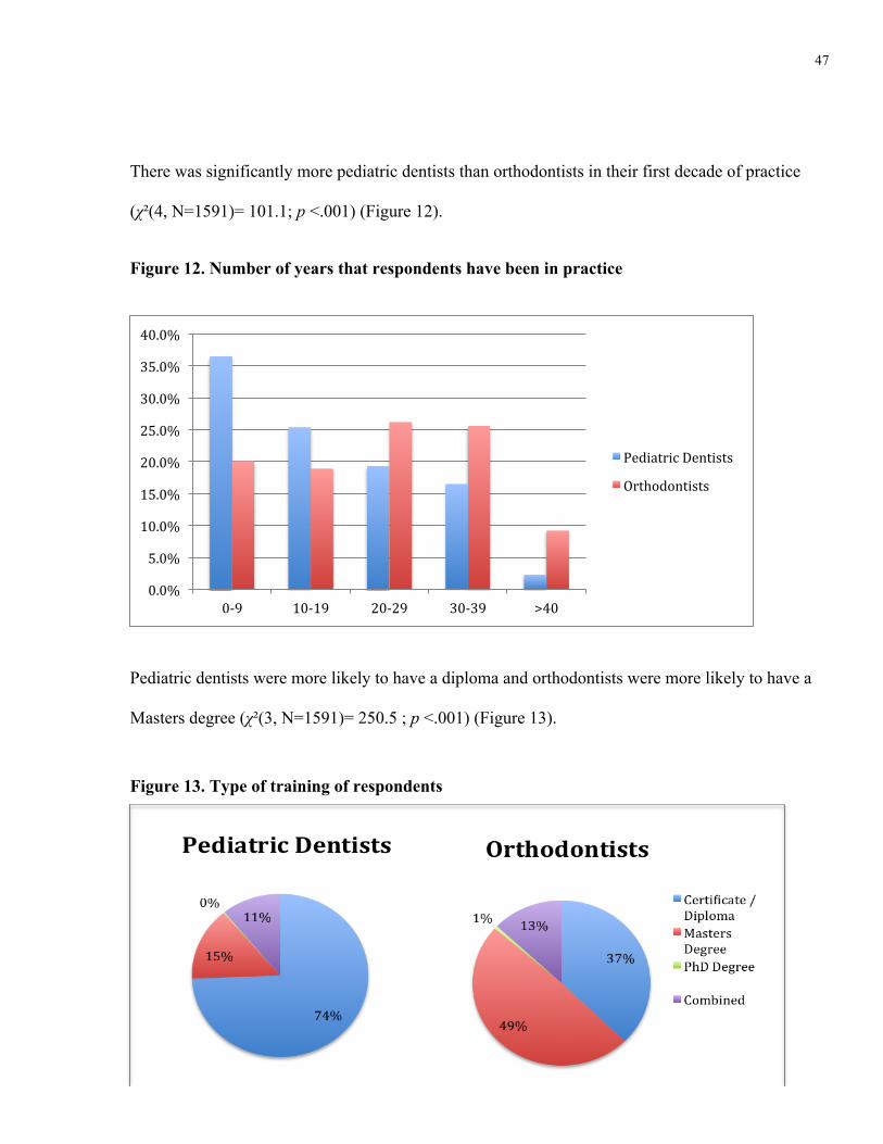
47
There was significantly more pediatric dentists than orthodontists in their first decade of practice
(χ²(4, N=1591)= 101.1; p <.001) (Figure 12).
Figure 12. Number of years that respondents have been in practice
Pediatric dentists were more likely to have a diploma and orthodontists were more likely to have a
Masters degree (χ²(3, N=1591)= 250.5 ; p <.001) (Figure 13).
0.0%
5.0%
10.0%
15.0%
20.0%
25.0%
30.0%
35.0%
40.0%
0-‐9 10-‐19 20-‐29 30-‐39 >40
Pediatric Dentists
Orthodontists
Figure 13. Type of training of respondents
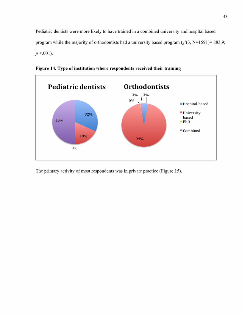
48
Pediatric dentists were more likely to have trained in a combined university and hospital based
program while the majority of orthodontists had a university based program (χ²(3, N=1591)= 883.9;
p <.001).
The primary activity of most respondents was in private practice (Figure 15).
Figure 14. Type of institution where respondents received their training
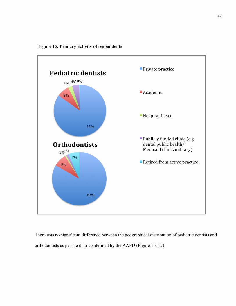
49
There was no significant difference between the geographical distribution of pediatric dentists and
orthodontists as per the districts defined by the AAPD (Figure 16, 17).
Figure 15. Primary activity of respondents
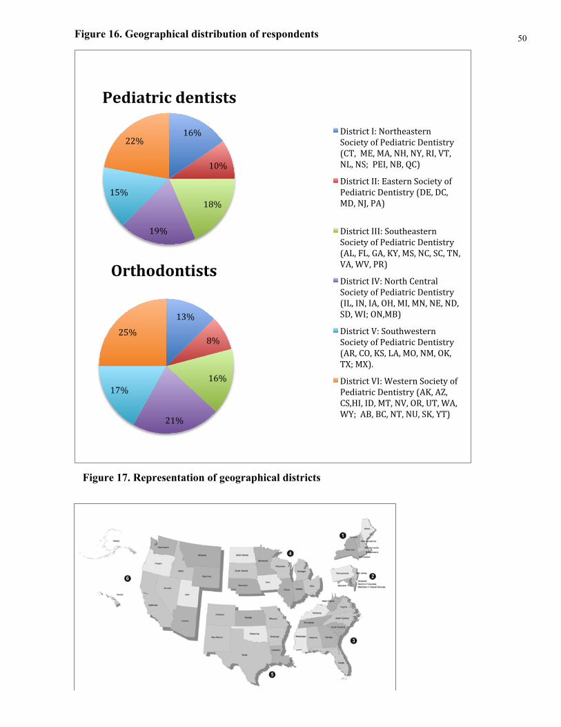
50
Figure 17. Representation of geographical districts
16%
10%
18%
19%
15%
22%
Pediatric dentists
District I: Northeastern Society of Pediatric Dentistry (CT, ME, MA, NH, NY, RI, VT, NL, NS; PEI, NB, QC)
District II: Eastern Society of Pediatric Dentistry (DE, DC, MD, NJ, PA)
District III: Southeastern Society of Pediatric Dentistry (AL, FL, GA, KY, MS, NC, SC, TN, VA, WV, PR)
District IV: North Central Society of Pediatric Dentistry (IL, IN, IA, OH, MI, MN, NE, ND, SD, WI; ON,MB)
District V: Southwestern Society of Pediatric Dentistry (AR, CO, KS, LA, MO, NM, OK, TX; MX).
District VI: Western Society of Pediatric Dentistry (AK, AZ, CS,HI, ID, MT, NV, OR, UT, WA, WY; AB, BC, NT, NU, SK, YT)
Figure 16. Geographical distribution of respondents
13%
8%
16%
21%
17%
25%
Orthodontists

51
3.3.2 Ectopic eruption cases
Figure 18. Survey case 1
A 7-year-old child presents for a recall appointment. Bilateral ectopic eruption of the first
permanent molar is observed on a routine bitewing radiograph. The condition is
asymptomatic.
Figure 19. Respondents’ classification of the ectopic eruption in case 1
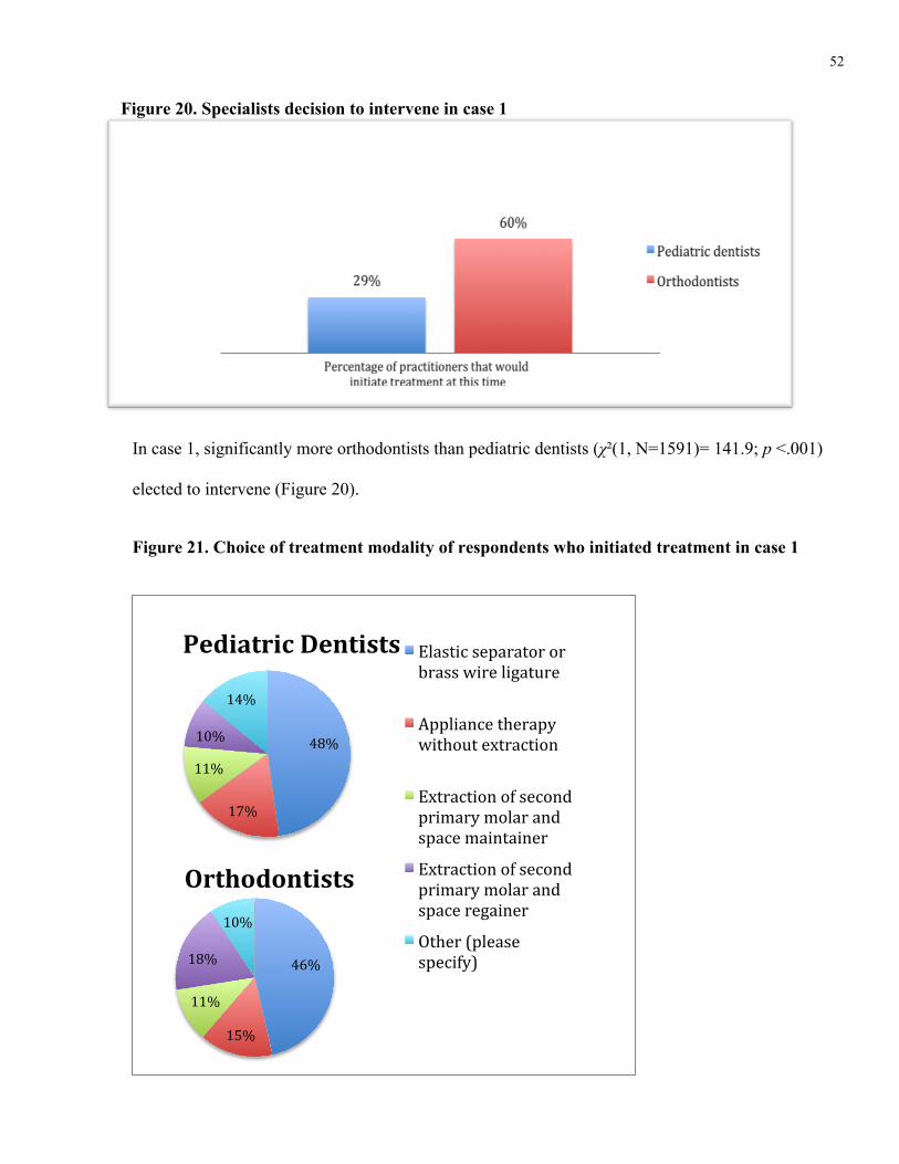
52
In case 1, significantly more orthodontists than pediatric dentists (χ²(1, N=1591)= 141.9; p <.001)
elected to intervene (Figure 20).
Figure 21. Choice of treatment modality of respondents who initiated treatment in case 1
48%
17%
11%
10%
14%
Pediatric Dentists Elastic separator or brass wire ligature
Appliance therapy without extraction
Extraction of second primary molar and space maintainer
Extraction of second primary molar and space regainer
Other (please specify) 46%
15%
11%
18%
10%
Orthodontists
Figure 20. Specialists decision to intervene in case 1

53
When asked, if this situation was unilateral, would your management be the same?; 96% of
pediatric dentists and 97% of orthodontists responded yes.
Figure 23. Specialist’s classification of ectopic eruption in case 2
A 9 -year-old child presents for a recall appointment. Unilateral ectopic eruption
of the first permanent molar is observed on a routine bitewing radiograph. The
condition is asymptomatic.
Figure 22. Survey case 2

54
There was no difference between specialists in case 2, where the ectopic eruption was diagnosed in
a 9-year-old patient, most practitioners elected to intervene at the time of diagnosis.
Figure 24. Specialists’ choice of treatment modality in case 2
39%
23%
12%
13%
13%
Pediatric Dentists
Elastic separator or brass wire ligature
Appliance therapy without extraction
Extraction of second primary molar and space maintainer
Extraction of second primary molar and space regainer
Other (please specify) 45%
19%
10%
19%
7%
Orthodontists
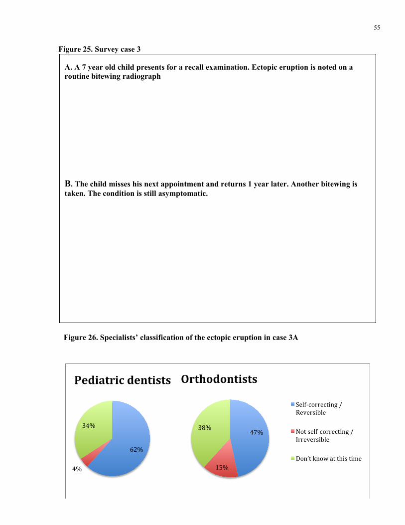
55
Figure 26. Specialists’ classification of the ectopic eruption in case 3A
1
Figure 25. Survey case 3
2
A. A 7 year old child presents for a recall examination. Ectopic eruption is noted on a routine bitewing radiograph
B. The child misses his next appointment and returns 1 year later. Another bitewing is taken. The condition is still asymptomatic.
62%
4%
34%
Pediatric dentists
Self-‐correcting / Reversible
Not self-‐correcting / Irreversible
Don’t know at this time
47%
15%
38%
Orthodontists

56
In case 3A, 91% of pediatric dentists and 84% of orthodontists opted to observe and follow up in 3
to 6 months. The modality of choice of those who elected to intervene was an elastic separator or
brass wire.
In case 3B, 81% of pediatric dentists and 82% of orthodontists elected to intervene.
Figure 27. Specialists’ classification of the ectopic eruption in case 3B
16%
78%
6%
Pediatric dentists
Self-‐correcting / Reversible
Not self-‐correcting / Irreversible
Don’t know at this time
15%
78%
7%
Orthodontists
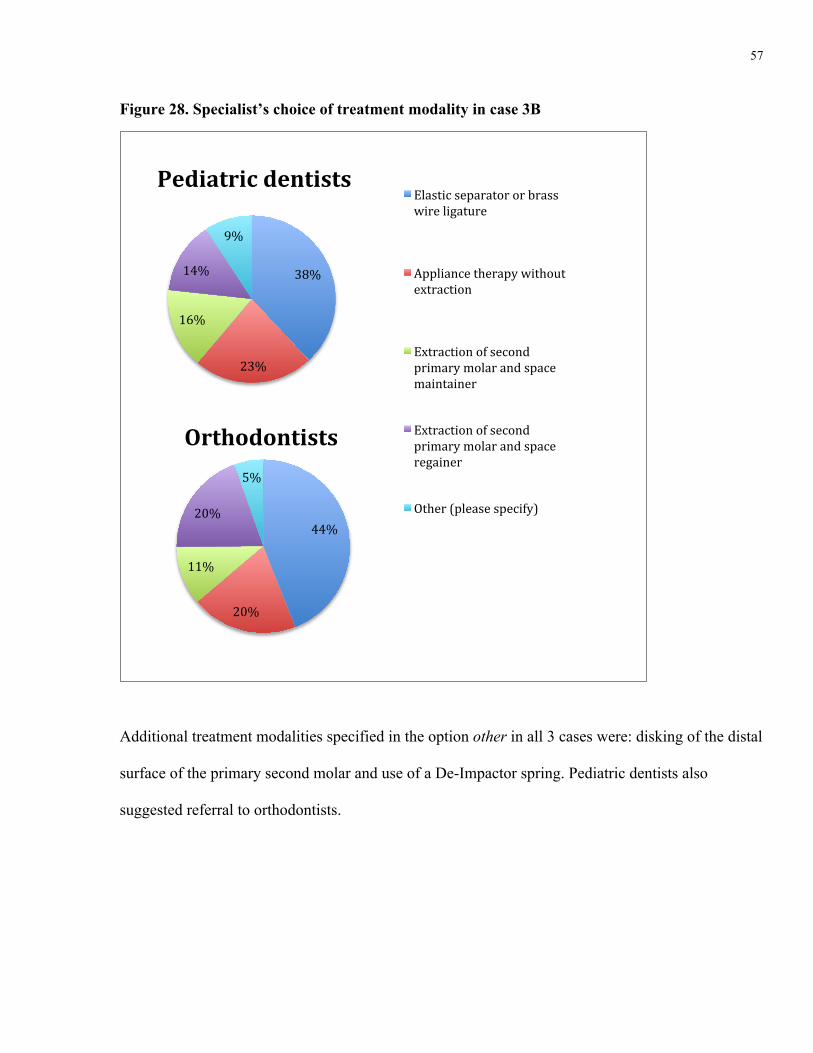
57
Additional treatment modalities specified in the option other in all 3 cases were: disking of the distal
surface of the primary second molar and use of a De-Impactor spring. Pediatric dentists also
suggested referral to orthodontists.
38%
23%
16%
14%
9%
Pediatric dentists Elastic separator or brass wire ligature
Appliance therapy without extraction
Extraction of second primary molar and space maintainer
Extraction of second primary molar and space regainer
Other (please specify) 44%
20%
11%
20%
5%
Orthodontists
Figure 28. Specialist’s choice of treatment modality in case 3B
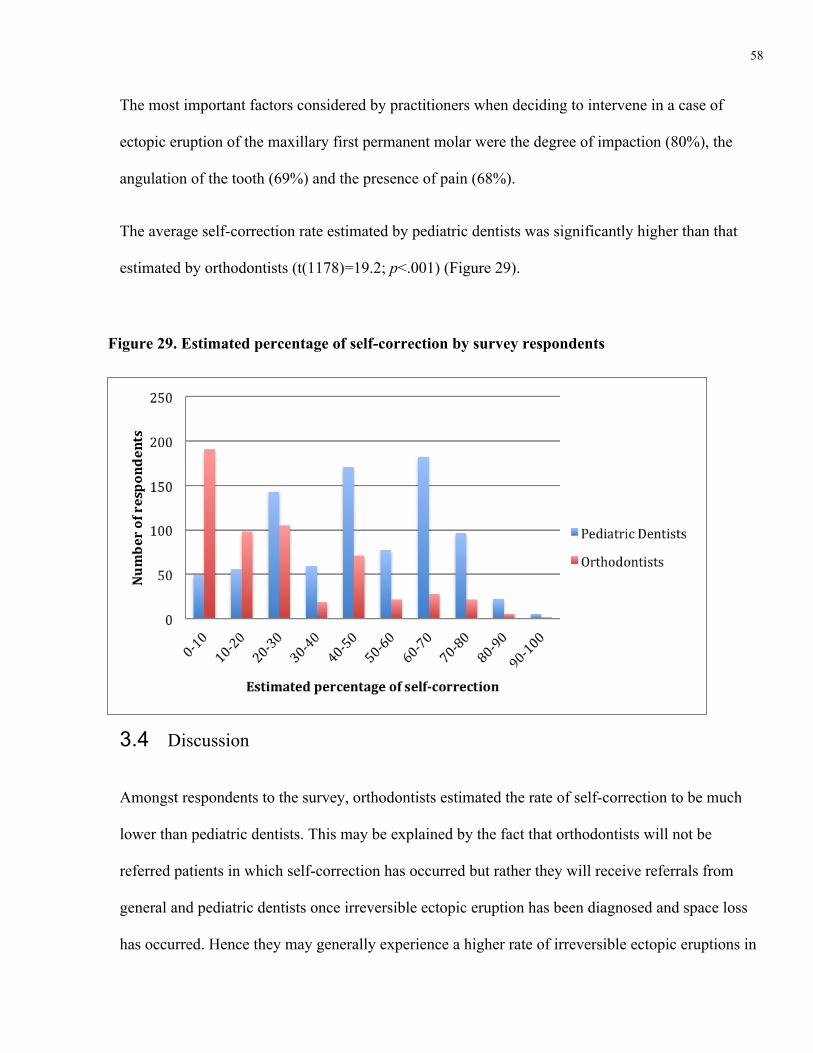
58
The most important factors considered by practitioners when deciding to intervene in a case of
ectopic eruption of the maxillary first permanent molar were the degree of impaction (80%), the
angulation of the tooth (69%) and the presence of pain (68%).
The average self-correction rate estimated by pediatric dentists was significantly higher than that
estimated by orthodontists (t(1178)=19.2; p<.001) (Figure 29).
3.4 Discussion
Amongst respondents to the survey, orthodontists estimated the rate of self-correction to be much
lower than pediatric dentists. This may be explained by the fact that orthodontists will not be
referred patients in which self-correction has occurred but rather they will receive referrals from
general and pediatric dentists once irreversible ectopic eruption has been diagnosed and space loss
has occurred. Hence they may generally experience a higher rate of irreversible ectopic eruptions in
Figure 29. Estimated percentage of self-correction by survey respondents

59
their practice. In fact, a few orthodontists commented that they receive referrals by the patient’s
treating dentist following the extraction of the primary second molar. The orthodontists’ inclination
to intervene earlier in the bilateral case may be due to the assumption that the cause of the ectopic
eruption is skeletal and not local. Indeed, at multiple occasions, orthodontists commented that they
suspect a maxillary deficiency when presented with an ectopic eruption.
The disparity in responses between pediatric dentists and orthodontists in the first survey case and in
the estimated percentage of self-correction may be the product of insufficient evidence-based
guidelines on the timing of intervention in cases of ectopic eruption. The development of validated
guidelines will help standardize treatment among practitioners and thus improve the quality of care
provided to patients. Additionally, theses guidelines should be readily available for all dentists as
many general practitioners are children’s primary care dentists.
In all three cases, the main modality of treatment chosen by the respondents who opted to intervene
was the elastic separator or brass wire technique. Separation is often indicated as a first treatment
option that is minimally invasive and cost efficient. (Kennedy & Turley, 1987). When separation is
not successful, appliance therapy is indicated. Of interest was the choice of treatment of many
clinicians who indicated disking the distal of the second primary molar as an appropriate
intervention. This technique has been recommended as a temporary treatment only in patients who
may need future orthodontic treatment including extractions or when the premolars are missing
(Kennedy & Turley, 1987). The author could not find any reliable literature supporting this
intervention.
The survey was based on diagnosing ectopic eruption from bite-wings because the radiographs used
were obtained from the retrospective chart review. As well, it was intended to reproduce the
presentation of ectopic eruption of the maxillary first permanent molar at the initial diagnosis. This
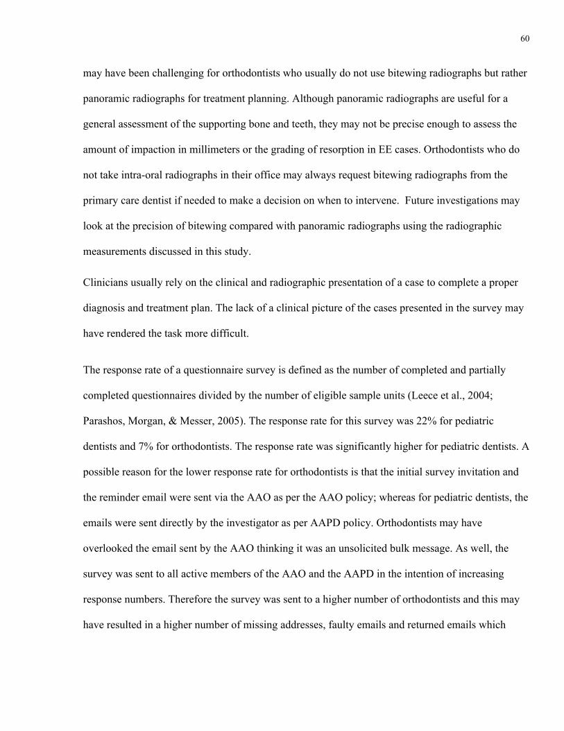
60
may have been challenging for orthodontists who usually do not use bitewing radiographs but rather
panoramic radiographs for treatment planning. Although panoramic radiographs are useful for a
general assessment of the supporting bone and teeth, they may not be precise enough to assess the
amount of impaction in millimeters or the grading of resorption in EE cases. Orthodontists who do
not take intra-oral radiographs in their office may always request bitewing radiographs from the
primary care dentist if needed to make a decision on when to intervene. Future investigations may
look at the precision of bitewing compared with panoramic radiographs using the radiographic
measurements discussed in this study.
Clinicians usually rely on the clinical and radiographic presentation of a case to complete a proper
diagnosis and treatment plan. The lack of a clinical picture of the cases presented in the survey may
have rendered the task more difficult.
The response rate of a questionnaire survey is defined as the number of completed and partially
completed questionnaires divided by the number of eligible sample units (Leece et al., 2004;
Parashos, Morgan, & Messer, 2005). The response rate for this survey was 22% for pediatric
dentists and 7% for orthodontists. The response rate was significantly higher for pediatric dentists. A
possible reason for the lower response rate for orthodontists is that the initial survey invitation and
the reminder email were sent via the AAO as per the AAO policy; whereas for pediatric dentists, the
emails were sent directly by the investigator as per AAPD policy. Orthodontists may have
overlooked the email sent by the AAO thinking it was an unsolicited bulk message. As well, the
survey was sent to all active members of the AAO and the AAPD in the intention of increasing
response numbers. Therefore the survey was sent to a higher number of orthodontists and this may
have resulted in a higher number of missing addresses, faulty emails and returned emails which
61
contributed to the lower survey response of orthodontists. Additionally, orthodontists may have felt
that this subject is of less interest to their practice and therefore be less inclined to respond.
The response rate for this survey was fairly low. A recent online survey of 1000 pediatric dentists
and 1000 orthodontists done at the University of Toronto’s department of pediatric dentistry yielded
a higher response rate for pediatric dentists (42%) and similar (7%) of orthodontists (Goldenberg,
2012). Recent surveys of pediatric dentists had response rates of 25 to 48% (Lee et al., 2012; Lopez-
Cepero, Amini, Pagano, Casamassimo, & Rashid, 2013; J. M. Russo, Russo, & Guelmann, 2006).
Surveys of orthodontists have had response rates ranging from 10 to 40% (Burke et al., 2012;
Madhavji, Araujo, Kim, & Buschang, 2011; Slack, Swift, Rossouw, & Phillips, 2013; Yang &
Kiyak, 1998). A web survey of orthodontists reported a very high response rate of 92%, which was
calculated by dividing the number of respondents by the number of clinicians who accessed the link.
Using the definition of response rate discussed previously, the response rate would be 3% (Huang,
Becerra, Walker, & Hovell, 2006).
There may be multiple reasons for the fairly low response rate in the present survey. It has been
suggested that survey response rates among health care professionals has been decreasing (McAvoy
& Kaner, 1996; Wiebe, Kaczorowski, & MacKay, 2012). Some clinicians have an office policy not
to participate in surveys (Wiebe, Kaczorowski, & MacKay, 2012). Additionally, the survey was sent
in May and the reminder 1 month later. The survey was open until September. This time period may
correspond with vacations for many practitioners and the emails may not have been read in time to
respond. As well, it is difficult to assess the accuracy of the email addresses provided by the
organizations and there may have been multiple faulty addresses.
Sending reminders to complete surveys has been shown to improve response rate (Braithwaite,
Emery, De Lusignan, & Sutton, 2003). In fact, in the present survey, survey responses increased

62
once the reminder email was sent. Due to the AAO policy, only one reminder was sent to
orthodontists. For consistency between both groups, only one reminder was sent to pediatric
dentists. Had there been an additional email reminder, the response rate may have increased.
When compared to traditional paper methods, internet surveys have relatively lower response rates
(Braithwaite, Emery, De Lusignan, & Sutton, 2003). Some clinicians may not be proficient with
technology or prefer to fill out the survey at a time where they do not have access to a computer
and/or internet and this may have deterred them from completing the survey.
A disadvantage of online surveys is the difficulty in controlling the sample size. In fact, some email
inboxes may be full or the email may be directly placed in a junk mail folder. As well, some
respondents may have previously received a survey request and declined it. In fact
SurveyMonkey™ has an option to opt out of any future survey. Recipients that had previously
“opted out” would have not received the survey but are unaccounted for. Additionally, some other
technical difficulties with the website were reported by some respondents, where they had to
subscribe to the site. This was fixed in a timely manner, but there may have been respondents that
encountered this problem and decided not to complete the survey. An option with web-based
surveys is to restrict to one responder per computer. This is done to avoid multiple answers from the
same respondents. Unfortunately, this may pose a problem if there are multiple respondents working
in the same office that attempt to complete the survey.
Due to previously reported low response rates, this survey was sent to all the active members of the
AAPD (4328) and the AAO (9679) with the intention to increase the number of responses. This
approach did result in a high number of responses, 1591, and increase the power of the analyses. At
the same time, it might have contributed to the low response rate by increasing the chances of faulty
email addresses blocked surveys and returned emails. The clinicians that responded self-selected

63
themselves, therefore there may be a systemic bias between respondents and non-respondents. Non-
responder bias is a major concern with any type of survey study. While it is best controlled by
maximizing response rates its effect must always be considered (Braithwaite, Emery, De Lusignan,
& Sutton, 2003). A high response rate is essential for the data to be considered representative of an
entire population (Tambor et al., 1993). Although opinions differ as to an optimal response rate to
eliminate non-response bias, the reported ideal range is between 70-80% (Parashos, Morgan, &
Messer, 2005; Tambor et al., 1993). However, a high response rate may still present with non-
respondent bias if there is a significant difference between respondents and non-respondents
(Tambor et al., 1993) and vice-versa, a survey reporting low response rate in a population where
there is little difference between non-respondents and respondents may be representative of that
population (Johnson & Wislar, 2012). One can argue that a healthcare professional survey may
differ from a general population survey by the fact that the group surveyed may have similar
demographics (Tambor et al., 1993). The existing variations between respondents and non-
respondents may not be related to the willingness to participate in the survey (Kellerman & Herold,
2001). The fairly similar responses between clinicians may be another indicator of the homogeneity
of the group and may indicate that a higher response rate may have yielded similar results.
3.5 Conclusions
1) Significant differences exist between pediatric dentists and orthodontists who responded to the
survey in regards to ectopic eruption of the first permanent molar.
2) Compared to pediatric dentists, orthodontists who responded to the survey estimated the rate of
self-correction to be much lower than pediatric dentists.
64
References
Ahern, N. R. (2005). Using the internet to conduct research. Nurse Researcher, 13(2), 55-70.
Baccetti, T. (1998). A clinical and statistical study of etiologic aspects related to associated tooth
anomalies in number, size, and position. Minerva Stomatologica, 47(12), 655-663.
Barberia-Leache E, Suarez-Clùa MC, Saavedra-Ontiveros D. (2005). Ectopic eruption of the
maxillary first permanent molar: Characteristics and occurrence in growing children. Angle
Orthod., 75(4):610-615.
Barry, D. T. (2001). Assessing culture via the internet: Methods and techniques for psychological
research. Cyberpsychology & Behavior : The Impact of the Internet, Multimedia and Virtual
Reality on Behavior and Society, 4(1), 17-21.
Bjerklin, K., Gleerup, A., & Kurol, J. (1995). Long-term treatment effects in children with ectopic
eruption of the maxillary first permanent molars. European Journal of Orthodontics, 17(4),
293-304.
Bjerklin K, Kurol J. (1981) Prevalence of ectopic eruption of the maxillary first permanent molar.
Swed Dent J. 1981;5(1):29-34.
Bjerklin, K., & Kurol, J. (1983). Ectopic eruption of the maxillary first permanent molar: Etiologic
factors. American Journal of Orthodontics, 84(2), 147-155.
Bjerklin, K., Kurol, J., & Valentin, J. (1992). Ectopic eruption of maxillary first permanent molars
and association with other tooth and developmental disturbances. European Journal of
Orthodontics, 14(5), 369-375.

65
Braithwaite, D., Emery, J., De Lusignan, S., & Sutton, S. (2003). Using the internet to conduct
surveys of health professionals: A valid alternative? Family Practice, 20(5), 545-551.
Burke, B., Hamdan, A. M., Tufekci, E., Shroff, B., Best, A. M., & Lindauer, S. J. (2012).
Perceptions of soft tissue laser use in orthodontics. The Angle Orthodontist, 82(1), 75-83.
Cantrell, M. A., & Lupinacci, P. (2007). Methodological issues in online data collection. Journal of
Advanced Nursing, 60(5), 544-549.
Canut, J. A., & Raga, C. (1983). Morphological analysis of cases with ectopic eruption of the
maxillary first permanent molar. European Journal of Orthodontics, 5(3), 249-253.
Carr, G. E., & Mink, J. R. (1965). Ectopic eruption of the first permanent maxillary molar in cleft lip
and palate children. ASDC Journal of Dentistry for Children, 32, 179-188.
Chapman, H. (1923). First upper permanent molar partially impacted against second deciduous
molars. Internat. J. Orthodont., Oral Surg. and Radiog., 9, 339-340, 341, 342, 343, 344,345.
Cheyne, V. D., & Wessels, K. E. (1947). Impaction of permanent first molar with resorption and
space loss in region of deciduous second molar. The Journal of the American Dental
Association, 35(11), 774-787.
Chintakanon K, Boonpinon P. (1998). Ectopic eruption of the first permanent molars: Prevalence
and etiologic factors. Angle Orthod., 68(2):153-160.
Duffy, M. E. (2002). Methodological issues in web-based research. Journal of Nursing Scholarship
: An Official Publication of Sigma Theta Tau International Honor Society of Nursing / Sigma
Theta Tau, 34(1), 83-88.
66
Eaton, J., & Struthers, C. W. (2002). Using the internet for organizational research: A study of
cynicism in the workplace. Cyberpsychology & Behavior : The Impact of the Internet,
Multimedia and Virtual Reality on Behavior and Society, 5(4), 305-313.
Gehm, S., & Crespi, P. V. (1997). Management of ectopic eruption of permanent molars.
Compendium of Continuing Education in Dentistry, 18(6), 561-6, 568.
Glenn, R. W. (1978). Ectopic eruption of permanent first molars: A simple interceptive method of
treatment. The Journal of the Nebraska Dental Association, 55(2), 11-14.
Goldenberg, L. (2012). A retrospective cohort study of fixed space maintainers and a survey of
their use by pediatric dentists and orthodontists . Unpublished Msc, University of Toronto,
University of Toronto.
Groper, J. N. (1992). Ectopic eruption of a mandibular first permanent molar: Report of an unusual
case. ASDC Journal of Dentistry for Children, 59(3), 228-230.
Gungor, H. C., & Altay, N. (1998). Ectopic eruption of maxillary first permanent molars: Treatment
options and report of two cases. The Journal of Clinical Pediatric Dentistry, 22(3), 211-216.
Halterman, C. W. (1982). A simple technique for the treatment of ectopically erupting permanent
first molars. The Journal of the American Dental Association, 105(6), 1031-1033.
Harrison LM, Michal BC. (1984) Treatment of ectopically erupting permanent molars. Dent Clin
North Am., 28(1):57-67.
Humphrey, W. P. (1962). Single technique for correcting an ectopically erupting first permanent
molar. J Dent Child, 29, 176.
67
Johnson, T. P., & Wislar, J. S. (2012). Response rates and nonresponse errors in surveys. JAMA :
The Journal of the American Medical Association, 307(17), 1805-1806.
Keeter, S., Miller, C., Kohut, A., Groves, R. M., & Presser, S. (2000). Consequences of reducing
nonresponse in a national telephone survey. Public Opinion Quarterly, 64(2), 125-148.
Kellerman, S. E., & Herold, J. (2001). Physician response to surveys. A review of the literature.
American Journal of Preventive Medicine, 20(1), 61-67.
Kennedy, D. B. (2008). Management of an ectopically erupting permanent mandibular molar: A
case report. Pediatric Dentistry, 30(1), 63-5.
Kim, Y. H., & Park, K. T. (2005). Simple treatment of ectopic eruption with a triangular wedging
spring. Pediatric Dentistry, 27(2), 143-145.
Kimmel, N. A., Gellin, M. E., Bohannan, H. M., & Kaplan, A. L. (1982). Ectopic eruption of
maxillary first permanent molars in different areas of the united states. ASDC Journal of
Dentistry for Children, 49(4), 294-299.
Kupietzky, A. (2000). Correction of ectopic eruption of permanent molars utilizing the brass wire
technique. Pediatric Dentistry, 22(5), 408-412.
Kurol, J., & Bjerklin, K. (1982). Ectopic eruption of maxillary first permanent molars: Familial
tendencies. Journal of Dentistry for Children (Chicago, Ill.), 49, 35-38.
Kurol, J., & Bjerklin, K. (1982). Resorption of maxillary second primary molars caused by ectopic
eruption of the maxillary first permanent molar: A longitudinal and histological study. ASDC
Journal of Dentistry for Children, 49(4), 273-279.
68
Kurol, J., & Bjerklin, K. (1984). Treatment of children with ectopic eruption of the maxillary first
permanent molar by cervical traction. American Journal of Orthodontics, 86(6), 483-492.
Kurol, J., & Bjerklin, K. (1986). Ectopic eruption of maxillary first permanent molars: A review.
ASDC Journal of Dentistry for Children, 53(3), 209-214.
Lee, J. Y., Caplan, D. J., Gizlice, Z., Ammerman, A., Agans, R., & Curran, A. E. (2012). US
pediatric dentists' counseling practices in addressing childhood obesity. Pediatric Dentistry,
34(3), 245-250.
Leece, P., Bhandari, M., Sprague, S., Swiontkowski, M. F., Schemitsch, E. H., Tornetta, P., et al.
(2004). Internet versus mailed questionnaires: A controlled comparison (2). Journal of
Medical Internet Research, 6(4), e39.
Lopez-Cepero, M., Amini, H., Pagano, G., Casamassimo, P., & Rashid, R. (2013). Advocacy
practices among U. S. pediatric dentists. Pediatric Dentistry, 35(2), E49-53.
Madhavji, A., Araujo, E. A., Kim, K. B., & Buschang, P. H. (2011). Attitudes, awareness, and
barriers toward evidence-based practice in orthodontics. American Journal of Orthodontics
and Dentofacial Orthopedics : Official Publication of the American Association of
Orthodontists, its Constituent Societies, and the American Board of Orthodontics, 140(3),
309-16.e2.
McAvoy, B. R., & Kaner, E. F. (1996). General practice postal surveys: A questionnaire too far?
BMJ (Clinical Research Ed.), 313(7059), 732-3; discussion 733-4.
Mooney, G. C., Morgan, A. G., Rodd, H. D., & North, S. (2007). Ectopic eruption of first permanent
molars: Presenting features and associations. European Archives of Paediatric Dentistry, 8(3),
69
153-157.
O’Meara, W. F. (1961). Ectopic eruption pattern in selected permanent teeth. J. Dent. Res., 41:607-
616.
Parashos, P., Morgan, M. V., & Messer, H. H. (2005). Response rate and nonresponse bias in a
questionnaire survey of dentists. Community Dentistry and Oral Epidemiology, 33(1), 9-16.
Pulver F. (1968). The etiology and prevalence of ectopic eruption of the maxillary first permanent
molar. ASDC J Dent Child., 35(2):138-146.
Russo, J. M., Russo, J. A., & Guelmann, M. (2006). Digital radiography: A survey of pediatric
dentists. Journal of Dentistry for Children (Chicago, Ill.), 73(3), 132-135.
Salzmann, J. A. (1957). Orthodontics: Principles and prevention. Philadelphia|bLippincott|c[1957]:
Lippincott|c[1957].
Schleyer, T. K., & Forrest, J. L. (2000). Methods for the design and administration of web-based
surveys. Journal of the American Medical Informatics Association : JAMIA, 7(4), 416-425.
Slack, M. E., Swift, E. J.,Jr, Rossouw, P. E., & Phillips, C. (2013). Tooth whitening in the
orthodontic practice: A survey of orthodontists. American Journal of Orthodontics and
Dentofacial Orthopedics : Official Publication of the American Association of Orthodontists,
its Constituent Societies, and the American Board of Orthodontics, 143(4 Suppl), S64-71.
Tambor, E. S., Chase, G. A., Faden, R. R., Geller, G., Hofman, K. J., & Holtzman, N. A. (1993).
Improving response rates through incentive and follow-up: The effect on a survey of
physicians' knowledge of genetics. American Journal of Public Health, 83(11), 1599-1603.
70
Venn, R. J. (1985). Ectopic eruption of permanent first molars: A clinical technique. The Journal of
Pedodontics, 10(1), 81-88.
Weinberger, S. J. (1992). Correction of bilateral ectopic eruption of first permanent molars using a
fixed appliance. Pediatric Dentistry, 14(6), 382-383.
Wiebe, E. R., Kaczorowski, J., & MacKay, J. (2012). Why are response rates in clinician surveys
declining? Canadian Family Physician Medecin De Famille Canadien, 58(4), e225-8.
Yang, E. Y., & Kiyak, H. A. (1998). Orthodontic treatment timing: A survey of orthodontists.
American Journal of Orthodontics & Dentofacial Orthopedics, 113(1), 96-103.
Yaseen, S. M., Naik, S., & Uloopi, K. S. (2011). Ectopic eruption - A review and case report.
Contemporary Clinical Dentistry, 2(1), 3-7.
Young DH. (1957) .Ectopic eruption of the first permanent molar. J Dent Child, 24:153-162.
Yuen, S., Chan, J., & Tay, F. (1985). Ectopic eruption of the maxillary permanent first molar: The
effect of increased mesial angulation on arch length. The Journal of the American Dental
Association, 111(3), 447-451.

71
APPENDIX A. Information letter to parents
Date
Dear Parents,
We are currently doing a dental study at Dr. Paul Andrews’ office on ectopic eruption of first
permanent molars. Ectopic eruption is a condition where a molar comes into the mouth at a different
angle and causes damage to a baby tooth. Often, this can only be seen on x-rays and does not cause
any problems to the child, but in certain cases additional treatment may be required. The purpose of
our study is to identify when these teeth need to be treated by the dentist. Your child, name of child,
has been previously diagnosed with ectopic eruption by Dr. Paul Andrews. For this study, a single
dentist will be looking at the dental records of approximately 50 children and collecting the
following information: sex, age at diagnosis of ectopic eruption and outcomes, as well as
information from the dental x-rays. All the information collected will be saved as a password
protected document and only the dentist performing the study will have access to it. It will be saved
on an anonymous basis and the name of your child will not be kept past the end of the study, which
may last up to one year.
72
With your permission, we would like to include the data from name of child dental file. If you do
not want your child’s dental file to be accessible for this study, or if you wish to learn more about
this study, please contact the office of Dr. Paul Andrews at (905) 270-4700 at any time but
preferably before 2 months from date of letter. Please contact the Office of Research Ethics at
[email protected] or 416-946-3273, if you have any questions or comments about your
rights as a participant in this study.
Sincerely,
Dr. Michael J. Sigal
Professor and Head, Discipline of Pediatric Dentistry
Director, Graduate Program in Pediatric Dentistry
Dr. Paul B. Andrews
Assistant Professor, Discipline of Pediatric Dentistry
Clinic Director, Graduate program in Pediatric Dentistry
Dr.Basma Dabbagh
Masters Student in Pediatric Dentistry
University of Toronto
73
APPENDIX B. Letter to survey recipients
Dear Doctor,
My name is Basma Dabbagh. I am currently a pediatric dental resident at the University of
Toronto, Toronto, Canada. As partial fulfillment of this program, I am presently conducting a survey
of pediatric dentists and orthodontists on the perceived incidence of self-correction of ectopically
erupting first molars and factors that are used to determine if and when intervention is warranted.
The survey consists of 3 cases of ectopically erupting molars and will require 10 minutes of your
time.
Your participation in this research will help determine whether; there is a consensus among
professionals on when to treat ectopic molars, whether this condition is under or over treated and
what the most common treatment is. The survey is available online at
www.surveymonkey.com/s/EctopicEruptionOrtho.
You are assured of complete confidentiality and anonymity in completing this survey. Your
responses will not be linked to your name in any way.
This research has been approved by the Research Ethics Board at The University of Toronto.
If you have any questions about your rights as a participant, you may contact the ethics office at
[email protected] or (416) 946-3273; or in regards to the survey itself, myself, Basma
Dabbagh at [email protected].
Please ensure that your responses are received no later than June 30th, 2012. Your participation in
this is greatly appreciated.

74
Sincerely,
Dr. Basma Dabbagh
MSc. Candidate
Pediatric Dentistry, University of Toronto
75
APPENDIX C. Survey of pediatric dentists
I. Demographics
What is your gender?
q Male
q Female
What is your age group?
q 20-29
q 30-39
q 40-49
q 50-59
q >60
How many years have you been practicing pediatric dentistry?
q 0-9
q 10-19
q 20-29
q 30-39
q >40
What type of pediatric dentistry training did you receive?
q Masters degree (3 or more years)
q Certificate (2 years)
In what type of institution did you receive your pediatric dentistry training?

76
q Hospital-based
q University- based
q PhD
q Combined
Which of the following best describes your current primary activity?
q Private practice
q Academic
q Hospital-based
q Publicly funded clinic (e.g. dental public health/Medicaid clinic/military)
q Retired from active practice
In which district do you currently practice?
q District I: Northeastern Society of Pediatric Dentistry
The Northeastern Society of Pediatric Dentistry represents the specialty of pediatric
dentistry in Connecticut, Maine, Massachusetts, New Hampshire, New York, Rhode
Island, Vermont, and the Canadian provinces of Newfoundland, Nova Scotia, Prince
Edward Island, New Brunswick, and Quebec.
q District II: Eastern Society of Pediatric Dentistry
The Eastern Society of Pediatric Dentistry represents the specialty of pediatric
dentistry in Delaware, District of Columbia, Maryland, New Jersey, Pennsylvania,
members in the Federal Services, and foreign countries not specifically cited.
q District III: Southeastern Society of Pediatric Dentistry
The Southeastern Society of Pediatric Dentistry represents the specialty of pediatric
dentistry in Alabama, Florida, Georgia, Kentucky, Mississippi, North Carolina,
77
South Carolina, Tennessee, Virginia, West Virginia and the Commonwealth of
Puerto Rico.
q District IV: North Central Society of Pediatric Dentistry
The North Central Society of Pediatric Dentistry represents the specialty of pediatric
dentistry in, Nebraska, Iowa, Minnesota, Ohio, Indiana, Michigan, North Dakota,
South Dakota, Illinois, Wisconsin. And the Canadian provinces of Manitoba and
Ontario.
q District V: Southwestern Society of Pediatric Dentistry
The Southwestern Society of Pediatric Dentistry represents the specialty of pediatric
dentistry in Colorado, Kansas, Missouri, New Mexico, Oklahoma, Arkansas,
Louisiana, Texas and Mexico.
q District VI: Western Society of Pediatric Dentistry
The Western Society of Pediatric Dentistry represents the specialty of pediatric
dentistry in Alaska, Arizona, California, Hawaii, Idaho, Montana, Nevada, Oregon,
Utah, Washington, Wyoming, and the Canadian provinces of Saskatchewan, Alberta,
British Columbia, Northwest Territories, Nunavut, and Yukon Territory.
II. Ectopic eruption cases
Case 1

78
A 7-year-old child presents for a recall appointment. Bilateral ectopic eruption of the first
permanent molar is observed on a routine bitewing radiograph. The condition is
asymptomatic.
How would you classify this ectopic eruption?
q Self-‐correcting / Reversible
q Not self-‐correcting / Irreversible
q Don’t know at this time
What would you do at this time?
q observe and follow-up in 3 to 6 months
q Initiate treatment
If you answered initiate treatment above, which treatment would you be more inclined to use?
q Elastic separator or brass wire ligature
q Appliance therapy without extraction
q Extraction of second primary molar and space maintainer
q Extraction of second primary molar and space regainer
q Other, please specify: ____________________________________________________________
If the situation was unilateral, would your management be the same?

79
q Yes
q No, please specify:
__________________________________________________________________________
__________________________________________________________________________

80
Case 2
A 9 -year-old child presents for a recall appointment. Unilateral ectopic eruption of the first
permanent molar is observed on a routine bitewing radiograph. The condition is
asymptomatic.
How would you classify this ectopic eruption?
q Self-‐correcting / Reversible
q Not self-‐correcting / Irreversible
q Don’t know at this time
What would you do at this time?
q Observe and follow-up in 3 to 6 months
q Initiate treatment
If you answered initiate treatment above, which treatment would you be more inclined to use?
q Elastic separator or brass wire ligature
q Appliance therapy without extraction

81
q Extraction of second primary molar and space maintainer
q Extraction of second primary molar and space regainer
q Other, please specify: ____________________________________________________________
The child returns 6 months later and there are no changes in the position of the permanent
first molar. The condition is still asymptomatic.
How would you classify this ectopic eruption?
q Self-‐correcting / Reversible
q Not self-‐correcting / Irreversible
q Don’t know at this time
What would you do at this time?
q Observe and follow-up in 3 to 6 months
q Initiate treatment
If you answered initiate treatment above, which treatment would you be more inclined to use?
q Elastic separator or brass wire ligature
q Appliance therapy without extraction
q Extraction of second primary molar and space maintainer

82
q Extraction of second primary molar and space regainer
q Other, please specify: ____________________________________________________________

83
Case 3
A 7-year- old child presents for a recall examination. Ectopic eruption is noted on a routine
bitewing radiograph.
How would you classify this ectopic eruption?
q Self-‐correcting / Reversible
q Not self-‐correcting / Irreversible
q Don’t know at this time
What would you do at this time?
q Observe and follow-up in 3 to 6 months
q Initiate treatment
If you answered initiate treatment above, which treatment would you be more inclined to use?
q Elastic separator or brass wire ligature
q Appliance therapy without extraction
q Extraction of second primary molar and space maintainer
q Extraction of second primary molar and space regainer
q Other, please specify: ____________________________________________________________

84
The child misses his next appointment and returns 1 year later. Another bitewing is taken.
The condition is still asymptomatic.
How would you classify this ectopic eruption?
q Self-‐correcting / Reversible
q Not self-‐correcting / Irreversible
q Don’t know at this time
What would you do at this time?
q Observe and follow-up in 3 to 6 months
q Initiate treatment
If you answered initiate treatment above, which treatment would you be more inclined to use?
q Elastic separator or brass wire ligature
q Appliance therapy without extraction
q Extraction of second primary molar and space maintainer
q Extraction of second primary molar and space regainer
q Other, please specify: ____________________________________________________________

85
III . General questions
In your opinion, what is the incidence of self-correction of ectopically erupting permanent first
molars?
________%
Which factor(s) do you consider when making a decision to intervene in the ectopic eruption
of a first permanent molar?
q Signs and/or symptoms of pain and/or infection of the primary tooth
q Amount of ledge of the enamel of the primary tooth
q Degree of resorption of the primary tooth
q Degree of impaction of the permanent molar (amount locked under the primary tooth)
q Angulation of the permanent molar
q Partial eruption in the mouth of the permanent molar
q Bilateral or unilateral ectopic eruption
q Age of patient
When making a decision to intervene in an ectopic eruption of a first permanent molar, please
rank the 3 most important factors (1-2-3) that you consider in order of importance:
¨ Signs and/or symptoms of pain and/or infection of the primary tooth
¨ Amount of ledge of the enamel of the primary tooth
¨ Degree of resorption of the primary tooth

86
¨ Degree of impaction of the permanent molar (amount locked under the primary tooth)
¨ Angulation of the permanent molar
¨ Partial eruption in the mouth of the permanent tooth
¨ Bilateral or unilateral ectopic eruption
¨ Age of patient
Any comments are appreciated:
________________________________________________________________________________
________________________________________________________________________________
________________________________________________________________________________
________________________________________________________________________________
____________________
Thank you for your time!
Demographic information

87
APPENDIX D. Survey of orthodontists
Survey of Orthodontists
I. Demographics
What is your gender?
q Male
q Female
What is your age group?
q 20-29
q 30-39
q 40-49
q 50-59
q >60
How many years have you been practicing orthodontics?
q 0-9
q 10-19
q 20-29
q 30-39
q >40
What type of orthodontic training did you receive?
q Masters degree (3 or more years)
q Certificate (2 years)

88
In what type of institution did you receive your orthodontic training?
q Hospital-based
q University- based
q PhD
q Combined
Which of the following best describes your current primary activity?
q Private practice
q Academic
q Hospital-based
q Publicly funded clinic (e.g. dental public health/Medicaid clinic/military)
q Retired from active practice
In which district do you currently practice?
q District I: Northeastern Society of Pediatric Dentistry
The Northeastern Society of Pediatric Dentistry represents the specialty of pediatric
dentistry in Connecticut, Maine, Massachusetts, New Hampshire, New York, Rhode
Island, Vermont, and the Canadian provinces of Newfoundland, Nova Scotia, Prince
Edward Island, New Brunswick, and Quebec.
q District II: Eastern Society of Pediatric Dentistry
The Eastern Society of Pediatric Dentistry represents the specialty of pediatric
dentistry in Delaware, District of Columbia, Maryland, New Jersey, Pennsylvania,
members in the Federal Services, and foreign countries not specifically cited.
q District III: Southeastern Society of Pediatric Dentistry
The Southeastern Society of Pediatric Dentistry represents the specialty of pediatric

89
dentistry in Alabama, Florida, Georgia, Kentucky, Mississippi, North Carolina,
South Carolina, Tennessee, Virginia, West Virginia and the Commonwealth of
Puerto Rico.
q District IV: North Central Society of Pediatric Dentistry
The North Central Society of Pediatric Dentistry represents the specialty of pediatric
dentistry in, Nebraska, Iowa, Minnesota, Ohio, Indiana, Michigan, North Dakota,
South Dakota, Illinois, Wisconsin. And the Canadian provinces of Manitoba and
Ontario.
q District V: Southwestern Society of Pediatric Dentistry
The Southwestern Society of Pediatric Dentistry represents the specialty of pediatric
dentistry in Colorado, Kansas, Missouri, New Mexico, Oklahoma, Arkansas,
Louisiana, Texas and Mexico.
q District VI: Western Society of Pediatric Dentistry
The Western Society of Pediatric Dentistry represents the specialty of pediatric
dentistry in Alaska, Arizona, California, Hawaii, Idaho, Montana, Nevada, Oregon,
Utah, Washington, Wyoming, and the Canadian provinces of Saskatchewan, Alberta,
British Columbia, Northwest Territories, Nunavut, and Yukon Territory.
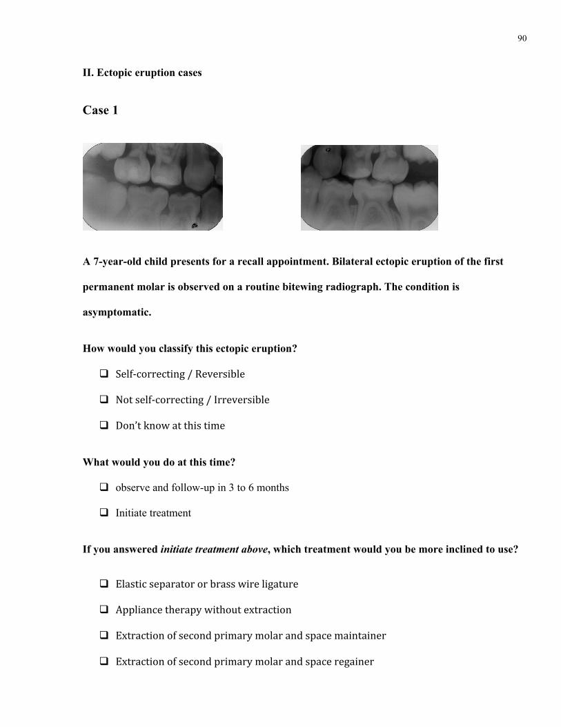
90
II. Ectopic eruption cases
Case 1
A 7-year-old child presents for a recall appointment. Bilateral ectopic eruption of the first
permanent molar is observed on a routine bitewing radiograph. The condition is
asymptomatic.
How would you classify this ectopic eruption?
q Self-‐correcting / Reversible
q Not self-‐correcting / Irreversible
q Don’t know at this time
What would you do at this time?
q observe and follow-up in 3 to 6 months
q Initiate treatment
If you answered initiate treatment above, which treatment would you be more inclined to use?
q Elastic separator or brass wire ligature
q Appliance therapy without extraction
q Extraction of second primary molar and space maintainer
q Extraction of second primary molar and space regainer

91
q Other, please specify: ____________________________________________________________
If the situation was unilateral, would your management be the same?
q Yes
q No, please specify:
__________________________________________________________________________
__________________________________________________________________________
Case 2
A 9 -year-old child presents for a recall appointment. Unilateral ectopic eruption of the first
permanent molar is observed on a routine bitewing radiograph. The condition is
asymptomatic.
How would you classify this ectopic eruption?
q Self-‐correcting / Reversible
q Not self-‐correcting / Irreversible
q Don’t know at this time

92
What would you do at this time?
q Observe and follow-up in 3 to 6 months
q Initiate treatment
If you answered initiate treatment above, which treatment would you be more inclined to use?
q Elastic separator or brass wire ligature
q Appliance therapy without extraction
q Extraction of second primary molar and space maintainer
q Extraction of second primary molar and space regainer
q Other, please specify: ____________________________________________________________
The child returns 6 months later and there are no changes in the position of the permanent
first molar. The condition is still asymptomatic.
How would you classify this ectopic eruption?
q Self-‐correcting / Reversible
q Not self-‐correcting / Irreversible
q Don’t know at this time
What would you do at this time?

93
q Observe and follow-up in 3 to 6 months
q Initiate treatment
If you answered initiate treatment above, which treatment would you be more inclined to use?
q Elastic separator or brass wire ligature
q Appliance therapy without extraction
q Extraction of second primary molar and space maintainer
q Extraction of second primary molar and space regainer
q Other, please specify: ____________________________________________________________

94
Case 3
A 7-year- old child presents for a recall examination. Ectopic eruption is noted on a routine
bitewing radiograph.
How would you classify this ectopic eruption?
q Self-‐correcting / Reversible
q Not self-‐correcting / Irreversible
q Don’t know at this time
What would you do at this time?
q Observe and follow-up in 3 to 6 months
q Initiate treatment
If you answered initiate treatment above, which treatment would you be more inclined to use?
q Elastic separator or brass wire ligature
q Appliance therapy without extraction
q Extraction of second primary molar and space maintainer
q Extraction of second primary molar and space regainer
q Other, please specify: ____________________________________________________________

95
The child misses his next appointment and returns 1 year later. Another bitewing is taken.
The condition is still asymptomatic.
How would you classify this ectopic eruption?
q Self-‐correcting / Reversible
q Not self-‐correcting / Irreversible
q Don’t know at this time
What would you do at this time?
q Observe and follow-up in 3 to 6 months
q Initiate treatment
If you answered initiate treatment above, which treatment would you be more inclined to use?
q Elastic separator or brass wire ligature
q Appliance therapy without extraction
q Extraction of second primary molar and space maintainer
q Extraction of second primary molar and space regainer
q Other, please specify: ____________________________________________________________
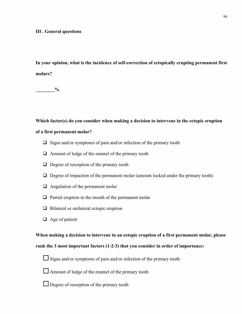
96
III . General questions
In your opinion, what is the incidence of self-correction of ectopically erupting permanent first
molars?
________%
Which factor(s) do you consider when making a decision to intervene in the ectopic eruption
of a first permanent molar?
q Signs and/or symptoms of pain and/or infection of the primary tooth
q Amount of ledge of the enamel of the primary tooth
q Degree of resorption of the primary tooth
q Degree of impaction of the permanent molar (amount locked under the primary tooth)
q Angulation of the permanent molar
q Partial eruption in the mouth of the permanent molar
q Bilateral or unilateral ectopic eruption
q Age of patient
When making a decision to intervene in an ectopic eruption of a first permanent molar, please
rank the 3 most important factors (1-2-3) that you consider in order of importance:
¨ Signs and/or symptoms of pain and/or infection of the primary tooth
¨ Amount of ledge of the enamel of the primary tooth
¨ Degree of resorption of the primary tooth

97
¨ Degree of impaction of the permanent molar (amount locked under the primary tooth)
¨ Angulation of the permanent molar
¨ Partial eruption in the mouth of the permanent tooth
¨ Bilateral or unilateral ectopic eruption
¨ Age of patient
Any comments are appreciated:
________________________________________________________________________________
________________________________________________________________________________
________________________________________________________________________________
________________________________________________________________________________
____________________
Thank you for your time!
Demographic information

98
APPENDIX E Manuscript submitted to NuSmile Graduate Research Student Award (AAPD)
Ectopic Eruption of the Permanent First Molar:
Prevalence and Prognostic Factors of Self-Correction
and
Survey of Specialists' Attitudes Regarding Intervention
Dabbagh B.*, Sigal M, Titley K, Tompson B, Andrews P
Faculty of Dentistry, University of Toronto
99
Abstract
Purpose: This study retrospectively assessed the incidence and predictive factors for self-correction and
occurrence of adverse events associated with ectopic eruption of permanent maxillary first molars, in a
sample population where no corrective intervention was initiated. The study also assessed prevailing attitudes
amongst surveyed pediatric dentists and orthodontists regarding indications and timing of intervention of
ectopically erupting permanent maxillary first molars. Methods: Charts of patients diagnosed with ectopic
eruption of the permanent maxillary first molar were assessed and radiographs were analyzed for predictive
factors reported in the literature. Outcome was determined as self-correcting: the permanent maxillary first
molar erupts into occlusion and the primary second molar is retained; or irreversible: the primary second
molar is lost prior to the eruption of the permanent maxillary first molar. An online survey presenting three
cases of ectopic eruption was sent to active members of the American Academy of Pediatric Dentistry, the
Canadian Academy of Pediatric Dentistry and the American Association of Orthodontists. Results: Sixty-
five ectopically erupting permanent maxillary first molars were included in the study. Forty-six (71%) self-
corrected, of which one third occurred after age 9. Space loss, with an average of approximately 3 mm,
occurred in 18 cases (28%), all of which were irreversible. Increased impaction magnitude (r(43)=0.59,
P<.001), degree of resorption of the primary second molar (r(57)=0.41, P=.001) and bilateral occurrence
(r(63)= 0.26, P=.03) were positively correlated with irreversibility. Multiple regression was positive for
males, increased impaction magnitude and bilateral occurrence as predictors of irreversible outcome. Nine
hundred and fifty pediatric dentists and 642 orthodontists responded to the survey. Orthodontists initiated
treatment more frequently than pediatric dentists in a case of bilateral ectopic eruption (x²(1, N=1178)=141.9,
P=.001) and estimated the rate of spontaneous self-correction to be much lower (t(1178)=19.2, P<.001).
Conclusion: Spontaneous self-correction occurred in 71% of cases. One third of self-corrections occurred
after 9 years of age and delaying treatment of ectopic eruption of permanent maxillary first molars may be a
viable option when uncertain of the outcome. Increased impaction and degree of resorption appear to be
reliable measures to predict irreversible outcome. Ectopic eruption of first permanent molars presenting

100
bilaterally, in males, with increased impaction magnitude are more likely to be irreversible and require early
intervention.
Introduction
Ectopic eruption of the first permanent molar is defined as a local eruption disturbance characterized by the
permanent tooth being locked under the distal contour of the second primary molar1. Ectopic eruption can
give rise to various degrees of resorption of the primary teeth. Two types of ectopic eruption of the first
permanent molar have been described: 1) self-correcting, where the ectopic molar unlocks from under the
second primary molar and erupts into occlusion; 2) irreversible, where the permanent molar remains locked
under the distal contour of the primary second molar until the latter exfoliates prematurely or treatment is
provided1. Interceptive orthodontic treatment can redirect the eruption path of the first permanent molar and
allow it to erupt into occlusion while maintaining the resorbed second primary molar. If interceptive
treatment is not provided in irreversible cases, the second primary molar may be lost prematurely in which
case space loss, crowding of the corresponding arch segment and impaction of the second premolar may
occur2. In these situations, future corrective treatment will be required, consisting of distalization and up
righting the first permanent molar, which may be lengthy and complicated2.
The rate of self-correction of ectopic eruption of permanent first molars reported in the literature ranges
between 59% to 69%1,3,4. Two articles have reported a self-correction rate outside that range of 91%5 and
6%6. These inconsistencies may be due to variations in study designs, diagnostic methods employed or early
implementation of treatment on what could in fact be self-correcting ectopic eruption cases.
In the absence of clinical symptoms, determining if the ectopic eruption of a first permanent molar will self-
correct or remain impacted and subsequently need intervention can be a challenge. Various criteria to aid in
assessing the type of ectopic eruption have been reported in the literature which can be used by the
practitioner to determine when and if intervention is necessary. Clinical factors that have been reported
included bilateral occurrence, age, and partial eruption of the first permanent molar. Bilateral occurrence of
101
ectopic eruption of the first permanent molar was significantly correlated with self-correction3. Bjerklin and
Kurol stated that in approximately 90% of cases, the type of ectopic eruption could be assessed during the
child’s 7th year of life4. Additionally, partial eruption of the crown in the mouth has been cited as a poor
prognostic factor for self-correction7.
The ectopic eruption of first permanent molars is usually diagnosed on routine bitewing radiographs. Some
authors have suggested radiographic factors which could indicate that intervention is warranted. Harrison and
Michal classified ectopic eruption as minimal versus severe lock7. Treatment was recommended when there
was a severe lock which was defined as the impaction of half or more of the width of the marginal ridge of
the first permanent molar under the second primary molar7. On the other hand, Chintakanon and Boonpinon
found no correlation between the severity of lock and the outcome of ectopic eruption of the first permanent
molar6. Pulver reported that the angle formed by the long axis of the first permanent molar and the occlusal
plane was more obtuse in irreversible cases5. In a flow-chart proposed by Kennedy and Turley, treatment was
recommended if there was an enamel ledge of 1 mm or greater on the distal of the second primary molar once
the permanent tooth was at the level of the cemento-enamel junction8. In a recent study, the magnitude of
impaction, defined as the distance between the maximal convexity of the permanent molar to the
prolongation of the distal aspect of the primary molar, was assessed in ectopic erupting permanent first
molars3(Figure 1).
The authors also defined a grading system to classify the degree of resorption of second primary molars. No
statistically significant correlation was found between the degree of resorption and the magnitude of
The magnitude of impaction was defined as the distance between
the maximal convexity of the permanent molar to the
prolongation of the distal aspect of the primary molar.
(Barberia-Leache, Suarez-Clùa, & Saavedra-Ontiveros, 2005)
Figure 1. Measurement of the magnitude of impaction
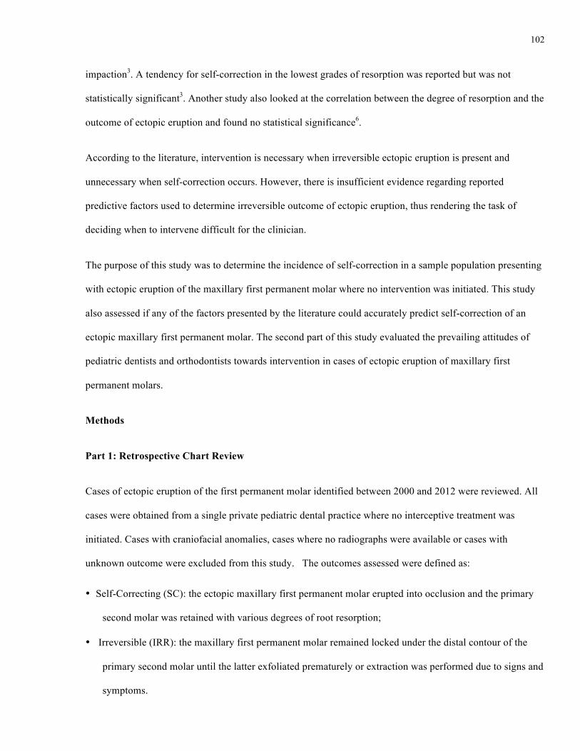
102
impaction3. A tendency for self-correction in the lowest grades of resorption was reported but was not
statistically significant3. Another study also looked at the correlation between the degree of resorption and the
outcome of ectopic eruption and found no statistical significance6.
According to the literature, intervention is necessary when irreversible ectopic eruption is present and
unnecessary when self-correction occurs. However, there is insufficient evidence regarding reported
predictive factors used to determine irreversible outcome of ectopic eruption, thus rendering the task of
deciding when to intervene difficult for the clinician.
The purpose of this study was to determine the incidence of self-correction in a sample population presenting
with ectopic eruption of the maxillary first permanent molar where no intervention was initiated. This study
also assessed if any of the factors presented by the literature could accurately predict self-correction of an
ectopic maxillary first permanent molar. The second part of this study evaluated the prevailing attitudes of
pediatric dentists and orthodontists towards intervention in cases of ectopic eruption of maxillary first
permanent molars.
Methods
Part 1: Retrospective Chart Review
Cases of ectopic eruption of the first permanent molar identified between 2000 and 2012 were reviewed. All
cases were obtained from a single private pediatric dental practice where no interceptive treatment was
initiated. Cases with craniofacial anomalies, cases where no radiographs were available or cases with
unknown outcome were excluded from this study. The outcomes assessed were defined as:
• Self-Correcting (SC): the ectopic maxillary first permanent molar erupted into occlusion and the primary
second molar was retained with various degrees of root resorption;
• Irreversible (IRR): the maxillary first permanent molar remained locked under the distal contour of the
primary second molar until the latter exfoliated prematurely or extraction was performed due to signs and
symptoms.
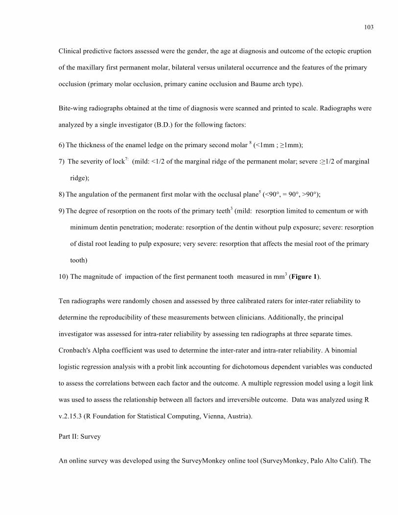
103
Clinical predictive factors assessed were the gender, the age at diagnosis and outcome of the ectopic eruption
of the maxillary first permanent molar, bilateral versus unilateral occurrence and the features of the primary
occlusion (primary molar occlusion, primary canine occlusion and Baume arch type).
Bite-wing radiographs obtained at the time of diagnosis were scanned and printed to scale. Radiographs were
analyzed by a single investigator (B.D.) for the following factors:
6) The thickness of the enamel ledge on the primary second molar 8 (<1mm ; ≥1mm);
7) The severity of lock7: (mild: <1/2 of the marginal ridge of the permanent molar; severe :≥1/2 of marginal
ridge);
8) The angulation of the permanent first molar with the occlusal plane5 (<90°, = 90°, >90°);
9) The degree of resorption on the roots of the primary teeth3 (mild: resorption limited to cementum or with
minimum dentin penetration; moderate: resorption of the dentin without pulp exposure; severe: resorption
of distal root leading to pulp exposure; very severe: resorption that affects the mesial root of the primary
tooth)
10) The magnitude of impaction of the first permanent tooth measured in mm3 (Figure 1).
Ten radiographs were randomly chosen and assessed by three calibrated raters for inter-rater reliability to
determine the reproducibility of these measurements between clinicians. Additionally, the principal
investigator was assessed for intra-rater reliability by assessing ten radiographs at three separate times.
Cronbach's Alpha coefficient was used to determine the inter-rater and intra-rater reliability. A binomial
logistic regression analysis with a probit link accounting for dichotomous dependent variables was conducted
to assess the correlations between each factor and the outcome. A multiple regression model using a logit link
was used to assess the relationship between all factors and irreversible outcome. Data was analyzed using R
v.2.15.3 (R Foundation for Statistical Computing, Vienna, Austria).
Part II: Survey
An online survey was developed using the SurveyMonkey online tool (SurveyMonkey, Palo Alto Calif). The
104
survey was sent to all active members of the American Academy of Pediatric Dentistry, the Canadian Academy
of Pediatric Dentistry and the American Association of Orthodontists. The survey included questions on
demographics and 3 cases of ectopic eruption for dentists to review and decide on intervention or observation.
The first case was characterized by a bilateral occurrence of the ectopic eruption of the maxillary first permanent
molar, the second case presented an ectopic eruption in a 9 year old child, and the third case was marked by an
ectopic eruption causing minimal resorption and impaction that increased over the course of a year. The survey
also contained general questions regarding factors used to determine if intervention is warranted in a case of
ectopic eruption of the first permanent molar. Pearson’s Chi-squared test was conducted to compare responses
between orthodontists and pediatric dentists.
Results
Part I: Retrospective Chart Review
A total of 66 cases of ectopic eruption of maxillary first permanent molars in 45 patients (20 males; 25
females) were identified. The occurrence of the ectopic eruption was bilateral in 21 and unilateral in 24
patients (16 right; 8 left). There was no statistical difference between right-sided and left-sided unilateral
ectopic eruption of the first permanent molar (χ²(2, N=24)= 5.733; p=.057). One tooth was excluded from the
study because of unknown outcome. Sixty–five teeth were included in the study. The rate of self-correction
of ectopic eruption (SC) of maxillary first permanent molars in this sample population where no intervention
was initiated was 71% (46/65). The rate of irreversible ectopic eruption (IRR) was 29%.
The average age at diagnosis of ectopic eruption was 7 years of age and there was no statistical difference
between self-correcting and irreversible cases. Fifteen (33%) cases of self-corrections occurred after age 9.
The average time between diagnosis and outcome was 1.3 years for self-correction cases and 0.8 years for
irreversible cases. This difference was statistically significant (t(37.817)= 2.15, p<.03). Fifteen cases (14 SC,
1IRR) were observed for more than 2 years from diagnosis.
The degree of resorption and the impaction magnitude were the only radiographic factors that showed a
strong inter-rater correlation between 3 different clinicians. The intra-rater correlation was acceptable for all
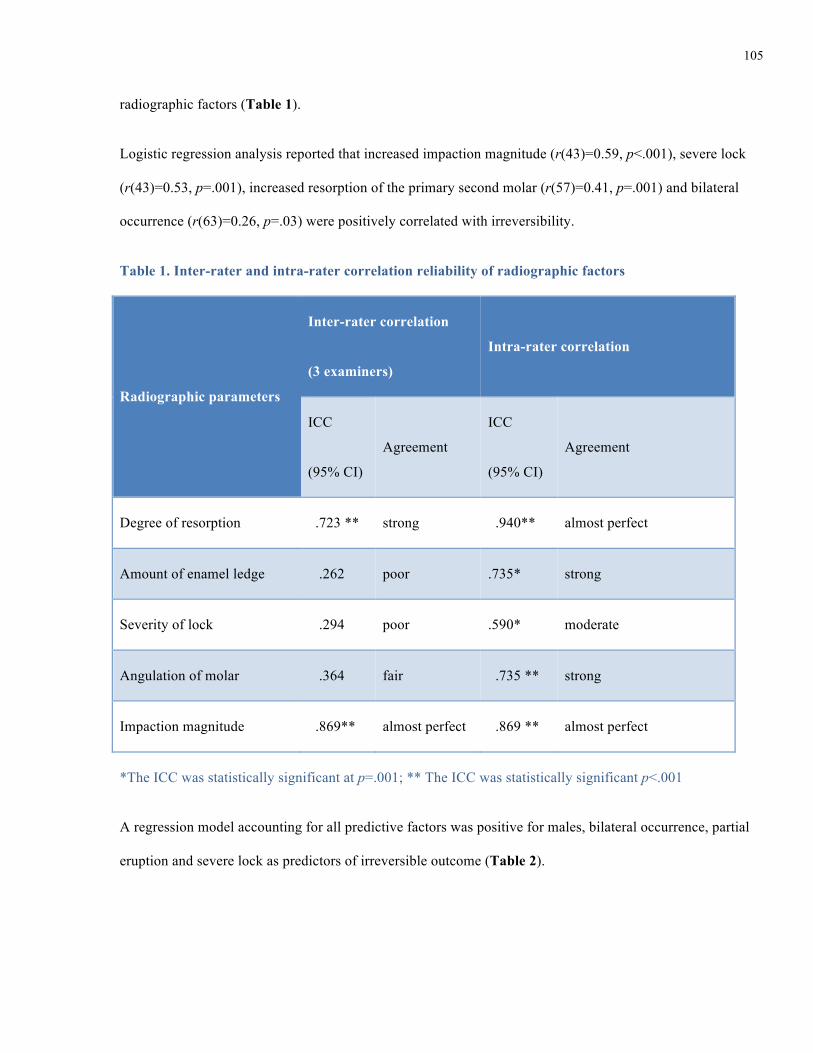
105
radiographic factors (Table 1).
Logistic regression analysis reported that increased impaction magnitude (r(43)=0.59, p<.001), severe lock
(r(43)=0.53, p=.001), increased resorption of the primary second molar (r(57)=0.41, p=.001) and bilateral
occurrence (r(63)=0.26, p=.03) were positively correlated with irreversibility.
Table 1. Inter-rater and intra-rater correlation reliability of radiographic factors
Radiographic parameters
Inter-rater correlation
(3 examiners)
Intra-rater correlation
ICC
(95% CI)
Agreement
ICC
(95% CI)
Agreement
Degree of resorption .723 ** strong .940** almost perfect
Amount of enamel ledge .262 poor .735* strong
Severity of lock .294 poor .590* moderate
Angulation of molar .364 fair .735 ** strong
Impaction magnitude .869** almost perfect .869 ** almost perfect
*The ICC was statistically significant at p=.001; ** The ICC was statistically significant p<.001
A regression model accounting for all predictive factors was positive for males, bilateral occurrence, partial
eruption and severe lock as predictors of irreversible outcome (Table 2).

106
Table 2. Regression of predictive factors for irreversible outcome
Variable B SE Z P
95% CI for B
Odds Ratio
Lower Upper
Intercept -1.10 1.35 -.81 .416 -3.75 1.54 --
Female Gender -2.83 1.26 -2.24 .025* -5.29 -.36 0.06
Bilateral 2.37 1.40 1.69 .091 -.37 5.11 10.69
Partial Eruption -1.83 1.14 -1.61 .107 -3.95 .29 0.16
Severe Lock 2.34 1.16 2.02 .042* .06 4.61 10.38
*Significant at p<.05
A second regression analysis was performed excluding radiographic factors found to be unreliable in the
inter-rater reliability correlation test (e.g. enamel ledge, angulation of the permanent molar and severity of
lock). This second model was positive for males, impaction value and bilateral occurrence as predictors of
irreversible outcome (Table 3).

107
Table 3. Regression of select reliable predictive factors for irreversible outcome
Variable B SE Z P
95% CI for B
Odds Ratio
Lower Upper
Intercept -12.62 6.22 -2.03 .04* -24.81 -0.43 --
Female Gender -4.23 2.20 -1.93 .05 -8.54 0.08 .01
Bilateral 4.97 2.80 1.77 .08 -0.52 10.46 144.03
Impaction magnitude 5.81 2.91 2.00 .046* 0.11 11.50 333.62
*Significant at p<.05
In the irreversible outcome group (n = 19), infection was observed in 2 cases (11%) and space loss was
observed in 18 cases (95%). The estimated average space loss was approximately 3.7mm (0 to 7.7mm). In the
self-correcting group, 5 primary second molars (11%) were extracted after the eruption of the permanent first
molar.
Part II: Survey
The total number of respondents was 1591, specifically 950 pediatric dentists and 641 orthodontists. The
respective response rate was 22% and 7%. The rate of response from pediatric dentists was significantly
higher than orthodontists (χ²(1, N=1591)= 801.7; p <.001).
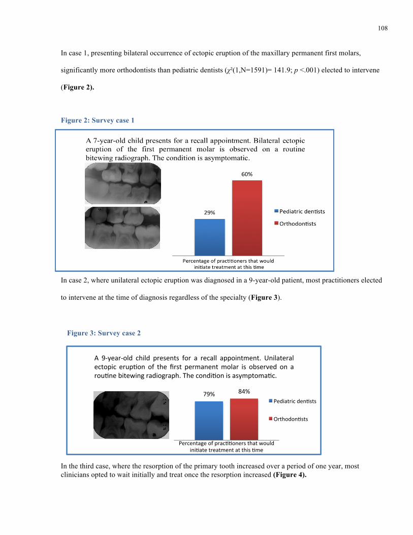
108
In case 1, presenting bilateral occurrence of ectopic eruption of the maxillary permanent first molars,
significantly more orthodontists than pediatric dentists (χ²(1,N=1591)= 141.9; p <.001) elected to intervene
(Figure 2).
In case 2, where unilateral ectopic eruption was diagnosed in a 9-year-old patient, most practitioners elected
to intervene at the time of diagnosis regardless of the specialty (Figure 3).
In the third case, where the resorption of the primary tooth increased over a period of one year, most clinicians opted to wait initially and treat once the resorption increased (Figure 4).
Figure 2. Survey case 1
79% 84%
Percentage of prac22oners that would ini2ate treatment at this 2me
A 9-‐year-‐old child presents for a recall appointment. Unilateral ectopic erup2on of the first permanent molar is observed on a rou2ne bitewing radiograph. The condi2on is asymptoma2c.
Pediatric den2sts
Orthodon2sts
Figure 2: Survey case 1
Figure 3: Survey case 2

109
The most important factors considered by practitioners when deciding to intervene in a case of ectopic
eruption of the maxillary first permanent molar were the degree of impaction (80%), the angulation of the
tooth (69%) and the presence of pain (68%). The average self-correction rate estimated by pediatric dentists
was significantly higher than orthodontists (t(1178)=192; p< .001) (Figure 5).
Figure 5: Perceived percentage of self-correction
0
50
100
150
200
250
Num
ber of respondents
Percentage
Pediatric Dentists
Orthodontists
9%
81%
16%
82%
Percentage of practitioners that would initiate treatment at time points A and B
A. A 7-‐year-‐old child presents for a recall examination. Ectopic eruption is noted on a routine bitewing radiograph B. The child misses his next appointment and returns 1 year later. Another bitewing is taken. The condition is still asymptomatic.
Pediatric dentists Orthodontists
A B
Figure 4: Survey case 3

110
Discussion
The 71% rate of self-correction in this study was similar to other reports. The delay in intervention did not
substantially increase the rate of self-correction. In this sample population, one third of the self-corrections
occurred after a 2 year observation period and after 9 years of age. Delaying treatment in these cases did not
cause any adverse effects. This indicates that delaying intervention when unsure of the type of ectopic
eruption can be a viable option and may prevent unnecessary treatment and cost.
An increase in right-sided unilateral occurrence has been reported previously3. In this sample, a higher
number of right-side ectopic eruptions was noted but the difference was not statistically significant. The
unilateral to bilateral occurrence ratio was similar in this population which is in accord with one study4 while
another report found a significant higher bilateral occurrence3.
The present study findings indicate that the higher grades of resorption are correlated with irreversible
outcome. Previous studies found no correlation and this may be due to a smaller sample size. The correlation
of the magnitude of impaction with the outcome had not been assessed previously and had the highest
correlation with irreversible outcome. Predictive measurements previously reported in the literature were
found to have poor statistical reliability. In fact the poor inter-rater reliability results for the enamel ledge
measurement, the permanent tooth angulation and the severity of lock indicates clinicians’ inability to
consistently reproduce these measurements and therefore they were excluded from the second regression
model. This model was found to be more accurate and applicable clinically and indicated that males
presenting with bilateral ectopic eruption of the maxillary first permanent molars and increased impaction
magnitude were more likely to have an irreversible outcome.
The retrospective chart review was limited by the data available in the charts as well as the quality and
availability of the radiographs. A future prospective cohort study looking at the impaction magnitude with
standardized radiographs may help in developing evidence based guidelines in the treatment of ectopic
eruption.

111
In the survey portion, orthodontists estimated the rate of self-correction to be much lower than pediatric
dentists. Orthodontists will usually see older patients and treat the consequences of irreversible ectopic
eruption once the primary second molar has been lost prematurely and therefore might experience a higher
rate of irreversible ectopic eruptions in their practice. In most cases, there was a general consensus on
treatment and this indicates that although there is no evidence based guidelines in the literature, most
respondents will intervene in the same manner in regards to ectopic eruption. The orthodontists’ inclination to
intervene in the bilateral case may be due to the assumption that the cause of the ectopic eruption is skeletal
and not local. The survey had a large sample with relatively homogeneous responses. However, a weakness
of the survey was the low response rate which may have resulted in self-selection bias.
Conclusions
Based on this study the following conclusions can be made:
4) Self-correction of ectopic eruption of maxillary first permanent molars may occur after the age of 9 and
delaying intervention can be a viable treatment option when unsure of the type of ectopic eruption.
5) Increased impaction magnitude and degree of resorption are reliable predictors of irreversible outcome of
ectopic eruption of first permanent molars.
6) Ectopic eruption of the maxillary first permanent molars presenting bilaterally, in males, with an
increased amount of impaction are more likely to be of the irreversible type and require early
intervention.
Acknowledgments
The authors wish to thank Drs. Hashim Nainar, Trang Nguyen, Darsi Perusini and Edward Yoon for their
assistance and support.
112
References
1. Young DH. Ectopic eruption of the first permanent molar. J Dent Child. 1957;24:153-162.
2. Yuen S, Chan J, Tay F. Ectopic eruption of the maxillary permanent first molar: The effect of increased
mesial angulation on arch length. J Am Dent Assoc. 1985;111(3):447-451.
3. Barberia-Leache E, Suarez-Clùa MC, Saavedra-Ontiveros D. Ectopic eruption of the maxillary first
permanent molar: Characteristics and occurrence in growing children. Angle Orthod. 2005;75(4):610-615.
4. Bjerklin K, Kurol J. Prevalence of ectopic eruption of the maxillary first permanent molar. Swed Dent J.
1981;5(1):29-34.
5. Pulver F. The etiology and prevalence of ectopic eruption of the maxillary first permanent molar. ASDC J
Dent Child. 1968;35(2):138-146.
6. Chintakanon K, Boonpinon P. Ectopic eruption of the first permanent molars: Prevalence and etiologic
factors. Angle Orthod. 1998;68(2):153-160.
7. Harrison LM, Michal BC. Treatment of ectopically erupting permanent molars. Dent Clin North Am.
1984;28(1):57-67.
8. Kennedy DB, Turley PK. The clinical management of ectopically erupting first permanent molars.
American journal of orthodontics and dentofacial orthopedics. 1987;92(4):336-345.
113
List of Figures and Tables
Figure 1. Measurement of the magnitude of impaction
Figure 2. Survey case 1
Figure 3. Survey case 2
Figure 4. Survey case 3
Figure 5. Perceived percentage of self-correction by specialists
Table 1. Inter-rater and intra-rater correlation reliability of radiographic factors
Table 2. Regression of predictive factors for irreversible outcome
Table 3. Regression of select predictive factors for irreversible outcome

114
p


