CHAPTER - 3
Evaluafion of Probiofic Bacieriato Inhibit the Growth of Vibrioharveyi in vifro and Improve
Survival of Penaeus monodonLarvae in vivo
60

CHAPTER - 3
Evaluation of probiotic bacteria to inhibit the growth of Vibrio harveyi
in vitro and improve survivalof Penaeus monodon larvae in vivo
3.1 Introduction
A major cause of shrimp larval mortality in hatcheries is infection by the Gram
negative bacterium Vibrio harveyi commonly known as luminescent bacteria due to the
characteristic blue-green light it emits. The organism has been reported as an
opportunistic pathogen to both juvenile and adult shrimp during culture where it
seriously threatens economic feasibility by causing mass mortalities. Extensive use of
antibiotics to control its incidence not only resulted in the emergence of multiple
antibiotic resistant strains (Karunasagar ct al. 1994, Abraham et al. 1997) but has also
caused grave environmental and human health concems (Holmstrom et al. 2003,
Nogueira-Lima et al. 2006) leading to their prohibition.
Such concerns have resulted in the exploration of biological control methods for
mitigating the adverse impact of pathogens, one of which includes the use of bacteria
live or dead as probiotics (Versehuere et al. 2000b, lrianto & Austin 2003). Probiotic
bacteria could prevent the establishment pathogenic bacteria by out-competing them for
adhesion and colonization sites in the intestines and other tissues of the animal (Vine et
al. 2004a). They could also produce inhibitory substances actively preventing pathogen
establishment (Verschuere et al. 2000b). When added to rearing water, they may act as
bioremediation agents improving water and sediment quality, augment nutrient cycling
in the system and initiate colonization of other beneficial microflora effecting an
overall positive impact on growth rates and productivity (Boyd & Massaut i999,
Prabhu et al. 1999). A wide array of both Gram-negative and Gram-positive bacteria
6|

with probiotic potential against a multitude of fish and shellfish pathogens have been
reported (lrianto & Austin 2002a, Balcazar et al. 2006).
Fluorescent pseudomonads produce phenazine and other antimicrobial compounds
which have broad spectrum activity against many phytopathogenic fungi and eubacteria
(Mavrodi et al. 2006). Pseudomonads have also been documented as the dominant flora
in the eggs and larvae of shrimps of successfully completed larval cycles in hatchery
systems (Singh et al. I989). Dopazo et al. (1988) demonstrated the antibacterial activity
of marine bacteria including Pseudontonas sp. against fish pathogens. Their ability to
control bacteria causing diseases of aquatic animals in culture and effect improvement
in productivity is gaining widespread acceptance. Pseudomonasfluorescens AH2 strain
was found to accord protection to rainbow trout from Vibrio anguillarum (Gram et al.
1999b) while Pscud0m0na.s' acruginosa was found to inhibit Shrimp pathogenic V.
harveyi, V. vulnificus, V. alginolyriczls, V. fluvialis and Aeromonas sp. (Chythanya et al.
2002, Vijayan et al. 2006). Their studies assigned the antimicrobial properties of the
pseudomonads to production of iron-chelating siderophores and pyocyanin.
Amongst Gram-positive bacteria, Bacillus sp. are widely used as probiotics for humans
and animals (Gatesoupe 1999, Duc et al. 2004). Although in terrestrial animals they are
presumed to be of telluric origin. Bacillus sp. are frequently isolated from the intestines
of healthy aquatic animals (Gatesoupe I999). Preemptive use of Bacillus sp. in Penaeus
monodon culture systems has yielded significantly better growth and survival in the
presence of pathogenic V. harveyi (Moriarty l998, Rengpipat et al. 2003). Recently,
Z-iaei-Nejad et al. (2006) obtained improved survival of F enneropenaeus lndlcus larvae
and adult maintained on a mixture of Bacillus spp. Micrococcus sp. is occasionally
isolated from intestines of healthy fish, but is regarded as a transient microflora as it is
not detected during all developmental stages (Sugita et al. 2002). A few strains
however have been shown to posses antibacterial properties against V. alginolyricus, V.
vulnlficus, Lactococcus garvieae, and Pastcurella piscida (Sugita et al. 2002,
Jayaprakash et al. 2005). A Gram-positive coccus Al-6 phenotypically similar to
62

Micr0c0cc'u.s' sp. could protect the fingerlings of rainbow trout from Aeromonas
sulmoizicicla infection (lrianto & Austin 2002b).
ln the present study we evaluated the capability of four microorganisms, Pseudonionas
MCC B102 and MCCBIO3, Bac:'l!us MCCBI 01, and .Micz'0c'0ccu.s' MCCB104 proposed
as potential probiotics (Jayaprakash et al. 2005, Vijayan et al. 2006) to confer
protection to Penaeus monodon larvae from the shrimp pathogen Vibrio harveyi.
3.2 Materials and Methods
3.2.1 Bacteria
Four putative probiotic bacterial isolates, P.s'eud0m0nas MCCBl02 (PS I 02) (Vijayan et
al. 2006'), Pseudomonas MCCBIO3 (Jayaprakash 2005), Micrococcus MCCBIO4
(Jayaprakash et al. 2005) and Bacillus MCCBl0l from the microbial culture collection
at National Centre for Aquatic Animal Health (NCAAH), Cochin University of Science
and Technology (CUSAT) were chosen for testing their probiotic potential against the
87 isolates Vibrio harveyi isolated in this study. All isolates were cryopresewed in 20
ppt salinity ZoBell’s marine broth 22l6E (ZB) with 10% glycerol at -80°C. Working
cultures were maintained in 20-ppt ZoBell’s marine agar (ZA) slants at 28°C.
3.2.2 Antagonism Assay
Antagonism of the culture as well as cell-free supernatants was tested following
Jayaprakash et al. (2005). Briefly, the probiotic bacterial cultures were grown in 20-ppt
salinity ZB at 28°C for 5 days. Six mm diameter discs from Whatman No.1 filter
papers (stack of3 filter papers) were prepared, sterilized at l2 I °C for I5 min and dried.
These discs were placed on ZA plates, previously swabbed with 0.5 ml ovemight
grown culture (I OD) of the target bacterial isolates. Aliquots (20 pl) of the four
63

robiotic bacterial cultures were dis ensed on to the discs se aratel in tri licate.P P P Y PAntagonism between the four putative probionts was also tested in the same way. The
plates were incubated for 24 h at 28°C and the formation of a zone of clearing around
the discs was considered as ositive indication of inhibito I activit .P '3 YAntagonism of cell free supernatant of the four probiotic bacteria was also tested. The
four probiotic cultures were grown in ZB for 5 days on a shaker (120 rpm) at 28°C.
Cells were pelleted by centrifugation (10000 x g. 4°C, l0 min), the pH of the
supernatant adjusted to 7.0 and then passed through 0.2 pm pore-size cellulose-acetate
membrane filter (Sartorius). inhibitory activity on the target microbial cultures was
detected by disc diffllsion method as described above and the zone of inhibition around
the discs recorded after 24 h using HiAntibiotic ZoneScale (Himedia).
3.2.3 Coculture Experiments
Co-culture experiments with Pseudomonas MC C B102 and V. harveyi MCCBI ll was
carried out following the method of Gram et al. (l999b_). They were precultured
separately in ZB at 28°C on a shaker at l20 rpm ovemight. From the above cultures, V.
harveyi was inoculated in 100 ml ZB to obtain an initial cell count of l03 cfu ml"
(approx), whereas the initial levels of PS?!-I6l'OI7?0I?0S' l\/lCCBl02 in those flasks were
104, 105, 10°, l07 and 10° cfu ml" respectively- All combinations were maintained in
duplicate and were repeated twice. The flasks were incubated at 28°C on a shaker (120
rpm), and samples (1 ml) were withdrawn at 24 h intervals for determination of cell
count. Counts of V. harveyi were estimated by using H&L medium (Hugh & Leifson
l953). Tubes containing 4 ml of H&L medium were inoculated with l ml aliquots of
serially ten-fold diluted culture and overlayed with sterile liquid paraffin and incubated
at 28°C for 24 h. The fermentative growth of V. ham/'e__vi caused a change in the pH of
the medium. The highest dilution, which showed growth, was used to calculate the
count of V. harveyi in the sample (l >< 10°, where n is the highest dilution which
showed fermentative reaction) and expressed as Log“, cfu ml". The cell count ot
64
\
Pseudomonas MCCBl02 was monitored on Pseudomonas isolation agar (PIA)
(Himedia) by spread plate method. Coculture experiments with P.s*eud0m0na.s'
MCCB103 were also carried out similarly.
Experiments with ll/licrococcus MCCBl04 and Bacillus MCCB10l with V. harveyi
MCCB1ll were carried out individually and in combination at the same initial cell
numbers as above. Counts of the pathogen were monitored by withdrawing daily 1 ml
samples which were serially diluted l0-fold and 0.2 ml aliquots spread plated on
Thiosulphate Citrate Bile salts Sucrose (TCBS) agar (Himedia) and ZA plates. The
plates were incubated at 28°C for 24 hours and colonies formed on TCBS were counted
and expressed as Log“) cfu ml'] of V. harveyi in the co-culture. Both the Gram-positive
bacteria (Bacillus MCC B101 and Micrococcus MCC B104) do not grow on TCBS agar
and also could be easily differentiated from V. lzarveyi on ZA. Micrococcus MCC B 104
formed yellow non—luminescent colonies while those fonned by Bacillus MCC B 101
apart from being non-luminescent were rough and white.
3.2.4 Effect of the probiotic bacteria t0 Peneaus monodon post larvae
Groups of 2000 P. monodon larvae at Protozoea 1 stage were introduced into six 100-L
F RP tanks and reared until metamorphosis to postlan/ac 30 at a commercial shrimp
hatchery. The effect of the four probiotics Bacillus MCCB101, Micrococcus
MCCBl04, Pseudomonas MCCBl02 and MCCB103 on the health and survival of
Penaeus morzodon larvae was assessed independently. The combined effect of Bacillus
MCCBl01 and Micrococcus MCCB104 (designated ‘Enterotrophotic’) was also
assessed. One group was maintained as control without any probiotic exposure. All
probiotics were precultured in ZB at 28°C for 5 days and added to the rearing water
every two days to obtain a final density of 106 cfu ml". The larvae were maintained on
a diet of Chaetoceros sp. (80000-130000 cells ml") from protozoea l to mysis lll and
thereafter on freshly hatched Arremia nauplli. Water in the tanks was topped up until
conversion to PL-l following which 25-30% water was exchanged daily.
65
Physieochemical parameters of the rearing water such as total ammonia, nitrite, pH,
salinity and temperature were monitored regularly following standard methods (APHA
et al. l995). The total heterotrophic bacterial population and total vibrio count of the
rearing water and larvae were monitored periodically by spread plating 0.2 ml aliquots
after I0-fold serial dilution on ZA and TCBS agar. Ten larvae from each group were
washed gently in sterile seawater to remove loosely adhering particles. The larvae were
lightly dried by blotting them on a sterile filter paper and niacerated in l ml of sterile
seawater. This suspension was serially diluted l0-fold and 0.2 ml aliquots were spread
plated on ZA and TCBS in duplicate. The plates were incubated at 28il °C for 24-72
hours and those having 30-300 colonies were taken for estimating the bacterial counts
which were expressed as cfu ml" and cfu larvae" for water and larval samples
respectively. From each of the final water and postlarval (PL-30) samples, a
representative of 20 colonies were isolated, streaked on ZA to purity and presumptively
grouped to genus/ family level following Oliver (I982). The health of the post larvae
(PL-303) was assessed (20 numbers from each group) and scored by microscopic
observation for features such as muscle opaqueness, deformities, size variation, gut
content, colour and condition of the hepatopanereas, epibiont fouling, intestinal
persistalsis, and muscle to gut ratio as per the FAO guidelines (FAO 2003). A formalin
stress test was also given by subjecting 100 larvae from each group to 100 ppm
formalin for I h and then monitoring them in normal rearing water for another 3 h.
Final survival was taken when the larvae reached PL-30 stage statistical significance
was arrived at by using the X2 test.
3.2.5 Evaluation of protection accorded by the probiotics to postlarvae of P.
monodon from Vibrio harveyi challenge
Three groups of 30 postlarvae (PL-24) from each of the treatments (probiotie treated
and control) above were transferred to plastic containers (3 L capacity). From the
control group two sets of 30 PL each were maintained. They were acclimatized for 24 h
following which an ovemight culture of V. harveyi MCCB 1 l 1 was added to water to
obtain I06 cfu ml" except to that of the negative control group. One group of 3 >< 30
66

from the control was not challenged. An ad-libitum feeding regime with freshly
hatched Arlemia nauplii and probiotic addition as mentioned above was continued. The
larvae were observed for l0 days. Assessment of the microbial population of water and
larvae was carried out on ZA and TCBS agar periodically as mentioned earlier. After
10 days of observation, a relative percent survival was calculated. Results were
statistically analysed using single-factor Al\lO\/A followed by least significant
difference (LSD) to compare different treatment groups.
Relative Percentage Sun/ival (RPS)
\\Qc\i
I [1 — (% mortality in the test + nortality in the control)] X 100l0O
3.3 Results
3.3.1 Antagonism Assay
ln the antagonism assay, the culture and cell-free supematants of putative probionts
Pseudomonas MCCBIOZ and MCCBlO3 inhibited all 87 isolates of Vibrio harveyi and
the Bacillus MCCBIOI. A clear Zone of inhibition in the range l2-15 mm was obtained
against V. harveyi with both the peudomonads. Micrococcus MCC B104 inhibited all V.
harveyi isolates and the two Pseudomonas spp. A turbid zone of inhibition in the range
of I8-22 mm was obtained against V. haweyi. Micrococrcus MCCBIO4 did not inhibit
Bacillus MCCBl0l. Neither the culture nor the cell-free supematants of Bacillus
MCCBIOI was inhibitory to any of the isolates tested. This made us to attempt the
effect of Micrococcus MCCBl04 and Bacillus MCCBIOI in combination as probionts
in Penaeus monodon larval rearing.
3.3.2 Coculture Experiments
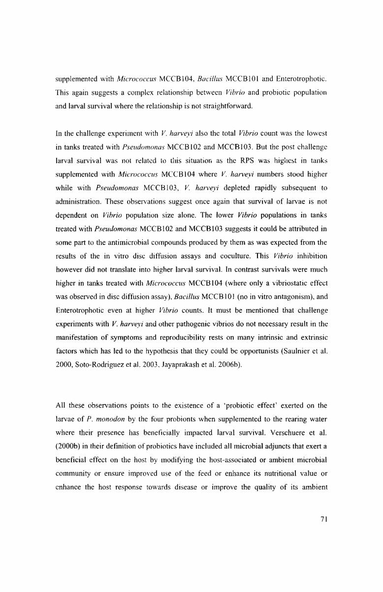
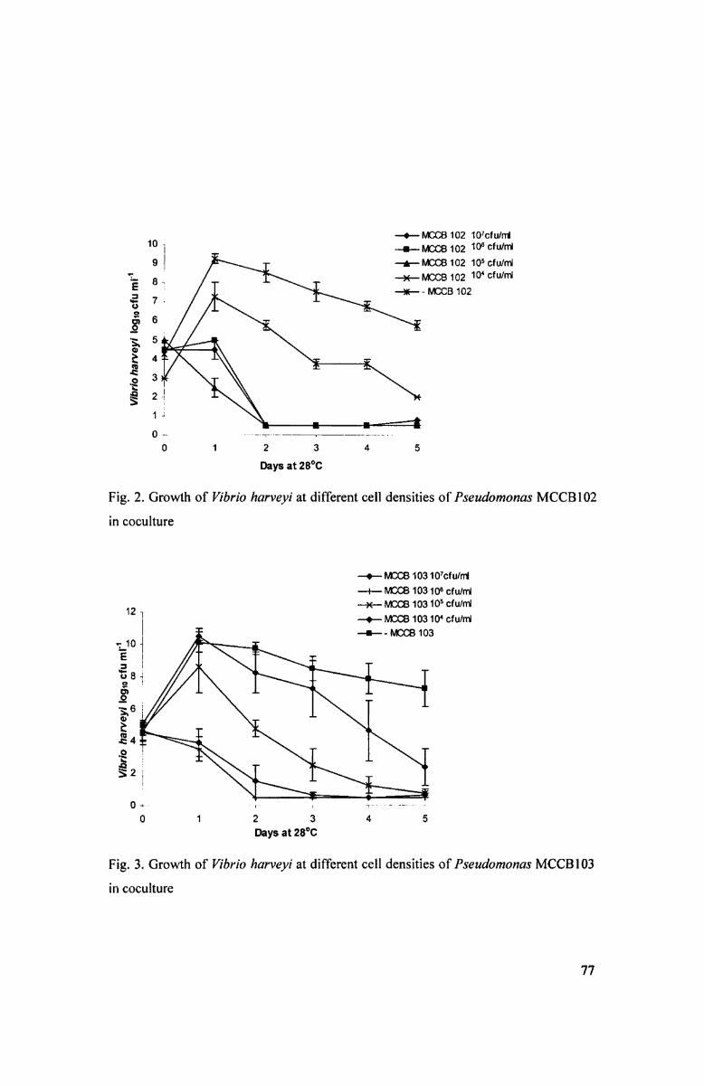
In the eoculture experiments of Pseudamonas MCCBIOZ with V. harveyi MCCBl ll
the latter became completely undetectable in 48 h at initial cell densities greater than
67
105 cfu ml" (Fig. 2). while similar situation with Pseudomonas MCCBIO3 was
obtained only at initial cell densities greater than 10“ cfu ml" (Fig. 3)- At lower initial
cell densities of the two pseudomonads, V. harveyl MCCBI 1 1 counts could reach up to
10.5 log cfu ml" in 24 h and remained well detectable even after five days. Both
pseudomonads could be recovered in high cell numbers on PIA even after 5 days from
all combinations (data not shown), which showed they were unaffected by V. l16H"V£?}~‘l
MCCBI l l. Bacillus MCCBIOI and Mz'cr0c0ccu.s- MCCBIO4 did not inhibit the growth
of V. liarveyi MCCBH1 even when inoculated at 108 cfil ml" initial cell densities
(Tables 4 & 5). ln coculture experiments of V. harveyi MCCBI ll with Micrococcus
MCCBIO4 and Bacillus MCC B101, counts of the Vibrio were found to increase and
remained unaffected even at 108 cfu ml" initial densities of the probionts. Akin to with
the pseudomonads, counts of Bacillus MCCBl0l and Micrococcus MCCBIO4 were
unaffected by V. harveyi MCC Bl l I in the cocultures.
3.3.3 Impact of probiotics on larval health and survival
Improved survival of P. monodon post larvae was obtained when their rearing water
was supplemented with all the probiotics individually as well as in combination of
Bacillus MCCBIOI and Mz'cr0c0ccus MCCBl04 (Enterotrophotic) (Table 6). Survival
was 70% or more in tanks treated with Micrococcus MCCBIO4, Bacillus MCCBIOI
and Enterotrophotic, while in those that received Pseudomonas MCCBIOZ and
MCCB103 it was 66.35 and 58.3 % respectively. The survival obtained with the
probiotic treatment groups were significantly higher (p<0.01, X2 = 121.9) in comparison
to the control. Based on X3 values. the order of significance was treatment with
Micrococcus MCCBIO4 followed by Bacillus MCC B101, Enterotrophotic,
Pseudomonas MCCBIOZ and lastly Pseudomonas MCCBl03. When 20 larvae from
each group were examined under the microscope all were transparent, with their guts
full, dark hepatopancreas, high intestinal peristalsis. no fouling or deformities on the
exoskeleton or gills, musclezgut ratio at the 6"‘ abdominal segment > 3:1, and uniform
size (%CV <15%) (Table 7). Total ammonia was consistently higher in all probiotic
68

treated tanks than the control (Table 8). When 100 post larvae (PL~3O) from each
treatment were subjected to formalin (100 ppm) stress test for I h, there were no
mortalities indicating good health of the larvae (Table 9). Total Vlbrlo population was
lowest in rearing water and larvae of tanks which were supplemented with
Pseuclomonas MCCBIOZ and MCCBIO3. Notably, Vibrio population was not detected
in the larvae of tanks supplemented with Pseudomonas MCCBIOZ until stage PL-8.
Vibrio population counts were similar in the control and tanks supplemented with
Bacillus MCCBIOI, Micrococcus MCCBIO4 and Enterotrophotic (Table I0).
Luminescent bacteria were not detected in any of the treatments and the control at any
stage during the experiment. The bacterial diversity in the larvae ofall treatment groups
and the control was high and presumptive generic level identification revealed the
isolates to belong to Pseudomonas sp., Vibrio sp., Photobacterium, Bacillus sp.,
_Plan0c0cc'i sp., Micrococcus sp., Acenitobacter sp. (Table l l).
3.3.4 Response of probiotic treated post larvae to challenge with Vibrio harveyiMCCBIII
When larvae from each of the probiotic treatment and the control were challenged with
V. harveyi at stage PL-25, 10-day post challenge survival (RPS) was highest in tanks
supplemented with Micrococcus MCCBIO4 (86.67) and Pseudomonas MCCBl02
(80.0) and MCBIO3 (86.67) (Table 12). An RPS of 66.67 and 36.67 was obtained in
tanks supplemented with Bacillus MCCBIOI and Enterotrophotic respectively. Using
single-factor ANOVA followed by LSD the sun/ivals obtained in tanks supplemented
with Bacillus MCCBl0l, Micrococcus MCCBIO4, Pseudomonas MCCBl02 and
MCCBl03 were observed to be significantly higher to that of control tanks (p < 0.05)
while Enterotrophotic treatment was found not significant (p > 0.05).
V. harveyi counts were estimated from the luminous bacterial counts obtained from
total plate count enumeration plates (Table 13). The challenge concentration obtained
in the tanks was as fixed at about I06 cfii ml-l and at this point they were the only
69

colonies appearing on TPC plates at the countable (30-300 colonies) higher dilutions.
Their numbers declined to about 10% of the colonies appearing on TPC plates by the
5"‘ day and no V. harveyi cells were detectable in the samples drawn on 10"‘ day after
challenge. Luminous colonies could not be recovered from any of the larval samples.
The luminescent bacterial counts in the rearing water detected through TCBS were one
log lower than that detected on ZoBcll’s agar. The total Vibrio count was found to
decline during the course of the experiment in the probiotie supplemented tanks.
Similar to the observations of the previous experiment, total Vibrio count was least in
tanks supplemented with Pseudomonas MC C B l 02 and MC CB l O3. Vibrio counts in the
larvae of control tanks which did not receive any probiotie supplement but challenged
with V. harveyi MCCBI ll was found to increase over the IO-day observation period.
Here too the bacterial diversity in the larvae of the final sample was high in all
treatment groups and the presumptive generic identification carried out revealed the
presence of Pseudomonas sp., Bacillus sp., Planococci sp., Micrococcus sp., Vibrio sp._,
and Phorobacterium sp (Table l4).
3.4 Discussion
The data generated through coculture experiments suggest that pseudomonads at cell
densities >10‘ cfu mr‘ for Pseudomtmas MCCBIOZ and >ro° cfu ml" for
Pseudomonas MCCBIO3 were required for elimination of V. harveyl from the system
suggesting that in vivo studies should be designed accordingly. ln contrast, Bacillus
MCCBIOI and Mic-r0c0ccu.s' MCCBIO4 could not inhibit V. harveyi in coculture. This
situation was observed more or less in the same order in vivo when introduced into the
larval rearing tanks. Clearly Vibrio population was found affected by the
pseudomonads and not by Mr'c'r0c0cc;'us MCC B104, Bacillus MCCBIOI or the
combination of the last two, Enterotrophotic. However larval survival at the end of the
experiment was not related to the vibriocidal property of the pseudomonads as the
survival in the tanks supplemented with them was 66.35 and 58.3 % (Pseudomonas
MCCBIOZ and MCCBl03 respectively) while it was more than 70% in tanks
70
supplemented with Micrococcus MCCBIO4, Bacillus MCCBl0l and Enterotrophotic.
This again suggests a complex relationship between Vibrio and probiotic population
and larval survival where the relationship is not straightforward.
In the challenge experiment with V. harveyi also the total Vibrio count was the lowest
in tanks treated with Pseudomonas MCCBIOZ and MCCBl03. But the post challenge
larval survival was not related to this situation as the RPS was highest in tanks
supplemented with Mz'cmc'0ccus MCCBIO4 where V. harveyi numbers stood higher
while with Pseudomonas MCC B103, V. harveyi depleted rapidly subsequent to
administration. These observations suggest once again that survival of larvae is not
dependent on Vibrio population size alone. The lower Vibrio populations in tanks
treated with Pseudomonas MCCBl02 and MCC B103 suggests it could be attributed in
some part to the antimicrobial compounds produced by them as was expected from the
results of the in vitro disc diffusion assays and coculture. This Vibrio inhibition
however did not translate into higher larval survival. In contrast survivals were much
higher in tanks treated with Micrococc-us MCCBIO4 (where only a vibriostatic effect
was observed in disc diffusion assay), Bacillus MCCB l 0l (no in vitro antagonism), and
Enterotrophotic even at higher Vibrio counts. It must be mentioned that challenge
experiments with V. harveyi and other pathogenic vibrios do not necessary result in the
manifestation of symptoms and reproducibility rests on many intrinsic and extrinsic
factors which has led to the hypothesis that they could be opportunists (Saulnier et al.
2000, Soto—Rodriguez et al. 2003. Jayaprakash et al. 2006b).
All these observations points to the existence of a ‘probiotic effect’ exerted on the
larvae of P. monodon by the four probionts when supplemented to the rearing water
where their presence has beneficially impacted larval survival. Verschuere et al.
(2000b) in their definition of probiotics have included all microbial adjuncts that exert a
beneficial effect on the host by modifying the host-associated or ambient microbial
community or ensure improved use of the feed or enhance its nutritional value or
enhance the host response towards disease or improve the quality of its ambient
7l

environment. This implies probiotics can favour the host animal in ways other than
antagonism of pathogens as Bacillus MCCBl()l which improved larval survival even
though it was not exhibiting in vitro antagonism to V. lzar'i=e_i->z'. Signal antagonists like
halogenated furanones from the alga, Delisea pulchra were found to interfere with cell
cell communication of V. harveyi which was found to reduce virulence to P. monodon
but did not affect the growth of the bacterium (Manefield ct al. 2000).
One of the noteworthy observations made in this study was the good survival and
metamorphosis of larva in the control tanks which were not supplemented with
probiotics at any point during the experiment. It could be reasoned that the overall
generic diversity in the system was higher which might have paved the way for
preventing colonization by V. harveyi in the larvae when challenged with the pathogen.
This could be the reason for not recovering V. harveyi from any of the larval samples
(including control) following challenge. Singh et al. ( I989) have linked higher generic
diversity to better survival and results of the present study further corroborate this. lt
was also observed that V. harveyi counts on TCBS were at least l log value lower
compared to those from ZA plates, therefore the ZA counts were chosen for analysis.
Addition of probiotic bacteria could also impact the physico-chemical parameters of the
water which can also impact the larvae. Although total ammonia was consistently
higher in all probiotic treated tanks compared to that in control. it did not appear to
influence the health or survival of the larvae.
In vitro inhibition of vibrios including V. harveyi by Pseudomonas aeruginosa and P.
,flu0rescens is well documented in literature (Dopazo et al. 1988, Toranzo & Torres
l996_, Gram et al. 1999b, Chythanya et al. 2002, Vijayan et al. 2006) but in vivo studies
are sparse. Gram et al. (l999b) report protection of rainbow trout from mortalities due
to V. anguillarzmz when P. fluorescens AH2 probiotic was added to the rearing water of
the fish. They however do not mention the status of populations of V. anguillarum or
other l/ibrio in their in vivo experiments. From the same group there was another report
where they could not obtain in vivo protection of salmon from Aeromonas salnionicida
caused funlneulosis with the probiont P. _/luroescens AH2 even though the latter
72

inhibited the pathogen in vitro antagonism assays. Protection to rainbow trout from
l~'luv0bacrerz'un-z columnare infections also could not be conferred by bath treatments
with an antagonistic Pseudomonas sp. MT5 (Suomalainen et al. 2005). The authors
were also unable to detect the probiont in the fish after treatment using a specific
detection length heterogeniety PCR (LH-PC R) assay. ln vivo experiments in protecting
fish and shellfish from pathogens using P. aeruglnosa have not come to our attention.
Nevertheless they are well documented to accord protection to plants from
phytopathogenic fungi (Mavrodi et al. 2006). Additionally, their inhibitory activity
against Staphylococcus sp. and Hellcobacrer pylori are well documented (Amnkumar
et al. 1997, Krausse et al. 2005). In this background. the improved survival compared to
the control we obtained with the two P. aeruginosa strains. MCCBIOZ and MCCBIO3
before and after challenge with V. harveyi MCCBl l I is significant.
Preemptive manipulation of bacterial flora of shrimp larval rearing environment with
probiotic strains with or without antagonism has been known to yield improved
survival (Versehuere et al. 2000a). The diversity and composition of the gut microflora
of early larval stages of shrimps and fishes reflects that of the ambient microflora
(Hansen & Olafsen 1999). Therefore preemptive manipulation of the microflora during
the early larval stages is likely to significantly impact survival and health of the larvae
not only in hatcheries but also in farming stages. This was demonstrated in the
experiments conducted by Ziaei-Nejad et al. (2006) in which they obtained improved
feed conversion ratio, specific growth rate and survival of F. indicus maintained on a
mixture of Bacillus spp. from sub-larval stages hatchery to adults in farms. The authors
however do not report any antagonistic activity of the Bacillus spp. used by them
against Vibrio spp. in their study nor are they clear on the Vibrio populations in the
water or shrimp following probiotic administration. They however report enhanced
enzyme activity in the gut of shrimp following feeding with Bacillus spp.
Supplementation of rearing water and feed of P. monodon adults by a probiotic strain
of Bacillus sp. resulted in improved survival and protection from challenge with V.
harveyl (Rengpipat ct al. i998). Feeding of shrimp with Bacillus sp. allowed their
domination over Vlbrlo in their intestines while Vlbi-1'0 spp dominated the intestinal
73

flora amongst control animals. The Bacillus MCCB10l strain employed in this study
was not antagonistic to V. hari=e}=1' in vitro, but it could significantly impact survival of
P. monodon larvae before and after challenge with the pathogen.
Micrococcus as a probiotie for fish and shellfish has only recently been identified
(lrianto & Austin 2002b, Jayaprakash et al. 2005). In the study of lrianto & Austin
(2002b) protection of rainbow trout from furunculosis could be obtained when
it/Iicrococcus Al~6 was administered along with feed. ln a later study they also showed
that inactivated cells of culture A l -6 could also accord protection to rainbow trout from
furunculosis (Irianto & Austin 2003). ln this study addition ofMicr0c0ccus MCCBIO4
to rearing water of P. monodon larvae was sufficient to improve survivals before and
after challenge. The lack of significance in the survival of larvae after challenge with
Vibrio harveyi MCCBI ll in the tank supplemented with Enterotrophotic cannot be
explained at this stage since independently both the probionts that went into the
combination had significantly improved survival. ln fact the survival in this treatment
before challenge was significantly higher than control.
One of the criteria for selecting candidate probionts is that they must be non toxic
(Verschuere et al. 2000b). From the higher survival and good health of the larvae
observed with all probiotie treatments, it was evident that they did not exhibit any
toxicity. Colonization of the intestine and other tissues of marine larvae takes place
during the first feeding stages and the diversity of the ambient flora determines the
microbial diversity in the animal (Hansen & Olafsen l999). If opportunists are
dominant, in the ambient water, then they will colonize and proliferate. Therefore it has
been suggested and experimentally validated that probiotie treatment at early stages
significantly impacts survival positively preventing opportunists from making
deleterious impact (Ringo & Vadstein 1998, Skjemio & Vadstein 1999). Furthermore
experiments conducted by preemptive treatment with selected bacterial strains in
gnotobiotie Artemia juveniles protected them from mortalities due to V. proteolyticus
although the probiotie strains did not inhibit the Vibrio in in vitro antagonism assays
(Verschuere et al. 1999, Versehuere et al. 2000a). Probiotic treatment of P. monodon
74

larvae and post larvae with Saccharomyces boulaidii, Lacrobacillus p/antarun-2 and
Bacillus subirlzs improved survival even though Vibrio populations were always present
in the tanks (Anikumari 2005). In this study we have observed that presence of vibrios
in the water and larvae (in all treatments including control) did not negatively impact
survival. Addition of probiotic V. pelagius also did not impact Vibrio populations in the
larvae of turbot, Scophrhalmus maximus; both probiotic treated and control had similar
TVC counts (Ringo & Vadstein 1998).
Generic diversity index in all the tanks were more or less the same which could have
contributed to lower mortalities in the control before and after challenge with V.
harveyi l\/lCCBlll. High generic diversity in the larvae has been linked to better
survivals (Singh et al. 1989) and the results of this study further validate the earlier
finding. Identification of bacterial diversity using phenotypic characters and fatty acid
methyl ester (FAME) analysis are not conclusive and stringer molecular methods may
be required (Verschuere et al. 1999).
Fate of probiotics in aquaculture and mechanism of their action are tnuch needed
information that would go a long way in optimizing their use. In the present study we
looked for the characteristic colonies of our probiotic strains on the enumeration plates,
but they could never be detected. We attempted to recover the pseudomonads by
plating samples on PIA and noticed counts rapidly tapering off over 2~3 days (data not
shown). It has been reported that V. pelagius, a probiotic strain applied to the water also
could not be detected in rearing water or gut of turbot after 7 days post application
(Ringo & Vadstein 1998). In other studies in fish and shellfish. the applied probiotics
could seldom be recovered (lrianto & Austin 2002b). Therefore. fate of probiotics in
aquaculture (in larviculture as well as in farms) needs further in-depth study using
specific detection techniques for tangible explanations. The present study proposes the
four strains Bacillus MCCBIOI, Pseudomonas MCCBIO2 and MCC B103, and
Mz'cr0c0ccu.s' MCCBIO4 as potential probionts which can accord protection to larvae
and post larvae of P. monodon from vibriosis in general and V. harveyi in particular.
The in vivo trials in this study were conducted in simulated hatchery mesocosms in 100
75

. FRP tanks at a commercial shrimp hatchery. The strengths of these probionts will
emerge when they are tested on a pilot scale in larger capacity commercial production
tanks.
76
10]9_l
?L-. 87
0910 cfu m
O)
7 ;
-55
Vibrio harveyi
—= Q -h
}»
2 .
0
L
—O—NKJCB102 10’cfu/ni_...__.~[;QB1g2 10° cfu/ml—A—I\/DCB102 105 cfu/ml
_)(_MQ(;B1g2 10‘ cfu/I11—)K—-MOCB 102
_.1 -.t._.e_ ._i___.. li_fii M 1O 1 2 3 4 5Fig. 2. Growth of Vibrio harveyi at different cell densities of Pseudomonas MCCBIO2
in coculture
Days at 28°C
—O— MCCB 10310’cfu/n1
Vibrio harveyi 0910 cfu m ___|\J -5 CD (D O NJ
¢ \Y§
—+—NCCB 10310° cfu/rri-—>e— MCCB103105 cfu/ml
-0- MCCB10310‘ cfu/ml-I--MCCB103
i
-n._
00 +___ . 1.. I . .. _ _ ..0 1 2 3 4 5Fig. 3. Growth of Vibrio harveyi at different cell densities of Pseudomonas MCCBIO3
in coculture
Days at 28°C
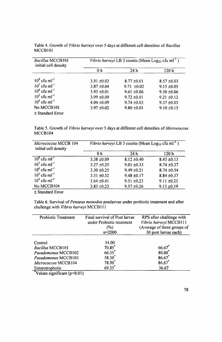
Table 4. Growth of Vibrio harveyi over 5 days at different cell densities of BacillusMCCBl01
Bacillus MCCBIOI Vibrio harveyi LB 3 counts (Mean Logm cfu ml“ )initial cell density M
0_h 2411 120 h
10* 6111 ml"
107 cfu mr‘106 cfu ml‘l
I105 cfu ml'104 cfu ml"No MCCB l 01i Standard Error
Table 5. Growth of Vibrio harveyi over 5 days at different cell densities of MicrococcusMCCB l 04
3.91 £0.033.87 i0.043.93 i0.0l3.99 i0.094.04 "£0.093.97 i0.02
8.77 i0.0l9.71 i0.029.61 i0.069.72 i0.0l9.74 -£0.039.80 "£0.01
8.57 i0.039.15 i0.059.30 i0.069.21 i0.129.37 i0.039.10 i0.l5
initial cell densityMicrococcus MCCB 104 Vibrio harvcgli LB 3 counts (Mean Logj6cfu_ml'l )
°h 7 "24 120th“10* cfu ml‘10’ cfu ml'
610 cfu ml'11105 cfu ml'
104 cfu ml"
No l_vlQCBl04
I W__g__ _ 3.38 i0.093.27 i0.253.30 i0.253.51 i0.523.64 i-0.413.83 i0.23
8.12 i0.409.01 i0.339.49 i0.2l9.48 i0. l 79.51 ;l:0.239.57 i0.26
8.43 i0.l38.74 i0.378.74 i0.548.84 i0.379.11 i0.259.13 i'0.l9
i Standard Error
Table 6. Survival of Penaeus monodon postlarvae under probiotic treatment and afterchallenge with Vibrio harveyi MCCBI1 1
Probiotic Treatment Final survival of Post larvae RPS after challenge withunder Probiotic treatment Vibrio harveyi MCCBI ll
(%) (Average of three groups ofK A n=2000 W _ 30 post larvae ea_ch)gControl 54.00Bacillus MCCBIOI 70.85‘ 66.67Pseudomonas MCCB 1 02 66.35‘ 80.00Pseudomonas MCCBl03 58.30 86.67Micrococcus MCCB 104 78.90 86.67Enterotrophotic W69.35 36.6Z_ 7
OO
Q
‘I’
Q
I
§
_'Values significant (p<0.01) M
78
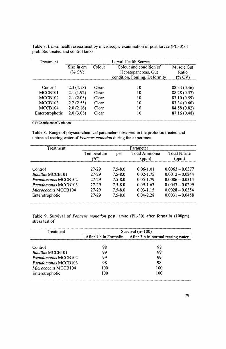
Table 7. Larval health assessment by microscopic examination of post larvae (PL30) ofprobiotic treated and control tanks
Treatment ____ -___ oLarvalH¢fll1h$<=<>r§sor
___ _ _ _ 0 _- _. _- ___-___ _ _ownditi<>11,FOulin8,P§f@:mi¢y.QMZV)
Size in cm Colour Colour and condition of Muscle:Gut(% CV) Hepatopancreas, Gut Ratio
ControlMCCB101MCCB102MCCB 103MCCB 104
Enterotrophotic
2.3 (4.18)2.1 (1.92)2.1 (2.05)2.2 (2.55)2.0 (2.16)2.0 (3.08)
ClearClearClearClearClearClear
101010101010
88.33 (0.46)88.28 (0.57)87.10 (0.59)87.34 (0.60)84.58 (0.82)87.16 (0.48)
CV: Coefficient of Variation
Table 8. Range of physico-chemical parameters observed in the probiotic treated anduntreated rearing water of Peaneus monodon during the experiment
Treatment _ _ _ _ _ _ p p __ fP_aram_eter_( 6 f Z é 6 _ _ é K _
_ 0 3 ._ __ _ £,Q1-__--, ._ _ <PPm>,_,.,<.Pnm>_ __Temperature pH Total Ammonia Total Nitrite
ControlBacillus MCCB 101Pseudomonas MCCB 102Pseudomonas MCCB 1 03Micrococcus MCCB104Enterotrophotic
27-2927-2927-2927-2927-2927-29
7.5-8.07.5-8.07.5-8.07.5-8.07.5-8.07.5-8.0
0.06-1.010.02-1.750.05-1.790.09-1.670.03-I .150.04-2.28
0.0063 — 0.03770.0012 — 0.02440.0086 — 0.03140.0043 -— 0.02990.0028 — 0.03540.0031 — 0.0458
Table 9. Survival of Penaeus monodon post larvae (PL-30) after formalin (100pm)stress test of
T1 T Treatment ‘T!’ T T T féfasui-vival(n?=1T00) K T T C T T T T 7 7 7it1 __ 1 T 1 i J 1 _ 1 pAfter1h(inF9*nna1in After3 hinnormal I'631‘lI‘lg!V3ECll_
ControlBacillus MCCB101Pseudomonas MCCB102Pseudomonas MCCB 103Micrococcus MCCB104Enterotrophotic
98999998100100
98999998100100
79

Table 10. Bacterial population in the rearing water and larvae at different stages duringthe larval cycle of probiotic supplemented and control (All counts are averages of 3samples plated in duplicate)
Treatment W { Rearingwater (>< 103cfu/ml)mp _ { WZoea-1 pZoea-37%? %Mysis-2 PL-2 CPL-8 PL-30%
Bacillus MCCB TPC101 TVCPseudomonas TPCMCCB 102 TVCPseudomonas TPCMCCB 103 TVCMicrococcus TPCMCCB 104 TVCEnterotrophotic TPC
TVCTPCTVC
Control
2700.042
1900.025200
0.008170ND170
0.033260ND
1200150
10000.191 100
0.2912001.79103256
0.15
57030086
ND49
ND160ND3108.84504.5
4001 1
2500.182400.633908.742013
3604.3
15012151.32500.953806.03909.042014
5505.22905.82907.54906.23505.83209.7
lsarvae (x 103 cfu/larvae)
Bacillus MCCB TPC101 TVCPseudomonas TPCMCCB 102 TVCPseudomonas TPCMCCB 103 TVCMicrococcus TPCMCCB 104 TVCEnterotrophotic TPC
TVCTPCTVC
Control
0.12ND0.14ND0.11ND
9.3 23.0 10.00.049 0.025 0.022
6.2ND11.0
0.0170.085 8.6ND 0.030.86ND 0.067 0.005 0.063
0.091ND
13.0
3.2ND0.12ND0.80ND10.0
1.8ND1.5
ND1.1
0.0211.0
7.4 11.0 11.00.035 0.009 0.028
12.00.130.70
1.80.761.4
0.008 0.0882.2 0.75
0.004 0.0890.57 0.900.03 0.0154.3 0.752.5 0.086.6 1.20.21 0.12
TPC: Total Plate Count TVC: Total Vibrio count ND: Not detected
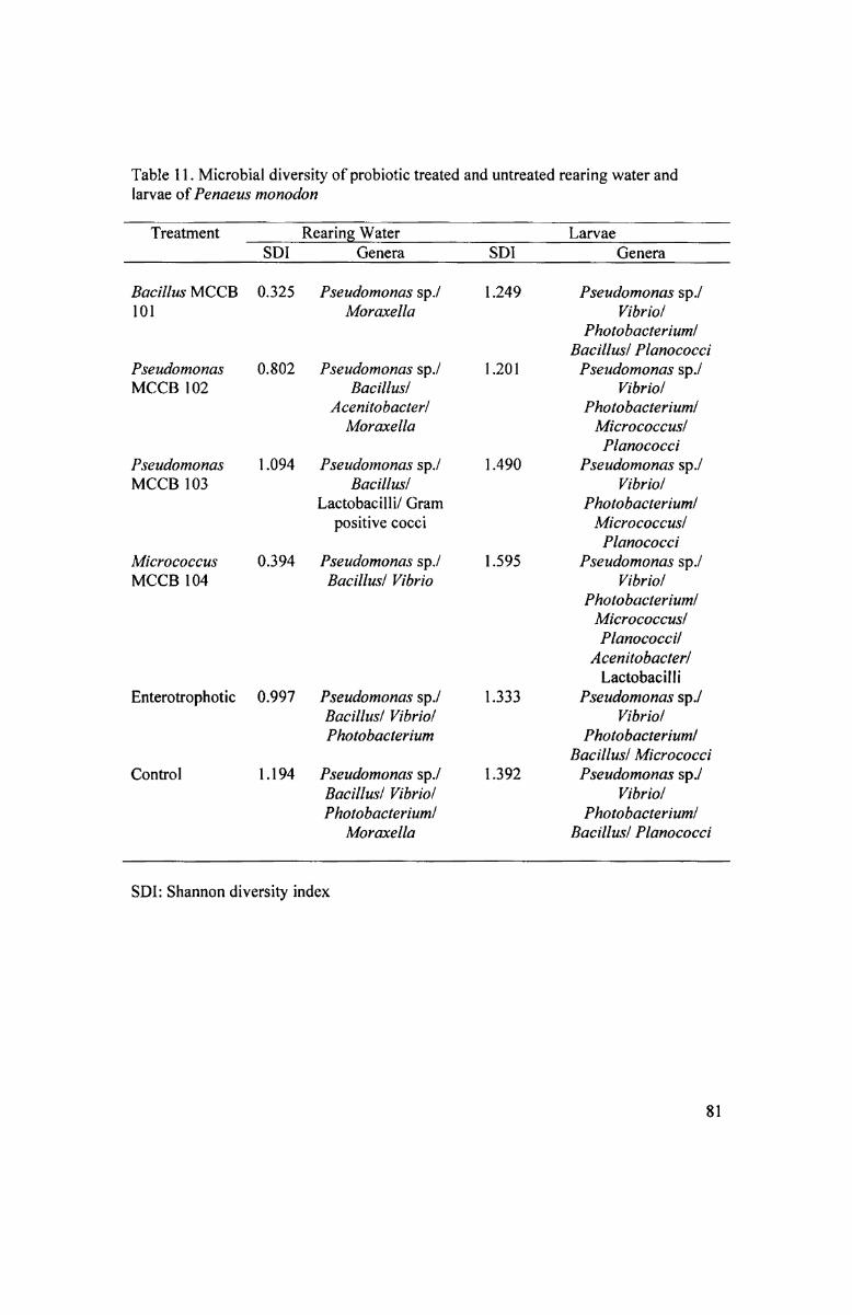
Table 11. Microbial diversity of probiotic treated and untreated rearing water andlarvae of Penaeus monodon
Treatment H WRearing WaterSDI Genera SDI Larvae fGenera
Bacillus MCCB101
PseudomonasMCCB 102
PseudomonasMCCB 103
MicrococcusMCCB 104
Enterotrophotic
Control
0.325
0.802
1.094
0.394
0.997
1.194
Pseudomonas sp./Moraxella
Pseudomonas sp./Bacillusl
Acem't0bacter/Moraxella
Pseudomonas sp./Bacillusl
Lactobacil1i/ Gram
positive cocci
Pseudomonas sp./Bacillusf Vibrio
Pseudomonas sp./Bacillusl VibriolPhotobacterium
Pseudomonas sp./Bacillus/ Vibri0/Photo bacteriuml
Moraxella
1.249
1.201
1.490
1.595
1.333
1.392
Pseudomonas sp./Vibri0/
Ph0t0bacterium/Bacillusl Planococcz
Pseudomonas sp./Vibri0/
Ph0t0bacterium/Mz'cr0c0ccus/Planococci
Pseudomonas sp./V ibriol
PhotobacteriumlMicr0c0ccus/Planococci
Pseudomonas sp./V ibriol
PhotobacteriumlMicrococcuslPlanococcil
Acenitobacter/Lactobacilli
Pseudomonas sp./Vibr2'0/
Ph0t0bacrerium/Bacillus/ Micrococcz
Pseudomonas sp./Vibri0/
PhotobacteriumfBacillus/ Planococcz
SDI: Shannon diversity index
81
U
O

Table 12. Survival of Penaeus monodon postlarvae after challenge with Vibrio harveyzMCCB] ll
Probiotic Treatment RPS after challenge with Vibrio harveyiMCCB] I1
_ it _ it t_ (Average Ofthree groups 01°30 POSI la“/R <=fl¢hL
Bacillus MCCB10lPseudomonas MCCB l 02Pseudomonas MCCB l 03Micrococcus MCCB l 04Enterotrophotic
Q
66.6780.0086.6786.6736.67
GG‘I
'Values significant (p<0.0l)

Table 13. Bacterial population in the rearing water and larvae after challenge withVibrio harveyi MCCBl11 (A11 counts are averages of 3 samples plated in duplicate)L 3 _Treatment 7 Rearing water (>< 10 cfu/ml)L . _ DELYI .I?2==1y5 Day 10
Bacillus MCCB 101 TPCTVCLBC
Pseudomonas MCCB 102 TPCTVCLBC
Pseudomonas MCCB 103 TPCTVCLBC
Micrococcus MCCB 104 TPCTVCLBC
Enterotrophotic TPCTVCLBCControl TPCTVCLBC
3258.33341.673258.332800.0366.672800.01916.67254.171916.672100.0516.672100.01491.67691.671491.673191.67182.50
3191.67
2141.6718.17
108.33975.05.58
118.33652.59.33
95.831100.025.83196.673475.017.08150.0
2608.3321.92249.17
3858.3310.00ND
2816.677.75ND
1683.337.33ND
2608.3312.50ND
2625.016.75ND
3400.064.17ND
Larvae‘ (x 103 cfu/larvae)
Bacillus MCCB 101 TPCTVC
Pseudomonas MCCB 102 TPCTVC
Pseudomonas MCCB 103 TPCTVC
Micrococcus MCCB 104 TPCTVC
Enterotrophotic TPCTVCControl TPCTVC
1.68ND1.400.023.000.1017.330.802.85ND1.930.35
14.420.437.250.0322.550.179.650.23
27.670.40
22.830.64
29.500.726.500.0818.180.5213.020.47
30.170.30
22.841.43
TPC: Total plate count TVC: Total Vibrio count LBC: Luminescent bacterial countND: Not detected‘Luminescent bacteria were not detected in any of the larval samples

Table 14. Microbial diversity of probiotic treated and untreated rearing water andlarvae of Penaeus monodon challenged with Vibrio harveyi MCCB1 1 1
it Treatment iRearing Water 0_ La“/a6 5{ SDI Genera Z SDI A Genera
Bacillus MCCB 0.731 Pseudomonas sp1 01 PhotobacteriumlPlanococcilMoraxella
1.151 Pseudomonas sp./Bacillus/
EnterobacteriaceaelPlanococci
1.4667 Pseudomonas sp./Planococcil
MicrococcuslAcenitobacterlPhoto bacterium
1.249 Pseudomonas sp./Bacillusl Vibriol
PseudomonasMCCB 102
PseudomonasMCCB 103
MicrococcusMCCB 104
Enterotrophotic 0.693 Pseudomonas sp./Vibriol
Photobacterium
Control 1.313 Pseudomonas sp./Bacillus! VibriolPh0t0bacterium/
PlanococcilStaphylococci
1.142
0.845
1.490
1.357
0.999
1.335
Pseudomonas sp./Bacillus/ Planococcil
Micrococcus
Pseudomonas sp./Vibri0/
PhotobacteriumlMicrococcus
Pseudomonas sp./Micrococcus/Bacillus
Pseudomonas sp./Vibriol
PhotobacteriumlMicrococcuslPlanococcilLactobacilli
Pseudomonas sp./Vibriol
PhotobacteriumlBacillusl Micrococci
Pseudomonas sp./EnterobacteriaceaelBacillusl Planococci
SDI: Shannon diversity index
84


