
REVIEW
Exploring NAD+ metabolism in host–pathogen interactions
Ines Mesquita1,2• Patrıcia Varela1,2
• Ana Belinha1,2•
Joana Gaifem1,2• Mireille Laforge4
• Baptiste Vergnes3•
Jerome Estaquier4,5• Ricardo Silvestre1,2
Received: 9 July 2015 / Revised: 27 November 2015 / Accepted: 14 December 2015
� Springer International Publishing 2015
Abstract Nicotinamide adenine dinucleotide (NAD?) is
a vital molecule found in all living cells. NAD? intracel-
lular levels are dictated by its synthesis, using the de novo
and/or salvage pathway, and through its catabolic use as
co-enzyme or co-substrate. The regulation of NAD?
metabolism has proven to be an adequate drug target for
several diseases, including cancer, neurodegenerative or
inflammatory diseases. Increasing interest has been given
to NAD? metabolism during innate and adaptive immune
responses suggesting that its modulation could also be
relevant during host–pathogen interactions. While the
maintenance of NAD? homeostatic levels assures an ade-
quate environment for host cell survival and proliferation,
fluctuations in NAD? or biosynthetic precursors bioavail-
ability have been described during host–pathogen
interactions, which will interfere with pathogen persistence
or clearance. Here, we review the double-edged sword of
NAD? metabolism during host–pathogen interactions
emphasizing its potential for treatment of infectious
diseases.
Keywords Nicotinamide adenine dinucleotide (NAD?) �Host-pathogen interaction � NAD?/NADH ratio �NADPH � Sirtuins � L-tryptophan
Nicotinamide adenine dinucleotide (NAD?) was initially
discovered by Sir Arthur Harden as a ‘cozymase’ for yeast
fermentation over 100 years ago. The succeeding work con-
tributed to the identification of NAD? as a player in hundreds
of biochemical reactions through its role in redox reactions.
NAD? is either consumed as a co-substrate by NAD?-con-
suming enzymes or used as an electron carrier in redox
reactions. Yet, the intracellular NAD?/NADH ratio is key to
the maintenance of an adequate metabolic status and cell
survival.Growing evidences indicate thatNAD? biosynthetic
pathways and metabolism are playing a major role in host–
pathogen interactions. In this review, we overview these
mechanisms highlighting the role of NAD?metabolism as an
attractive therapeutic target for microbe infections.
NAD1 biosynthesis: where the tale begins
NAD1 biosynthesis in mammalian cells
The biosynthesis of NAD? in mammals occurs through
two different pathways: the de novo and the salvage
pathways (Fig. 1). The de novo pathway begins with the
uptake and conversion of dietary L-tryptophan in N-for-
mylkynurenine, which is mediated by the rate-limiting
indoleamine 2,3-dioxygenase (IDO) or tryptophan 2,3-
& Jerome Estaquier
& Ricardo Silvestre
1 Microbiology and Infection Research Domain, Life and
Health Sciences Research Institute (ICVS), School of Health
Sciences, University of Minho, Braga, Portugal
2 ICVS/3B’s-PT Government Associate Laboratory,
Braga/Guimaraes, Portugal
3 MIVEGEC (IRD 224-CNRS 5290-Universite Montpellier),
Institut de Recherche pour le Developpement (IRD),
Montpellier, France
4 CNRS FR 3636, Universite Paris Descartes, 75006 Paris,
France
5 Centre de Recherche du CHU de Quebec, Universite Laval,
Quebec G1V 4G2, Canada
Cell. Mol. Life Sci.
DOI 10.1007/s00018-015-2119-4 Cellular and Molecular Life Sciences
123
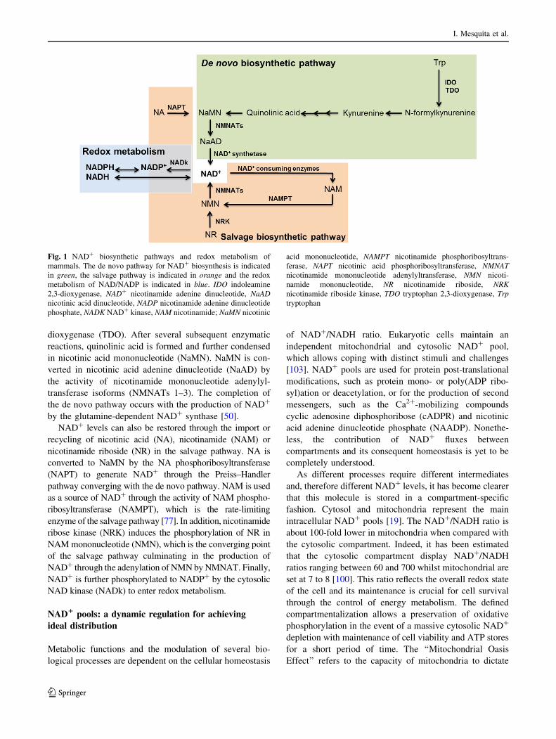
dioxygenase (TDO). After several subsequent enzymatic
reactions, quinolinic acid is formed and further condensed
in nicotinic acid mononucleotide (NaMN). NaMN is con-
verted in nicotinic acid adenine dinucleotide (NaAD) by
the activity of nicotinamide mononucleotide adenylyl-
transferase isoforms (NMNATs 1–3). The completion of
the de novo pathway occurs with the production of NAD?
by the glutamine-dependent NAD? synthase [50].
NAD? levels can also be restored through the import or
recycling of nicotinic acid (NA), nicotinamide (NAM) or
nicotinamide riboside (NR) in the salvage pathway. NA is
converted to NaMN by the NA phosphoribosyltransferase
(NAPT) to generate NAD? through the Preiss–Handler
pathway converging with the de novo pathway. NAM is used
as a source of NAD? through the activity of NAM phospho-
ribosyltransferase (NAMPT), which is the rate-limiting
enzyme of the salvage pathway [77]. In addition, nicotinamide
ribose kinase (NRK) induces the phosphorylation of NR in
NAMmononucleotide (NMN), which is the converging point
of the salvage pathway culminating in the production of
NAD? through the adenylation of NMNbyNMNAT. Finally,
NAD? is further phosphorylated to NADP? by the cytosolic
NAD kinase (NADk) to enter redox metabolism.
NAD1 pools: a dynamic regulation for achieving
ideal distribution
Metabolic functions and the modulation of several bio-
logical processes are dependent on the cellular homeostasis
of NAD?/NADH ratio. Eukaryotic cells maintain an
independent mitochondrial and cytosolic NAD? pool,
which allows coping with distinct stimuli and challenges
[103]. NAD? pools are used for protein post-translational
modifications, such as protein mono- or poly(ADP ribo-
syl)ation or deacetylation, or for the production of second
messengers, such as the Ca2?-mobilizing compounds
cyclic adenosine diphosphoribose (cADPR) and nicotinic
acid adenine dinucleotide phosphate (NAADP). Nonethe-
less, the contribution of NAD? fluxes between
compartments and its consequent homeostasis is yet to be
completely understood.
As different processes require different intermediates
and, therefore different NAD? levels, it has become clearer
that this molecule is stored in a compartment-specific
fashion. Cytosol and mitochondria represent the main
intracellular NAD? pools [19]. The NAD?/NADH ratio is
about 100-fold lower in mitochondria when compared with
the cytosolic compartment. Indeed, it has been estimated
that the cytosolic compartment display NAD?/NADH
ratios ranging between 60 and 700 whilst mitochondrial are
set at 7 to 8 [100]. This ratio reflects the overall redox state
of the cell and its maintenance is crucial for cell survival
through the control of energy metabolism. The defined
compartmentalization allows a preservation of oxidative
phosphorylation in the event of a massive cytosolic NAD?
depletion with maintenance of cell viability and ATP stores
for a short period of time. The ‘‘Mitochondrial Oasis
Effect’’ refers to the capacity of mitochondria to dictate
Fig. 1 NAD? biosynthetic pathways and redox metabolism of
mammals. The de novo pathway for NAD? biosynthesis is indicated
in green, the salvage pathway is indicated in orange and the redox
metabolism of NAD/NADP is indicated in blue. IDO indoleamine
2,3-dioxygenase, NAD? nicotinamide adenine dinucleotide, NaAD
nicotinic acid dinucleotide, NADP nicotinamide adenine dinucleotide
phosphate, NADK NAD? kinase, NAM nicotinamide; NaMN nicotinic
acid mononucleotide, NAMPT nicotinamide phosphoribosyltrans-
ferase, NAPT nicotinic acid phosphoribosyltransferase, NMNAT
nicotinamide mononucleotide adenylyltransferase, NMN nicoti-
namide mononucleotide, NR nicotinamide riboside, NRK
nicotinamide riboside kinase, TDO tryptophan 2,3-dioxygenase, Trp
tryptophan
I. Mesquita et al.
123

cell survival through maintenance of specific NAD? pools,
even after depletion of nuclear and cytosolic ones. This
protection was shown to be dependent on NAMPT activity
via mitochondrial SIRT3 and SIRT4 [103].
The compartmentalization of NAD? synthesis is
achieved by tightly controlled localization of the three
NMNAT isoforms; nuclear NMNAT1, cytosolic NMNAT2
and mitochondrial NMNAT3 [58]. The rigorous subcellu-
lar localization of mammalian NMNAT isoforms suggests
a predominant role for this enzyme in determining the
subcellular NAD? pool distribution. NMNAT1 is the most
efficient enzyme involved in the forward and reverse
equilibrium reaction that originates adenylyltransference or
pyrophosphorylysis, respectively. Although NMNAT1
utilizes nicotinamide mononucleotide (NMN) as a major
precursor for NAD? synthesis, it was shown that
NMNAT2 displays a high affinity for nicotinic acid
mononucleotide (NaMN) that originates nicotinic acid
adenine dinucleotide (NAAD), whilst hNMNAT3 was
demonstrated to be the isoform with lower selectivity for
purine nucleotides [46]. Although it is still debated whether
NMNAT1-synthesized NAD? may be exchanged between
the nucleus and cytoplasm via nuclear pores, it is known
that NAD? synthesis is independently regulated in these
compartments and the cytoplasmic NAD? pool is main-
tained primarily by NMNAT2. In mammals, mitochondrial
NAD? is not exchangeable with the cytosol and thus
NMNAT3 presumably participates in the maintenance of
the organelle nucleotide pool [18, 45]. Indeed, it has been
reported that FK866, a well-known inhibitor of rate-limit-
ing NAMPT, does not affect mitochondrial NAD? pool,
possibly indicating that this enzyme is not a major regu-
lator of NAD? in this organelle [70].
NAD1 metabolism in pathogens: evolution
towards auxotrophy
NAD? metabolic networks present a remarkable intrinsic
complexity and evolutionary variability [91]. Notably, the
biosynthesis of NAD? has evolved in several pathogenic
organisms towards auxotrophy or to a restriction in the
capacity to use a biosynthetic precursor. Several microor-
ganisms encoded enzymes ae capable to utilize NAD? from
infected hosts. Candida glabrata, a fungus that lacks the
genes for de novo synthesis, is aNAD? auxotroph possessing
a functioning salvage pathway that requires the uptake of
external sources of NAD? or precursors from the host cell
milieu [20]. Haemophilus influenzae is a gram-negative
bacterium that possesses an absolute need for NAD? due to a
lack of de novo biosynthetic enzymes or of salvaging NAM,
niacin or other intermediates of the Preiss–Handler pathway.
As NAD? cannot be taken up into the bacterium cytosolic
compartment as an intact molecule, previous studies have
established that NMN and NR are the biochemical sources
for NAD? in H. influenzae. Two proteins were identified to
play a key role in the uptake of NAD?; the outer membrane
lipoprotein e(P4) and a periplasmic NAD nucleotidase
(NadN). The e(P4) outer membrane protein and the NadN
periplasmic enzyme convert NAD? to NMN and NR [38].
The latter is able to cross the inner membrane to the cyto-
plasm, where NadR recycles it back to NAD? by
phosphorylating NR to NMN that is further adenylated to
NAD? [44, 87]. Shigella spp., the pathological agent of
bacillary dysentery, lacks a de novo pathway for the syn-
thesis of NAD? and therefore requires nicotinic acid for
growth [51]. As most prokaryotes, Shigella converts L-as-
partate into the precursor forNAD? synthesis quinolinic acid
depending on the enzyme complex composed by quinolate
synthase (NadA) and L-aspartate oxidase (NadB). Quinolinic
acid is subsequently converted into nicotinic acid mononu-
cleotide by quinolinate quinolinic acid concentration
decreases the intracellular spreading of Shigella, which
confirmed the occurrence of a selective pressure towards the
inactivation of the nadA and nadB genes during evolution
[72, 73]. Therefore, the available intracellular concentration
of NA is not limiting for bacterial growth and in fact the
reintroduction of functional copies of nadA and nadB into
this strain restored the ability to synthesize quinolate, but
resulted in strong attenuation of virulence, thus defining the
nadA and nadB as an anti-virulent loci [73]. Comparative
genomic studies have established that Leishmania protozoan
parasites are also NAD? auxotrophic organisms. Exogenous
supplementation of NA, NAMor NR precursors increase the
intracellular NAD? content in Leishmania parasites [26]. In
the case of Mycobacterium tuberculosis, NAD? synthesis
relies on both pathways, with the common enzyme being
NAD? synthase [99].
Several pathogens encode a nicotinamidase to convert
NAM in NA for NAD? synthesis. Leishmania nicotinami-
dase deletion led to a reduction of 70 % in NAD? content,
affecting both promastigote growth and the establishment
of infection in mice [26]. This enzyme should further pre-
vent the accumulation of anti-leishmanial NAM [85] by
recycling it to NAD?. In addition, Borrelia burgdorferi and
Brucella abortus nicotinamidases were shown to be
essential for bacteria replication and infectivity [40]. These
examples support that the inhibition of nicotinamidase may
drive specific microbicidal effects towards intracellular
pathogens. NMNAT encoded by Plasmodium (PfNMNAT)
is quite divergent from the human homologs but share
significant homology with bacterial counterparts. The
inhibition of PfNMNAT results in the arrest of parasite
growth in earlier events, thus indicating an importance of
this biosynthetic enzyme in the development of Plasmod-
ium parasites [63]. Therefore, NAM, nicotinamidase and
NMNAT activities are determinants for pathogen survival
Exploring NAD? metabolism in host–pathogen interactions
123

in its mammalian host. These observations highlight that
NAD? is essential for parasite growth being required for the
activity of several key substrates.
The importance of NAD1 in host–pathogeninteractions
Modulation of host NAD1 levels by intracellular
pathogens
Fluctuations in NAD? levels in infected cells have been
described for different classes of intracellular pathogens.
Peripheral blood lymphocytes from HIV-infected individ-
uals display a decrease of intracellular NAD? levels, which
may be reverted by exogenous administration of NAM
[62]. In contrast, Plasmodium-infected erythrocytes display
higher NAD? levels than uninfected ones. This increase
appears to be mediated by an increase in NAMPT and
NAPT activity in infected cells, which allows the produc-
tion of NAD? through NAM and NA salvage, respectively
[107]. Leishmania infantum induced a transitory NADH
increase immediately after infection that was posteriorly
reverted to higher NAD?/NADH ratio once the infection is
established [56]. Therefore, the modulation of host NAD?
levels may vary accordingly to the infectious agent and
probably the type of host cell due to their intrinsic meta-
bolic requirements. Group A streptococci (Streptococcus
pyogenes or GAS) represent a remarkable case of intra-
cellular NAD? modulation. GAS NAD? glycohydrolase
has the ability to cleave NAD? producing nicotinamide
and ADP-ribose but also cyclic ADP-ribose (cADPR) upon
being injected in the cytosol of an infected host cell [95].
This results in a profound depletion of cellular NAD? and
ATP levels, leading to growth arrest and cell death [54]. As
a consequence of depletion of host cell energy stores
through the enzymatic action of NADase, GAS has proven
to disrupt several innate processes of immune defense. As
such, NAD? glycohydrolase activity modifies several
NAD?-dependent host cell responses including poly (ADP-
ribose) polymerase (PARP)-1 activity [13], preventing
phagolysosome acidification [3] and autophagy killing
[64], which contributes to treatment failure, relapse and
chronic persistence. Further studies in different types of
pathogens are required to fully understand if the modula-
tion of host NAD? levels by intracellular pathogens is
imperative for successful colonization.
Enzymes involved in NAD1 synthesis contribute
to the immune response against pathogens
Tryptophan catabolism, which ultimately results in NAD?
production, has been shown to have a major role in the
regulation of immune responses. The immunosuppressive
effects of IDO have been vastly associated with impaired
proliferation, induction of apoptosis and induction of reg-
ulatory T cells [71]. However, several pathogenic species
are tryptophan auxotrophs, such as Chlamydia, Leishmania
or Toxoplasma gondii. Thus, tryptophan depletion by IDO
may further impact their intracellular survival [5]. IFN-c-nduced IDO was also demonstrated to be responsible for
inhibition of Staphylococcus aureus replication, the major
causative agent of cerebral abscesses [83]. Recently, CD4
T cells were shown to contain M. tuberculosis growth by
starving out of tryptophan [108]. Although M. tuberculosis
can synthesize tryptophan under immune stress, blocking
the bacterial tryptophan synthesis restored the efficacy of
the immune system to kill the mycobacteria. Tryptophan
catabolism by IDO is also a central mechanism for limiting
tissue damage. The blockage of IDO was shown to atten-
uate T. gondii replication in the lung due to decreased
inflammatory tissue damage [60]. Distinctively, in a
Clostridium difficile infection model, IDO-/- mice showed
increased immunopathology, as evidenced by increased
mucosal destruction, cecal hemorrhage and higher levels of
neutrophil-driven IFN-c production. Therefore, tryptophan
catabolism by IDO is a central mechanism for limiting
tissue damage and for decreasing C. difficile bacterial
burden, consequently restricting the observed pathology
[22]. The absence of IDO was also correlated with a sup-
pression of LP-BM5 murine leukemia virus replication via
upregulation of type I IFNs [35]. IDO was found to be
increased during HIV infection, which was associated with
the dysfunction of CD8 immune T cells in controlling
pathogens, the loss of Th22 cells and a consequent shift to
Treg cells [4, 16]. Several other viral infections, including
hepatitis B and C as well as influenza infections display an
increased expression and activation of IDO, where trypto-
phan metabolism appears to have a crucial role in the fight
against pathogens [81]. If in one hand the depletion of
tryptophan may alter the phenotype of immune cells driv-
ing them towards immunosuppression, on the other hand it
may have a severe impact on the growth of intracellular
pathogens.
Besides IDO, other enzymes in the biosynthetic pathway
of NAD? synthesis have been demonstrated to play a role
during infection. NAMPT was shown to inhibit HIV
replication at an early step through abrogation of the
integration of proviral DNA [97]. However, an increase in
NAMPT expression could prevent HIV-1 replication rather
than inhibiting it [14]. NAMPT targeting could provide
strong anti-inflammatory effects leading to a decrease of
the inflammatory tissue damage, without compromising
host defense as exemplified during S. aureus infection [78].
Overall, a special attention should be paid to the dual
effect of targeting host enzymes involved in NAD?
I. Mesquita et al.
123

synthesis, especially now that novel IDO and NAMPT
inhibitors are being tested for cancer chemotherapy [102],
which may increase the potential immunosuppression of
the patients.
The importance of NAD1-consuming proteinsin infected cells
NAD? is a cofactor for three classes of proteins: sirtuins,
PARPs and membrane proteins CD38/CD157, where it
contributes as a source of ADP-ribose. These NAD?-con-
suming proteins are considered metabolic sensors with a
vital role in energy metabolism, cell survival, proliferation
and effector functions. The mechanistic action of NAD?-
consuming proteins during host–pathogen interaction is
illustrated in Fig. 2.
cADP-ribose synthases
These ectoenzymes, known as lymphocyte antigens CD38
and CD157, are multifunctional proteins involved in the
generation of second messengers in intracellular signaling.
cADP-synthases are the major NAD?-regulating proteins:
for each cADP-ribose molecule produced, around 100
NAD? molecules are broken [17, 21]. Hence, CD38 is a
main cellular NADase in mammalian tissues being a crit-
ical regulator of NAD? levels by modulating its
bioavailability. Under homeostatic conditions, very little
NAD? is found free in the serum of normal mice [42].
Previous studies have shown that NAD? is consistently
released or actively transported to the extracellular medium
and rapidly catabolized by CD38 to maintain its levels to a
minimum [84]. Nevertheless, upon damage or infection,
local levels of extracellular NAD? can rise quite dramati-
cally, leading to an increased activity of CD38. The
generated cADP-ribose and nicotinic acid adenine dinu-
cleotide phosphate (NAADP) contribute to Ca2?
mobilization [47] and will enhance the ability of mono-
cytes, neutrophils and dendritic cells to migrate to sites
where danger was felt and secondary lymphoid tissues in
response to chemokines [69]. Studies performed with
CD38-/- mice demonstrated its crucial role in the regu-
lation of both innate and adaptive immune responses
against infections [69]. As illustrative examples, CD38-/-
mice are more susceptible to infection by S. pneumoniae
[68] and M. avium [98], while presenting a decrease hep-
atic elimination of Entamoeba histolytica [24] due to
reduced neutrophil recruitment and limited inflammatory
response. A similar failure to induce an appropriate
inflammatory response was observed in Naegleria fowleri-
induced primary amoebic meningoencephalitis [12].
Therefore, understanding how to alter NAD? extracellular
levels or CD38 enzyme activity is an exciting prospect in
the modulation of inflammatory responses during
infections.
PARPs
NAD? is also used as a substrate for mono- or poly(ADP-
ribosyl)ation (PARylation) reactions mediated by ADP-ri-
bose transferases (ARTs) or poly(ADP-ribose)
polymerases, respectively. ARTs catalyze the formation of
mono(ADP-ribosyl)ation, but generally the attached ADP-
riboses are built as polymers by PARPs, which are the most
common ADP-ribosyltransferases [8]. PARP-1, one of five
confirmed PARPs, is the most abundant and highly
expressed nuclear enzymes widely involved in DNA-
damage response, apoptosis, chromatin stabilization and
epigenetic modifications in mammalians [82]. Overactivity
of PARP-1 driven by DNA strand breaks or metabolic
insults leads to NAD? exhaustion and bioenergetic failure
[89] inducing caspase-independent apoptosis [106]. Indeed,
this phenomenon was estimated to contribute for a 75 %
depletion of NAD? [31]. Some pathogens were shown to
take advantage of the loss of PARP-1 function to its own
advantage. As example, Chlamydia trachomatis release a
protease-like activity factor (CPAF) leading to the cleavage
of PARP-1, assuring a reduced inflammatory response to
membrane-damaged cells [104]. In opposition, PARP-1
activation was detected in the brains of Vietnamese
patients with fatal Plasmodium falciparum malaria [52].
Interestingly, the use of PARP-1 inhibitor, 3-aminobenza-
mide, was protective against meningitis-associated central
nervous system complications resulting from Streptococcus
pneumoniae infection [43]. However, the role of PARP-1
in the integration of retroviral DNA and consequent steps
of retroviral infections, as HIV and Moloney murine leu-
kemia virus, remains controversial [2, 88]. Whereas some
groups found that PARP-1 by decreasing the intracellular
levels of NAD? in infected host cells contribute for the
maintenance of infection [30], others report a viral tran-
scriptional repression through epigenetic mechanisms [7].
The exacerbated activation of PARP-1 was shown to be
associated with NAD? depletion, followed by the opening
of mitochondrial permeability transition (MPT) pore [1]. In
parallel, the disturbance of mitochondria homeostasis and
the rupture of membrane potential cause mitochondrial and
cellular NAD? depletion, culminating in cell death.
Recently, the role of NAD? in different types of cell death
has been vastly addressed [25]. The modulation of mito-
chondria damage by pathogens has also been demonstrated
in several studies [55, 79]. Because mitochondria are also
the primary site for reactive oxygen species (ROS) pro-
duction, which constitute essential microbicidal molecules,
it seems likely that NAD? modulation at the mitochondrial
Exploring NAD? metabolism in host–pathogen interactions
123

level may have an impact on microbe infections, but fur-
ther studies are required.
Sirtuins
The mammalian sirtuin family comprises seven members,
named SIRT1-7 [53], with distinct subcellular localizations
reverting acetyl modifications of lysine residues or acting
as ADP-rybosiltransferases in histones and other proteins.
Sirtuins are activated in situations of energy deficit and
prompt the utilization of non-carbohydrate energy sources,
such as fatty acids [36]. The induced metabolic shift allows
the organism to increase the efficiency of energy
production.
SIRT1 was shown to be upregulated in hepatitis B virus
(HBV) infected-liver cells. Its pharmacological inhibition
by sirtinol was associated with a suppression of viral DNA
replication, suggesting that SIRT1 inhibitors might be used
as novel therapies to treat HBV infection [76]. The over-
expression of hepatitis C virus (HCV) core proteins in
HepG2 cells leads to an alteration in the cellular redox
state, with decreased NAD?/NADH ratio. This imbalance
was suggested to derive from a decreased signaling in the
SIRT1-AMPK pathway, contributing to the hepatic
Fig. 2 Mechanistic action of NAD?-consuming proteins during
host–pathogen interaction. The three major classes of NAD?-
consuming proteins are involved in NAD? breakdown and utilization,
which originates functional and metabolic alterations upon challenge.
NAD? cleavage in NAADP and cADP-ribose is catalyzed by the
ectoenzymes CD38 and CD157. When extracellular NAD? content is
increased, a high NAD? turnover results in increased concentration of
NAADP and cADP-ribose, which contribute for Ca2? mobilization.
This phenomenon leads to the production of secondary messengers,
activation of intracellular signaling cascades and consequent chemo-
taxis of immune cells (for example, dendritic cells, monocytes and
neutrophils) towards chemokine gradient. PARPs respond to DNA
damage by infectious agents originating poly(ADP-rybosyl)ation of
targeted proteins. This may have a role in chromatin stabilization and
epigenetic modifications. Furthermore, PARP activation results in
NAD? depletion and cell apoptosis due to decreased energy
availability. The seven isoforms of sirtuins are spread in the nucleus
(SIRT1, SIRT6 and SIRT7), the cytosol (SIRT2) and the mitochon-
dria (SIRT3, SIRT4 and SIRT5), although SIRT1 may shuttle
between the nuclear and cytosolic compartment and SIRT2 was
already shown to be able to translocate to the nucleus. Upon sirtuin
modulation by the presence of an infectious agent, NAD? is degraded
and ultimately SIRT1 activity originates mitochondrial biogenesis
and increased lipid oxidation. These processes lead to an increase of
intracellular ATP levels, in an attempt to restore energy homeostasis.
Sirtuins and PARPs are also able to cause epigenetic modifications in
cellular DNA, which may also contribute to metabolism modulation.
ATP adenosine triphosphate, ADP adenosine diphosphate, cADP
cyclic adenosine diphosphate, NAADP nicotinic acid adenine dinu-
cleotide phosphate, NAD? nicotinamide adenine dinucleotide, SIRT
sirtuin, PARP poly-(ADP-ribose) polymerase
I. Mesquita et al.
123

metabolic disorder and influencing disease progression and
anti-viral therapy efficacy [105]. Along the same line,
Moreira and colleagues demonstrated the role of energy
sensors AMPK and SIRT1 in Leishmania parasites survival
and proliferation [56]. NAD?/NADH fluctuations during
the course of infection reflected a correlation between
SIRT1 activity and host metabolism driving pathogen
persistence. Conversely, SIRT1 knockdown or inhibition
by NAM and sirtinol in Kaposi’s Sarcoma-Associated
Herpesvirus (KSHV)-infected cells resulted in increased
concentration of infectious virions, which could reactivate
the virus from the latent stage [32, 48]. Finally, the
infection of human biliary epithelial cells with Cryp-
tosporidium parvum, a coccidian parasite, resulted in
higher SIRT1 expression, in a let-71-dependent manner
that ultimately regulates NF-jB-driven innate immune
response [101].
Interestingly, Listeria monocytogenes infection was
impaired through H3K18 deacetylation-dependent fashion
in SIRT2 knockout mice or blocking SIRT2 activity [23].
The role played by SIRT2 during infection appears to be
pathogen-specific since the modulation of SIRT2 activity
in vivo did not affect chronic infection with M. tubercu-
losis [11]. Therefore, it is critical to further explore the
implications of sirtuin modulation during infections,
especially since several sirtuin activators/inhibitors are in
the biopharmaceutical pipeline to tackle metabolic, car-
diovascular, neurodegenerative and neoplastic diseases.
The importance of NADP(H) in host–pathogeninteractions
In contrast to NAD?/NADH, the NADP?/NADPH ratio
must be maintained at very low levels. NADPH, the
reduced form of NADP?, is known to provide reducing
equivalents for anabolic reactions, such as fatty acid
biosynthesis. It is also vital for protecting cells against
reactive oxygen species (ROS), produced notably by the
mitochondrial metabolism, through its role as cofactor for
NADPH-dependent glutathione reductases, which ulti-
mately ensures the regeneration of reduced glutathione
(GSH). During host–pathogens interactions, NADPH is
further used as an electron donor for NADPH oxidases
activation that play both effector and signalling roles in the
course of infection, through generation of microbicidal
ROS [34, 66, 96]. Seven NADPH oxidases isoforms have
been characterized in humans; the five NOX enzymes
produce superoxide anion, while the two Dual Oxidase
enzymes (DUOX1-2) generate hydrogen peroxide in a
Ca2?-dependent manner (reviewed in [75]). NADPH oxi-
dase NOX2, found in the membranes of neutrophils and
macrophages phagosomes, has been extensively studied.
During pathogen phagocytosis, this membrane-linked
complex (composed of gp91phox; p22 phox; p47 phox; p40phox; p67 phox; and small GTPase Rac) associates, resulting
in the oxidation of NADPH to NADP? with the con-
comitant production of superoxide (O2�-) from oxygen and
the remaining downstream ROS to eliminate invading
pathogens [67]. DUOX 1 and 2 are expressed in the
epithelial surfaces of salivary glands, airways and along the
gastrointestinal tract. The initial evidences of the in vivo
role of DUOX enzymes in the crosstalk gut-microbiota
were first provided in a Drosophila gut infection model
system. The knockdown of DUOX on flies was shown to
severely increase the susceptibility to gut infections (re-
viewed in [41]). Accordingly, Ha et al. [29] demonstrated
that DUOX activity is essential for the maintenance of
homeostasis in the fly gut in an infectious context through
the development of an oxidative burst that is capable of
limiting microbial proliferation. The tight control of
DUOX enzymes allows the host gut–microbe homeostasis
by efficiently controlling infection while tolerating com-
mensal microbes [28]. In this context, the differential
sensing of gut microbiota by innate immune sensors (Nod
and Toll-like receptors) as well as the activation of distinct
signalling pathways (Myd88, TRIF or NF-jB) has been
shown to play a critical role by controlling the expression
and activity of DUOX enzymes [33, 90]. Similarly,
DUOX-derived ROS has been demonstrated to be essential
for the innate immune response against bacterial or viral
infections in airway mucosa [37, 39, 49, 92]. Thus, DUOX
enzymes work in close collaboration with innate immune
recognition receptors to develop an efficient innate immune
response to viral or bacterial pathogens [37, 39]. However,
its role in controlling parasite infections has never been
addressed.
The beneficial versus detrimental role of NADPH oxi-
dase activation in infectious contexts has been recently
addressed. While some pathogenic bacteria, viruses and
parasites have developed different means to limit NADPH
oxidase activation and escape oxidative burst [93, 96],
others seem to use NADPH oxidase activation and ROS
production for their own benefit [34, 66]. Moreover, this is
not limited to phagocytic cells. NOX/DUOX enzymes in
the lung epithelium has been shown to participate in the
host defense against respiratory viruses [27]. In opposition,
hepatic NOX proteins during chronic hepatitis C virus were
associated with exacerbated oxidative stress that leads to
hepatocellular carcinoma [15], while ROS-generating
NOX5 are essential for HTLV-I virus mediated T cell
transformation phenotype [86].
For pathogens, the maintenance of high levels of
NADPH is considered fundamental for survival. If in one
hand, the pathogens have to adapt to the oxidative burst
naturally present in the phagosomes, on the other this
Exploring NAD? metabolism in host–pathogen interactions
123
hostile environment may be aggravated by antiparasitic
drugs act that act through generation of oxidative stress.
Similarly to what happens in host cells, the majority of the
antioxidant cofactor NADPH produced by parasites and
bacteria derives from the concomitant action of G6PDH
and 6PGD in the pentose phosphate pathway (PPP), which
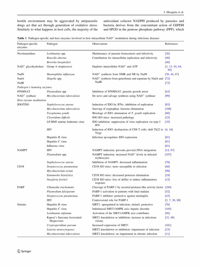
Table 1 Pathogen-specific and host enzymes involved in host intracellular NAD? modulation during infectious diseases
Pathogen-specific
enzymes
Pathogen Observations References
Nicotinamidase Leishmania spp. Maintenance of parasite homeostasis and infectivity [26]
Brucella abortus Contribution for intracellular replication and infectivity [40]
Borrelia burgdorferi [40]
NAD? glycohydrolase Group A streptococci Depletes intracellular NAD? and ATP [3, 13, 54, 64,
95]
NadN Haemophilus influenzae NAD? synthesis from NMR and NR by NadN [38, 44, 87]
NadA Shigella spp. NAD? synthesis from quinolinate and aspartate by NadA and
NadB
[72]
NadB [73]
Pathogen’s homolog enzymes
PfNMNAT Plasmodium spp. Inhibition of PfNMNAT: parasite growth arrest [63]
NAD? synthase Mycobacterium tuberculosis De novo and salvage synthesis using NAD? synthase [99]
Host enzyme modulation
IDO/TDO Staphylococcus aureus Induction of IDO by IFNy: inhibition of replication [83]
Mycobacterium tuberculosis Starving of tryptophan: bacteria elimination [108]
Toxoplasma gondii Blockage of IDO: attenuation of T. gondii replication [60]
Clostridium difficile IDO KO mice: increased pathology [22]
LP-BM5 murine leukemia virus IDO inhibition: suppression of virus replication via type I
IFN
[35]
HIV Induction of IDO: dysfunction of CD8 T cells; shift Th22 to
Tregs
[4, 16]
Hepatitis B virus Infection up-regulates IDO expression [81]
Hepatitis C virus [81]
Influenza virus [81]
NAMPT HIV NAMPT induction: prevents proviral DNA integration [14, 97]
Plasmodium spp. NAMPT induction: increased NAD? levels in infected
erythrocytes
[107]
Staphylococcus aureus Inhibition of NAMPT: decreased inflammation [78]
CD38 Streptococcus pneumoniae CD38 KO mice: more susceptible to infection [68]
Mycobacterium avium [98]
Entamoeba histolytica CD38 KO mice: decreased protozoa elimination [24]
Naegleria fowleri CD38 KO mice: loss of ability to induce inflammatory
response
[12]
PARP Chlamydia trachomatis Cleavage of PARP-1 by secreted protease-like activity factor [104]
Plasmodium falciparum PARP-1 activation in patients with fatal malaria [52]
Streptococcus pneumoniae PARP-1 inhibitor: protective against meningitis [43]
HIV Controversial role for PARP-1 [2, 7, 30, 88]
Sirtuins Hepatitis B virus SIRT1: upregulated in infection; sirtinol: protective [76]
Hepatitis C virus Imbalanced SIRT1/AMPK axis: hepatic disorder [105]
Leishmania infantum Activation of the SIRT1/AMPK axis contributes [56]
Kaposi’s Sarcoma-Associated
Herpesvirus
SIRT1 knockdown or inhibition: increase in infectious
virions
[32, 48]
Cryptosporidium parvum Increased expression of SIRT1 [101]
Listeria monocytogenes SIRT2 knockdown or inhibition: impairment of infection [23]
Mycobacterium tuberculosis SIRT2 knockdown: no impairment in chronic infection [11]
I. Mesquita et al.
123
makes this metabolism an attractive target for weakening
of pathogen’s defenses. G6PDH deficiency is one of the
most common enzymopathy found in humans, affecting
over 400 million people. This genetic disorder is more
frequent in Africa and it mostly affects red blood cells
(RBC) that are unable to produce sufficient NADPH levels
and become therefore highly susceptible to oxidative stress.
Paradoxically, G6PDH-deficient persons are more resistant
to Plasmodium infections in Africa. The possible expla-
nations are that chronic oxidative stress generated in
G6PDH-deficient infected RBCs limit infection by Plas-
modium or that these RBCs cannot sustain a normal
infection and will be rapidly eliminated by macrophages
[10, 59]. All these examples underline a complex role of
NADPH in host/pathogen interactions, with both a role in
the generation and the resistance to produced ROS during
infection.
Concluding remarks
The biosynthetic pathways that culminate with NAD?
production are currently being used to fight non-infectious
diseases, demonstrating its importance in novel drug design
[3, 9, 57, 80]. It is possible to draw an analogy of this
principle for infectious diseases. The remaining outstand-
ing questions are: (1) Would it be more efficient to target
host and/or pathogens NAD? metabolism? (2) Upon
blockage of a biosynthetic enzyme and consequent NAD?
depletion, are the pathogens able to evade elimination by
upregulating other enzymes or by retrieving NAD? from
other sources? (3) Will the targeting of such an important
molecule affect not only the host infected cells, but also
bystander or non-infected cells? A rapid and considerable
drop in NAD? levels may cause massive cell death. More
importantly, do the pathogen’s enzymes differ signifi-
cantly, in terms of homology, from host’s ones, allowing its
specific target? Or should the therapeutic approaches focus
only in pathogen-specific and unique enzymes, as the
nicotinamidases? It is also important to acknowledge the
importance of maintaining NAD? levels for the activation
of metabolic sensors, as the sirtuins, and downstream sig-
naling pathways. The correct functioning of host cells
depends on the intricate connection between processes that
drive cell survival, proliferation and host defense. The
modulation of NAD? levels is predicted to affect effector
functions of immune cells and, consequently, the clearance
or persistence of infections. Therefore, the study of NAD?
biology may be a promising approach for the discovery of
new targets against infectious diseases. Table 1 synthetizes
the major mechanisms used by pathogens to modulate host
NAD? levels. Remarkably, the first trials targeting NAD?
biology in infectious diseases go back to 1945, where
nicotinamide was explored as an anti-M. tuberculosis
agent, and later on during the 1990’s as an anti-HIV drug
[61]. Although all of this information had fallen into
obscurity, the past decade has seen the renaissance of tar-
geting NAD? biology to tackle infectious diseases, which
has been accompanied by the arrival of new structure-
based chemical modulators [6, 65, 74, 80, 94]. Nonetheless,
the investigation of NAD? metabolome is taking its first
steps and it is expected to convey important updates
regarding the interface between metabolism and immunity.
Acknowledgments JG was supported by PD/BD/106053/2015. BV
was supported by IRD (Institut de Recherche pour le Developpement)
institutional funding. JE was supported by a European Community’s
Seventh Framework Program under grant agreement No. 602773
(Project KINDRED), an ANR grant (LEISH-APO, France) and a
Partenariat Hubert Curien (PHC) (program Volubilis, MA/11/262). JE
also thanks the Canada Research Chair program for his support. RS
thank FCT—Foundation for Science and Technology—for their
Investigator FCT Grant (IF/00021/2014)
Compliance with ethical standards
Conflict of interest The authors have declared that no competing
interests exist.
References
1. Alano CC, Garnier P, Ying W, Higashi Y, Kauppinen TM,
Swanson RA (2010) NAD? depletion is necessary and sufficient
for poly(ADP-ribose) polymerase-1-mediated neuronal death.
J Neurosci 30:2967–2978
2. Ariumi Y, Turelli P, Masutani M, Trono D (2005) DNA damage
sensors ATM, ATR, DNA-PKcs, and PARP-1 are dispensable
for human immunodeficiency virus type 1 integration. J Virol
79:2973–2978
3. Bastiat-Sempe B, Love JF, Lomayesva N, Wessels MR (2014)
Streptolysin O and NAD-glycohydrolase prevent phagolyso-
some acidification and promote group a streptococcus survival
in macrophages. mBio 5:e01690-01614
4. Boasso A (2011) Wounding the immune system with its own
blade: HIV-induced tryptophan catabolism and pathogenesis.
Curr Med Chem 18:2247–2256
5. Brown SA, Palmer KL, Whiteley M (2008) Revisiting the host
as a growth medium. Nat Rev Microbiol 6:657–666
6. Bruzzone S, Parenti MD, Grozio A, Ballestrero A, Bauer I, Del
Rio A, Nencioni A (2013) Rejuvenating sirtuins: the rise of a
new family of cancer drug targets. Curr Pharm Des 19:614–623
7. Bueno MT, Reyes D, Valdes L, Saheba A, Urias E, Mendoza C
et al (2013) Poly(ADP-ribose) polymerase 1 promotes tran-
scriptional repression of integrated retroviruses. J Virol
87:2496–2507
8. Burkle A (2005) Poly(ADP-ribose). The most elaborate
metabolite of NAD?. FEBS J 272:4576–4589
9. Cagnetta A, Soncini D, Caffa I, Acharya C, Acharya P, Adamia
S et al (2015) Apo866 increases anti-tumor activity of cyclos-
porin-a by inducing mitochondrial and endoplasmic reticulum
stress in leukemia cells. Clin Cancer Res 21(17):3934–3945
10. Cappellini MD, Fiorelli G (2008) Glucose-6-phosphate dehy-
drogenase deficiency. Lancet 371:64–74
11. Cardoso F, Castro F, Moreira-Teixeira L, Sousa J, Torrado E,
Silvestre R et al (2015) Myeloid sirtuin 2 expression does not
Exploring NAD? metabolism in host–pathogen interactions
123
impact long-term Mycobacterium tuberculosis control. PLoS
One 10:e0131904
12. Cervantes-Sandoval I, Serrano-Luna Jde J, Garcia-Latorre E,
Tsutsumi V, Shibayama M (2008) Characterization of brain
inflammation during primary amoebic meningoencephalitis.
Parasitol Int 57:307–313
13. Chandrasekaran S, Caparon MG (2015) The Streptococcus
pyogenes NAD glycohydrolase modulates epithelial cell
PARylation and HMGB1 release. Cell Microbiol
17(9):1376–1390
14. Chen XY, Zhang HS, Wu TC, Sang WW, Ruan Z (2013) Down-
regulation of NAMPT expression by miR-182 is involved in
Tat-induced HIV-1 long terminal repeat (LTR) transactivation.
Int J Biochem Cell Biol 45:292–298
15. Choi J, Corder NL, Koduru B, Wang Y (2014) Oxidative stress
and hepatic Nox proteins in chronic hepatitis C and hepatocel-
lular carcinoma. Free Radic Biol Med 72:267–284
16. Cumont MC, Monceaux V, Viollet L, Lay S, Parker R, Hurtrel
B, Estaquier J (2007) TGF-beta in intestinal lymphoid organs
contributes to the death of armed effector CD8 T cells and is
associated with the absence of virus containment in rhesus
macaques infected with the simian immunodeficiency virus. Cell
Death Differ 14:1747–1758
17. de Toledo FG, Cheng J, Liang M, Chini EN, Dousa TP (2000)
ADP-Ribosyl cyclase in rat vascular smooth muscle cells:
properties and regulation. Circ Res 86:1153–1159
18. Di Stefano M, Conforti L (2013) Diversification of NAD bio-
logical role: the importance of location. FEBS J 280:4711–4728
19. Dolle C, Niere M, Lohndal E, Ziegler M (2010) Visualization
of subcellular NAD pools and intra-organellar protein local-
ization by poly-ADP-ribose formation. Cell Mol Life Sci
67:433–443
20. Domergue R, Castano I, De Las Penas A, Zupancic M, Lockatell
V, Hebel JR et al (2005) Nicotinic acid limitation regulates
silencing of Candida adhesins during UTI. Science 308:866–870
21. Dousa TP, Chini EN, Beers KW (1996) Adenine nucleotide
diphosphates: emerging second messengers acting via intracel-
lular Ca2? release. Am J Physiol 271:C1007–C1024
22. El-Zaatari M, Chang YM, Zhang M, Franz M, Shreiner A,
McDermott AJ et al (2014) Tryptophan catabolism restricts IFN-
gamma-expressing neutrophils and Clostridium difficile
immunopathology. J Immunol 193:807–816
23. Eskandarian HA, Impens F, Nahori MA, Soubigou G, Coppee
JY, Cossart P, Hamon MA (2013) A role for SIRT2-dependent
histone H3K18 deacetylation in bacterial infection. Science
341:1238858
24. Estrada-Figueroa LA, Ramirez-Jimenez Y, Osorio-Trujillo C,
Shibayama M, Navarro-Garcia F, Garcia-Tovar C, Talamas-
Rohana P (2011) Absence of CD38 delays arrival of neutrophils
to the liver and innate immune response development during
hepatic amoebiasis by Entamoeba histolytica. Parasite Immunol
33:661–668
25. Fouquerel E, Sobol RW (2014) ARTD1 (PARP1) activation and
NAD(?) in DNA repair and cell death. DNA Repair (Amst)
23:27–32
26. Gazanion E, Garcia D, Silvestre R, Gerard C, Guichou JF,
Labesse G et al (2011) The Leishmania nicotinamidase is
essential for NAD? production and parasite proliferation. Mol
Microbiol 82:21–38
27. Grandvaux N, Mariani M, Fink K (2015) Lung epithelial NOX/
DUOX and respiratory virus infections. Clin Sci (Lond)
128:337–347
28. Ha EM, Lee KA, Seo YY, Kim SH, Lim JH, Oh BH et al (2009)
Coordination of multiple dual oxidase-regulatory pathways in
responses to commensal and infectious microbes in Drosophila
gut. Nat Immunol 10:949–957
29. Ha EM, Oh CT, Bae YS, Lee WJ (2005) A direct role for dual
oxidase in Drosophila gut immunity. Science 310:847–850
30. Ha HC, Juluri K, Zhou Y, Leung S, Hermankova M, Snyder SH
(2001) Poly(ADP-ribose) polymerase-1 is required for efficient
HIV-1 integration. Proc Natl Acad Sci USA 98:3364–3368
31. Hassa PO, Haenni SS, Elser M, Hottiger MO (2006) Nuclear
ADP-ribosylation reactions in mammalian cells: Where are we
today and where are we going? Microbiol Mol Biol Rev
70:789–829
32. He M, Gao SJ (2014) A novel role of SIRT1 in gammaher-
pesvirus latency and replication. Cell Cycle 13:3328–3330
33. Hill T 3rd, Xu C, Harper RW (2010) IFNgamma mediates
DUOX2 expression via a STAT-independent signaling pathway.
Biochem Biophys Res Commun 395:270–274
34. Hogan D, Wheeler RT (2014) The complex roles of NADPH
oxidases in fungal infection. Cell Microbiol 16:1156–1167
35. Hoshi M, Saito K, Hara A, Taguchi A, Ohtaki H, Tanaka R et al
(2010) The absence of IDO upregulates type I IFN production,
resulting in suppression of viral replication in the retrovirus-
infected mouse. J Immunol 185:3305–3312
36. Houtkooper RH, Pirinen E, Auwerx J (2012) Sirtuins as regu-
lators of metabolism and healthspan. Nat Rev Mol Cell Biol
13:225–238
37. Joo JH, Ryu JH, Kim CH, Kim HJ, Suh MS, Kim JO et al (2012)
Dual oxidase 2 is essential for the toll-like receptor 5-mediated
inflammatory response in airway mucosa. Antioxid Redox Sig-
nal 16:57–70
38. Kemmer G, Reilly TJ, Schmidt-Brauns J, Zlotnik GW, Green
BA, Fiske MJ et al (2001) NadN and e (P4) are essential for
utilization of NAD and nicotinamide mononucleotide but not
nicotinamide riboside in Haemophilus influenzae. J Bacteriol
183:3974–3981
39. Kim HJ, Kim CH, Kim MJ, Ryu JH, Seong SY, Kim S et al
(2015) The Induction of pattern-recognition receptor expression
against influenza A virus through Duox2-derived reactive oxy-
gen species in Nasal Mucosa. Am J Respir Cell Mol Biol
53:525–535
40. Kim S, Kurokawa D, Watanabe K, Makino S, Shirahata T,
Watarai M (2004) Brucella abortus nicotinamidase (PncA)
contributes to its intracellular replication and infectivity in mice.
FEMS Microbiol Lett 234:289–295
41. Kim SH, Lee WJ (2014) Role of DUOX in gut inflammation:
lessons from Drosophila model of gut–microbiota interactions.
Front Cell Infect Microbiol 3:116
42. Kim UH, Kim MK, Kim JS, Han MK, Park BH, Kim HR (1993)
Purification and characterization of NAD glycohydrolase from
rabbit erythrocytes. Arch Biochem Biophys 305:147–152
43. Koedel U, Winkler F, Angele B, Fontana A, Pfister HW (2002)
Meningitis-associated central nervous system complications are
mediated by the activation of poly(ADP-ribose) polymerase.
J Cereb Blood Flow Metab 22:39–49
44. Kurnasov OV, Polanuyer BM, Ananta S, Sloutsky R, Tam A,
Gerdes SY, Osterman AL (2002) Ribosylnicotinamide kinase
domain of NadR protein: identification and implications in NAD
biosynthesis. J Bacteriol 184:6906–6917
45. Lau C, Dolle C, Gossmann TI, Agledal L, Niere M, Ziegler M
(2010) Isoform-specific targeting and interaction domains in
human nicotinamide mononucleotide adenylyltransferases.
J Biol Chem 285:18868–18876
46. Lau C, Niere M, Ziegler M (2009) The NMN/NaMN adeny-
lyltransferase (NMNAT) protein family. Front Biosci
(Landmark Ed) 14:410–431
47. Lee HC (2006) Structure and enzymatic functions of human
CD38. Mol Med 12:317–323
48. Li Q, He M, Zhou F, Ye F, Gao SJ (2014) Activation of
Kaposi’s sarcoma-associated herpesvirus (KSHV) by inhibitors
I. Mesquita et al.
123

of class III histone deacetylases: identification of sirtuin 1 as a
regulator of the KSHV life cycle. J Virol 88:6355–6367
49. Lu H, Wu Q, Yang H (2015) DUOX2 promotes the elimination
of the Klebsiella pneumoniae strain K5 from T24 cells through
the reactive oxygen species pathway. Int J Mol Med 36:551–558
50. Magni G, Orsomando G, Raffelli N, Ruggieri S (2008) Enzy-
mology of mammalian NAD metabolism in health and disease.
Front Biosci 13:6135–6154
51. Mantis NJ, Sansonetti PJ (1996) The nadB gene of Salmonella
typhimurium complements the nicotinic acid auxotrophy of
Shigella flexneri. Mol Gen Genet 252:626–629
52. Medana IM, Mai NT, Day NP, Hien TT, Bethell D, Phu NH et al
(2001) Cellular stress and injury responses in the brains of adult
Vietnamese patients with fatal Plasmodium falciparum malaria.
Neuropathol Appl Neurobiol 27:421–433
53. Michan S, Sinclair D (2007) Sirtuins in mammals: insights into
their biological function. Biochem J 404:1–13
54. Michos A, Gryllos I, Hakansson A, Srivastava A, Kokkotou E,
Wessels MR (2006) Enhancement of streptolysin O activity and
intrinsic cytotoxic effects of the group A streptococcal toxin,
NAD-glycohydrolase. J Biol Chem 281:8216–8223
55. Mocarski ES, Guo H, Kaiser WJ (2015) Necroptosis: the Trojan
horse in cell autonomous antiviral host defense. Virology
479–480:160–166
56. Moreira D, Rodrigues V, Abengozar M, Rivas L, Rial E,
Laforge M et al (2015) Leishmania infantum modulates host
macrophage mitochondrial metabolism by hijacking the SIRT1-
AMPK axis. PLoS Pathog 11:e1004684
57. Moreno-Vinasco L, Quijada H, Sammani S, Siegler J, Letsiou E,
Deaton R et al (2014) Nicotinamide phosphoribosyltransferase
inhibitor is a novel therapeutic candidate in murine models of
inflammatory lung injury. Am J Respir Cell Mol Biol
51:223–228
58. Mori V, Amici A, Mazzola F, Di Stefano M, Conforti L, Magni
G et al (2014) Metabolic profiling of alternative NAD biosyn-
thetic routes in mouse tissues. PLoS One 9:e113939
59. Muller S (2004) Redox and antioxidant systems of the malaria
parasite Plasmodium falciparum. Mol Microbiol 53:1291–1305
60. Murakami Y, Hoshi M, Hara A, Takemura M, Arioka Y,
Yamamoto Y et al (2012) Inhibition of increased indoleamine
2,3-dioxygenase activity attenuates Toxoplasma gondii replica-
tion in the lung during acute infection. Cytokine 59:245–251
61. Murray MF (2003) Nicotinamide: an oral antimicrobial agent
with activity against both Mycobacterium tuberculosis and
human immunodeficiency virus. Clin Infect Dis 36:453–460
62. Murray MF, Nghiem M, Srinivasan A (1995) HIV infection
decreases intracellular nicotinamide adenine dinucleotide
[NAD]. Biochem Biophys Res Commun 212:126–131
63. O’Hara JK, Kerwin LJ, Cobbold SA, Tai J, Bedell TA, Reider
PJ, Llinas M (2014) Targeting NAD? metabolism in the human
malaria parasite Plasmodium falciparum. PLoS One 9:e94061
64. O’Seaghdha M, Wessels MR (2013) Streptolysin O and its co-
toxin NAD-glycohydrolase protect group A Streptococcus from
Xenophagic killing. PLoS Pathog 9:e1003394
65. Olesen UH, Thougaard AV, Jensen PB, Sehested M (2010) A
preclinical study on the rescue of normal tissue by nicotinic acid
in high-dose treatment with APO866, a specific nicotinamide
phosphoribosyltransferase inhibitor. Mol Cancer Ther
9:1609–1617
66. Paiva CN, Bozza MT (2014) Are reactive oxygen species
always detrimental to pathogens? Antioxid Redox Signal
20:1000–1037
67. Panday A, Sahoo MK, Osorio D, Batra S (2015) NADPH oxi-
dases: an overview from structure to innate immunity-associated
pathologies. Cell Mol Immunol 12:5–23
68. Partida-Sanchez S, Cockayne DA, Monard S, Jacobson EL,
Oppenheimer N, Garvy B et al (2001) Cyclic ADP-ribose pro-
duction by CD38 regulates intracellular calcium release,
extracellular calcium influx and chemotaxis in neutrophils and is
required for bacterial clearance in vivo. Nat Med 7:1209–1216
69. Partida-Sanchez S, Rivero-Nava L, Shi G, Lund FE (2007)
CD38: an ecto-enzyme at the crossroads of innate and adaptive
immune responses. Adv Exp Med Biol 590:171–183
70. Pittelli M, Formentini L, Faraco G, Lapucci A, Rapizzi E,
Cialdai F et al (2010) Inhibition of nicotinamide phosphoribo-
syltransferase: cellular bioenergetics reveals a mitochondrial
insensitive NAD pool. J Biol Chem 285:34106–34114
71. Prendergast GC, Smith C, Thomas S, Mandik-Nayak L, Laury-
Kleintop L, Metz R, Muller AJ (2014) Indoleamine 2,3-dioxy-
genase pathways of pathogenic inflammation and immune
escape in cancer. Cancer Immunol Immunother 63:721–735
72. Prunier AL, Schuch R, Fernandez RE, Maurelli AT (2007)
Genetic structure of the nadA and nadB antivirulence loci in
Shigella spp. J Bacteriol 189:6482–6486
73. Prunier AL, Schuch R, Fernandez RE, Mumy KL, Kohler H,
McCormick BA, Maurelli AT (2007) nadA and nadB of Shigella
flexneri 5a are antivirulence loci responsible for the synthesis of
quinolinate, a small molecule inhibitor of Shigella pathogenic-
ity. Microbiology 153:2363–2372
74. Pulla VK, Sriram DS, Soni V, Viswanadha S, Sriram D,
Yogeeswari P (2015) Targeting NAMPT for therapeutic inter-
vention in cancer and inflammation: structure-based drug design
and biological screening. Chem Biol Drug Des 86(4):881–894
75. Rada B, Leto TL (2008) Oxidative innate immune defenses by
Nox/Duox family NADPH oxidases. Contrib Microbiol
15:164–187
76. Ren JH, Tao Y, Zhang ZZ, Chen WX, Cai XF, Chen K et al
(2014) Sirtuin 1 regulates hepatitis B virus transcription and
replication by targeting transcription factor AP-1. J Virol
88:2442–2451
77. Revollo JR, Korner A, Mills KF, Satoh A, Wang T, Garten A
et al (2007) Nampt/PBEF/Visfatin regulates insulin secretion in
beta cells as a systemic NAD biosynthetic enzyme. Cell Metab
6:363–375
78. Roberts KJ, Cross A, Vasieva O, Moots RJ, Edwards SW (2013)
Inhibition of pre-B cell colony-enhancing factor (PBEF/
NAMPT/visfatin) decreases the ability of human neutrophils to
generate reactive oxidants but does not impair bacterial killing.
J Leukoc Biol 94:481–492
79. Rodrigues V, Cordeiro-da-Silva A, Laforge M, Ouaissi A, Sil-
vestre R, Estaquier J (2012) Modulation of mammalian
apoptotic pathways by intracellular protozoan parasites. Cell
Microbiol 14:325–333
80. Sampath D, Zabka TS, Misner DL, O’Brien T, Dragovich PS
(2015) Inhibition of nicotinamide phosphoribosyltransferase
(NAMPT) as a therapeutic strategy in cancer. Pharmacol Ther
151:16–31
81. Schmidt SV, Schultze JL (2014) New insights into IDO biology
in bacterial and viral infections. Front Immunol 5:384
82. Schreiber V, Dantzer F, Ame JC, de Murcia G (2006) Poly(-
ADP-ribose): novel functions for an old molecule. Nat Rev Mol
Cell Biol 7:517–528
83. Schroten H, Spors B, Hucke C, Stins M, Kim KS, Adam R,
Daubener W (2001) Potential role of human brain microvascular
endothelial cells in the pathogenesis of brain abscess: inhibition
of Staphylococcus aureus by activation of indoleamine 2,3-
dioxygenase. Neuropediatrics 32:206–210
84. Seman M, Adriouch S, Haag F, Koch-Nolte F (2004) Ecto-ADP-
ribosyltransferases (ARTs): emerging actors in cell communi-
cation and signaling. Curr Med Chem 11:857–872
Exploring NAD? metabolism in host–pathogen interactions
123
85. Sereno D, Alegre AM, Silvestre R, Vergnes B, Ouaissi A (2005)
In vitro antileishmanial activity of nicotinamide. Antimicrob
Agents Chemother 49:808–812
86. Shigemura T, Shiohara M, Kato M, Furuta S, Kaneda K,
Morishita K et al (2015) Superoxide-generating Nox5alpha is
functionally required for the human T-cell leukemia virus
Type 1-induced cell transformation phenotype. J Virol
89:9080–9089
87. Singh SK, Kurnasov OV, Chen B, Robinson H, Grishin NV,
Osterman AL, Zhang H (2002) Crystal structure of Haemophilus
influenzae NadR protein. A bifunctional enzyme endowed with
NMN adenyltransferase and ribosylnicotinimide kinase activi-
ties. J Biol Chem 277:33291–33299
88. Siva AC, Bushman F (2002) Poly(ADP-ribose) polymerase 1 is
not strictly required for infection of murine cells by retroviruses.
J Virol 76:11904–11910
89. Sodhi RK, Singh N, Jaggi AS (2010) Poly(ADP-ribose) poly-
merase-1 (PARP-1) and its therapeutic implications. Vascul
Pharmacol 53:77–87
90. Sommer F, Backhed F (2015) The gut microbiota engages dif-
ferent signaling pathways to induce Duox2 expression in the
ileum and colon epithelium. Mucosal Immunol 8:372–379
91. Sorci L, Kurnasov O, Rodionov D, Osterman A (2010) Geno-
mics and enzymology of NAD biosynthesis. In: Mander L, Liu
H-W (eds) Comprehensive natural products II. Elsevier, Oxford,
pp 213–257
92. Strengert M, Jennings R, Davanture S, Hayes P, Gabriel G,
Knaus UG (2014) Mucosal reactive oxygen species are required
for antiviral response: role of Duox in influenza a virus infec-
tion. Antioxid Redox Signal 20:2695–2709
93. Sun K, Metzger DW (2014) Influenza infection suppresses
NADPH oxidase-dependent phagocytic bacterial clearance and
enhances susceptibility to secondary methicillin-resistant Sta-
phylococcus aureus infection. J Immunol 192:3301–3307
94. Takeuchi M, Niimi T, Masumoto M, Orita M, Yokota H,
Yamamoto T (2014) Discovery of a novel nicotinamide phos-
phoribosyl transferase (NAMPT) inhibitor via in silico
screening. Biol Pharm Bull 37:31–36
95. Tatsuno I, Isaka M, Minami M, Hasegawa T (2010) NADase as
a target molecule of in vivo suppression of the toxicity in the
invasive M-1 group A Streptococcal isolates. BMC Microbiol
10:144
96. Van Assche T, Deschacht M, da Luz RA, Maes L, Cos P (2011)
Leishmania-macrophage interactions: insights into the redox
biology. Free Radic Biol Med 51:337–351
97. Van den Bergh R, Florence E, Vlieghe E, Boonefaes T, Grooten
J, Houthuys E et al (2010) Transcriptome analysis of monocyte-
HIV interactions. Retrovirology 7:53
98. Viegas MS, do Carmo A, Silva T, Seco F, Serra V, Lacerda M,
Martins TC (2007) CD38 plays a role in effective containment
of mycobacteria within granulomata and polarization of Th1
immune responses against Mycobacterium avium. Microbes
Infect 9:847–854
99. Vilcheze C, Weinrick B, Wong KW, Chen B, Jacobs WR Jr
(2010) NAD? auxotrophy is bacteriocidal for the tubercle
bacilli. Mol Microbiol 76:365–377
100. Williamson DH, Lund P, Krebs HA (1967) The redox state of
free nicotinamide-adenine dinucleotide in the cytoplasm and
mitochondria of rat liver. Biochem J 103:514–527
101. Xie H, Lei N, Gong AY, Chen XM, Hu G (2014) Cryp-
tosporidium parvum induces SIRT1 expression in host epithelial
cells through downregulating let-7i. Hum Immunol 75:760–765
102. Yamahira A, Narita M, Iwabuchi M, Uchiyama T, Iwaya S,
Ohiwa R et al (2014) Activation of the leukemia plasmacytoid
dendritic cell line PMDC05 by Toho-1, a novel IDO inhibitor.
Anticancer Res 34:4021–4028
103. Yang H, Yang T, Baur JA, Perez E, Matsui T, Carmona JJ et al
(2007) Nutrient-sensitive mitochondrial NAD? levels dictate
cell survival. Cell 130:1095–1107
104. Yu H, Schwarzer K, Forster M, Kniemeyer O, Forsbach-Birk V,
Straube E, Rodel J (2010) Role of high-mobility group box 1
protein and poly(ADP-ribose) polymerase 1 degradation in
Chlamydia trachomatis-induced cytopathicity. Infect Immun
78:3288–3297
105. Yu JW, Sun LJ, Liu W, Zhao YH, Kang P, Yan BZ (2013)
Hepatitis C virus core protein induces hepatic metabolism dis-
orders through down-regulation of the SIRT1-AMPK signaling
pathway. Int J Infect Dis 17:e539–e545
106. Yu SW, Wang H, Poitras MF, Coombs C, Bowers WJ, Federoff
HJ et al (2002) Mediation of poly(ADP-ribose) polymerase-1-
dependent cell death by apoptosis-inducing factor. Science
297:259–263
107. Zerez CR, Roth EF Jr, Schulman S, Tanaka KR (1990)
Increased nicotinamide adenine dinucleotide content and syn-
thesis in Plasmodium falciparum-infected human erythrocytes.
Blood 75:1705–1710
108. Zhang YJ, Reddy MC, Ioerger TR, Rothchild AC, Dartois V,
Schuster BM et al (2013) Tryptophan biosynthesis protects
mycobacteria from CD4 T-cell-mediated killing. Cell
155:1296–1308
I. Mesquita et al.
123


