

EYE TRAUMA: INCIDENCE
• 2.5 million eye injuries per year in U.S.
• 40,000–60,000 of eye injuries lead to visual loss
Introduction

Final visual outcome of many ocular emergencies
depends on prompt, appropriate triage,
diagnosis, and treatment.
Introduction

VISION HISTORY
• Is one eye affected, or both?
• What is your current level of vision?
• Was vision normal prior to trauma?
Evaluation
ADDITIONAL HISTORY
• What symptoms do you have other than decreased vision?
• How long have you had symptoms?
• Have you had any eye surgery prior to trauma?
• Details of trauma?
Evaluation
COMPLETE EYE EXAMINATION
• Vision• External exam• Pupils• Motility exam• Anterior segment• Ophthalmoscopy• Intraocular pressure• Peripheral vision
Evaluation
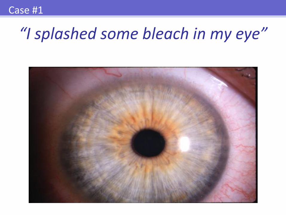
“I splashed some bleach in my eye”
Case #1

CHEMICAL BURNS
• A vision-threatening emergency
• Immediate irrigation essential
Treatment: Chemical Burns

Treatment: Chemical Burns
Acute and chronic stages of alkali burn

Treatment: Chemical Burns
Irrigation of chemical burns should begin immediately following contact with the substance and continue upon arrival at the
emergency department.

CHEMICAL BURNS: INITIAL MANAGEMENT
• Instill topical anesthetic
• Check for and remove foreign bodies
• Institute copious irrigation
Treatment: Chemical Burns
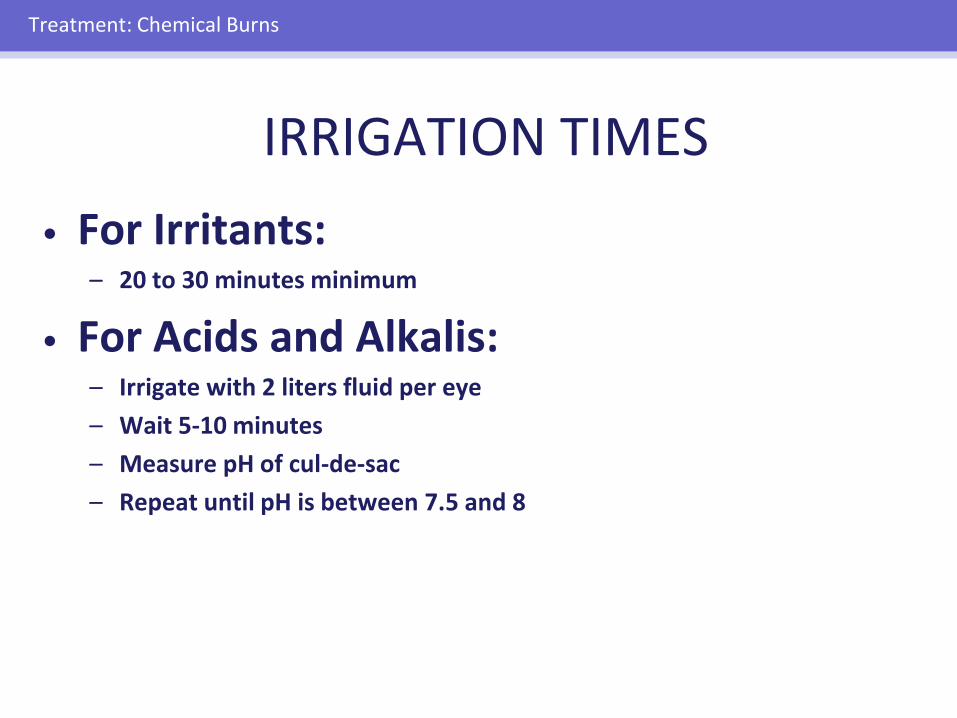
IRRIGATION TIMES
• For Irritants: – 20 to 30 minutes minimum
• For Acids and Alkalis:– Irrigate with 2 liters fluid per eye
– Wait 5-10 minutes
– Measure pH of cul-de-sac
– Repeat until pH is between 7.5 and 8
Treatment: Chemical Burns
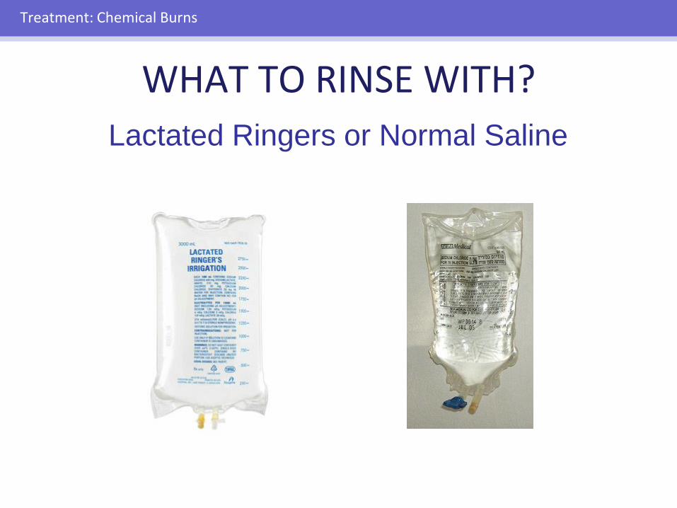
WHAT TO RINSE WITH?
Treatment: Chemical Burns
Lactated Ringers or Normal Saline

Treatment: Chemical Burns
Ocular irrigation

Treatment: Chemical Burns
The Morgan Lens®
CHEMICAL BURNS: TREATMENT FOLLOWING IRRIGATION
• Instill topical cycloplegic and topical antibiotic
• Shield eye
• Refer promptly to ophthalmologist
Treatment: Chemical Burns

“I always wear my safety goggles… just not today”
Case #2
Marked lid swelling after blunt trauma may conceal a ruptured globe.
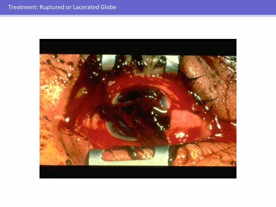
Treatment: Ruptured or Lacerated Globe

• Severe blunt trauma
• Sharp object
• Metal-on-metal contact
Treatment: Ruptured or Lacerated Globe
SUSPECT A RUPTURED GLOBE IF
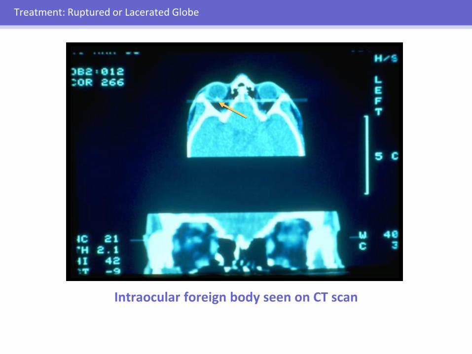
Intraocular foreign body seen on CT scan
Treatment: Ruptured or Lacerated Globe
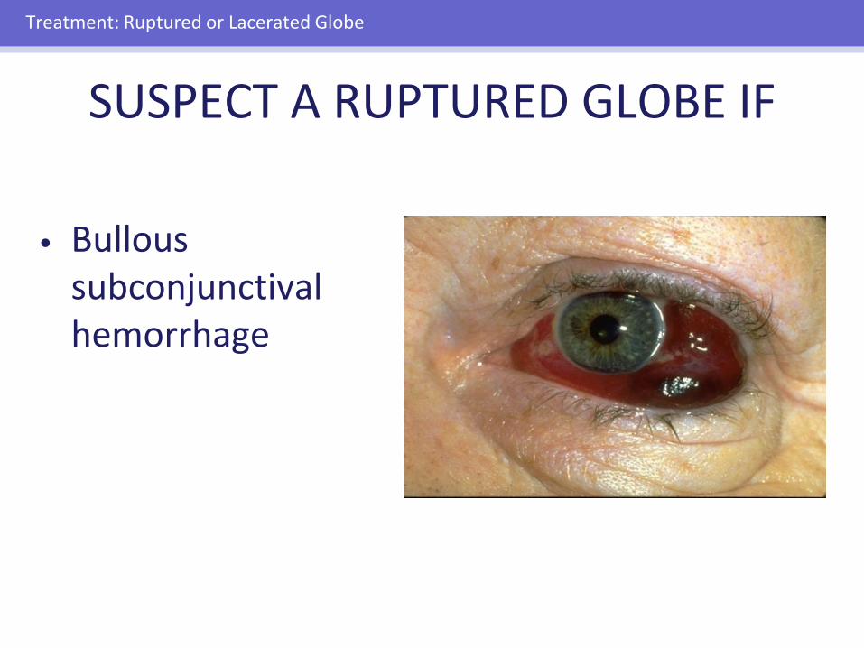
• Bullous subconjunctival hemorrhage
Treatment: Ruptured or Lacerated Globe
SUSPECT A RUPTURED GLOBE IF

• Uveal prolapse (iris or ciliary body)
Treatment: Ruptured or Lacerated Globe
SUSPECT A RUPTURED GLOBE IF

• Irregular pupil
Treatment: Ruptured or Lacerated Globe
SUSPECT A RUPTURED GLOBE IF

• Hyphema
• Vitreous hemorrhage
Treatment: Ruptured or Lacerated Globe
SUSPECT A RUPTURED GLOBE IF

• Lens opacity
Treatment: Ruptured or Lacerated Globe
SUSPECT A RUPTURED GLOBE IF

RUPTURED GLOBE
• Suspect if intraocular pressure is lowered
• Evaluate cautiously to avoid extrusion of intraocular contents
Treatment: Ruptured or Lacerated Globe
IF GLOBE RUPTURE OR LACERATION IS SUSPECTED
• Stop examination
• Shield the eye (do not patch)
• Give tetanus prophylaxis
• Refer immediately to ophthalmologist
Treatment: Ruptured or Lacerated Globe

Protective eye shields
Treatment: Ruptured or Lacerated Globe

Case #3
“I was hit in the eye by a plastic BB”
HYPHEMA: MANAGEMENT
• Assume globe is potentially ruptured
• Shield eye and refer to ophthalmologist
• Ophthalmologic management:– Restricted activity
– Protective metal shield
– Topical cycloplegic and corticosteroids
– Possibly systemic corticosteroids or antifibrinolytic agents
Treatment: Hyphema
HYPHEMA: COMPLICATIONS
• Rebleeding into anterior chamber
• Glaucoma
• Associated ocular injuries in 25% of patients
Treatment: Hyphema
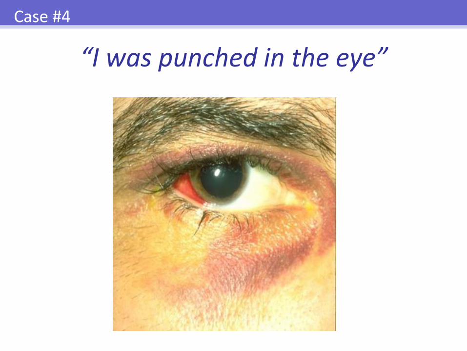
“I was punched in the eye”
Case #4

Treatment: Orbital Trauma
SUBCONJUCTIVAL HEMORRHAGE•Re-assurance
•Artificial tears

• Bullous subconjunctival hemorrhage
• Proptosis
• Corneal exposure
• Elevated intraocular pressure
Treatment: Orbital Trauma
SEVERE ORBITAL HEMORRHAGE

• Assess ocular motility
• Assess sensation over cheek and lip
• Palpate for bony abnormality of orbital rim
Treatment: Orbital Trauma
ORBITAL FRACTURES

X-ray of skull CT scan
(Waters or Caldwell view) (Coronal and Sagittal views)
Treatment: Orbital Trauma
ORBITAL TRAUMA: BLOW-OUT FRACTURES
• Surgery if persistent, nontransient diplopia or poor cosmesis
• Must rule out occult ocular trauma
Treatment: Orbital Trauma
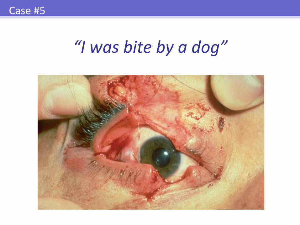
“I was bite by a dog”
Case #5
LID LACERATIONS
• Can result from sharp or blunt trauma
• Rule out associated ocular injury
Treatment: Lid Lacerations
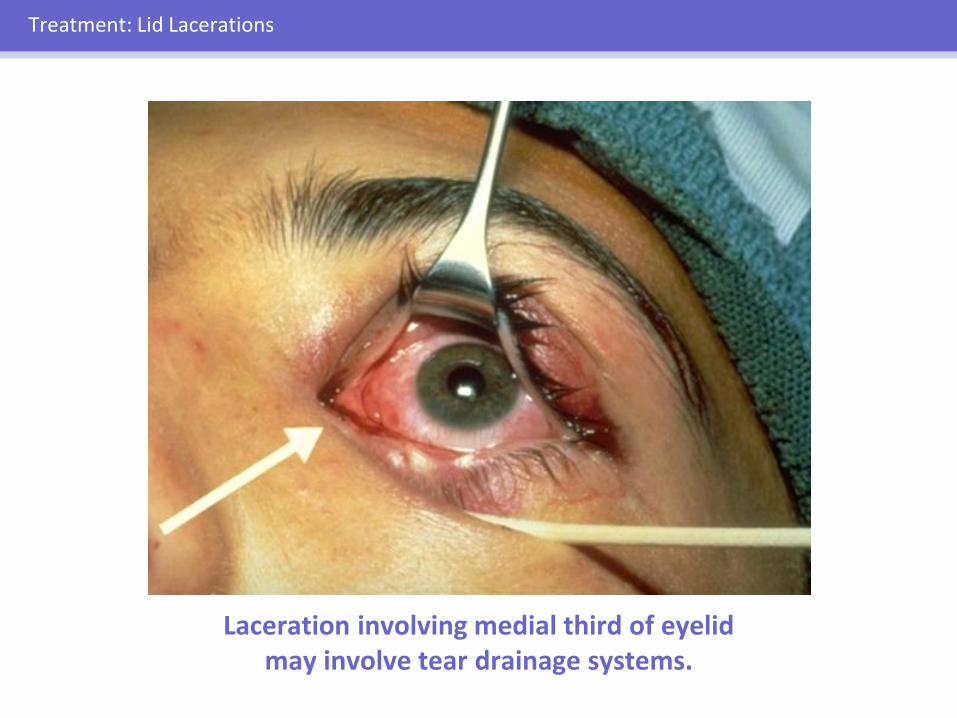
Laceration involving medial third of eyelid may involve tear drainage systems.
Treatment: Lid Lacerations

SUPERFICIAL LID LACERATIONS
• Avoid lid margin retraction
• Remove superficial foreign bodies
• Rule out deeper foreign bodies
• Give tetanus prophylaxis
Treatment: Lid Lacerations
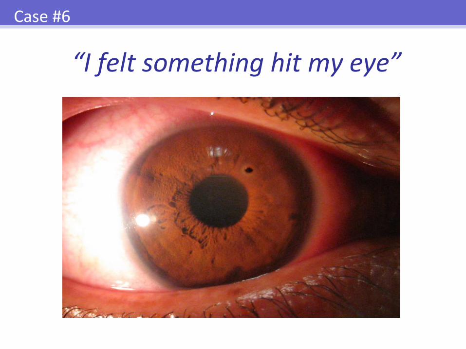
“I felt something hit my eye”
Case #6

CORNEAL ABRASIONS: SYMPTOMS
• Foreign-body sensation
• Pain
• Tearing
• Photophobia
Treatment: Corneal Abrasions and Foreign Bodies
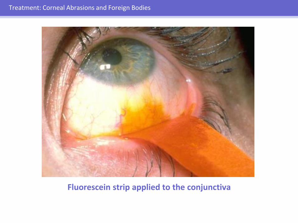
Treatment: Corneal Abrasions and Foreign Bodies
Fluorescein strip applied to the conjunctiva

Treatment: Corneal Abrasions and Foreign Bodies
Corneal abrasion seen in blue illumination
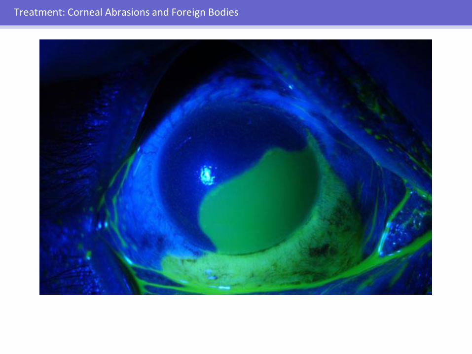
Treatment: Corneal Abrasions and Foreign Bodies
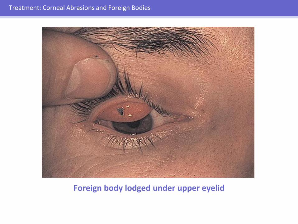
Treatment: Corneal Abrasions and Foreign Bodies
Foreign body lodged under upper eyelid

Treatment: Corneal Abrasions and Foreign Bodies
Corneal foreign body

Removal of corneal foreign body using magnification
Treatment: Corneal Abrasions and Foreign Bodies

Treatment: Corneal Abrasions and Foreign Bodies
Rust ring after removal of corneal foreign body (slit-lamp view)

CORNEAL ABRASIONS:TREATMENT
• Topical cycloplegic
• Topical antibiotic
• Pressure patch over eye is an option
• Systemic analgesics often needed
Treatment: Corneal Abrasions and Foreign Bodies
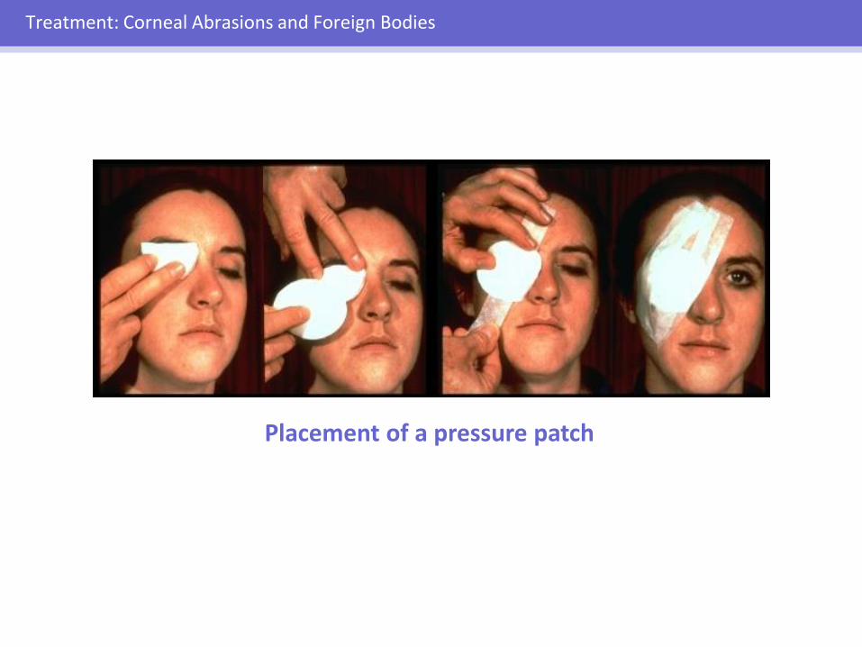
Treatment: Corneal Abrasions and Foreign Bodies
Placement of a pressure patch

CORNEAL ABRASIONS:CONTACT LENS WEARERS
• Remove contact lens
• Antibiotics for Gram-negative organisms
• Do not patch
• Follow up with ophthalmologist in 24 hours
Treatment: Corneal Abrasions and Foreign Bodies
CORNEAL ABRASIONS:FOLLOW-UP
• Follow up in 24 hours
• Refer to ophthalmologist if – Not healed in 24 hours
– Abrasion is related to contact lens wear
– White corneal infiltrate develops
Treatment: Corneal Abrasions and Foreign Bodies
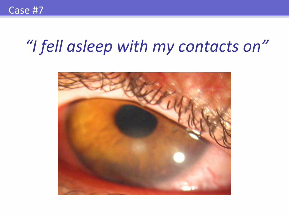
“I fell asleep with my contacts on”
Case #7

• Pain, foreign-body sensation
• Decreased vision
• Corneal infiltrate
Treatment: Keratitis
CORNEAL INFLAMMATION OR INFECTION

• STOP Contact Lenses!
• Consider starting fourth-generation fluoroquinolone (Ex: Vigamox, Moxeza, Besivance, Zymar, Zymaxid)
• Urgent vs. Stat referral
Treatment: Keratitis
CORNEAL INFLAMMATION OR INFECTION

Case #8
“My eye hurts, my vision is blurry, and I’m nauseous”

• Severe ocular pain
• Decreased vision
• Headache, nausea/vomiting
• Halos around lights
• Pupil moderately dilated
• Hazy cornea
• Elevated IOP
Treatment: Angle Closure
ACUTE ANGLE-CLOSURE GLAUCOMA: SIGNS & SYMPTOMS

ACUTE ANGLE-CLOSURE GLAUCOMA: INITIAL TREATMENT
• Timolol maleate 0.5% drops
• Apraclonidine 0.5% drops
• Pilocarpine 2% drops
• Acetazolamide 500 mg IV or po, or dorzolamide 2% drops
• IV Mannitol
Treatment: Angle Closure

“My eyelid is red, swollen, and tender”
Case #9
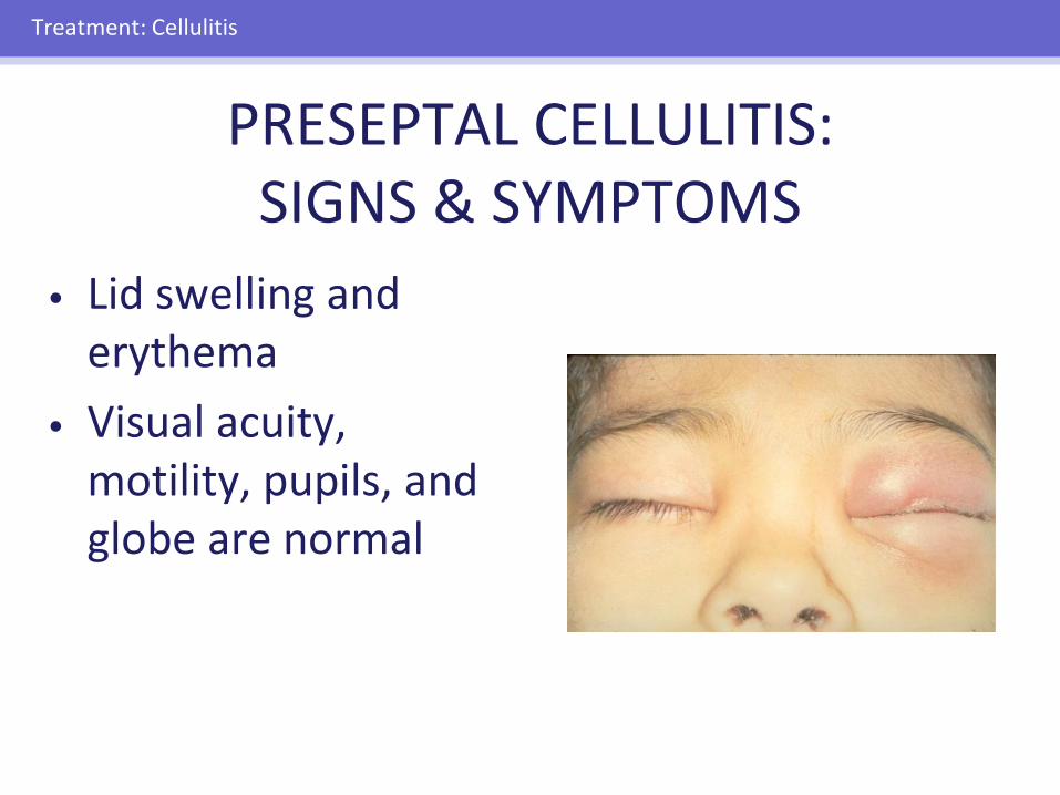
• Lid swelling and erythema
• Visual acuity, motility, pupils, and globe are normal
Treatment: Cellulitis
PRESEPTAL CELLULITIS: SIGNS & SYMPTOMS
PRESEPTAL CELLULITIS: MANAGEMENT CONSIDERATIONS
• Warm compresses
• Systemic antibiotics
• X-rays if history of trauma/sinus disease
Treatment: Cellulitis

• Pain
• Decreased vision
• Impaired ocular motility
• Afferent pupillarydefect
• Proptosis
• Optic nerve swelling
Treatment: Cellulitis
ORBITAL CELLULITIS: SIGNS AND SYMPTOMS
ORBITAL CELLULITIS: MANAGEMENT
• Immediate treatment
• Nasopharynx and blood cultures
• Intravenous antibiotics
• Surgery may be necessary
• Rule out mucormycosis in immunocompromised patients
Treatment: Cellulitis

“I can’t see out of my eye”
Case #10

SUDDEN, NONTRAUMATIC, MONOCULAR VISION LOSS
• Most often caused by vascular occlusion– Vasculopathic risk factors
– Vein: most common, better prognosis
– Artery: less common, worse prognosis
• Less commonly caused by retinal or optic nerve lesions– Retinal Detachment: Sx. flashes, floaters or curtain blocking vision,
refer to ophthalmology promptly
– Optic Neuritis: younger patients, MS association
Treatment: Sudden Vision Loss

Treatment: Sudden Vision Loss
Central retinal artery occlusion (CRAO) Cherry Red Spot

• Unilateral loss of vision
• Afferent pupillary defect
• Optic nerve swelling
• Scalp/forehead tenderness
• +/- Chewing pain
• +/- Polymyalgiarheumatica
Treatment: Sudden Vision Loss
TEMPORAL ARTERITIS: SIGNS AND SYMPTOMS

TEMPORAL ARTERITIS: MANAGEMENT
• Obtain ESR and C-reactive protein
• Administer systemic corticosteroids
• Perform temporal artery biopsy
Treatment: Sudden Vision Loss
EYE TRAUMA: PATIENT CARE/ PRESERVATION OF VISION
• Timely, accurate emergency diagnosis and treatment
• Appropriate ophthalmologic referral
Summary


