
COPYRIGHT © BY THE JOURNAL OF BONE AND JOINT SURGERY, INCORPORATED
VERBERNE ET AL.
THE ACCURACY OF IMAGING TECHNIQUES IN THE ASSESSMENT OF PERIPROSTHETIC HIP INFECTION: A SYSTEMATIC REVIEW AND
META-ANALYSIS
http://dx.doi.org/10.2106/JBJS.15.00898
Page 1
Appendix
Fig. E-1 Methodological quality of the included studies with QUADAS-2.
Fig. E-2 Diagnostic accuracy of bone scintigraphy for the detection of periprosthetic hip infection. The values are given as the mean with the 95% confidence interval. The diamond and vertical lines indicate the pooled estimate and the 95% confidence interval.

COPYRIGHT © BY THE JOURNAL OF BONE AND JOINT SURGERY, INCORPORATED
VERBERNE ET AL.
THE ACCURACY OF IMAGING TECHNIQUES IN THE ASSESSMENT OF PERIPROSTHETIC HIP INFECTION: A SYSTEMATIC REVIEW AND
META-ANALYSIS
http://dx.doi.org/10.2106/JBJS.15.00898
Page 2
Fig. E-3 Diagnostic accuracy of combined bone and gallium scintigraphy for the detection of periprosthetic hip infection. The values are given as the mean with the 95% confidence interval. The diamond and vertical lines indicate the pooled estimate and the 95% confidence interval.
Fig. E-4 Diagnostic accuracy of combined bone and leukocyte scintigraphy for the detection of periprosthetic hip infection. The values are given as the mean with the 95% confidence interval. The diamond and vertical lines indicate the pooled estimate and the 95% confidence interval.

COPYRIGHT © BY THE JOURNAL OF BONE AND JOINT SURGERY, INCORPORATED
VERBERNE ET AL.
THE ACCURACY OF IMAGING TECHNIQUES IN THE ASSESSMENT OF PERIPROSTHETIC HIP INFECTION: A SYSTEMATIC REVIEW AND
META-ANALYSIS
http://dx.doi.org/10.2106/JBJS.15.00898
Page 3
Fig. E-5 Diagnostic accuracy of leukocyte scintigraphy for the detection of periprosthetic hip infection. The values are given as the mean with the 95% confidence interval. The diamond and vertical lines indicate the pooled estimate and the 95% confidence interval.
Fig. E-6 Diagnostic accuracy of combined leukocyte and bone marrow scintigraphy for the detection of periprosthetic hip infection. The values are given as the mean with the 95% confidence interval. The diamond and vertical lines indicate the pooled estimate and the 95% confidence interval.
COPYRIGHT © BY THE JOURNAL OF BONE AND JOINT SURGERY, INCORPORATED
VERBERNE ET AL.
THE ACCURACY OF IMAGING TECHNIQUES IN THE ASSESSMENT OF PERIPROSTHETIC HIP INFECTION: A SYSTEMATIC REVIEW AND
META-ANALYSIS
http://dx.doi.org/10.2106/JBJS.15.00898
Page 4
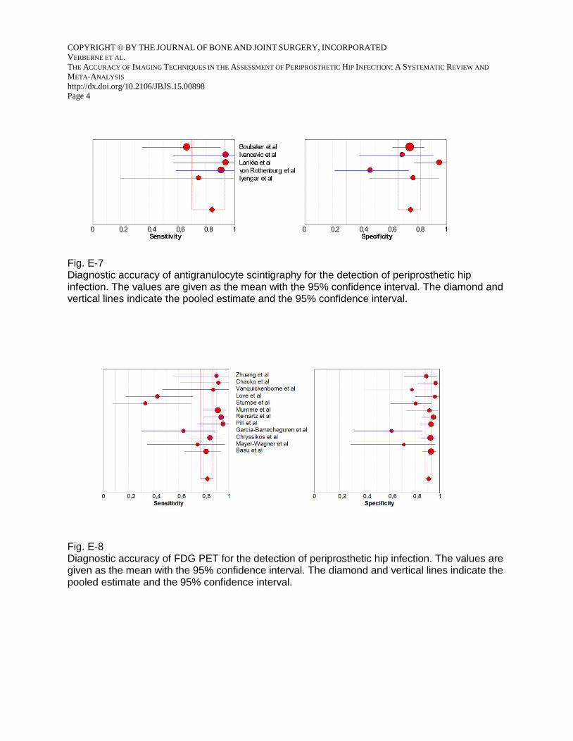
Fig. E-7 Diagnostic accuracy of antigranulocyte scintigraphy for the detection of periprosthetic hip infection. The values are given as the mean with the 95% confidence interval. The diamond and vertical lines indicate the pooled estimate and the 95% confidence interval.
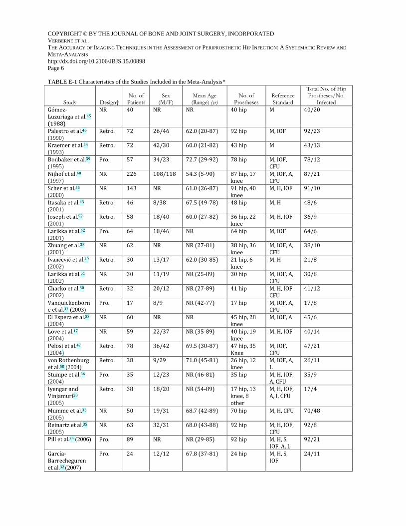
Fig. E-8 Diagnostic accuracy of FDG PET for the detection of periprosthetic hip infection. The values are given as the mean with the 95% confidence interval. The diamond and vertical lines indicate the pooled estimate and the 95% confidence interval.

COPYRIGHT © BY THE JOURNAL OF BONE AND JOINT SURGERY, INCORPORATED
VERBERNE ET AL.
THE ACCURACY OF IMAGING TECHNIQUES IN THE ASSESSMENT OF PERIPROSTHETIC HIP INFECTION: A SYSTEMATIC REVIEW AND
META-ANALYSIS
http://dx.doi.org/10.2106/JBJS.15.00898
Page 5
Fig. E-9 PRISMA 2009 flow diagram.
COPYRIGHT © BY THE JOURNAL OF BONE AND JOINT SURGERY, INCORPORATED
VERBERNE ET AL.
THE ACCURACY OF IMAGING TECHNIQUES IN THE ASSESSMENT OF PERIPROSTHETIC HIP INFECTION: A SYSTEMATIC REVIEW AND
META-ANALYSIS
http://dx.doi.org/10.2106/JBJS.15.00898
Page 6
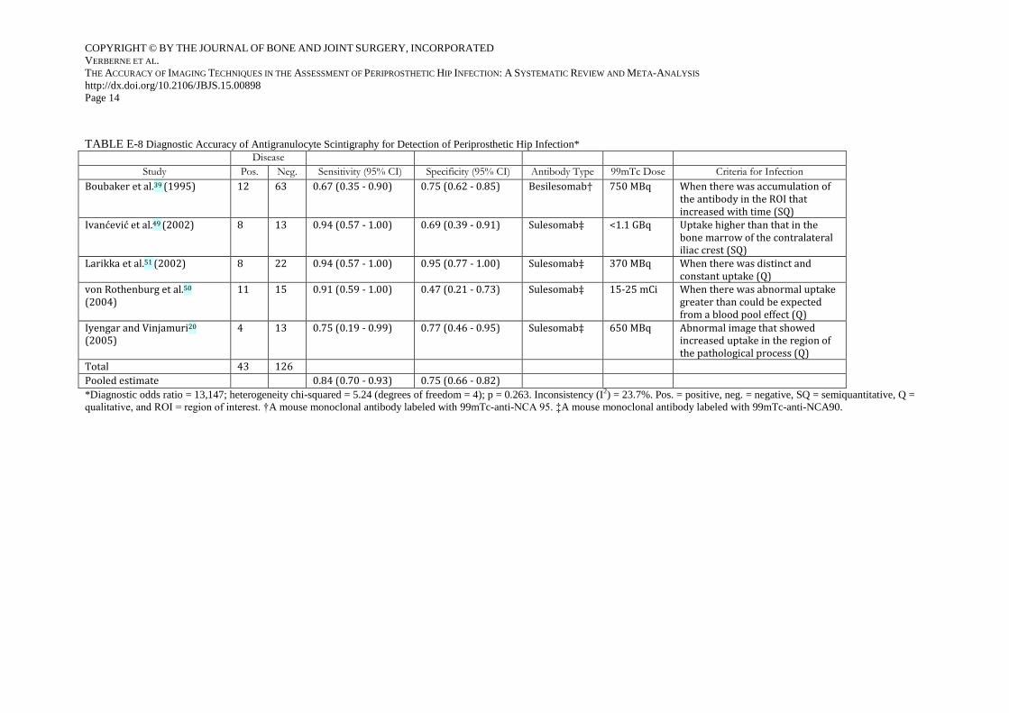
TABLE E-1 Characteristics of the Studies Included in the Meta-Analysis*
Study Design†
No. of Patients
Sex (M/F)
Mean Age (Range) (yr)
No. of Prostheses
Reference Standard
Total No. of Hip Prostheses/No.
Infected
Gómez-Luzuriaga et al.45
(1988)
NR 40 NR NR 40 hip M 40/20
Palestro et al.46
(1990)
Retro. 72 26/46 62.0 (20-87) 92 hip M, IOF 92/23
Kraemer et al.54
(1993)
Retro. 72 42/30 60.0 (21-82) 43 hip M 43/13
Boubaker et al.39
(1995)
Pro. 57 34/23 72.7 (29-92) 78 hip M, IOF, CFU
78/12
Nijhof et al.40
(1997)
NR 226 108/118 54.3 (5-90) 87 hip, 17 knee
M, IOF, A, CFU
87/21
Scher et al.55
(2000)
NR 143 NR 61.0 (26-87) 91 hip, 40 knee
M, H, IOF 91/10
Itasaka et al.43
(2001)
Retro. 46 8/38 67.5 (49-78) 48 hip M, H 48/6
Joseph et al.52
(2001)
Retro. 58 18/40 60.0 (27-82) 36 hip, 22 knee
M, H, IOF 36/9
Larikka et al.42
(2001)
Pro. 64 18/46 NR 64 hip M, IOF 64/6
Zhuang et al.38
(2001)
NR 62 NR NR (27-81) 38 hip, 36 knee
M, IOF, A, CFU
38/10
Ivanćević et al.49
(2002)
Retro. 30 13/17 62.0 (30-85) 21 hip, 6 knee
M, H 21/8
Larikka et al.51
(2002)
NR 30 11/19 NR (25-89) 30 hip M, IOF, A, CFU
30/8
Chacko et al.30
(2002)
Retro. 32 20/12 NR (27-89) 41 hip M, H, IOF, CFU
41/12
Vanquickenborne et al.37 (2003)
Pro. 17 8/9 NR (42-77) 17 hip M, IOF, A, CFU
17/8
El Espera et al.53
(2004)
NR 60 NR NR 45 hip, 28 knee
M, IOF, A 45/6
Love et al.17
(2004)
NR 59 22/37 NR (35-89) 40 hip, 19 knee
M, H, IOF 40/14
Pelosi et al.47
(2004)
Retro. 78 36/42 69.5 (30-87) 47 hip, 35 Knee
M, IOF, CFU
47/21
von Rothenburg et al.50 (2004)
Retro. 38 9/29 71.0 (45-81) 26 hip, 12 knee
M, IOF, A, L
26/11
Stumpe et al.36
(2004)
Pro. 35 12/23 NR (46-81) 35 hip M, H, IOF, A, CFU
35/9
Iyengar and Vinjamuri20
(2005)
Retro. 38 18/20 NR (54-89) 17 hip, 13 knee, 8 other
M, H, IOF, A, I, CFU
17/4
Mumme et al.33
(2005)
NR 50 19/31 68.7 (42-89) 70 hip M, H, CFU 70/48
Reinartz et al.35
(2005)
NR 63 32/31 68.0 (43-88) 92 hip M, H, IOF, CFU
92/8
Pill et al.34 (2006) Pro. 89 NR NR (29-85) 92 hip M, H, S, IOF, A, L
92/21
García-Barrecheguren et al.32 (2007)
Pro. 24 12/12 67.8 (37-81) 24 hip M, H, S, IOF
24/11

COPYRIGHT © BY THE JOURNAL OF BONE AND JOINT SURGERY, INCORPORATED
VERBERNE ET AL.
THE ACCURACY OF IMAGING TECHNIQUES IN THE ASSESSMENT OF PERIPROSTHETIC HIP INFECTION: A SYSTEMATIC REVIEW AND
META-ANALYSIS
http://dx.doi.org/10.2106/JBJS.15.00898
Page 7
Simonsen et al.48
(2007)
Retro. 66 25/41 NR (47-95) 76 hip M, H, IOF, CFU
76/27
Chryssikos et al.31 (2008)
Pro. 113 54/59 NR (31-87) 127 hip M, H, S, IOF, A, L
127/33
Fuster et al.44
(2008)
Pro. 70 28/42 68.0 (NR) 70 hip M, CFU 70/12
Nagoya et al.41
(2008)
NR 46 18/28 NR (28-81) 46 hip M, H, IOF 46/17
Mayer-Wagner et al.21 (2010)
NR 32 13/19 NR (45-90) 15 hip, 22 knee
M 15/8
Basu et al.29
(2014)
Pro. 221 112/109 57.0 (18-84) 134 hip, 87 knee
M, H, IOF 134/33
Kim et al.19
(2014)
Retro. 164 53/111 65.0 (17-82) 71 hip, 93 knee
M, H, IOF, CFU, A
71/26
Total 2,195 1,753/475
*Retro. = retrospective, pro. = prospective, M = microbiology, H = histology, IOF = intraoperative findings, A = aspiration, L =
laboratory (erythrocyte sedimentation rate and C-reactive protein), S = sinus tract, I = imaging, CFU = clinical follow-up of at
least 6 months, and NR = not recorded. †Explicit notation in study.

COPYRIGHT © BY THE JOURNAL OF BONE AND JOINT SURGERY, INCORPORATED
VERBERNE ET AL.
THE ACCURACY OF IMAGING TECHNIQUES IN THE ASSESSMENT OF PERIPROSTHETIC HIP INFECTION: A SYSTEMATIC REVIEW AND META-ANALYSIS
http://dx.doi.org/10.2106/JBJS.15.00898
Page 8
TABLE E-2 Characteristics of the Reference Test(s) and Implants*
Study Hip Prostheses
Fixation (C, U, H)
Total Hip / Hybrid Hip Prosthesis
Mean Age of Hip Prostheses (Range)
Imaging: Minimal Time Postop.
Minimal Duration of Follow-up (mo)
Gómez-Luzuriaga et al.45 NR NR 40/0 4.3 y (1-11 y) >12 mo NR
Palestro et al.46 P68, R24 C92, U0, H0 92/0 4.6 y (1 wk-17 y) >1 wk >6
Kraemer et al.54 NR NR NR 4.5 y (8 mo-13 y) >8 mo NR
Boubaker et al.39 NR C59, U19, H0 NR 6.3 y (7-17 y) >7 mo >8
Nijhof et al.40 NR NR 87/0 NR NR >12
Scher et al.55 NR NR 91/0 47 mo NR NR
Itasaka et al.43 NR C48, U0, H0 48/0 10.9 y (2-18 y) >2 y >24
Joseph et al.52 NR NR 36/0 NR NR NR
Larikka et al.42 NR NR 64/0 6.6 y† (2 mo-22 y) >2 mo >12
Zhuang et al.38 NR NR NR NR (3 mo-8 y) >3 mo >12
Ivanćević et al.49 NR NR 21/0 NR NR >6
Larikka et al.51 NR NR 30/0 12 mo (1 wk-12 y) >1 wk >12
Chacko et al.30 NR NR NR NR >12 mo >9
Vanquickenborne et al.37 P10, R7 NR 17/0 NR (0-14 y) >0 mo >6
El Espera et al.53 NR NR NR 5.4 y ( – ) NR NR
Love et al.17 P30, R10 C14, U10, H16 37/3 NR (1 mo-20 y) >1 mo NR
Pelosi et al.47 NR NR 47/0 NR NR >12
von Rothenburg et al.50 NR C3, U23, H0 NR NR(2 mo-10 y) >2 mo NR
Stumpe et al.36 P26, R9 C18, U17, H0 NR 71 mo (12-260 mo ) >1 y >6
Iyengar and Vinjamuri20 NR NR NR NR NR >12
Mumme et al.33 NR C51, U17, H2 NR 9.2 y, SD 5.7 >12 mo >9
Reinartz et al.35 NR C60, U32, H0 NR NR (1-31 mo) >6 mo (FDG-PET) >9
Pill et al.34 NR NR NR NR NR NR
García-Barrecheguren et al.32 NR NR NR NR >6 mo NR
Simonsen et al.48 P68, R8 C35, U19, H22 62/14 NR 1.5-2 y (0.07-14 y) >12
Chryssikos et al.31 NR NR NR NR >12 mo NR
Fuster et al.44 NR NR 70/0 46 mo >3 mo >12
Nagoya et al.41 NR NR 30/16 8.5 y (1 mo-28 y) >1 mo NR
Mayer-Wagner et al.21 NR NR NR NR NR NR
Basu et al.29 NR NR NR 5.8 y (FDG PET) and 6.4 y (LS) NR >6
Kim et al.19 NR NR NR 3.0 y† (2 wk-32 y) >2 wk >12
*P = primary implant, R = revision, NR = not recorded, C = cemented hip prostheses, U = uncemented hip prostheses, H = hybrid hip prostheses, SD = standard deviation, FDG PET =
fluorodeoxyglucose positron emission tomography, and LS = leukocyte scintigraphy. †The value is given as the median.

COPYRIGHT © BY THE JOURNAL OF BONE AND JOINT SURGERY, INCORPORATED
VERBERNE ET AL.
THE ACCURACY OF IMAGING TECHNIQUES IN THE ASSESSMENT OF PERIPROSTHETIC HIP INFECTION: A SYSTEMATIC REVIEW AND META-ANALYSIS
http://dx.doi.org/10.2106/JBJS.15.00898
Page 9
TABLE E-3 Diagnostic Accuracy of Bone Scintigraphy for Detection of Periprosthetic Hip Infection*
Disease
Study Pos. Neg. Sensitivity (95% CI)
Specificity (95% CI) Tracer Dose (MBq) Criteria for Infection
Boubaker et al.39
(1995)
12 66 0.92 (0.62 - 1.00) 0.64 (0.51 - 0.75) 99mTc-DPD 740 Uptake in both the early and delayed phases
Nijhof et al.40(1997) 13 47 0.96 (0.70 - 1.00) 0.15 (0.06 - 0.28) 99mTc-MDP 600 Uptake in both the blood pool and late phases
Itasaka et al.43 (2001) 6 42 0.83 (0.36 - 1.00) 0.79 (0.63 - 0.90) 99mTc-MDP NR Diffuse increased uptake femoral and/or acetabular (no early phase)
Larikka et al.42 (2001) 6 58 0.92 (0.47 - 1.00) 0.83 (0.71 - 0.91) 99mTc-HDP 550 Uptake in arterial and soft phases
Stumpe et al.36 (2004) 9 25 0.56 (0.21 - 0.86) 0.88 (0.69 - 0.97) 99mTc-DPD 700 Increased uptake in all 3 phases
Reinartz et al.35 2005) 25 67 0.68 (0.46 - 0.85) 0.76 (0.64 - 0.86) 99mTc-HDP 733 Wilson criteria: uptake in both the blood pool and late phases
Mumme et al.33 (2005) 40 30 0.78 (0.62 - 0.89) 0.70 (0.51 - 0.85) 99mTc-HDP 750 Wilson Criteria: uptake in both the blood pool and late phases
Nagoya et al.41 (2008) 17 29 0.88 (0.64 - 0.99) 0.90 (0.73 - 0.98) 99mTc-MDP 740 Increased uptake in all 3 phases
Total 128 364
Pooled estimate 0.80 (0.72 - 0.86) 0.69 (0.64 - 0.73)
*Diagnostic odds ratio = 11,006; heterogeneity chi-squared = 6.66 (degrees of freedom = 7); p = 0.465. Inconsistency (I2) = 0.0%. Pos. = positive, neg. = negative, 95% CI = 95% confidence
interval, 99mTc = 99m-technetium, MDP = methylene diphosphonate, HDP = hydroxymethylene diphosphonate, and DPD = dicarboxy diphosphonate.

COPYRIGHT © BY THE JOURNAL OF BONE AND JOINT SURGERY, INCORPORATED
VERBERNE ET AL.
THE ACCURACY OF IMAGING TECHNIQUES IN THE ASSESSMENT OF PERIPROSTHETIC HIP INFECTION: A SYSTEMATIC REVIEW AND META-ANALYSIS
http://dx.doi.org/10.2106/JBJS.15.00898
Page 10
TABLE E-4 Diagnostic Accuracy of Combined Bone and Gallium Scintigraphy for Detection of Periprosthetic Hip Infection*
Disease
Study Pos. Neg. Sensitivity
(95% CI) Specificity
(95% CI) Tracer Dose Criteria for Infection
Gómez-Luzuriaga et al.45 (1988)
20 20 0.70 (0.46-0.88) 0.90 (0.68-0.99) Tc99m/Ga67 NR When Ga67 image was spatially incongruent with that of Tc99m, or, if spatially congruent, of greater intensity
Kraemer et al.54 (1993) 13 30 0.38 (0.14-0.68) 0.98 (0.86-1.00) Tc99m/Ga67 NR When the gallium uptake was spatially incongruent or spatially congruent but greater than the Tc99m uptake
Itasaka et al.43 (2001) 6 42 0.67 (0.22 -0.96) 0.99 (0.90-1.00) Tc99m/Ga67 NR When Ga67 image showed increased activity of a different distribution (incongruency) or of a relatively greater activity than the focus of the Tc99m scans
Total 49 72
Pooled estimate 0.59 (0.42-0.74) 0.97 (0.91-0.99)
*Diagnostic odds ratio = 34,173; heterogeneity chi-squared = 1.23 (degrees of freedom = 2); p = 0.542. Inconsistency (I2) = 0.0%. Pos. = positive, neg. = negative, Tc99m = 99m-technetium,
Ga67 = gallium-67, and NR = not recorded.

COPYRIGHT © BY THE JOURNAL OF BONE AND JOINT SURGERY, INCORPORATED
VERBERNE ET AL.
THE ACCURACY OF IMAGING TECHNIQUES IN THE ASSESSMENT OF PERIPROSTHETIC HIP INFECTION: A SYSTEMATIC REVIEW AND META-ANALYSIS
http://dx.doi.org/10.2106/JBJS.15.00898
Page 11
TABLE E-5 Diagnostic Accuracy of Combined Bone and Leukocyte Scintigraphy for Detection of Periprosthetic Hip Infection*
Disease
Study Pos. Neg. Sensitivity (95% CI) Specificity (95% CI) Tracer Dose (MBq) Criteria for Infection
Scher et al.55 (2000) 10 81 0.60 (0.26 - 0.88) 0.93 (0.85 - 0.97) 99mTc-HDP/ 111In-Oxine
925 (25 mCi)/14.8-18.5 (400-500 µCi)
When indium scan showed hyperactivity in a different distribution (incongruency) or a relativity greater intensity than the activity on the Tc99 scan
Larikka et al.42 (2001) 5 59 0.91 (0.41 - 1.00) 0.98 (0.91 - 1.00) 99mTc-HDP/ 99mTc-HMPAO
550/370 When periprosthetic leukocyte uptake intensity was higher than that of the bone metabolic image in at least 1 zone, or if uptake was incongruent
Vanquickenborne et al.37 (2003)
8 9 0.88 (0.47 - 1.00) 0.95 (0.60 - 1.00) 99mTc-MDP/ 99mTc-HMPAO
740/185 Moderately or substantial uptake around the hip prosthesis that was higher uptake than that of the contralateral hip region, but only when the lesions were congruous on both scans
Total 23 149
Pooled estimate 0.77 (0.55 - 0.91) 0.95 (0.90 - 0.98)
*Diagnostic odds ratio = 67.336; heterogeneity chi-squared = 3.62 (degrees of freedom = 2); p = 0.164. Inconsistency (I2) = 44.7%. Tc99m = 99m-technetium, HDP = hydroxymethylene
diphosphonate, and 99Tc-HMPAO = hexamethylpropyleneamine oxime.

COPYRIGHT © BY THE JOURNAL OF BONE AND JOINT SURGERY, INCORPORATED
VERBERNE ET AL.
THE ACCURACY OF IMAGING TECHNIQUES IN THE ASSESSMENT OF PERIPROSTHETIC HIP INFECTION: A SYSTEMATIC REVIEW AND META-ANALYSIS
http://dx.doi.org/10.2106/JBJS.15.00898
Page 12
TABLE E-6 Diagnostic Accuracy of Leukocyte Scintigraphy for Detection of Periprosthetic Hip Infection*
Disease
Study Pos. Neg. Sensitivity (95% CI) Specificity (95% CI) Tracer Dose Criteria for Infection
Gómez-Luzuriaga et al.45 (1988)
20 20 0.95 (0.75 - 1.00) 0.90 (0.68 - 0.99) 111In-Oxine NR More intense uptake than that of the contiguous bone and negative otherwise
Palestro et al.46 (1990) 23 69 0.87 (0.66 - 0.97) 0.94 (0.86 - 0.98) 111In-Oxine 18.5 MBq (500 µCi) Activity in the headzone, regardless of activity in any other zone
Pelosi et al.47 (2004) 21 34 0.95 (0.76 - 1.00) 0.97 (0.85 - 1.00) 99Tc-HMPAO 430-600 MBq SQ: Klate > Kearly by at least 10%
Simonsen et al.48
(2007)
27 49 0.81 (0.62 - 0.94) 0.94 (0.83 - 0.99) 111In-Oxine or 99Tc-HMPAO
36 MBq/628 MBq† When periprosthetic uptake was greater than the activity in surrounding bone tissue and in the contralateral site
Fuster et al.44 (2008) 12 58 0.83 (0.52 - 0.98) 0.57 (0.43 - 0.70) 99Tc-HMPAO 185 MBq Any extramedullary periprosthetic focal uptake
Kim et al.19 (2014) 22 49 0.86 (0.65 - 0.97) 0.86 (0.73 - 0.94) 99Tc-HMPAO 740-1100 MBq Increased uptake in the periprosthetic area or if the foci in nearby soft tissue had greater activity than the background soft tissue activity
Total 125 279
Pooled estimate 0.88 (0.81 - 0.93) 0.85 (0.80 - 0.89)
*Diagnostic odds ratio = 60,329; heterogeneity chi-squared = 11.57 (degrees of freedom = 5); p = 0.041. Inconsistency (I2) = 56.8%. Pos. = positive, neg. = negative, NR = not recorded, SQ =
semiquantitative, K = suspected region of infection/reference region (bone marrow), and HMPAO = hexamethylpropyleneamine oxime. †Mean dose.

COPYRIGHT © BY THE JOURNAL OF BONE AND JOINT SURGERY, INCORPORATED
VERBERNE ET AL.
THE ACCURACY OF IMAGING TECHNIQUES IN THE ASSESSMENT OF PERIPROSTHETIC HIP INFECTION: A SYSTEMATIC REVIEW AND META-ANALYSIS
http://dx.doi.org/10.2106/JBJS.15.00898
Page 13
TABLE E-7 Diagnostic Accuracy of Combined Leukocyte and Bone Marrow Scintigraphy for Detection of Periprosthetic Hip Infection*
Disease
Study Pos. Neg. Sensitivity
(95% CI) Specificity
(95% CI) Tracer Dose Criteria for Infection
Palestro et al.46 (1990) 10 40 0.95 (0.63 - 1.00) 0.98 (0.87 - 1.00) 111In-Oxine/99mTc-SC
18.5 MBq (500 µCi)/370 MBq (10 mCi)
Incongruent labeled leukocyte and sulfur colloid images
Joseph et al.52 (2001) 9 27 0.33 (0.07 - 0.70) 0.98 (0.84 - 1.00) 111In-Oxine/99mTc-SC
500 µCi /10 µCi Pattern of activity on the indium image that was not matched on the colloid images
Love et al.17 (2004) 14 26 0.97 (0.72 - 1.00) 0.88 (0.70 - 0.98) 111In-Oxine/99mTc-SC
18.5 MBq (500 µCi)/370 MBq (10 mCi)
Periprosthetic activity on the indium image without corresponding activity on the marrow scan, regardless of intensity or location
El Espera et al.53 (2004) 6 39 0.67 (0.22 - 0.96) 0.97 (0.87 - 1.00) 111In-Oxine/99mTc-SC
30 MBq/185 MBq When increased activity was observed on the leukocyte image, without corresponding uptake on the bone marrow images (incongruent patterns)
Pill et al.34 (2006) 10 41 0.50 (0.19 - 0.81) 0.95 (0.83 - 0.99) 111In-Oxine/99mTc-SC
500 µCi/15 mCi When activity on the leukocyte image was observed, without corresponding activity on the bone marrow scans
Fuster et al.44 (2008) 12 58 0.92 (0.62 - 1.00) 0.98 (0.91 - 1.00) 99Tc-HMPAO/99mTc-SC
185 MBq / 370 MBq All uptake of leukocytes inconsistent with the bone marrow images
Basu et al.29 (2014) 13 46 0.38 (0.14 - 0.68) 0.96 (0.85 - 0.99) 111In-Oxine/99mTc-SC
500 µCi/555 MBq When activity in the periprosthetic region on the leukocyte image was observed, without corresponding activity on the bone marrow images
Total 74 277
Pooled estimate 0.69 (0.58 - 0.79) 0.96 (0.93 - 0.98)
*Diagnostic odds ratio = 65,303; heterogeneity chi-squared = 9.54 (degrees of freedom = 6); p = 0.145. Inconsistency (I2) = 37.1%. Pos. = positive, neg. = negative, 99mTc-SC = 99mTc-sulfur
colloid, and HMPAO = hexamethylpropyleneamine oxime.
COPYRIGHT © BY THE JOURNAL OF BONE AND JOINT SURGERY, INCORPORATED
VERBERNE ET AL.
THE ACCURACY OF IMAGING TECHNIQUES IN THE ASSESSMENT OF PERIPROSTHETIC HIP INFECTION: A SYSTEMATIC REVIEW AND META-ANALYSIS
http://dx.doi.org/10.2106/JBJS.15.00898
Page 14
TABLE E-8 Diagnostic Accuracy of Antigranulocyte Scintigraphy for Detection of Periprosthetic Hip Infection*
Disease
Study Pos. Neg. Sensitivity (95% CI) Specificity (95% CI) Antibody Type 99mTc Dose Criteria for Infection
Boubaker et al.39 (1995) 12 63 0.67 (0.35 - 0.90) 0.75 (0.62 - 0.85) Besilesomab† 750 MBq When there was accumulation of the antibody in the ROI that increased with time (SQ)
Ivanćević et al.49 (2002) 8 13 0.94 (0.57 - 1.00) 0.69 (0.39 - 0.91) Sulesomab‡ <1.1 GBq Uptake higher than that in the bone marrow of the contralateral iliac crest (SQ)
Larikka et al.51 (2002) 8 22 0.94 (0.57 - 1.00) 0.95 (0.77 - 1.00) Sulesomab‡ 370 MBq When there was distinct and constant uptake (Q)
von Rothenburg et al.50
(2004)
11 15 0.91 (0.59 - 1.00) 0.47 (0.21 - 0.73) Sulesomab‡ 15-25 mCi When there was abnormal uptake greater than could be expected from a blood pool effect (Q)
Iyengar and Vinjamuri20
(2005)
4 13 0.75 (0.19 - 0.99) 0.77 (0.46 - 0.95) Sulesomab‡ 650 MBq Abnormal image that showed increased uptake in the region of the pathological process (Q)
Total 43 126
Pooled estimate 0.84 (0.70 - 0.93) 0.75 (0.66 - 0.82)
*Diagnostic odds ratio = 13,147; heterogeneity chi-squared = 5.24 (degrees of freedom = 4); p = 0.263. Inconsistency (I2) = 23.7%. Pos. = positive, neg. = negative, SQ = semiquantitative, Q =
qualitative, and ROI = region of interest. †A mouse monoclonal antibody labeled with 99mTc-anti-NCA 95. ‡A mouse monoclonal antibody labeled with 99mTc-anti-NCA90.

COPYRIGHT © BY THE JOURNAL OF BONE AND JOINT SURGERY, INCORPORATED
VERBERNE ET AL.
THE ACCURACY OF IMAGING TECHNIQUES IN THE ASSESSMENT OF PERIPROSTHETIC HIP INFECTION: A SYSTEMATIC REVIEW AND META-ANALYSIS
http://dx.doi.org/10.2106/JBJS.15.00898
Page 15
TABLE E-9 Diagnostic Accuracy of FDG PET for Detection of Periprosthetic Hip Infection
Disease
Study Pos. Neg. Sensitivity(95% CI) Specificity
(95% CI) Tracer Dose Criteria for Infection
Zhuang et al.38 (2001) 10 28 0.90 (0.55 - 1.00) 0.89 (0.72 - 0.98) 18F-FDG 4.22 - 4.56 MBq/kg
Increased uptake in the BPI compared with PST, not if uptake was limited to only femoral head or neck
Chacko et al.30 (2002) 12 29 0.92 (0.62 - 1.00) 0.97 (0.82 - 1.00) 18F-FDG 2.52 MBq/kg Uptake in the BPI
Vanquickenborne et al.37
(2003)
8 9 0.88 (0.47 - 1.00) 0.78 (0.40 - 0.97) 18F-FDG 370 MBq Moderately or substantially higher uptake than contralateral distal femur
Love et al.17 (2004) 14 26 0.43 (0.18 - 0.71) 0.96 (0.80 - 1.00) 18F-FDG 150 – 20 MBq Semiquantitative analysis of BPI activity in femur with T/B ratio
Stumpe et al.36 (2004) 9 26 0.33 (0.07 - 0.70) 0.81 (0.61 - 0.93) 18F-FDG 300 - 400 MBq Diffusely increased uptake in BPI, less than or comparable with bladder uptake
Mumme et al.33 (2005) 46 24 0.91 (0.79 - 0.98) 0.92 (0.73 - 0.99) 18F-FDG 200-250 MBq Increased uptake in the BPI and PST
Reinartz et al.35 (2005) 33 59 0.94 (0.80 - 0.99) 0.95 (0.86 - 0.99) 18F-FDG 283 ± 38 MBq Uptake in the PST
Pill et al.34 (2006) 21 71 0.95 (0.76 - 1.00) 0.93 (0.84 - 0.98) 18F-FDG 140 µCi/kg Uptake in BPI, not if uptake was limited to only PST or neck of prosthesis
García-Barrecheguren et al.32 (2007)
11 13 0.64 (0.31 - 0.89) 0.62 (0.32 - 0.86) 18F-FDG 10 - 15 mCi Uptake in BPI, more than PST, fistulous or synovial uptake
Chryssikos et al.31 (2008) 33 94 0.85 (0.68 - 0.95) 0.93 (0.85 - 0.97) 18F-FDG 140 µCi/kg Uptake in BPI, not if uptake was limited to only PST or neck of prosthesis
Mayer-Wagner et al.21
(2010)
8 7 0.75 (0.35 - 0.97) 0.71 (0.29 - 0.96) 18F-FDG 180 MBq Uptake in BPI, not if uptake was limited to tip of stem or neck
Basu et al.29 (2014) 33 101 0.82 (0.65 - 0.93) 0.93 (0.86 - 0.97) 18F-FDG 0.14 mCi/kg Uptake in BPI in middle portion of stem, not if uptake was limited to only PST, neck, or synovial uptake
Total 238 487
Pooled estimate 0.83 (0.77 - 0.87) 0.91 (0.89 - 0.94)
*Diagnostic odds ratio = 38,189; heterogeneity chi-squared = 36.28 (degrees of freedom = 11); p < 0.0001. Inconsistency (I2) = 69.7%. 18F-FDG = fluorodeoxyglucose, BPI = bone-prosthesis
interface, T/B = target-to-background, and PST = periprosthetic soft tissue.

COPYRIGHT © BY THE JOURNAL OF BONE AND JOINT SURGERY, INCORPORATED
VERBERNE ET AL.
THE ACCURACY OF IMAGING TECHNIQUES IN THE ASSESSMENT OF PERIPROSTHETIC HIP INFECTION: A SYSTEMATIC REVIEW AND META-ANALYSIS
http://dx.doi.org/10.2106/JBJS.15.00898
Page 16
TABLE E-10 Search String Used for Identifying Studies on Diagnostic Imaging in Diagnosing Periprosthetic Hip Infection
MEDLINE: (((((((("Hip"[Mesh] OR "Hip Joint"[Mesh]) OR "Femur"[Mesh]) OR "Acetabulum"[Mesh]) OR (hip[tiab]) OR (hips[tiab]) OR (femur*[tiab])
OR (femoral[tiab]) OR (coxa*[tiab]) OR (Trochanter*[tiab]) OR (acetabul*[tiab]) OR (Cotyloid Cavit*[tiab]) OR (cups[tiab]) OR (cup[tiab]) OR
(stem[tiab]) OR (stems[tiab]) OR (socket*[tiab]))) AND ((((((((((((("Prostheses and Implants"[Mesh:noexp]) OR "Joint
Prosthesis"[Mesh:noexp]) OR "Hip Prosthesis"[Mesh]) OR "Metal-on-Metal Joint Prostheses"[Mesh]) OR "Prosthesis
Implantation"[Mesh:noexp]) OR "Arthroplasty, Replacement"[Mesh:noexp])) OR prosthe*[tiab]) OR implant*[tiab]) OR arthroplast*[tiab]) OR
replac*[tiab])) OR metal-on-metal[tiab])) AND ((((((((((((((("Infection"[Mesh]) OR "Biofilms"[Mesh]) OR "Bacterial Adhesion"[Mesh]) OR
"Adhesins, Bacterial"[Mesh]) OR "Bacterial Infections"[Mesh])) OR infect*[tiab]) OR sepsi*[tiab]) OR septi*[tiab]) OR bacterie*[tiab]) OR
bacteria*[tiab]) OR biofilm*[tiab]) OR (bacterem*[tiab] OR bacteraem*[tiab]))) OR inflammat*[tiab]))) AND ((((((((("Magnetic Resonance
Imaging"[Mesh] OR ("magnetic resonance"[tiab] AND (image[tiab] OR images[tiab] OR imaging[tiab])) OR zeugmatograph*[tiab] OR
MRI*[tiab] OR NMR*[tiab] OR Magnetization*[tiab] OR Magnetisation*[tiab] OR fMRI*[tiab] OR MR imag*[tiab])) OR (Magnetic
Resonance Spectroscopy[Mesh] AND (chemical shift[tiab] AND imag*[tiab])))) OR (((((("ultrasonography"[Subheading] OR
"Ultrasonography"[Mesh])) OR ultraso*[tiab]) OR echograph*[tiab]) OR sonograph*[tiab]) OR echotomograph*[tiab])) OR
((((((((("Radionuclide Imaging"[Mesh]) OR "radionuclide imaging"[Subheading]) OR "Radiopharmaceuticals"[Pharmacological Action])) OR
(scintigraph*[tiab] OR scintiphotograph*[tiab])) OR (radionuclid*[tiab] OR radioisotop*[tiab])) OR Gamma Camera Imag*[tiab]) OR
(indium[tiab] OR in-111[tiab] OR 111in*[tiab])) OR (99mtc[tiab] OR 99m[tiab]) OR scintiscan*[tiab])) OR (((((((((("Positron-Emission
Tomography"[Mesh]) OR "Tomography, Emission-Computed"[Mesh])) OR "Tomography"[Mesh:noexp])) OR PET[tiab]) OR
tomograph*[tiab]) OR ((("Fluorodeoxyglucose F18"[Mesh]) OR "Deoxyglucose"[Mesh]))) OR (deoxyglucose[tiab] OR deoxy-glucose[tiab] OR
2deoxyglucose[tiab])) OR (fluorin*[tiab] OR fluoro[tiab] OR fluorodeoxyglucose[tiab] OR 18F[tiab] OR 18FDG[tiab] OR F-18DG[tiab]))) OR
((((("Tomography, X-Ray Computed"[Mesh]) OR (compute*[tiab] AND tomograph*[tiab])) OR (CT[tiab] OR CTs[tiab] OR CT's[tiab])) OR
electron beam[tiab]) OR Tomodensitometr*[tiab])) OR (((((((((((((("Radiography"[Mesh]) OR "radiography"[Subheading])) OR
radiograph*[tiab]) OR x-ray*[tiab]) OR x-radiograph*[tiab]) OR xray*[tiab]) OR Zonograph*[tiab]) OR fluoroscop*[tiab]) OR
arthrograph*[tiab]) OR roentgen*[tiab]) OR rontgen*[tiab])) OR image subtraction*[tiab]))
EMBASE: (('radiography'/exp OR ('fluoroscopy'/exp OR 'xray'/exp OR 'image subtraction'/exp) OR (xradiograph*:ab,ti OR xray*:ab,ti OR zonograph*:ab,ti
OR fluoroscop*:ab,ti OR arthrograph*:ab,ti OR roentgen*:ab,ti OR rontgen*:ab,ti) OR (image NEAR/1 (subtraction OR subtractions)):ab,ti) OR
('computer assisted tomography'/exp OR (compute*:ab,ti AND tomograph*:ab,ti) OR (ct*:ab,ti OR (electron AND beam:ab,ti) OR
tomodensitometr*:ab,ti)) OR (('emission tomography'/exp OR 'tomography'/de OR 'fluorodeoxyglucose f 18'/exp OR 'deoxyglucose'/exp) OR

COPYRIGHT © BY THE JOURNAL OF BONE AND JOINT SURGERY, INCORPORATED
VERBERNE ET AL.
THE ACCURACY OF IMAGING TECHNIQUES IN THE ASSESSMENT OF PERIPROSTHETIC HIP INFECTION: A SYSTEMATIC REVIEW AND META-ANALYSIS
http://dx.doi.org/10.2106/JBJS.15.00898
Page 17
(pet:ab,ti OR tomograph*:ab,ti OR deoxyglucose:ab,ti OR 2deoxyglucose:ab,ti OR fluorin*:ab,ti OR fluoro:ab,ti OR fluorodeoxyglucose:ab,ti
OR 18f:ab,ti OR 18fdg:ab,ti OR f18dg:ab,ti)) OR (('radioisotope diagnosis'/exp OR 'radiopharmaceutical agent'/exp OR 'radioisotope'/exp) OR
(scintigraph*:ab,ti OR scintiphotograph*:ab,ti OR radionuclid*:ab,ti OR radioisotop*:ab,ti OR indium:ab,ti OR in111:ab,ti OR 111in*:ab,ti OR
99mtc:ab,ti OR 99m:ab,ti) OR ('gamma camera image':ab,ti OR 'gamma camera images':ab,ti OR 'gamma camera imaging':ab,ti)) OR
('echography'/exp OR (ultraso*:ab,ti OR echograph*:ab,ti OR sonograph*:ab,ti OR echotomograph*:ab,ti)) OR ('nuclear magnetic resonance
imaging'/exp OR (magnetic AND resonance:ab,ti AND (image:ab,ti OR images:ab,ti OR imaging:ab,ti) OR zeugmatograph*:ab,ti OR mri*:ab,ti
OR nmr*:ab,ti OR magnetization*:ab,ti OR magnetisation*:ab,ti OR fmri*:ab,ti OR mr AND imag*:ab,ti))) AND (('hip prosthesis'/exp OR 'joint
prosthesis'/de OR 'metal on metal joint prosthesis'/exp OR 'prosthesis'/de OR 'implantation'/de OR 'metal implantation'/exp OR 'hip
arthroplasty'/exp) OR (prosthe*:ab,ti OR implant*:ab,ti OR arthroplast*:ab,ti OR replac*:ab,ti OR 'metal on metal':ab,ti)) AND
(('acetabulum'/exp OR 'hip'/exp OR 'femur'/exp) OR (hip:ab,ti OR hips:ab,ti OR femur*:ab,ti OR femoral:ab,ti OR coxa*:ab,ti OR
trochanter*:ab,ti OR acetabul*:ab,ti OR cups:ab,ti OR cup:ab,ti OR stem:ab,ti OR stems:ab,ti OR socket*:ab,ti) OR (cotyloid:ab,ti AND
cavit*:ab,ti)) AND (('infection'/exp OR 'biofilm'/exp OR 'bacterium adherence'/exp OR 'adhesin'/exp) OR (infect*:ab,ti OR sepsi*:ab,ti OR
septi*:ab,ti OR bacterie*:ab,ti OR bacteria*:ab,ti OR biofilm*:ab,ti OR bacterem*:ab,ti AND orbacteraem*:ab,ti OR inflammat*:ab,ti))

COPYRIGHT © BY THE JOURNAL OF BONE AND JOINT SURGERY, INCORPORATED
VERBERNE ET AL.
THE ACCURACY OF IMAGING TECHNIQUES IN THE ASSESSMENT OF PERIPROSTHETIC HIP INFECTION: A SYSTEMATIC REVIEW AND META-ANALYSIS
http://dx.doi.org/10.2106/JBJS.15.00898
Page 18
TABLE E-11 Results of Search for Studies on Diagnostic Imaging in Diagnosing Periprosthetic Hip Infection
No. Search String
MEDLINE* (no. of studies)
Embase* (no. of studies)
1 Hip prostheses 74,103 91,982
2 Infected hip prostheses 11,341 16,476
3 Radiography or MRI or CT or scintigraphy or ultrasonography 2,305,863 3,834,824
4 No. 1 and no. 2 and no. 3 3,451 3,344
*Search conducted on April 1, 2015.

COPYRIGHT © BY THE JOURNAL OF BONE AND JOINT SURGERY, INCORPORATED
VERBERNE ET AL.
THE ACCURACY OF IMAGING TECHNIQUES IN THE ASSESSMENT OF PERIPROSTHETIC HIP INFECTION: A SYSTEMATIC REVIEW AND META-ANALYSIS
http://dx.doi.org/10.2106/JBJS.15.00898
Page 19
TABLE E-12 Analysis of the Interpretation Criteria Used in the Included Studies of FDG PET for Diagnosing Periprosthetic Hip Infection Using the Z-Test*
Criteria for Positivity for Index Test Sensitivity
(95% CI) Comparison
(p value) Specificity
(95% CI) Comparison
(p value)
1. BPI without limitation of location of uptake 0.87 (0.79 - 0.92) 0.91 (0.85 - 0.95)
2. BPI with limitation of location of uptake 0.79 (0.71 - 0.86) 0.91 (0.88 - 0.94)
1 versus 2 0.2973 1.0000
3. BPI uptake compared with uptake in bladder 0.33 (0.07 - 0.70) 0.81 (0.63 - 0.93)
4. BPI uptake not compared with uptake in bladder 0.85 (0.79 - 0.89) 0.92 (0.89 - 0.94)
3 versus 4 0.0064 0.0714
5. Only uptake in PST 0.94 (0.80 - 0.99) 0.95 (0.86 - 0.99)
6. Uptake not only in PST 0.81 (0.75 - 0.86) 0.91 (0.88 - 0.93)
7. BPI uptake > uptake PST 0.76 (0.53 - 0.92) 0.81 (0.65 - 0.91)
8. BPI without comparison uptake PST 0.83 (0.78 - 0.88) 0.92 (0.89 - 0.95)
5 versus 6 0.1207 0.3843
5 versus 7 0.1135 0.0588
5 versus 8 0.1648 0.4985
6 versus 7 0.6315 0.0594
7 versus 8 0.4855 0.0403
9. BPI uptake only in femoral component 0.81 (0.65 - 0.91) 0.92 (0.85 - 0.96)
10. BPI uptake not limited to uptake in femoral component
0.83 (0.77 - 0.88) 0.91 (0.88 - 0.94)
9 versus 10 0.7759 0.7570
*FDG PET = fluorodeoxyglucose positron emission tomography, BPI = bone-prosthesis interface, and PST = periprosthetic soft tissue.
COPYRIGHT © BY THE JOURNAL OF BONE AND JOINT SURGERY, INCORPORATED
VERBERNE ET AL.
THE ACCURACY OF IMAGING TECHNIQUES IN THE ASSESSMENT OF PERIPROSTHETIC HIP INFECTION: A SYSTEMATIC REVIEW AND META-ANALYSIS
http://dx.doi.org/10.2106/JBJS.15.00898
Page 20
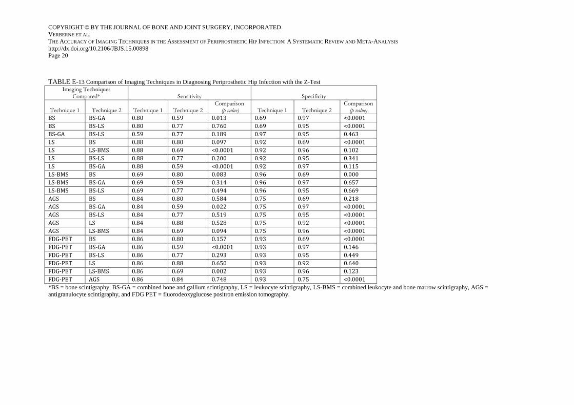
TABLE E-13 Comparison of Imaging Techniques in Diagnosing Periprosthetic Hip Infection with the Z-Test
Imaging Techniques Compared* Sensitivity Specificity
Technique 1 Technique 2 Technique 1 Technique 2 Comparison
(p value) Technique 1 Technique 2 Comparison
(p value)
BS BS-GA 0.80 0.59 0.013 0.69 0.97 <0.0001
BS BS-LS 0.80 0.77 0.760 0.69 0.95 <0.0001
BS-GA BS-LS 0.59 0.77 0.189 0.97 0.95 0.463
LS BS 0.88 0.80 0.097 0.92 0.69 <0.0001
LS LS-BMS 0.88 0.69 <0.0001 0.92 0.96 0.102
LS BS-LS 0.88 0.77 0.200 0.92 0.95 0.341
LS BS-GA 0.88 0.59 <0.0001 0.92 0.97 0.115
LS-BMS BS 0.69 0.80 0.083 0.96 0.69 0.000
LS-BMS BS-GA 0.69 0.59 0.314 0.96 0.97 0.657
LS-BMS BS-LS 0.69 0.77 0.494 0.96 0.95 0.669
AGS BS 0.84 0.80 0.584 0.75 0.69 0.218
AGS BS-GA 0.84 0.59 0.022 0.75 0.97 <0.0001
AGS BS-LS 0.84 0.77 0.519 0.75 0.95 <0.0001
AGS LS 0.84 0.88 0.528 0.75 0.92 <0.0001
AGS LS-BMS 0.84 0.69 0.094 0.75 0.96 <0.0001
FDG-PET BS 0.86 0.80 0.157 0.93 0.69 <0.0001
FDG-PET BS-GA 0.86 0.59 <0.0001 0.93 0.97 0.146
FDG-PET BS-LS 0.86 0.77 0.293 0.93 0.95 0.449
FDG-PET LS 0.86 0.88 0.650 0.93 0.92 0.640
FDG-PET LS-BMS 0.86 0.69 0.002 0.93 0.96 0.123
FDG-PET AGS 0.86 0.84 0.748 0.93 0.75 <0.0001
*BS = bone scintigraphy, BS-GA = combined bone and gallium scintigraphy, LS = leukocyte scintigraphy, LS-BMS = combined leukocyte and bone marrow scintigraphy, AGS =
antigranulocyte scintigraphy, and FDG PET = fluorodeoxyglucose positron emission tomography.

COPYRIGHT © BY THE JOURNAL OF BONE AND JOINT SURGERY, INCORPORATED
VERBERNE ET AL.
THE ACCURACY OF IMAGING TECHNIQUES IN THE ASSESSMENT OF PERIPROSTHETIC HIP INFECTION: A SYSTEMATIC REVIEW AND META-ANALYSIS
http://dx.doi.org/10.2106/JBJS.15.00898
Page 21
TABLE E-14 QUADAS-2 Evaluation
Risk of Bias* Applicability Concerns*
Study
Patient Selection Index Test
Reference Standard
Flow and Timing
Patient Selection Index Test
Reference Standard
Gómez-Luzuruaga et al.45 2 2 0 2 0 2 1
Palestro et al.46 0 1 1 0 0 1 1
Kraemer et al.54 0 0 0 0 1 1 1
Boubaker et al.39 0 2 0 0 1 1 1
Nijhof et al.40 0 2 0 1 2 1 1
Scher et al.55 1 0 0 1 1 1 1
Itasaka et al.43 0 0 1 0 1 1 1
Joseph et al.52 1 1 2 1 1 1 1
Larikka et al.42 1 0 2 1 1 1 1
Zhuang et al.382 1 1 1 0 1 1 1
Ivanćević et al.49 1 1 1 1 1 1 1
Larikka et al.51 0 2 0 1 1 1 1
Chacko et al.30 0 1 0 0 1 1 1
Vanquickenborne et al.37 2 2 2 2 1 1 0
El Espera et al.53 0 1 1 1 1 1 1
Love et al.17 2 1 0 0 0 1 1
Pelosi et al.47 2 1 0 0 1 1 1
von Rothenburg et al.50 0 0 0 0 1 1 1
Stumpe et al.36 1 1 0 1 1 1 1
Iyengar and Vinjamuri20 0 0 2 2 0 1 1
Mumme et al.33 0 1 0 1 1 1 1
Reinartz et al.35 1 1 0 1 1 1 1
Pill et al.34 0 0 2 0 1 2 2
García-Barrecheguren et al.32 2 0 0 1 1 1 1

COPYRIGHT © BY THE JOURNAL OF BONE AND JOINT SURGERY, INCORPORATED
VERBERNE ET AL.
THE ACCURACY OF IMAGING TECHNIQUES IN THE ASSESSMENT OF PERIPROSTHETIC HIP INFECTION: A SYSTEMATIC REVIEW AND META-ANALYSIS
http://dx.doi.org/10.2106/JBJS.15.00898
Page 22
Simonsen et al.48 0 0 0 0 1 0 1
Chryssikos et al.31 0 2 0 0 1 1 1
Fuster et al.44 0 0 1 1 1 1 1
Nagoya et al.41 1 0 0 1 1 1 1
Mayer-Wagner et al.21 2 1 0 1 1 1 1
Basu et al.29 1 1 0 1 1 1 0
Kim et al.19 0 1 1 0 1 1 1
*0 indicates unclear risk, 1 indicates low risk, and 2 indicates high risk.


