
Fluorescent Angiography: Practical uses in the Clinical Setting
Charles Andersen MD, FACS, MAPWCAChief Vascular/Endovascular/ Limb Preservation Surgery Service
(Emeritus) Chief of Wound Care Service
Madigan Army Medical CenterClinical Professor of Surgery UW, USUHS
Disclosures
• No relevant financial relationship reported

Perspective
•Practical Uses in the clinical setting• Fluorescent Angiographic Studies provide information that can be
useful in the clinical setting when combined with other clinical information
• Fluorescent Angiography has become an important tool in our Wound Care and Limb Preservation Service
t

Traditional measurement of Tissue Perfusion
• Clinical Judgment• Physical exam• ABIs, Toe Pressures, Toe Wave Forms• Forefoot PVR• Duplex scan• tcP02• SPP
Measurement of Tissue Perfusion• Current methods utilized to evaluate tissue perfusion are often limited by
• medial calcinosis• scarring• wounds• prior amputations• infection
• Current methods can be technically challenging, costly and time consuming and don’t measure global perfusion of the foot• Fluorescence Angiography offers an additional option to measure tissue perfusion

Clinical Role of Fluorescence Angiography
• Utilized in outpatient clinic • Is there adequate perfusion to heal a wound• Does the patient require revascularization to heal a wound or prior to a minor
foot amputation?• Was revascularization successful in improving perfusion to the foot?• Predict what level of minor foot amputation will heal?
• Utilized in OR üSpy assisted Amputation
üPrevention of suture line complications

Fluorescence Angiography
Visualize and quantitate micro-circulation

Fluorescence Angiography
Fluorescent dye ICG) is injected IV
The injected agent lights up blood flowing through the veins and arteries in real time, and the camera captures live images of the patient’s vasculature.
These images can be captured on a computer screen, analyzed and saved and printed for medical reference.

Practical Uses in the Clinical SettingHeel Ulcers
Fluorescent Angiography provides perfusion assessment of the Heel that can’t be obtained with traditional methods of measuring perfusion
Perfusion Assessment in Heel Ulcers
• Measurement of tissue perfusion can help assess the healing potential in patients with heel ulceration• Traditional methods of measuring tissue perfusion are a poor indicator
of heel perfusion. • Fluorescence angiography can measure tissue perfusion in a heel ulcer
Malik R, Pinto P, Bogaisky M, et al. Older adults with heel ulcers in the acute care setting: frequency of noninvasive vascular assessment, surgical intervention, and 1-year mortality. J Am Med Dir Assoc. 2013;14(12):916-919.
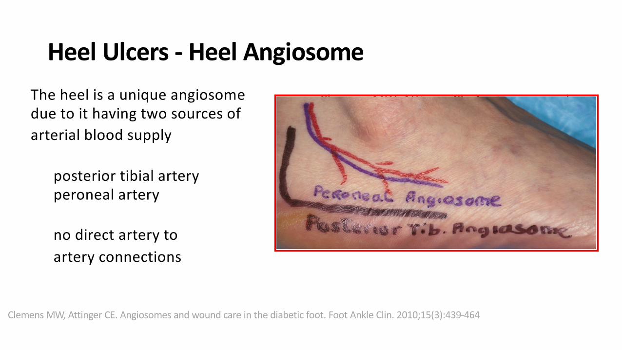
Heel Ulcers - Heel AngiosomeThe heel is a unique angiosomedue to it having two sources of arterial blood supply
posterior tibial artery peroneal artery
no direct artery to artery connections
Clemens MW, Attinger CE. Angiosomes and wound care in the diabetic foot. Foot Ankle Clin. 2010;15(Clemens MW, Attinger CE. Angiosomes and wound care in the diabetic foot. Foot Ankle Clin. 2010;15(3):439
Clemens MW, Attinger CE. Angiosomes and wound care in the diabetic foot. Foot Ankle Clin. 2010;15(3):439-464

Heel Ulcers“Orphan Heal Syndrome”• Regional malperfusion of the heel has been termed
“Orphan Heel Syndrome”• Most common in patients with diabetes and/or renal failure
The role of fluorescein angiography in the management of orphan heel syndrome Authors: Nicole Byerley, DPM*, Col (Ret) Charles A. Andersen, MD, FACS, FAPWCA1, Mario N. Ponticello, DPM, FACFAS, FAPWCA2, LTC, MC, Peter Kreishman, MD3 The Journal of DiabeticFoot Complications, 2016
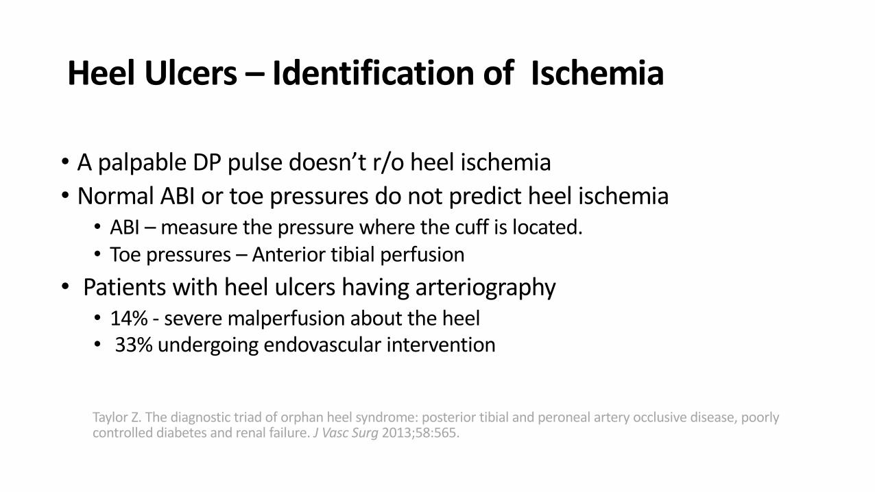
Heel Ulcers – Identification of Ischemia
• A palpable DP pulse doesn’t r/o heel ischemia• Normal ABI or toe pressures do not predict heel ischemia• ABI – measure the pressure where the cuff is located.• Toe pressures – Anterior tibial perfusion
• Patients with heel ulcers having arteriography• 14% - severe malperfusion about the heel • 33% undergoing endovascular intervention
Taylor Z. The diagnostic triad of orphan heel syndrome: posterior tibial and peroneal artery occlusive disease, poorly controlled diabetes and renal failure. J Vasc Surg 2013;58:565.

Case Study
• 81 year- old female with a history of poorly-controlled insulin-dependent DM type II with neuropathy with a painful right postero-lateral heel ulceration that had been present for three weeks• Physical Exam è non-palpable pedal pulses• ABI – 0.5

Presentation

Pre RevascularizationFluorescence Angiogram
• Pre revascularization No fluorescence in wound bed and minimal inflammatory response• Heel demonstrates blotchy uptake
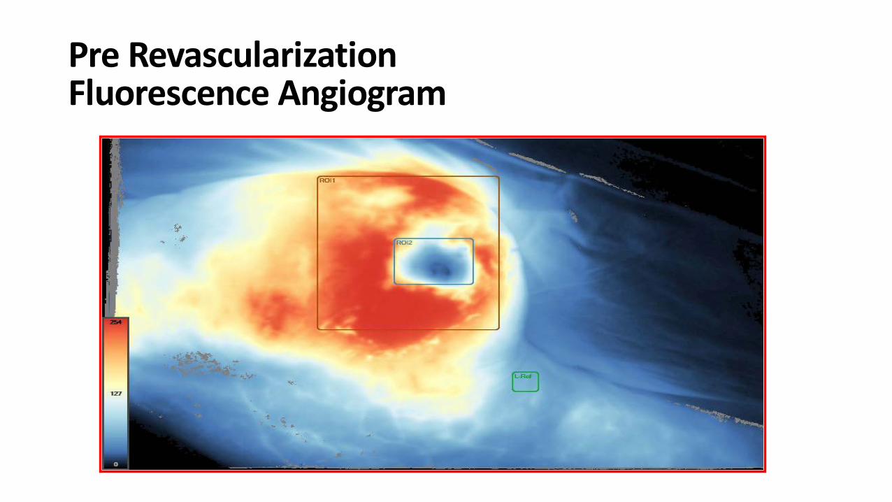
Pre RevascularizationFluorescence Angiogram

Revascularization
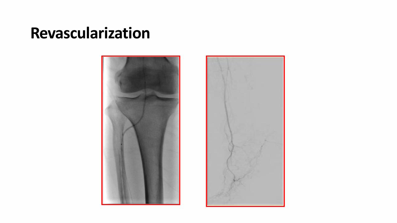
• An arteriogram demonstrated popliteal occlusion and severe infra-popliteal disease • Popliteal stent and angioplasty of the tibial peroneal trunk• The only runoff vessel was a peroneal artery reconstituting the distal
dosalis pedis artery. The posterior tibial artery was totally occluded• Indirect revascularization – Is there perfusion to the heel?

Revascularization
Revascularization
Revascularization
• Post Stenting, arterial flow to the foot was improved with a triphasic dorsalis pedis arterial signal and after several days a monophasic posterior tibial signal. • Question – what is the perfusion to the heel
• Fluorescence angiography demonstrated improved perfusion to the heel with an improved inflammatory response and increased uptake to the wound bed.

Post Revascularization - Fluorescence Angiogram

One Month Post Revascularizations

Changes in Perfusion
Global Ingress Wound Ingress6/21/2016 2.4 8.47/22/2016 7.0 7.79/01/2016 1.9 15.1

Healed Ulcer
Ulcer healed 6 weeks post stenting

Practical use Fluorescence Angiography in Heel Ulcers
• Documented severe ischemia of heel and the need for revascularization• Documented adequate perfusion to the heel following indirect
revascularization• With indirect revascularization, documented the increased perfusion
over time• Appropriate identification of regional ischemia and revascularization
can prevent major amputation or support calcanectomy in more severe ulceration

Practical Uses in the Clinical SettingDigital Amputations• Significant incidence of readmission and revision to higher levels of
amputation following digital amputations• Significant incidence of suture line complications• Fluorescence angiography pre and intra op may decrease these
complications
Predictors of hospital readmissions after lower extremity amputations www.jvascsurg.org/article/S0741-5214(15)01967-9
Andersen et al publication pending
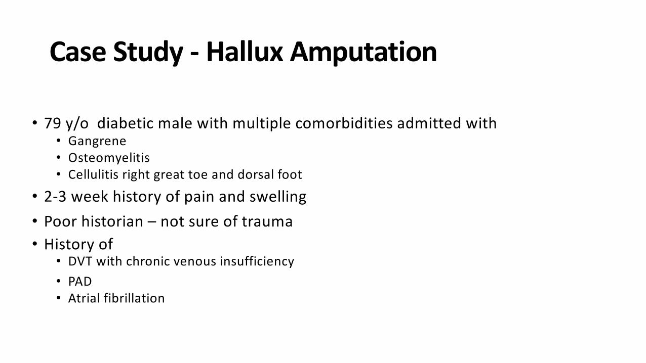
Case Study - Hallux Amputation
• 79 y/o diabetic male with multiple comorbidities admitted with • Gangrene• Osteomyelitis• Cellulitis right great toe and dorsal foot
• 2-3 week history of pain and swelling• Poor historian – not sure of trauma• History of
• DVT with chronic venous insufficiency• PAD• Atrial fibrillation

Case Study - continued• Positive blood cultures for MRSA• Started on broad spectrum IV antibiotics with resolution of cellulitis
on dorsum of foot• Vascular assessment• Fluorescence angiography• Right hallux amputation

Pre-op
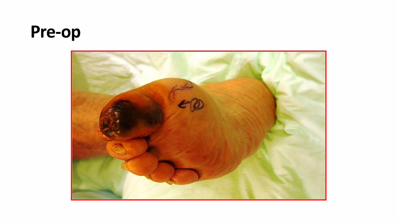
Pre-op

Pre-op MRI
• Abscess surrounding the flexor hallucis longus• Tendon is concerning for infective tenosynovitis •Moderate osteoarthritis of the right foot

Pre op Vascular Assessment
• ABIs – non compressible vessels with ABIs greater than 1.5• Biphasic wave forms at right ankle• Right toe pressure not obtainable
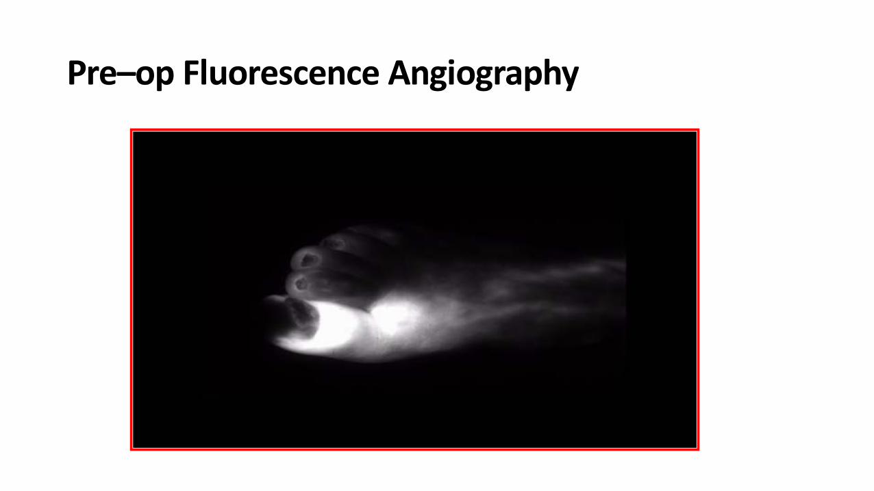
Pre–op Fluorescence Angiography
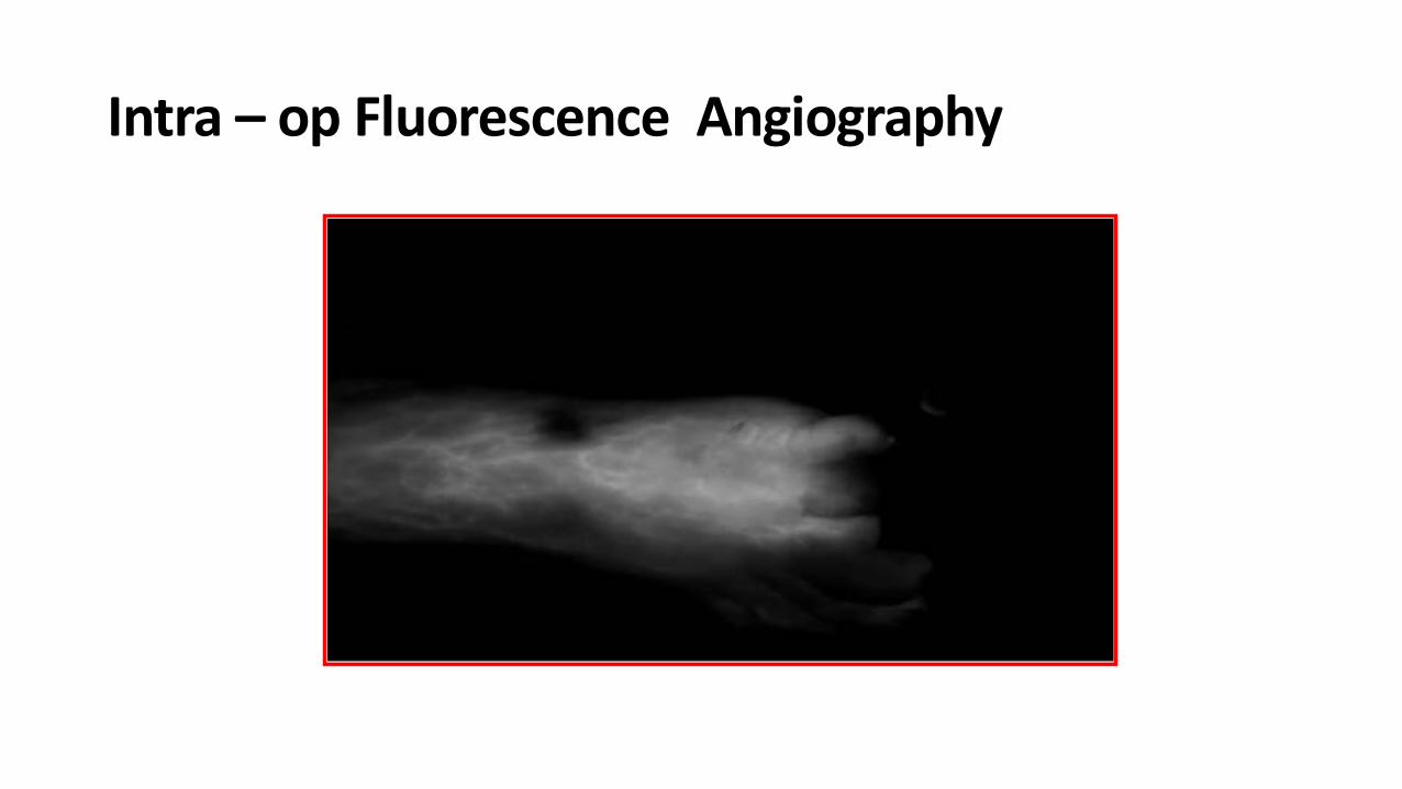
Intra – op Fluorescence Angiography

Amputation
Flap revision following intra-opFluorescence Angiography

Amputation
Amputation following excision of ischemic distal flap

Amputation
Flap Closure

Fluorescent Angiography Following Closure
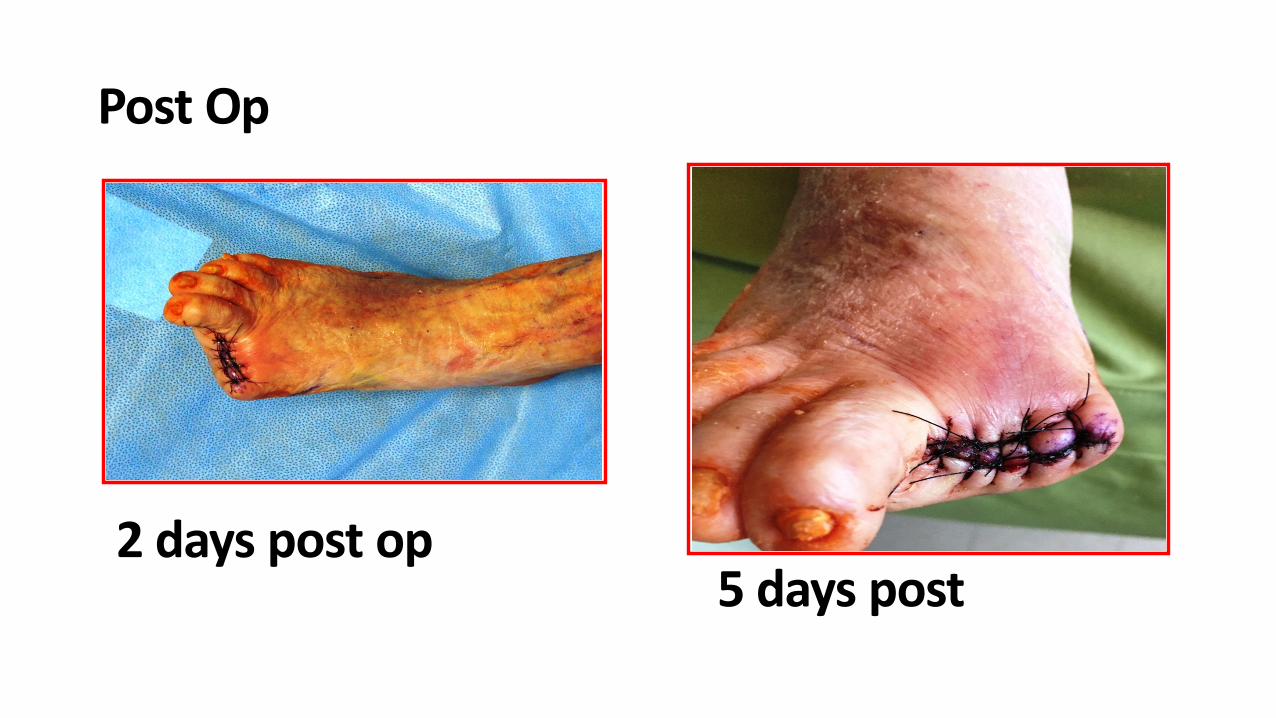
Post Op
2 days post op5 days post
Practical Uses in the Clinical SettingTMA• Historically TMAs have up to a 50% incidence of suture line
complications• Can the use of Fluorescence Angiography decrease the suture line
complication rate?
Case Study - TMA • Severe poorly controlled diabetes• S/P hallux amputation with severe deformity with recurrent
ulceration and cellulitis• Vascular studies and fluorescent angiography demonstrated adequate
perfusion to heal a TMA• Elective TMA
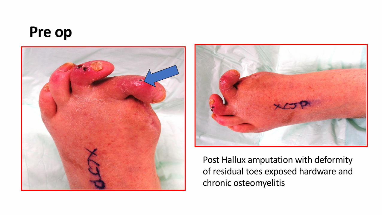
Pre op
Post Hallux amputation with deformity of residual toes exposed hardware and chronic osteomyelitis

Fluorescent Angiography Assisted Amputations
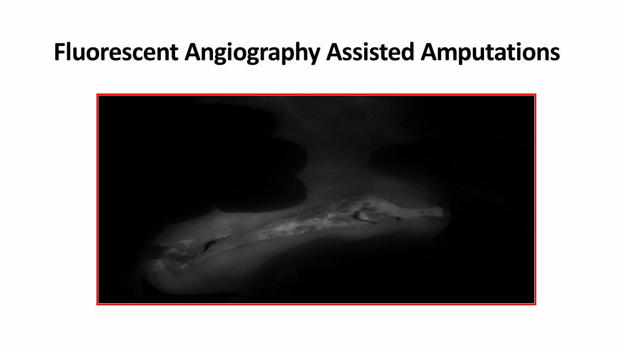
Fluorescent Angiography Assisted Amputations

Suture Line Modification

TMA

Ten days post op

Two months post op

Conclusions
• Fluorescence Angiography has become an important component of our Limb Preservation/Wound Care practice• Fast and accurate evaluation of tissue perfusion of the foot• Fluorescence Angiography can assess the need for
revascularization and document the post procedure results• Assesses perfusion to determine amputation level• Help prevent suture line complications with amputations

References
• Braun JD, Trinidad-Hernandez M, Perry D, Armstrong DG, Mills JL. Early quantitative evaluation of indocyanine green angiography in patients with critical limb ischemia. J Vasc Surg 2013;-:1-6.
• Gurtner GC, Jones GE, Neligan PC, Newman MI, Phillips BT, Sacks JM, Zenn MR. Intraoperative laser angiography using the SPY system: review of the literature and recommendations for use. Ann Surg Innov Res. 2013 Jan 7;7(1):1.
• Perry D, Bharara M, Armstrong, DG, Mills, J. Intraoperative Fluorescence Vascular Angiography: During Tibial Bypass. Journal of Diabetes Science and Technology. Volume 6, Issue 1, January 2012.
• Taylor Z. The diagnostic triad of orphan heel syndrome: posterior tibial and peroneal artery occlusive disease, poorly controlled diabetes and renal failure. J Vasc Surg 2013;58:565.
• The role of fluorescein angiography in the management of orphan heel syndrome Authors: Nicole Byerley, DPM*, Col (Ret) Charles A. Andersen, MD, FACS, FAPWCA1, Mario N. Ponticello, DPM, FACFAS, FAPWCA2, LTC, MC, Peter Kreishman, MD3 The Journal of Diabetic Foot Complications, 2016


Fluorescent Angiography: Practical uses in the Clinical Setting
Charles Andersen MD, FACS, MAPWCAChief Vascular/Endovascular/ Limb Preservation Surgery Service
(Emeritus) Chief of Wound Care Service
Madigan Army Medical CenterClinical Professor of Surgery UW, USUHS


