Galactose-functionalized dendritic siRNA-nanovector to
potentiate hepatitis C inhibition in liver cells
Journal: Nanoscale
Manuscript ID: NR-ART-05-2015-002898.R2
Article Type: Paper
Date Submitted by the Author: 12-Aug-2015
Complete List of Authors: Lakshminarayanan, Abirami; Indian Institute of Science, Organic Chemistry and Microbiology and Cell Biology Reddy, Uma; Indian Institute of Science, Microbiology and Cell Biology Raghav, Nallani; Indian Institute of Science, Department of Physics Ravi, Vijay; Indian Institute of Science, Department of Physics Kumar, Anuj; Indian Institute of Science, Microbiology and Cell Biology
Maiti, Prabal; Indian Institute of Science, Department of Physics Sood, A. K.; Indian Institute of Science, Physics Jayaraman, Narayanaswamy; Indian Institute of Science, Department of Organic Chemistry Das, Saumitra; Indian Institute of Science, Microbiology and Cell Biology
Nanoscale
Nanoscale
ARTICLE
This journal is © The Royal Society of Chemistry 20xx Nanoscale., 2015, xx, xx | 1
Please do not adjust margins
Please do not adjust margins
a.Department of Organic Chemistry, Indian Institute of Science, Bangalore 560012,
India. b.
Department of Microbiology and Cell Biology, Indian Institute of Science,
Bangalore 560012, India. c.Department of Physics; Indian Institute of Science, Bangalore 560012, India.
*Corresponding authors: [email protected],
Electronic Supplementary Information (ESI): Spectral data and experimental details See DOI: 10.1039/x0xx00000x
Received 00th January 20xx,
Accepted 00th January 20xx
DOI: 10.1039/x0xx00000x
www.rsc.org/
Galactose-functionalized dendritic
siRNA-nanovector to potentiate hepatitis
C inhibition in liver cells
Abirami Lakshminarayanana,b, B. Uma Reddyb, Nallani Raghavc, Vijay Kumar Ravic, Anuj Kumarb, Prabal K. Maiti*c, A. K. Sood*c, N. Jayaraman*a, Saumitra Das*b,
RNAi based antiviral strategy holds the promise to impede hepatitic C viral
(HCV) infection overcoming the problem of emergence of drug resistant
variants, usually encountered in the interferon free direct-acting antiviral
therapy. Targeted delivery of siRNA helps minimize adverse ‘off-target’
effects and maximize the efficacy of therapeutic response. Herein, we report
the delivery of siRNA against the conserved 5’untranslated region (UTR) of
HCV RNA using liver-targeted dendritic nano-vector functionalized with
galactopyranoside ligand (DG). Physico-chemical characterizations revealed
finer details of complexation of DG with siRNA, whereas molecular dynamics
simulations demonstrated sugar moieties projecting “out” in the complex.
Preferential delivery of siRNA to liver was achieved through highly specific
ligand–receptor interaction between dendritic galactose and
asialoglycoprotein receptor. The siRNA-DG complex exhibited perinuclear
localization in liver cells and colocalized with viral proteins. The
histopathological studies showed the systemic tolerance and
biocompatibility of DG. Further, whole body imaging and
immunohistochemistry studies confirmed the preferential delivery of the
nucleic acid to mice liver. Significant decrease in HCV RNA levels (up to 75 %)
was achieved in HCV subgenomic replicon and full length HCV-JFH1
infectious cell culture systems. The multidisciplinary approach provides the
‘proof of concept’ for restricted delivery of therapeutic siRNAs using a target
oriented dendritic nano-vector.
Introduction
Hepatitis C virus (HCV) is an important human pathogen
primarily infecting the liver cells. The cascade of events caused
by the entry of HCV positive strand RNA to hepatocytes
includes Internal Ribosome Entry Site (IRES) mediated
translation to produce an assortment of viral proteins, of
which few are known to be involved in the virion assembly
along the lipid droplets of ER.1,2 Virus entry into the cells is
aided further by envelope proteins E1 and E2 interacting with
hepatocytes.1Detailed investigations to understand important
events of HCV life cycle have allowed exploring various
strategies against this virus. Several conventional approaches
are reported to combat HCV infection which include (i)
blocking the virus entry;3,4 (ii) inhibiting interaction of essential
host factors, such as, La, miR 122 with HCV RNA by use of
Page 1 of 11 Nanoscale
ARTICLE Nanoscale
2 | Nanoscale, 2015, xx, xx This journal is © The Royal Society of Chemistry 20xx
Please do not adjust margins
Please do not adjust margins
peptides,5,6 miravirsin;6,7 to inhibit 5’ IRES mediated
translation, (iii) by inhibiting replication using protease or
polymerase inhibitors, such as, boceprevir, telaprevir,
sofosbouvir8 and small molecules like punicalin, punicalagin,9
(iv) inhibiting virion assembly and release.10,11 However,
interferon based therapy combined with serine protease
inhibitors is prominent in current treatment against HCV.
Despite high potency of different antivirals, emergence of drug
resistant variants of HCV is a major concern.12 In order to
overcome drug resistance, alternate approaches have
emerged in the past few years to inhibit viral translation and
replication processes, such as, use of antisense
oligonucleotides,13,14 DNAzymes,15 siRNAs16,17 and targeting
the conserved 5’-untranslated region (UTR) of HCV.
The use of siRNA to inhibit HCV in JFH-1 infectious cell
culture showed viral clearance rates up to 80%.16 Further,
delivery of short hairpin RNA (shRNA), using Sendai virosome,
showed specific inhibition of HCV IRES mediated translation in
vivo. The membrane fusion glycoprotein of the virosome,
having exposed galactose residues specifically targets
hepatocytes and mediates localized delivery of the shRNA.14 A
nanosome-siRNA formulation was also demonstrated recently
to inhibit the viral replication.17 These studies exemplify the
importance of siRNA mediated inhibition to combat HCV
infection and highlight the advantages of targeted delivery.
Target specific delivery of siRNA assumes greater
importance, especially in the in vivo systems, in order to
enhance inhibitory activities, minimize off-target effects and
avoid adverse effects on healthy organs. Although the use of
pseudoviruses, virus-like particles, adenoviruses and virosomes
were studied as delivery systems for antiviral agents; inherent
immunogenicity and virulence of these vectors raise concerns
in their therapeutic developments.19 Non-viral vectors have
emerged as an efficient alternative to deliver nucleic acids
both in vitro and in vivo.19 A recent entry to the repertoire of
non-viral vectors is the dendritic macromolecule or
dendrimers, characterized by the presence of branches-upon-
branches structural motif.20 We have demonstrated earlier the
efficient nucleic acid delivery and non-toxic properties of novel
synthetic dendritic vectors, namely, the poly(propyl ether
imine) (PETIM) dendrimers.21-23 In an effort to deliver antiviral
siRNA against HCV specifically to liver cells, we undertook a
study of the liver target-specific PETIM dendrimer as a delivery
system. Target-specific nature of the dendrimer delivery
system took advantage of the ligand-receptor interaction
between a sugar ligand, namely, galactoside ligand and its
cognate asialoglycoprotein receptor (ASGPR), which is
expressed abundantly in the liver cells.24 In the present study,
a third generation galactose-functionalized PETIM dendrimer
(DG) was synthesized and studied for its ability to interact with
siRNA by biophysical techniques and molecular dynamics (MD)
simulations. Immunofluorescence based confocal microscopy
showed that the delivered siRNA co-localized with the HCV
viral proteins NS5B and NS3 which participate in HCV
replication. The study demonstrates, for the first time, that
siRNA delivered using DG is localized in the perinuclear region
which is the site for HCV replication. The target-specific PETIM
dendrimer is found to deliver a chosen anti-HCV siRNA into the
liver cells preferentially and inhibit HCV proliferation.
Methods
Synthesis of galactose functionalized PETIM dendrimer.
Benzobromogalactose25 (0.50 g, 0.76 mmol) in CH2Cl2(30 mL)
was added to a solution of dendritic alcohol21,26 (0.050 g, 9.6
µmol) Ag2CO3 (0.20 g,0.72 mmol) and AgClO4 (0.050g,
0.085mmol), stirred for 30 h at room temperature, filtered,
filtrate concentrated in vacuo and purified by column
chromatography (Al2O3, CH2Cl2:MeOH = 95:5). The resulting
product in tetrahydrofuran : MeOH (1:1) (10 mL) was added
with NaOMe in MeOH (1 M) (0.02 mL), stirred at room
temperature for 16 h, neutralized with Amberlite ion-exchange
(H+) resin, filtered, concentrated in vacuo and the resulting
solution subjected to dialysis (MW cut-off 3.5 kD) to afford DG,
as a foamy solid. Yield: 0.057 g (57%, after two steps); 1H NMR
(D2O, 400 MHz) δ 1.87-1.92 (br, 132 H, CH2-CH2-CH2),3.15 (br,
132 H, N-CH2-CH2 and CH2-CH2-N ), 3.37 (t, 24 H, J= 8.4 Hz,),
3.47-3.62 (br, 228 H), 3.62-3.80 (br, 72 H), 4.26 (d, 24 H, J= 4
Hz); 13C NMR (D2O, 100 MHz) δ 23.3, 31.3, 36.9, 60.9, 68.6,
70.0, 70.7, 72.8, 75.1, 102.7.
UV-Visible spectroscopy.siRNA (1 µM) in 1 X siRNA buffer
(Dharmacon) was titrated with DG (25 mg mL-1) and the
resultant solution equilibrated for 20 min. before the UV-
visible spectrum was recorded. Titrations were carried out up
to a weight ratio (R) of 100 in a 10 mm stoppered quartz
cuvette at a constant temperature of 25 oC. UV-Vis absorption
spectra were recorded on Perkin-Elmer Lambda35
spectrophotometer.
Ethidium bromide displacement assay. A solution of siRNA (1
µM) in siRNA buffer was incubated for 10 min with EB at a
molar ratio of 6:1 siRNA:EB. The fluorescence spectrum was
recorded by exciting at 520 nm and recording the emission in
the 535-800 nm region using 10 mm quartz cuvette on Varian
Carey Eclipse Fluorimeter. The siRNA-EB solution was titrated
with DG (25 mg mL-1) in siRNA buffer and fluorescence
spectrum recorded after 20 min. equilibration.
Zeta potential measurements. Aqueous solutions of siRNA-
dendrimer complexes at R were prepared by mixing siRNA
(100 nM) with required amounts of DG (10 mg mL-1) in siRNA
buffer and equlibrated at 25 oC for 45 min. Zeta potential and
electrophoretic mobility measurements were carried out on
Brookhaven ZetaPals instrument at a controlled temperature
of 25 ± 0.5 oC. Each data was taken as an average of three runs
with each run comprising of ten measurements. The zeta
potential of siRNA alone (100 nM) and dendrimer alone (193
µM) were also measured.
Atomic Force Microscopy. DG (0.77 nM), siRNA (50 nM) and its
complexes at R 1, 50 and 100 were prepared for AFM imaging
by drop-casting 10 μL of each sample on mica, drying under
Page 2 of 11Nanoscale
Nanoscale ARTICLE
This journal is © The Royal Society of Chemistry 20xx Nanoscale., 2015, xx, xx | 3
Please do not adjust margins
Please do not adjust margins
vacuum and imaging using VeecodiInnova AFM (Bruker, USA)
under tapping mode in air. The resonance frequency of
tapping mode cantilever was set at 300 kHz and scanning rate
was fixed 1Hz. The z-height distributions were analyzed using
Nanoscope Analysis software.
Molecular dynamics simulations. The PETIM dendrimer was built
using dendrimer builder toolkit (DBT).27 The beta galactose
structure was Gaussian optimized with HF 6-31g basis set and was
added to the hydroxy terminated PETIM dendrimer to make the
galactose functionalized PETIM dendrimer. Using the antechamber
module of AMBER 12,28 the RESP charges are calculated and GAFF
atom types are assigned for the dendrimer.29 ff99bsc030 with
parmbsc031 correction was used to describe inter and intra
molecular interaction involving siRNA molecule. The solute (DG
and siRNA) was solvated using xleap module of AMBER
keeping a buffer of 15 Å on all sides. TIP3P water model was
used for solvation.32 Of the non-protonated dendrimer and
siRNA, only siRNA required having counter-ions to charge
neutralize the system. In non-protonated cases the system was
charge neutralized with 44 Na+ ions. The system was then
energy minimized for 1000 steps using steepest descent
method and further 2000 steps by conjugate gradient method,
while the solute was fixed using a harmonic constraint of 500
kcal/mol/Å2, to eliminate bad contacts with solvent. Further
3000 steps of conjugate gradient minimization was carried out,
reducing the harmonic constraint by 5 kcal/mol/Å2 at every
600 steps from 20 kcal/mol/Å2 to 0 kcal/mol/Å2. Subsequently,
an equilibration run of 50 ps in NPT ensemble was done before
going into production run of 100 ns in NPT ensemble.33-35 All
the simulations were carried out using PMEMD module of
AMBER12.36
Cell culture. Cell monolayers were maintained in Dulbecco’s
modified Eagle’s medium (DMEM) (Sigma Aldrich)
supplemented with 10% heat-inactivated foetal bovine serum
(FBS) (Invitrogen) and 1% penicillin-streptomycin at 37 oC in 5%
CO2. For stable maintenance of replicon 2a harbouring Huh7
cell line, hygromycin B (25 µg mL-1) was used as an additive
supplement.
Transfection of siRNA-DG complexes in mammalian cells.
1x10⁵ cells were grown on sterilized 18mm cover slips in a 12
well plate to 65% confluency. DG-siRNA complexes were
prepared at various ratios using 50 nM siRNA and required
amount of dendrimer by incubating in Opti-MEM (Sigma
Aldrich, India) for 45min. The cells were incubated with the
complexes in Opti-MEM for 6h. After transfection, cells were
replenished with complete growth medium. In the case of
experiments where free sugars were used, a stock of 1mM D-
galactose or D-mannose was prepared by dissolving the
appropriate amount of sugar in sterile double distilled water
and the solution was filtered through 0.2 µM pores before use.
Cells were pre-incubated with the free sugars in Opti-MEM for
20 min. before the addition of the complexes for transfection.
Confocal microscopy and immunofluorescence. Post
transfection, cells were washed with PBS, fixed with 4%
paraformaldehyde solution, permeated with 0.1% Triton X and
the nuclei stained with DAPI. After blocking in 3% BSA (Bovine
serum albumin) solution for 40 min., cells were washed with
PBS and mounted onto slides using glycerol. Fluorescent
images were obtained by using excitation wavelengths of 350
nm, 488 nm and 657 nm (Zeiss Confocal microscope). Images
were processed using ZEN10 software and quantified using
Image J. Immunofluorescence was carried out by incubating
transfected cells, with NS5B or NS3 primary antibody (Thermo
Fischer, India) diluted to 1:200 in 3% BSA for 2 h at 4 oC. Cells
were then washed with PBS and incubated at room
temperature with secondary antibodies at 1:500 dilution
(Alexa 488 tagged antiRabbit antibody for NS5B and Alexa 657
tagged antiMouse antibody for NS3) for 30 min.
Real Time PCR to assess HCV RNA levels.14 Huh 7 cells
harbouring the HCV were transfected with siHCV-DG
complexes as described previously. At the required time, cells
were harvested in TRIzol reagent (Sigma Aldrich), total cellular
RNA was extracted and reverse transcribed with HCV forward
primer and GAPDH reverse primer. Resulting cDNA (1:10
diluted) was subjected to quantitative PCR using a SYBR green
real-time assay mixture (Thermo Scientific).
In vitro transcription of JFH1 RNA. The HCV-pJFH1construct
was linearized with restriction enzyme XbaI. The linear DNA
was used as a template for in vitro transcription to synthesize
JFH1 RNA using Ribomax Large scale RNA production T7 kit
(Promega).
JFH 1 replication inhibition assay.37 Huh 7.5 cells were
transfected with 25nM and 50nM of siHCV using DG. Four
hours later, cells were transfected with HCV-JFH1 RNA using
Lipofectamine 2000 (Invitrogen). After 48 h and 72 h, cells
were harvested for total cellular RNA isolation. The relative
levels of HCV RNA were quantified by real time PCR.
Haemolysis Assay.38 500 µL blood was drawn by ocular
bleeding into microfuge tubes containing 50 µL of 6% EDTA as
anticoagulant. The whole blood was centrifuged at 10000 rpm
to separate plasma and leukocytes. 1.78x10⁵ cells were
suspended in 1 mL of PBS and required amount of dendrimer
(1 mg, 5 mg and 10 mg) solution added to the RBCs. Triton X at
1% v/v was added in order to induce 100% haemolysis. The
suspensions were incubated in a 37 oC water bath. After
incubation time, the RBC suspensions were centrifuged at
10000 rpm for 5 min., supernatant collected and absorbance
recorded at 540 nm. Percentage haemolysis was calculated as
follows:
% Haemolysis = (Absorbance of sample-Absorbance of
control)/(Absorbance of Trition X-Absorbance of control) * 100
Page 3 of 11 Nanoscale
ARTICLE Nanoscale
4 | Nanoscale, 2015, xx, xx This journal is © The Royal Society of Chemistry 20xx
Please do not adjust margins
Please do not adjust margins
In vivo toxicity and histopathology. The mice were divided
into six groups of three mice each. Group 1 served as control;
mice were administered with 100 µL of sterile distilled water.
Mice in groups 2 to 6 were injected with 100 µL DG at 50, 100,
200, 350 and 500 mg/kg body weight (b.w.). All mice were
observed for morphological and behavioural responses, food-
water intake, body weight changes and mortality for 14 days.
Subsequently, they were sacrificed, organs were excised and
fixed in 10% NBF (neutral buffered formalin) for 72 h. The
tissues were then dehydrated in graded alcohol series of
ethanol, mounted in paraffin blocks, sectioned to 5 µm
thickness, deparaffinized, rehydrated and stained with
haematoxylin-eosin, and examined using Zeiss microscope.
In vivo bioimaging and immunohistochemistry. Mice were
injected with DG-HCVLucDNA or DG-HCVLuc-siRNA complexes
via tail vein injection. Luciferase expression in different organs
of the mice was tracked by Xenogen In Vivo Imaging System
(IVIS) by intraperitonial administered of luciferin (150 mg/kg
b.w.). The mice were sacrificed on the 5th day post treatment,
tissue samples isolated and processed as described above. The
presence of luciferase protein in tissues was detected using
primary antibody (1:100 dilution, goat polyclonal antibody
G7451, Promega) and horseradish peroxidase tagged
secondary antibody (1:200 dilution anti-goat IgG). The sections
were treated with 3,3′-diaminobenzidine stained with
haematoxylin and imaged.
Ethical clearance. All Animal experiments were performed in compliance with the relevant laws and institutional guidelines and approved by the ‘Institutional Animal Ethics Committee’ (IAEC) Indian Institute of Science. Healthy Balb/c mice (4-7 weeks old) were selected from the inbred colony and maintained under a 16:8 light: dark cycle at a temperature of 20±1 °C. The animals were fed
with food pellets and water ad libitum.
Results
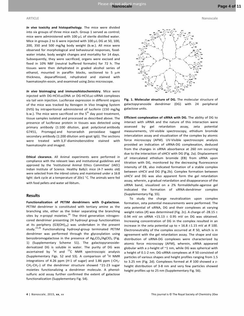
Functionalization of PETIM dendrimers with D-galactose.
PETIM dendrimer is constituted with tertiary amine as the
branching site, ether as the linker separating the branching
sites by n-propyl moieties.26 The third generation nitrogen-
cored dendrimer presenting 24 hydroxyl group functionalities
at its periphery (G3(OH)24) was undertaken in the present
study.23,26 Functionalizing hydroxyl-group terminated PETIM
dendrimer was performed through the glycosylation using
benzobromogalactose in the presence of Ag2CO3/AgClO4 (Fig.
1) (Supplementary Scheme S1). The galactopyranoside-
derivatized DG is soluble in water. The purity of DG was
ascertained by 1H and 13C NMR spectroscopic analysis
(Supplementary Figs. S2 and S3). A comparison of 1H NMR
integrations of 4.26 ppm (H-1 of sugar) and 1.86 ppm (-CH2-
CH2-CH2-) of the dendrimer structure showed ~21-23 sugar
moieties functionalizing a dendrimer molecule. A phenol-
sulfuric acid assay further confirmed the extent of galactose
functionalization (Supplementary Fig. S4).
Fig. 1. Molecular structure of DG. The molecular structure of
galactopyranoside dendrimer (DG) with 24 peripheral
galactose units.
Efficient complexation of siRNA with DG. The ability of DG to
interact with siRNA and the nature of this interaction were
assessed by gel retardation assay, zeta potential
measurements, UV-visible spectroscopy, ethidium bromide
intercalation assay and visualization of the complex by atomic
force microscopy (AFM). UV-Visible spectroscopic analysis
provided an indication of siRNA-DG complexation, deduced
from the changes in siRNA absorbance at 260 nm occurring
due to the interaction of siHCV with DG (Fig. 2a). Displacement
of intercalated ethidium bromide (EB) from siRNA upon
titration with DG, monitored by the decreasing fluorescence
intensity of EB, also indicated formation of a stable complex
between siHCV and DG (Fig.2b). Complex formation between
siHCV and DG was also apparent form the gel retardation
assay, wherein, a gradual retardation and disappearance of the
siRNA band, visualized on a 2% formaldehyde-agarose gel
indicated the formation of siRNA-dendrimer complex
(Supplementary Fig. S5).
To study the charge neutralization upon complex
formation, zeta potential measurements were performed. The
zeta potential of siRNA, DG and their complexes at varying
weight ratios (R) was determined (Fig. 2c). A charge of -28.15 ±
0.94 mV on siRNA +15.13 ± 0.95 mV on DG was obtained.
Increasing concentration of DG in the complex resulted in an
increase in the zeta potential up to + 16.8 ±1.15 mV at R 100.
Electroneutrality of the complex occurred at R 50, which is in
agreement with the gel retardation assay. The shape and size
distribution of siRNA-DG complexes were characterized by
atomic force microscopy (AFM), wherein, siRNA appeared
globular with a z-height of ~1 nm, while DG was spherical with
a height of 0.1-2 nm. DG-siRNA complexes at R 50 consisted of
particles of various shapes and height profiles ranging from 1.5
to 3.25 nm (Fig. 2d). Complexes formed at R 100 showed a z-
height distribution of 3-8 nm and very few particles showed
height profiles up to 25 nm (Supplementary Fig. S6).
Page 4 of 11Nanoscale

Nanoscale ARTICLE
This journal is © The Royal Society of Chemistry 20xx Nanoscale., 2015, xx, xx | 5
Please do not adjust margins
Please do not adjust margins
Fig. 2. Characterizations of siRNA-DG complexes. (a) UV-Visible spectroscopy of siHCV and siHCV-DG complexes at different weight ratios of siRNA-DG complexes, R: 10, 20, 30, 40, 50, 60, 70, 80, 90 and 100. (b) Fluorescence emission profile of EB bound to siHCV, and, upon titration with DG at R 10, 30, 50, 70, 90 and 100. (c) Zeta potential measurements of siRNA, DG and their complexes in 1X siRNA buffer. 100 nM siRNA was used in each of the measurements. Complexes at R10, 20, 30, 40, 50, 60, 70, 80, 90, 100 as well as DG alone (193 µM) was assessed for their zeta potential and electroneutrality was found to be at R 50. (d) AFM height siRNA-DG complexes at R 50 Scale bar = 300 nm. Height profile of these complexes was 1.5-3.25 nm.
Structural analysis of siRNA-DG complexation by MD
simulation. In order to understand the complexation and to
visualize the dendriplexes, a fully atomistic MD simulation of
the siRNA-DG complexation was performed. Simulation studies
revealed that in case the dendrimer is not protonated, there is
no complexation with siRNA. At physiological pH, tertiary
amines at the outer shells (penultimate shell) are protonated,
as pKa values are known to be in the range of 8-9. Lower pH is
required generally for the tertiary amines at the interior and
core for their protonation. In the MD studies, tertiary amines
at the outer shells were protonated, as would be expected in
experimental studies conducted at pH 7.4.39,40 When
dendrimer amines are protonated, formation of a very tight
complex was observed, demonstrating the role of electrostatic
interactions in the complexation process. Fig. 3a,b shows
molecular level picture of the siRNA-DG complex as a function
of simulation time from 0 to 100 ns. Simulations also revealed
that the dendrimer preferentially binds to the major groove
during complexation with siRNA. Instantaneous snapshots of
the complex (Supplementary Fig.S7) shown at few ns interval
reveal major groove binding nature of the dendrimer.
A graphical representation of the sizes of the complexes, as
well as sizes of the siRNA and DG in the complex as the
complexation proceeds is shown in Fig. 3c. The radius of
gyration (Rg) of the siRNA-DG complex was found to be ~2 nm
and that of siRNA alone and dendrimer alone were 1.8 nm and
Fig. 3. MD simulation studies of siHCV-DG complexes. (a)
Instantaneous snapshots of the siRNA- DG (protonated)
complex at different time interval. The red spheres are the
hydroxyl oxygens of the galactose caps, and the blue spheres
are the nitrogens in the dendrimer. (b) Instantaneous
snapshots of the complex of the siRNA- DG (non protonanted)
complex at different time interval. (c) The radius of gyration
(Rg) as a function of simulation time for DG non-protonated
complex (pink), DG protonated complex (red), siRNA alone in
the non-protonated dendrimer complex (light blue), siRNA
alone in the protonated dendrimer complex (green), non-
protonated dendrimer alone in the complex (grey) and
protonated dendrimer alone in the complex (blue). (d) Time
evolution of the distance between the centre of mass of
dendrimer and siRNA for both the protonated and non-
protonated case.
Page 5 of 11 Nanoscale
ARTICLE Nanoscale
6 | Nanoscale, 2015, xx, xx This journal is © The Royal Society of Chemistry 20xx
Please do not adjust margins
Please do not adjust margins
1.3 nm, respectively. The Rg of siRNA remained almost
constant in all the conditions studied. In order to verify the
complex formation between the protonated dendrimer and
siRNA, the radial distribution of the dendrimer amines as well
as sugar groups and phosphates of the siRNA were studied
(Supplementary Fig. S8). Further, a plot of the distance
between the centre of mass (COM) of dendrimer and siRNA as
a function of simulation time showed that in the case of non-
protonated dendrimer, the dendrimer moves away from the
siRNA over simulation time. In contrast the protonated
dendrimer binds tightly on the surface of siRNA, as seen by the
very low COM distance between the two (Fig. 3d).
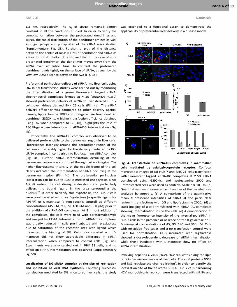
Preferential perinuclear delivery of siRNA into liver cells using
DG. Initial transfection studies were carried out by monitoring
the internalization of a green fluorescent tagged siRNA.
Electroneutral complexes formed at R 50 (siRNA:DG =1:50)
showed preferential delivery of siRNA to liver derived Huh 7
cells over kidney derived BHK 21 cells (Fig. 4a). The siRNA
delivery efficiency was compared to other delivery agents,
namely, lipofectamine 2000 and non-galactose functionalized
dendrimer G3(OH)24. A higher transfection efficiency obtained
using DG when compared to G3(OH)24 highlights the role of
ASGPR-galactose interaction in siRNA-DG internalization (Fig.
4b).
Importantly, the siRNA-DG complex was observed to be
delivered preferentially to the perinuclear region in liver cells.
Fluorescence intensity around the perinuclear region of the
cell was considerably higher for the delivery mediated by DG-
siRNA complex, in comparison to lipofectamine-siRNA complex
(Fig. 4c). Further, siRNA internalization occurring at the
perinuclear region was confirmed through z-stack imaging. The
higher fluorescence intensity at the middle frame of the cell
clearly indicated the internalization of siRNA occurring at the
perinuclear region (Fig. 4d). The preferential perinuclear
localization can be due to ASGPR mediated endocytosis, since
ASGPR enters the cell during endocytosis and particularly
delivers the bound ligand in the area surrounding the
nucleus.41 In order to verify this hypothesis, the Huh 7 cells
were pre-incubated with free D-galactose (a specific ligand for
ASGPR) or D-mannose (a non-specific control) at different
concentrations (45 µM, 90 µM, 180 µM and 360 µM) prior to
the addition of siRNA-DG complexes. At 8 h post addition of
the complexes, the cells were fixed with paraformaldehyde
and imaged by CLSM. Internalization of siRNA-DG complexes
was greatly reduced in cells pre-incubated with D-galactose
due to saturation of the receptor sites with ligand which
prevented the binding of DG. Cells pre-incubated with D-
mannose did not show appreciable difference in siRNA
internalization when compared to control cells (Fig. 4e).
Experiments were also carried out in BHK 21 cells, and no
effect on siRNA internalization was observed (Supplementary
Fig. S9).
Localization of DG-siRNA complex at the site of replication
and inhibition of viral RNA synthesis. Following successful
transfection mediated by DG in cultured liver cells, the study
was extended to a functional assay, to demonstrate the
applicability of preferential liver delivery in a disease model
Fig. 4. Transfection of siRNA-DG complexes in mammalian
cells mediated by asialoglycoprotein receptor. Confocal
microscopic images of (a) Huh 7 and BHK 21 cells transfected
with fluorescent tagged siRNA-DG complexes at R 50. siRNA
transfected using G3(OH)24 and lipofectamine 2000 and
untransfected cells were used as controls. Scale bar 10 µm; (b)
Quantitative mean fluorescence intensities of the transfections
analysed by Image J. (c) A comparison of the quantitative
mean fluorescence intensities of siRNA at the perinuclear
region in transfections with DG and lipofectamine 2000. (d) z-
stack imaging of a cell transfected with siRNA-DG complexes
showing internalization inside the cells. (e) A quantification of
the mean fluorescence intensity of the internalized siRNA in
Huh 7 cells in the presence or absence of free D-galactose or D-
Mannose at concentrations of 45, 90, 180 and 360 µM. Cells
with no added free sugar and a no transfection control were
used for normalization. Cells incubated with D-galactose
showed a dose–dependent decrease of siRNA internalization
while those incubated with D-Mannose show no effect on
siRNA internalization.
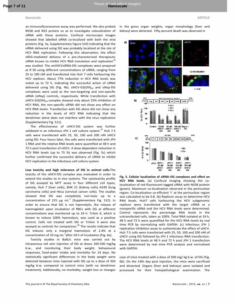
involving hepatitis C virus (HCV). HCV replicates along the lipid
rafts in perinuclear region of liver cells. The viral proteins NS5B
and NS3 regulate the viral replication1. In order to identify the
localization site of the delivered siRNA, Huh 7 cells harbouring
HCV monocistronic replicon were transfected with siRNA and
Page 6 of 11Nanoscale
Nanoscale ARTICLE
This journal is © The Royal Society of Chemistry 20xx Nanoscale., 2015, xx, xx | 7
Please do not adjust margins
Please do not adjust margins
an immunofluorescence assay was performed. We also probed
NS5B and NS3 protein so as to investigate colocalization of
siRNA with these proteins. Confocal microscopic images
showed that labelled siRNA co-localized with both the viral
proteins (Fig. 5a, Supplementary Figure S10) indicating that the
siRNA delivered using DG was probably localized at the site of
HCV RNA replication. Following this observation, the effect
ofDG-mediated delivery of a pre-characterized therapeutic
siRNA known to inhibit HCV RNA translation and replication18
was studied. The antiHCVsiRNA-DG complexes were prepared
at R 50 using different concentrations of siRNA, ranging from
25 to 100 nM and transfected into Huh 7 cells harbouring the
HCV replicon. About 77% reduction in HCV RNA levels was
noted up to 72 h, indicating the successful action of siRNA
delivered using DG (Fig. 4b). siHCV-G3(OH)24 and siNsp-DG
complexes were used as the non-targeting and non-specific
siRNA (siNsp) controls, respectively. While transfection with
siHCV-G3(OH)24 complex showed only about 25% inhibition of
HCV RNA, the non-specific siRNA did not show any effect on
HCV RNA levels. Transfection with DG alone did not show any
reduction in the levels of HCV RNA indicating that the
dendrimer alone does not interfere with the virus replication
(Supplementary Fig. S11).
The effectiveness of siHCV-DG system was further
validated in an infectious JFH 1 cell culture system.37 Huh 7.5
cells were transfected with 25, 50, 100 and 200 nM siHCV
using DG. Four hours later, the cells were transfected with JFH
1 RNA and the relative RNA levels were quantified at 48 h and
72 h post transfection of siHCV. A dose dependent reduction in
HCV RNA levels (up to 75 %) was observed (Fig. 5c) which
further confirmed the successful delivery of siRNA to inhibit
HCV replication in the infectious cell culture system.
Low toxicity and high tolerance of DG in animal cells.The
toxicity of the siHCV-DG complex was evaluated in order to
extend the studies to in vivo systems. The cytotoxicity profile
of DG assayed by MTT assay in four different cell types,
namely, Huh 7 (liver cells), BHK 21 (kidney cells) A549 (lung
carcinoma cells) and HeLa (cervical cancer cells). The studies
showed that DG was completely non-toxic up to a
concentration of 225 µg mL-1 (Supplementary Fig. S12). In
order to ensure that DG is not haemolytic, the release of
haemoglobin upon incubation of RBCs with DG at different
concentrations was monitored up to 24 h. Triton X, which is
known to induce 100% haemolysis, was used as a positive
control. Cells not treated with DG or Trition X were also
assayed as controls for comparison.38 The results indicate that
DG induces only a marginal haemolysis of 2.4% at a
concentration of 10 mg mL-1after 24 h of incubation (Fig. 6a).
Toxicity studies in Balb/c mice was carried out by
intravenous tail vein injection of DG at doses 100-500 mg/kg
b.w., and monitoring their body weight, behavioural
responses, food-water intake and mortality for 14 days. No
statistically significant differences in the body weight were
detected between mice injected with DG up to a dose of 350
mg/kg b.w. compared to control mice (with no dendrimer
treatment). Additionally, no morbidity, weight loss or changes
in the gross organ weights, organ morphology (liver and
kidney) were detected. Fifty percent death was observed in
Fig. 5. Cellular localization of siRNA–DG complexes and effect on
HCV RNA levels. (a) Confocal imaging showing the co-localization of red fluorescent tagged siRNA with NS5B protein (green). Maximum co-localization observed in the perinuclear region. Co-localization co-efficient ‘r’ at the perinuclear region was calculated to be 0.8. (b) Replicon assay to determine HCV RNA levels. Huh7 cells harbouring the HCV subgenomic replicon were transfected with the target siRNA or a nonspecific siRNA and the HCV RNA levels were determined. Control represents the percentage RNA levels in the untransfected cells, taken as 100%. Total RNA isolated at 24 h, 48 h and 72 h were quantified for the HCV RNA levels by real time PCR by normalizing with GAPDH. (c) Infectious JFH 1 replication inhibition assay to authenticate the effect of siHCV. Huh 7.5 cells were transfected with 25, 50, 100 and 200 nM of siHCV using DG followed by JFH 1 infectious RNA transfection. The HCV RNA levels at 48 h and 72 h post JFH 1 transfection were determined by real time PCR analysis and normalized with GAPDH.
case of mice treated with a dose of 500 mg/ kg b.w. of DG (Fig.
6b). On the 14th day post injection, the mice were sacrificed
and dissected. Organs (liver and kidneys) were isolated and
processed for their histopathological examination. The
Page 7 of 11 Nanoscale
ARTICLE Nanoscale
8 | Nanoscale, 2015, xx, xx This journal is © The Royal Society of Chemistry 20xx
Please do not adjust margins
Please do not adjust margins
haematoxylin and eosin-staining based histopathological
studies showed no alterations in the histoarchitecture of liver
as compared to control groups up to a dose of 350 mg/kg b.w.
The tissues of mice treated with a dose of 500 mg/ kg showed
larger inter cellular spaces between the hepatocytes (Fig. 6c).
These results clearly indicate that intravenous administration
of DG is non-toxic up to a dose of 350 mg/kg b.w.
Fig. 6. In vivo tolerance and biocompatibility of DG.(a) Haemolysis assay to determine the blood tolerance of DG. RBCs were incubated with different concentrations of DG at 37oC and the percentage of haemoglobin released into the supernatant at a given time point was determined spectrophotometrically. Triton X, a known haemolysis inducer was used as a positive control. (b) In vivo toxicity assay in Balb/c female mice. 5-7 weeks old mice were injected with dendrimer via tail vein injection and their body weight, food intake and general behaviour was monitored for 14 days. (c) Histopathology studies of the (i) liver and (ii) kidney tissue samples isolated from control mice, (iii) liver and (iv) kidney tissue samples of mice treated with dendrimer at a dose of 350 mg/kg body weight, (v) liver and (vi) kidney tissue samples of mice treated with dendrimer at a dose of 500 mg/kg body weight. No difference in the treated samples when compared to the control indicated that the dendrimers are non-toxic.
Preferential delivery of DG-siRNA complex into mouse liver.
Upon determining the biocompatible nature of DG, the
biodistribution of DNA-DG complexes (5’HCVIRESLuc) was
studied by in vivo bioluminescence imaging (BLI). Balb/c mice
(5-6 weeks old) were injected intravenously with HCVLucDNA-
DG complexes in the presence or absence of siHCV at R 50.
After 48 h, the mice were injected with luciferase substrate
luciferin and whole body imaging was carried out to track the
luciferase expression in the mice. BLI showed that the DNA
was delivered to the mice liver in greater amounts compared
to other organs indicating preferential liver targeting of (Fig.
7a). The results of the bio-imaging were further confirmed by
immunohistochemistry (IHC) studies which showed higher
levels of luciferase localized in the liver and brain tissues when
compared to other organs (Supplementary Fig. S13). In the
presence of siHCV, luciferase expression was greatly reduced
compared to the control. Liver tissues of mice injected with
siHCV-HCVLuc-DG complexes showed a dose dependent
decrease in luciferase expression compared to those injected
only with HCVLuc-DG complexes (Fig. 7a). The results of BLI
were confirmed by IHC wherein, luciferase antibody was used
to probe the luciferase production in the mice liver tissues.
The formation of a black-brown precipitate in tissues of mice
treated with HCVLuc-DG complexes indicated successful
delivery of DNA to the liver (Fig. 7b). Tissues of mice treated
with siHCV-HCVLuc-DG complexes showed a net decrease in
luciferase production indicated by the lower amounts of
brown precipitate formed. These results were consistent with
that observed in the bioluminescence imaging assay. Further,
the use of a non-specific siRNA in the DG-HCVLuc complex did
not affect the luciferase production reiterating the specificity
of siHCV to the HCV 5’UTR. Mice injected with either DNA
alone or DG alone were used as controls to rule out
background signals. Taken together, the results clearly
establish the potential use of DG for delivery of therapeutic
siRNA to inhibit HCV infection.
Fig. 7. Whole body imaging of Balb/c mice showing preferential
localization of HCV Luc DNA to the liver and inhibition of luciferase
expression in the presence of siHCV. (a) In vivo bio-imaging of 5-6 weeks old Balb/c mice injected with luciferase DNA (5’HCVIRES-Luc)-DG complexes and HCVLuc-siHCV-DG complexes monitored after 48 h injection. Maximum luminescence obtained in the liver region for mice injected with HCVLuc-DG complexes. Mice injected with HCVLuc-
Page 8 of 11Nanoscale
Nanoscale ARTICLE
This journal is © The Royal Society of Chemistry 20xx Nanoscale., 2015, xx, xx | 9
Please do not adjust margins
Please do not adjust margins
siHCV-DG complexes at R 50 and R 100 show decreased luminescence. (b) Immunohistochemistry (IHC) studies of mice liver tissues. The brown-black precipitate formed upon reaction of horseradish peroxidase (HRP) with the luciferase antibody can be visualized. Tissues of mice treated with DG alone was used as control. A non-specific siRNA-DG complex used as control had no effect on the production of luciferase in mice liver tissues.
Discussion
Our study provides a ‘proof–of-concept’ approach for the use
of siRNA technology for combating HCV infection based on a
newly developed dendritic galactoside nanovector.
Functionalization of the dendrimer with galactose units
imparted the ability of target-oriented delivery which is one of
the key attributes to the successful action of the desired
antiviral. The formation of a stable complex with intact siRNA
by the delivery vector is essential to ensure crossing of cellular
barriers and successful action of siRNA at target site to down
regulate gene expression.19 Gel retardation assay, zeta
potential measurements, ethidium bromide displacement
assay and UV-Vis spectroscopy confirmed the formation of
stable siRNA-DG complexes. Atomic force microscopy imaging
showed that majority of DG particles had height profiles below
1 nm, similar to that observed in the case of PAMAM42 and
PETIM dendrimers.21 The shape of siRNA and its non-
aggregating behaviour was consistent with the reports of
siRNA AFM imaging.43,44 The morphologies of DG-siRNA
complexes at different weight ratios were visualized as dome
shaped, spherical and globular structures. Particle size
distribution at R 50 was also monodispersed, possibly as a
result of net neutral charge of the complex, as determined by
the zeta potential measurements.
MD simulation showed that most of the galactose residues
are projected away from the siRNA whereas dendrimer
nitrogens (highlighted as blue spheres in Fig. 3a) are involved
in electrostatic complex formation with siRNA. The galactose
residues are free to interact with asialoglycoprotein receptor
(ASGPR) when these complexes are used for the transfection.
The radius of gyration (Rg) values obtained from simulation
studies are in reasonable agreement with those obtained from
AFM imaging. For example, the z-height profile of the complex
varied between 1.5-3.25 nm which is in the same range of the
Rg value of the complex. The dendrimer structure was seen to
undergo a change becoming more compact and orienting itself
such that to accommodate itself into the major groove. The
constant value of Rg for siRNA indicates that siRNA maintains
its structural integrity in the complex. The analysis of the radial
distribution function (RDF) of the dendrimer amines as well as,
galactose oxygens with respect to phosphate groups of siRNA
indicated complex formation between the protonated
dendrimer and siRNA. This was further confirmed by the
analysis of the change in the distance between the COM of
dendrimer and siRNA with respect to simulation time.
Initial nucleic acid delivery experiments involved the
transfection of fluorescent-tagged siRNA using DG at three
ratios, R 25, R 50 and R 100. Comparison of these three ratios
indicated that the delivery at R 50 was more efficient. The use
of low N/P ratios to mediate effective siRNA transfection was
demonstrated recently44 and our observation was on similar
lines of this study. Due to the electroneutrality of the siRNA-
DG complex at R 50, entry inside the cell would occur
preferentially by receptor-mediated endocytosis, involving
ASGPR-galactoside interaction. A comparison of transfection
with native hydroxyl functionalized dendrimer and
lipofectamine, showed that DG preferentially localized in the
perinuclear region of liver cells with much higher fluorescence
intensities. The z-stack imaging helped rule out surface
adherence and ascertain ASGPR mediated endocytosis in liver
cells. ASGPR belongs to the class of receptors that enter the
cells during endocytosis and deliver bound ligand in the area
surrounding the nucleus. Saunier and co-workers had earlier
shown co-localization of GFP-labelled ASGPR and red
fluorescence labelled galactoprotein which binds to ASGPR at
the perinuclear region.41 These studies showed that ASGPR
internalized along with its bound ligand in the perinuclear
regions of the cell. The present study demonstrates that
siRNA, delivered specifically to the liver cells by receptor
mediated endocytosis, is localized in the perinuclear area.
The role of ASGPR in internalization was further validated
by competition assay involving the pre-incubation of liver cells
with D-galactose or D-mannose. Ligand-receptor interactions
are highly specific due to the ability of receptor to recognize
subtle conformational differences of the ligand which alters
the ligand-binding affinity.46 ASGPR specifically binds to D-
galactose over D-mannose by conformational specific
hydrogen bonding interactions. Hence cells pre-incubation
with D-galactose showed decreased siRNA uptake compared to
incubation with D-mannose.
Perinuclear localization of siRNA gains importance to
inhibit HCV RNA replication, since it is known that the
replication occurs along the lipid droplets of the endoplasmic
reticulum in the perinuclear region of hepatocytes.1,2 The viral
proteins NS5B, the RNA dependent RNA polymerase (RdRp)
and NS3 which exhibits RNA helicase activity are known to
regulate the viral replication.1 Both proteins are a part of the
HCV replication complex. We demonstrated here siRNA tagged
with a fluorophore co-localizes with NS5B and NS3 proteins of
the HCV virus replication complex. The proximity of siRNA
localization at the site of viral RNA replication ensures the
effective action of siRNA, which was proven by delivering
siHCV using DG in replicon harbouring cells and probing the
viral RNA levels. The siRNA against HCV inhibits the replication
of viral mRNA through targeted binding to the 5’ -UTR of HCV
RNA. The use of 5’shDNA (short hairpin DNA), a precursor for
the in situ generation of the siRNA to inhibit HCV IRES
mediated translation and hence HCV RNA replication
inhibition, was demonstrated earlier.14 In the present study,
inhibition of HCV RNA up to ~77% was obtained by the use of
siRNA-DG delivery system. The preferential perinuclear
delivery of siRNA in the vicinity of HCV virus replication site
might contribute to the decrease in HCV RNA levels.
Page 9 of 11 Nanoscale
ARTICLE Nanoscale
10 | Nanoscale, 2015, xx, xx This journal is © The Royal Society of Chemistry 20xx
Please do not adjust margins
Please do not adjust margins
Dendrimer based delivery systems are injected
intravenously for studies in vivo, encountering blood in the
first instance.41 In order to verify that intravenously injected
compounds do not cause a haemolysis by rupturing the RBC
membranes, a haemolytic assay was performed. The
haemolysis induced by DG was observed to be negligible when
compared to the dendritic vectors like PAMAM dendrimers,
(60% haemolysis), PPI dendrimer (80% haemolysis) and
polyethylenimine (PEI) based polymers (17-30%
haemolysis).41,47-49 These results indicated that DG is safe to be
injected intravenously. Further experiments in female Balb/c
mice showed acute tolerance dose for DG up to 350 mg/kg
b.w. A comparison with reported studies on other dendritic
systems47 shows DG to be much less toxic, since even a dose of
350 mg/kg b.w. is very well tolerated by mice, which, to our
knowledge, is perhaps the most non-toxic synthetic delivery
vector in vivo reported so far.
In vivo whole body imaging showed a preferential bio
distribution of the complexes to the liver. This observation was
confirmed further by immunohistochemistry (IHC) of the
isolated mice organs. Localization of large quantities of
luciferase enzyme in the liver when compared to other organs
indicated the preferential targeting of DG to the mouse liver.
Previous studies using radiolabeled glycolipids and PAMAM
dendrimers showed that liver uptake of delivery vehicle 15 min
to 1 h post i.v. injection was 60-90%. Minor or negligible
uptake by other organs was also reported.49 The present study
establishes the targeted delivery and successful expression of
a reporter gene using a non-radiolabeled dendrimer, even
after 84 h post injection. Reports of siRNA being delivered to
tumour cells in vivo using engineered PAMAM or PPI
dendrimers showed only a modest success.50,51 Interestingly,
luminescence signals were observed in the brain tissues which
were further validated by IHC experiments. Crossing of the
blood-brain barrier by hydroxyl-terminated PAMAM
dendrimers was demonstrated recently.52 Experiments are
currently under progress to decipher the mechanism and
specificity for delivery in to neuronal cells. The use of siRNA in
the complexes resulted in a net decrease in the luciferase
production probed by BLI, as well as, IHC assays. The siRNA
binds to the 5’-UTR and prevents the HCV IRES mediated
translation of the subsequent luciferase gene, thereby
reducing the luminescence signal, especially in the liver. This
indicated the successful action of siHCV delivered
preferentially to the liver cells by using DG.
Conclusion
RNAi technology and dendrimer based delivery are being used
as popular strategies to combat diseases, such as, cancer and
HIV.53-55 Significant progress has been made to implement this
technology to overcome HCV infection.54,56 Our results
establish that siRNA-DG is a biocompatible system for
preferential liver targeted delivery. The ability of DG to deliver
siRNA in the perinuclear region near the HCV replication
complex and observation of a significant decrease in JFH 1 RNA
levels authenticates the siRNA-DG complex as an efficient
delivery system. Our study shows DG as a targeted delivery
vector can be used for the effective delivery of antivirals
against HCV. The novel siRNA-DG nano-system appears to be a
promising option for HCV related therapeutics.
Acknowledgements
Authors would like to thank Ralf Bartenschlager, Charles M.
Rice and Takaji Wakita for sharing plasmid constructs and cell
lines. We thank the Central Animal Facility, Confocal imaging
facility, Department of Microbiology and Cell Biology and
Centre for Nanoscience and Engineering (CeNSE), IISc. Current
work was supported by a grant from the Department of
Biotechnology, India to SD, NJ, AKS, PM; Science and
Engineering Research Board, DST, India to U.R and NJ. AL and
AK thank Council of Scientific and Industrial Research (CSIR),
India for a research fellowship. SD also acknowledges J. C Bose
fellowship from DST, India.
References
1 D. Moradpur, F. Penin and C. M. Rice, Nat. Rev. Micorbiol., 2007, 5, 453.
2 Y. Ma, M. Anantpadma, J. M. Timpe, S. Shanmugam, S. M. Singh, S. M. Lemon and M. Yi, J. Virol., 2011, 85, 86.
3 S. Das, R. K. Shetty, A. Kumar, R. N. Shridharan, R. Tatineni, G. Chi, A. Mukherjee, S. Das, S. M. Subbarao and A. A. Karande, PLoS One, 2013, 8, e53619.
4 C. Ji, Y. Liu, C. Pamulapati, S. Bohini, G. Fertig, M. Schraeml, W. Rubas, M. Brandt, S. Ries, H. Ma and K. Klumpp, Hepatology, 2015, 61, 1136.
5 A. K. Manna, A. Kumar, U. Ray, S. Das, G. Basu and S. Roy, Antiviral Res., 2013, 97, 223.
6 T. J. Liang and M. G. Ghany, N. Eng. J. Med. 2013, 368, 1907. 7 R. E. Lanford, E. S. Hildebrandt-Eriksen, A. Petri, R. Persson,
M. Lindow, M. E. Munk, S. Kauppinen and H. Ørum, Science, 2010, 327, 198.
8 P. J. Pockros and J. S. Au, Clin. Pharm. Therapy., 2014, 95, 78. 9 B. U. Reddy, R. Mullick, A. Kumar, G. Sudha, N. Srinivasan
and S. Das, Sci Rep., 2014, 24, 5411. 10 T. L. Foster, G. S. Thompson, A. P. Kalverda, J. Kankanala, M.
Bentham, L. F. Wetherill, J. Thompson, A. M. Barker, D. Clarke, M. Noerenberg, A. R. Pearson, D. J. Rowlands, S. W. Homans, M. Harris, R. Foster and S. Griffin. Hepatology, 2014, 59, 408.
11 Y. Gao, X. Yu, B. Xue, F. Zhou, X. Wang, D. Yang, N. Liu, L. Xu, X. Fang and H. Zhu, PLoS One, 2014, 28, e90333.
12 E. Poveda, D. L. Wyles, A. Mena, J. D. Pedreira, A. Castro-Iglesias and E. Cachay, Antiviral Res., 2014, 108, 181.
13 M. M. Verstegen, Q. Pan and L. J. van der Laan, Adv. Exp.
Med. Biol., 2015, 848, 1. 14 N. Subramanian, P. Mani, S. Roy, S. V. Gnanasundram, D. P.
Sarkar and S. Das, J. Gen. Virol., 2009, 90, 1812. 15 S. Roy, N. Gupta, N. Subramanian, T. Mondal, A. C. Banerjea
and S. Das, J. Gen. Virol., 2008, 89, 1579. 16 C. Chevalier, A. Saunier, Y. Benureau, D. Fléchet, D.
Delgrange, F. Colbère-Garapin, C. Wychowski and A. Martin, Mol. Therapy, 2013, 2, 1452.
17 P. K. Chandra, A. K. Kundu, S. Hazari, S. Chandra, L. Bao, T. Ooms, G. F. Morris, T. Wu, T. K. Mandal and S. Dash, Mol.
Therapy, 2012, 20, 1724. 18 R. Kanasty, J. R. Dorkin, A. Vegas, and D. Anderson, Nature
Mat., 2013, 12, 967. 19 J.-P. Behr, Acc. Chem. Res., 2012, 45, 980.
Page 10 of 11Nanoscale
Nanoscale ARTICLE
This journal is © The Royal Society of Chemistry 20xx Nanoscale., 2015, xx, xx | 11
Please do not adjust margins
Please do not adjust margins
20 X. Liu, P. Rocchic and L. Peng, New J. Chem. 2012, 36, 256. 21 A. Lakshminarayanan V. K. Ravi. R. Tatineni,Y. B. R. D. Rajesh,
V. Maingi, K. S. Vasu, N. Madhusudhan, P. K. Maiti, A. K. Sood, S. Das, N. Jayaraman, Bioconjugate Chem., 2013, 24, 1612.
22 U. P. Thankappan, S. N. Madhusudhana, A. Desai, G. Jayamurugan, Y. B. R. D. Rajesh, N. Jayaraman, Bioconjugate
Chem., 2011, 22, 115. 23 T. R. Krishna, M. Belwal, U. S. Tatu and N. Jayaraman,
Tetrahedron, 2005, 61, 4281. 24 L. A. J. M. Sliedregt, P. C. N. Rensen, E. T. Rump, P. J. van
Santbrink, M. K. Bijsterbosch, A. R. P. M. Valentijn, G. A. van der Marel, J. H. van Boom, T. J. C. van Berkel and E. A. L. Biessen, J. Med. Chem., 1999, 42, 609.
25 R. K. Ness, H. G. Fletcher Jr. and C. S. Hudson, C. S. J. Am.
Chem. Soc., 1950, 72, 2200. 26 T. R. Krishna and N. Jayaraman, J. Org. Chem., 2003, 68,
9694. 27 V. Maingi, V. Jain, P. V. Bharatam and P. K. Maiti, J. Comput.
Chem., 2012, 33, 1997-2011. 28 D.A. Case, T.A. Darden, T.E. Cheatham, III, C.L. Simmerling, J.
Wang, R.E. Duke, R. Luo, R.C. Walker, W. Zhang, K.M. Merz, B. Roberts, S. Hayik, A. Roitberg, G. Seabra, J. Swails, A.W. Götz, I. Kolossváry, K.F. Wong, F. Paesani, J. Vanicek, R.M. Wolf, J. Liu, X. Wu, S.R. Brozell, T. Steinbrecher, H. Gohlke, Q. Cai, X. Ye, J. Wang, M.-J. Hsieh, G. Cui, D.R. Roe, D.H. Mathews, M.G. Seetin, R. Salomon-Ferrer, C. Sagui, V. Babin, T. Luchko, S. Gusarov, A. Kovalenko, and P.A. Kollman (2012), AMBER 12, University of California, San Francisco.
29 J. M. Wang, R. M. Wolf, J. W. Caldwell, P. A. Kollman and D. A. Case, J. Comput. Chem., 2004, 25, 1157-1174.
30 J. Wang, P. Cieplak and P. A. Kollman, J. Comput. Chem., 2000, 21, 1049-1074.
31 A. Perez, I. Marchan, D. Svozil, J. Sponer, T. E. Cheatham, III, C. A. Laughton and M. Orozco, Biophys. J. 2007, 92, 3817-3829.
32 W. L. Jorgensen, J. Chandrasekhar, J. D. Madura, R. W. Impey and M. L. Klein, J. Chem. Phys. 1983, 79, 926-935.
33 B. Nandy and P. K. Maiti, J. Phys. Chem. B, 2011, 115, 217. 34 V. Vasumati and P. K. Maiti, Macromolecules, 2010, 43, 8264. 35 P. K. Maiti and B. Bagchi, Nano Lett., 2006, 6, 2478. 36 R. Salomon-Ferrer, A. W. Goetz, D. Poole, S. L. Grand and R.
C. Walker, J. Chem. Theory Comput., 2013, 9, 3878. 37 T. Kato,T. Date, A. Murayama, K. Morikawa, D. Akazawa and
T. Wakita, Nat. Protoc., 2006, 1, 2334. 38 B. Klajnert, S. Pikala, and M. Bryszewska, Proc. R. Soc. A,
2010, 466, 1527. 39 R. C. van Duijvenbode, M. Borkovec and G. J. M. Koper,
Polymer, 1998, 39, 2657-2664. 40 D. Cakara, J. Kleimann and M. Borkovec, Macromolecules,
2003, 36, 4201-4207. 41 B. Saunier,M. Triyatni, L. Ulianich, P. Maruvada, P. Yenand L.
D. Kohn, J. Virol. 2003, 77, 546. 42 J. Li, L. T. Piehler, D. Qin, J. R. Baker and D. A. Tomalia,
Langmuir, 2000, 16, 5613. 43 H. G. Abdelhady, Y. –L. Lin, H. Sun and M. E. H. El-Sayed, PLoS
One, 2013, 8, e61710 44 P. Ofek, W. Fischer, M. Calderón, R. Haag and R. Satchi-
Fainaro, FASEB J., 2010, 24, 3122. 45 M. Zheng, G. M. Pavan, M. Neeb, A. K. Schaper, A. Danai, G.
Klebe, O. M. Merkel and T. Kissel, ACS Nano, 2012, 6, 9447. 46 N. Jayaraman, Chem. Soc. Rev., 2009, 38, 3463. 47 K. L. Allion, Y. Xie, N. El-Gendy, C. J. Berkland and M. L.
Forrest, Adv. Drug Deliv. Rev., 2009, 61, 457. 48 V. Russ, M. Gunther, A. Halama, M. Ogris and E. Wnage, J.
Control. Release, 2008, 132, 131.
49 N. Malik, R. Wiwattanapatapee, R. Klopsch, K. Lorenz, H. Frey, J. W. Weener, E. W. Meijer, W. Paulus and R. Duncan,J. Control. Release, 2000, 65, 133.
50 O. Taratula, O. B. Garbuzenko, P. Kirkpatrick, I. Pandya, R. Savla, V. P. Pozharov, H. He and T. Minko, J. Control. Release, 2009, 140, 284.
51 X. Liu, C. Liu, J. Zhou, C. Chen, F.Qu, J. J. Rossi, P. Rocchi and L. Peng, Nanoscale, 2015, 7, 3867.
52 M. K. Mishra, C. A. Beaty, W. G. Lesniak, S. P. Kambhapati, F. Zhang, M. A. Wilson, M. E. Blue, J. C. Troncoso, S. Kannan, M. V. Johnston, W. A. Baumgartner and R. M. Kannan, ACS
Nano, 2014, 8, 2134. 53 D. H. Kim and J. J. Rossi, Nat. Rev. Genetics, 2007, 8, 173. 54 J. Haasnoot, E. M. Westerhout and B. Berkhout, Nat.
Biotechnol., 2007, 25, 1435. 55 B. Nandy, D. H. Bindu, N. M Dixit and P. K Maiti, J. Chem.
Phys., 2013, 139, 024905. 56 D. Grimm and M. A. Kay, Gene Therapy, 2006, 13, 563.
Page 11 of 11 Nanoscale


