RESEARCH PAPER
Green synthesis of palladium nanoparticlesusing broth of Cinnamomum camphora leaf
Xin Yang Æ Qingbiao Li Æ Huixuan Wang Æ Jiale Huang Æ Liqin Lin ÆWenta Wang Æ Daohua Sun Æ Yuanbo Su Æ James Berya Opiyo ÆLuwei Hong Æ Yuanpeng Wang Æ Ning He Æ Lishan Jia
Received: 25 October 2008 / Accepted: 2 June 2009 / Published online: 19 June 2009
� Springer Science+Business Media B.V. 2009
Abstract The development of dependable, environ-
mentally benign processes for the synthesis of nano-
scale materials is an important aspect of nano-
technology. In the present study, we report one-pot
biogenic fabrication of palladium nanoparticles
by a simple procedure using broth of Cinnamomum
camphora leaf without extra surfactant, capping agent,
and/or template. The mean size of palladium nanopar-
ticles, ranging from 3.2 to 6.0 nm, could be facilely
controlled by merely varying the initial concentration
of the palladium ions. The polyols components and the
heterocyclic components were believed to be respon-
sible for the reduction of palladium ions and the
stabilization of palladium nanoparticles, respectively.
Keywords Palladium � Nanoparticles �Synthesis � Green � Nanobiotechnology
Introduction
Nanoscale materials have received considerable
attention because their structure and properties differ
significantly from those of atoms and molecules as
well as those of bulk materials (Rosei 2004) The
synthesis of nanomaterials with the desired quality is
one of the most exciting aspects in modern nanosci-
ence and nanotechnology (Bhattacharya and Gupta
2005). Moreover, ultrafine transition metal nanopar-
ticles have attracted great interest due to their unique
physical, chemical, and thermodynamic properties
that have made them useful in such diverse fields as
catalysis (Narayanan and El-Sayed 2005), electronics
(Cui and Lieber 2001), optics (Eychmuller 2000), and
even in biological and medical science (Salata 2004).
In the last decade, the utilization of biological
systems has emerged as a novel and reliable method
for the synthesis of nanoparticles due to a growing
need to develop eco-friendly processes in nanomate-
rials syntheses (Bhattacharya and Gupta 2005; Mandal
et al. 2006). A great deal of effort has been put into the
biosynthesis of metal nanoparticles, using bacteria
(Klaus et al. 1999; Fu et al. 2000; Nair and Pradeep
2002; Yong et al. 2002; Zhang et al. 2005; Fu et al.
2006), fungi (Mukherjee et al. 2001a, b; Ahmad et al.
2005), and plant (Gardea-Torresdey et al. 1999, 2002,
2003; Shankar et al. 2003a, b; Shankar et al. 2004a, b;
Ankamwar et al. 2005; Chandran et al. 2006; Huang
et al. 2007; Nadagouda and Varma 2008). From these
studies, biosynthetic method employing live plant or
X. Yang � Q. Li (&) � H. Wang � J. Huang �L. Lin � W. Wang � D. Sun � Y. Su � J. B. Opiyo �L. Hong � Y. Wang � N. He � L. Jia
Department of Chemical and Biochemical Engineering,
College of Chemistry and Chemical Engineering, Xiamen
University, Xiamen 361005, People’s Republic of China
e-mail: [email protected]
X. Yang � Q. Li � H. Wang � J. Huang �L. Lin � W. Wang � Y. Su
National Engineering Laboratory for Green Chemical
Productions of Alcohols, Ethers and Esters, Key
Laboratory for Chemical Biology of Fujian Province,
Xiamen University, Xiamen 361005, People’s
Republic of China
123
J Nanopart Res (2010) 12:1589–1598
DOI 10.1007/s11051-009-9675-1
plant extract has emerged a simple and viable alter-
native to traditional chemical procedures and physical
methods only in recent years. Gardea-Torresdey et al.
(1999, 2002, 2003) firstly reported the preparation of
gold and silver nanoparticles by living plants. Sastry
et al. attained biosynthesis of metal nanoparticles by
plant leaf extract (Shankar et al. 2003a, b; Shankar
et al. 2004a, b; Ankamwar et al. 2005; Chandran et al.
2006). Our group demonstrated that sundried Cinna-
momum camphora (C. camphora) leaf could be used to
synthesize silver and gold nanoparticles in aqueous
solutions at ambient conditions (Huang et al. 2007).
The above synthetic techniques by plant extract or
biomass exhibits the prospective application of plant
bioresource for synthesis of metal nanoparticles.
Recently, the advances in fabrication of ultrafine
palladium nanoparticles (PdNPs) have gained great
importance due to their application both in heteroge-
neous and homogeneous catalysis, due to their high
surface-to-volume ratio and their high surface energy
(Narayanan and El-Sayed 2005). The usual synthetic
methodologies for producing PdNPs involve chemical
reduction of Pd(II) by alcohol (Teranishi and Miyake
1998), NaBH4/ascorbic acid (Jana et al. 2000), N2H4
(Yonezawa et al. 2001), PEG (Luo et al. 2005),
CNCH2COOK (Wang et al. 2004), or ascorbic acid
(Sun et al. 2007), reduction of Pd(OAc)2 by dimeth-
ylamine-borane in supercritical carbon dioxide
(Kameo et al. 2003), thermally induced reduction of
Pd(Fod)2 in o-xylene (Ho and Chi 2004), and sono-
chemical reduction of Pd(NO3)2 (Nemamcha et al.
2006). Nevertheless, most of these processes were
performed in the presence of various stabilizers to
prevent the formation of undesired agglomerates or
aggregates of PdNPs. Additionally, there are few
reports concerning biological production of PdNPs by
plant extract or biomass, where the biomass was
usually found to act as both reducing agent and
stabilizer. The only success was the recent demonstra-
tion by Nadagouda and Varma (2008), who showed
production of PdNPs using coffee and tea extract.
Unfortunately, they offered relatively scarce informa-
tion regarding PdNPs.
In this study, broth of C. camphora leaf was first
used to synthesize PdNPs at ambient conditions.
Ultrafine PdNPs were obtained and their characteris-
tics were examined by various techniques. It is
important to point out that size control of PdNPs
could be achieved by simply altering the initial
concentration of Pd(II) ions. As is commonly known,
C. camphora has been widely cultivated in South
China. And it has been reported by our group that some
antitumor components could be extracted from
C. camphora leaf (Su et al. 2006). Therefore, we deem
that some pharmaceutically valuable molecules could
be retained after bioreduction and the as-prepared
PdNPs might have potential application in nanomed-
icines. The approach appears to be a facile alternative
to conventional methods of producing PdNPs.
Experimental
Materials
Palladium chloride (PdCl2) was purchased from
Sinopharm Chemical Reagent Co. Ltd, China and
was used as received. C. camphora leaf used for the
bioreduction was prepared using the same procedures
as those in our previous study (Huang et al. 2007).
Synthesis of PdNPs
In order to obtain the broth of C. camphora leaf, the
carefully weighted biomass, 4.0 g, was added to
200 ml deionized water in round-bottom flasks of
500 ml capacity. The mixtures were boiled for 5 min,
and thereafter filtrated and the filtrate was used for
further experiments. In a typical synthesis for PdNPs,
an appropriate amount of concentrated aqueous PdCl2solution (0.226 mol L-1) was added to 50 ml filtrate in
conical flasks of 100 ml capacity at room temperature
to obtain 1 9 10-3 mol L-1, 3 9 10-3 mol L-1, and
5 9 10-3 mol L-1 PdCl2 solutions, respectively. The
flasks were thereafter shaken at a rotation rate of
150 rpm in the dark at 30 �C.
Characterization of samples
UV–visible spectroscopy analysis was carried out on an
UNICAM UV-300 spectrophotometer (Thermo Spec-
tronic) over wavelengths from 250 to 800 nm at a
resolution of 1 nm. Equivalent amounts of the suspen-
sion (0.5 ml) were diluted in a constant volume of
deionized water (5 ml) and subsequently analyzed at
room temperature. X-ray diffraction (XRD) measure-
ment was performed on an X’Pert Pro X-ray Diffrac-
tometer (PANalytical BV, The Netherlands) operated at
1590 J Nanopart Res (2010) 12:1589–1598
123
a voltage of 40 kV and a current of 30 mA with Cu Karadiation. After the reaction, the resulting solutions were
centrifuged at a speed of 12,000 rpm for 10 min and the
precipitates were dried at 60 �C overnight and collected
for the determination of the formation of palladium.
Transmission electron microscopy (TEM) and
high resolution transmission electron microscopy
(HRTEM) images were obtained on a JEOL JEM-
2100 Microscope at 200 keV. Samples for TEM were
prepared by placing a drop of the suspension on
carbon coated copper grids and allowing water to
completely evaporate. Size distribution and average
size of the nanoparticles were estimated on the basis
of TEM micrographs with the assistance of Sigma-
Scan Pro software (SPSS Inc, Version 4.01.003).
Energy dispersive X-ray (EDX) and selected-area
electron diffraction (SAED) analysis of PdNPs were
performed on a Tecnai F30 Microscope.
X-ray photoelectron spectroscopy (XPS) analysis
was performed on an American Perkin-Elmer PHI-
1600 with a monochromatised microfocused Al X-ray
source. The binding energy was calibrated by C1 s as
reference energy (C1 s = 284.5 eV). The colloidal
solution of PdNPs was centrifuged at a speed of
12,000 rpm for 10 min. After removal of the colorless
supernatant, the precipitates were then redispersed in
pure water to form a new suspension of PdNPs.
Sample for XPS analysis was fabricated by dropping
the suspension onto clean silicon and allowing water
to completely evaporate.
The leaf biomass before bioreduction, the biomass
residue after bioreduction and the resulting palladium
nanoparticles were analyzed by Fourier transform
infrared (FTIR) Nicolet Avatar 660 (Nicolet, USA).
The leaf biomass before bioreduction was attained by
completely drying of the leaf biomass at 60 �C. The
biomass residue after bioreduction and the resulting
palladium nanoparticles were obtained by centrifuging
the resulting solution at 12,000 rpm for 10 min, and
then the supernatant and the precipitates were com-
pletely dried at the same temperature, respectively.
Results and discussion
Formation of PdNPs by broth of C. camphora leaf
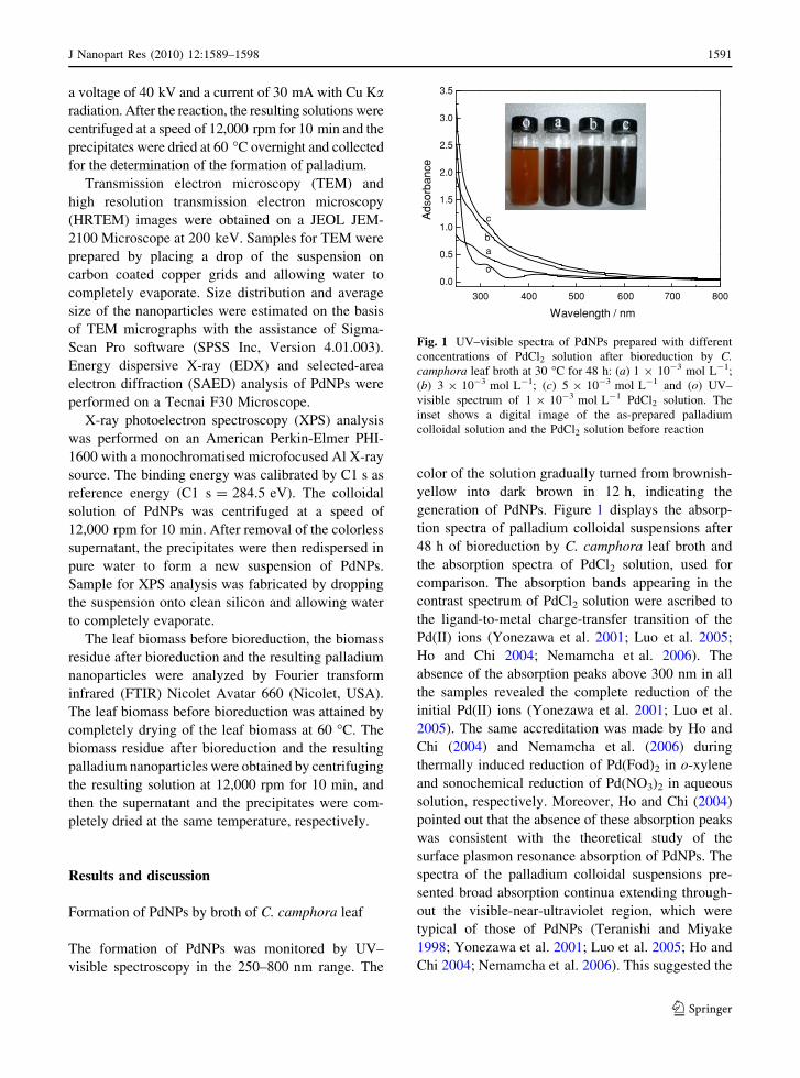
The formation of PdNPs was monitored by UV–
visible spectroscopy in the 250–800 nm range. The
color of the solution gradually turned from brownish-
yellow into dark brown in 12 h, indicating the
generation of PdNPs. Figure 1 displays the absorp-
tion spectra of palladium colloidal suspensions after
48 h of bioreduction by C. camphora leaf broth and
the absorption spectra of PdCl2 solution, used for
comparison. The absorption bands appearing in the
contrast spectrum of PdCl2 solution were ascribed to
the ligand-to-metal charge-transfer transition of the
Pd(II) ions (Yonezawa et al. 2001; Luo et al. 2005;
Ho and Chi 2004; Nemamcha et al. 2006). The
absence of the absorption peaks above 300 nm in all
the samples revealed the complete reduction of the
initial Pd(II) ions (Yonezawa et al. 2001; Luo et al.
2005). The same accreditation was made by Ho and
Chi (2004) and Nemamcha et al. (2006) during
thermally induced reduction of Pd(Fod)2 in o-xylene
and sonochemical reduction of Pd(NO3)2 in aqueous
solution, respectively. Moreover, Ho and Chi (2004)
pointed out that the absence of these absorption peaks
was consistent with the theoretical study of the
surface plasmon resonance absorption of PdNPs. The
spectra of the palladium colloidal suspensions pre-
sented broad absorption continua extending through-
out the visible-near-ultraviolet region, which were
typical of those of PdNPs (Teranishi and Miyake
1998; Yonezawa et al. 2001; Luo et al. 2005; Ho and
Chi 2004; Nemamcha et al. 2006). This suggested the
300 400 500 600 700 800
0.0
0.5
1.0
1.5
2.0
2.5
3.0
3.5
Ads
orba
nce
Wavelength / nm
o
a
b
c
Fig. 1 UV–visible spectra of PdNPs prepared with different
concentrations of PdCl2 solution after bioreduction by C.camphora leaf broth at 30 �C for 48 h: (a) 1 9 10-3 mol L-1;
(b) 3 9 10-3 mol L-1; (c) 5 9 10-3 mol L-1 and (o) UV–
visible spectrum of 1 9 10-3 mol L-1 PdCl2 solution. The
inset shows a digital image of the as-prepared palladium
colloidal solution and the PdCl2 solution before reaction
J Nanopart Res (2010) 12:1589–1598 1591
123
formation of palladium colloid having a particle size
of less than 10 nm, as described by Nemamcha et al.
(2006).
Characterization of PdNPs
The morphology and size of PdNPs in the colloidal
solutions and their size distribution were investigated
by TEM. Figure 2 displays the TEM images of typical
nanoparticles synthesized with C. camphora leaf and
PdCl2 solution of different concentrations at various
magnifications. The initial concentration of the Pd(II)
was found to have a vital impact on size and size
distribution of PdNPs. For sample A (1 9 10-3
mol L-1), the resulting PdNPs were well dispersed
but quantitatively sparse (Fig. 2a–c). It might result
from the relative insufficiency of the palladium
precursor. In addition, some particles with irregular
contour could be observed probably due to the effect of
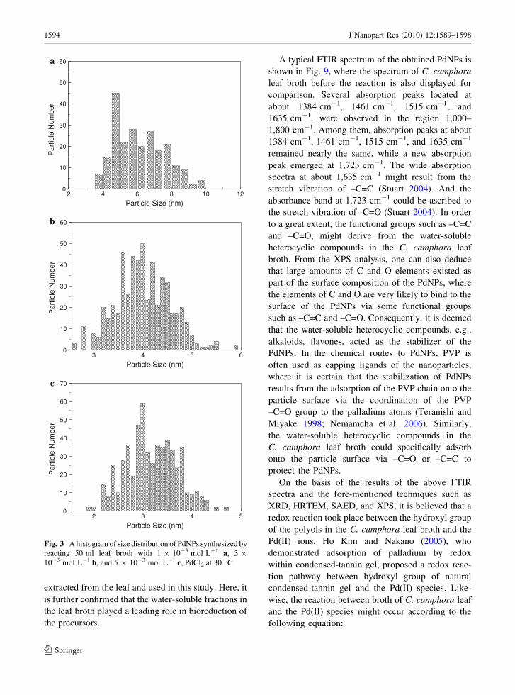
Ostwald ripening. According to size distribution of the
PdNPs shown in Fig. 3a, the nanoparticles, mostly
ranging from 3.6 to 9.9 nm in size, have a mean
diameter of 6.0 nm. However, a further increase of
initial Pd(II) concentration led to the formation of
quasi-spherical nanoparticles with a narrower size
distribution (Fig. 3b, c). Interestingly, the inter-parti-
cle distance of PdNPs was more uniformly dispersed
and well aligned (Fig. 2d–i). The nanoparticles mainly
range from 3.0 to 5.0 nm and from 2.2 to 4.3 nm in
size, with mean particle diameters of 4.0 nm and
3.2 nm for samples B (3 9 10-3 mol L-1) and C
(5 9 10-3 mol L-1), respectively. Apparently, it was
found that the higher the concentration of the PdCl2solution, the smaller the PdNPs formed.
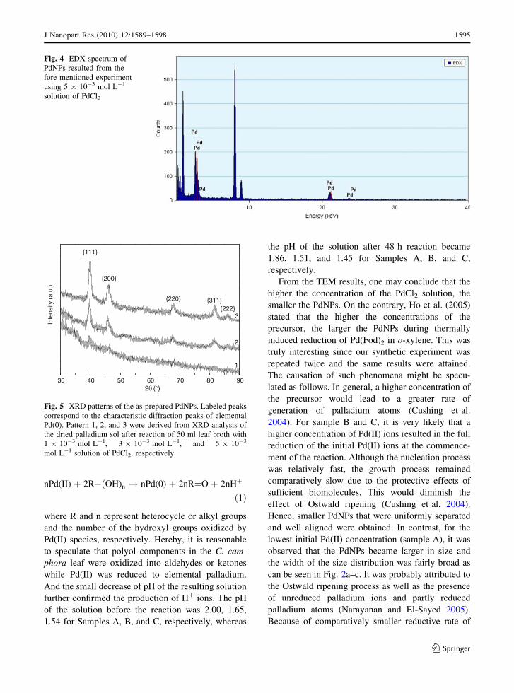
The particles were verified to contain a great deal
of palladium according to EDX analysis in Fig. 4. The
crystalline phase of the synthesized nanoparticles was
confirmed by XRD (Fig. 5) and HRTEM (Fig. 6a).
From the XRD patterns, prominent Bragg reflections
at 2h values of 40.0, 46.1, 67.9, 82.0, and 86.0 were
observed, which correspond to the {111}, {200},
{220}, {311}, and {222} Bragg reflections of face-
centered cubic (fcc) PdNPs. All the palladium nano-
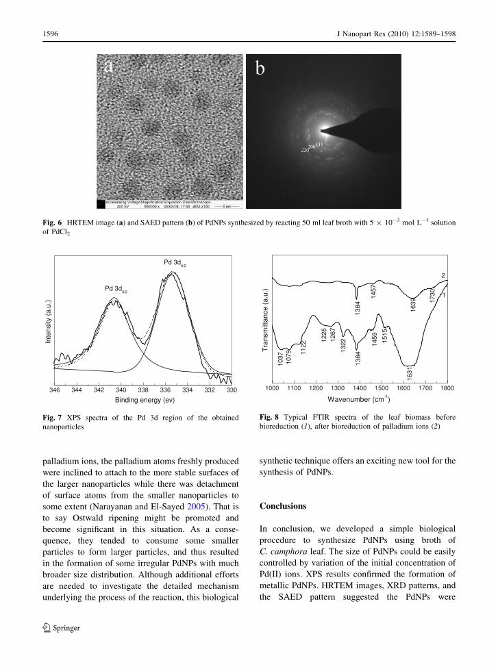
particles in the HRTEM image showed lattice images,
indicating that they are single crystallites. Figure 6b
shows the SAED pattern recorded from the corre-
sponding PdNPs. The ring-like diffraction pattern
could be indexed on the basis of the fcc structure of
palladium. Ring 1, 2, 3 arise due to reflections from
{111}, {200}, {220} lattice planes of fcc palladium,
respectively. HRTEM images, XRD patterns, and the
SAED pattern thus clearly show that the PdNPs are
crystalline in nature.
An XPS spectrum of the Pd 3d region (Pd 3d5/2
and Pd 3d3/2) for the obtained nanoparticles is shown
in Fig. 7. The binding energy values are 335.4 ev for
Pd 3d5/2 and 340.6 ev for Pd 3d3/2. The observed
binding energy values for Pd 3d coincide with the
reported data of Pd(0) within the experimental errors
(Moulder et al. 1992). Quantitavive surface comopo-
sition of the PdNPs determined by XPS is represented
by C (45.8 atom%), O(33.7 atom%), Si (16.2 atom%),
N (2.0 atom%), Pd (1.7 atom%), and Cl (0.6 atom%).
Thereamong the three elements (C, O, N) originate
from the biomass residue of C. camphora leaf broth.
The trace amounts of Cl, which was not fully
removed, may originate from the metal precursor.
Formation mechanism of PdNPs
Representative FTIR spectra of C. camphora leaf
broth before and after the reaction are presented in
Fig. 8. Some pronounced absorbance bands centered
at 1037 cm-1, 1079 cm-1, 1122 cm-1, 1226 cm-1,
1267 cm-1, 1322 cm-1, 1384 cm-1, 1459 cm-1,
1515 cm-1, 1631 cm-1, 1730 cm-1 are observed in
the region 1,000–1,800 cm-1. Therein the absorbance
bands at 1037 cm-1, 1079 cm-1, and 1122 cm-1 in
Curve 1 could be assigned to the stretch vibration of
–C–O (Stuart 2004). The sharpest band at 1,631 cm-1
might result from conjugated –C=C (Stuart 2004). The
absorbance band at 1,730 cm-1 in Curve 2 could be
assigned to the stretch vibration of -C=O (Stuart 2004).
The comparison provided information concerning the
chemical transformation of the functional groups
involved in reduction of palladium ions. In the first
place, it is clear that the absorbance bands at
1037 cm-1, 1079 cm-1, and 1122 cm-1 were weak-
ened after the reaction. These bands might be assigned
to the stretch vibration of –C–O (Stuart 2004).
Previously, our group demonstrated that various
components such as alkaloids, flavones, hydroxybenz-
enes, anthracenes, steroids, terpenoids, coumarins,
lactones, linalools, polysaccharides, amino acids, and
proteins existed in C. camphora leaf (Su et al. 2006).
Thereby, we could infer that the polyols such as
flavones, terpenoids, and polysaccharides in the broth
played a critical role in reduction of Pd(II) ions. In the
1592 J Nanopart Res (2010) 12:1589–1598
123

next place, the freshly appeared band at 1,730 cm-1 in
Curve 2 could be ascribed to the stretch vibration of
carbonyl groups. Hence, it is speculated that the
polyols are oxidized to aldehydes or ketones and
induce the formation of the band 1,730 cm-1. In our
previous study, the disappearance of the band at
1,109 cm-1 after bioreduction implied that the polyols
from the sundried C. camphora leaf were mainly
responsible for the reduction of silver ions or chloroa-
urate ions (Huang et al. 2007). Owing to this result and
considering the application of as-prepared PdNPs in
catalysis, the water-soluble components were
Fig. 2 TEM images of PdNPs after bioreduction. 50 ml leaf broth reacted with a–c 1 9 10-3 mol L-1, d–f 3 9 10-3 mol L-1 and
g–i 5 9 10-3 mol L-1 PdCl2 at 30 �C, respectively
J Nanopart Res (2010) 12:1589–1598 1593
123
extracted from the leaf and used in this study. Here, it
is further confirmed that the water-soluble fractions in
the leaf broth played a leading role in bioreduction of
the precursors.
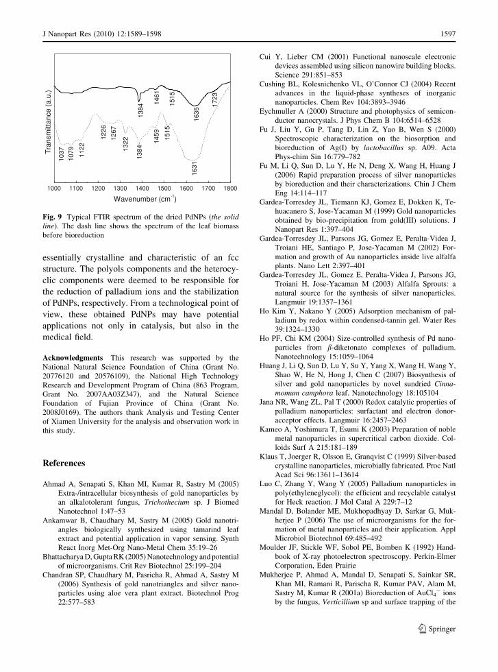
A typical FTIR spectrum of the obtained PdNPs is
shown in Fig. 9, where the spectrum of C. camphora
leaf broth before the reaction is also displayed for
comparison. Several absorption peaks located at
about 1384 cm-1, 1461 cm-1, 1515 cm-1, and
1635 cm-1, were observed in the region 1,000–
1,800 cm-1. Among them, absorption peaks at about
1384 cm-1, 1461 cm-1, 1515 cm-1, and 1635 cm-1
remained nearly the same, while a new absorption
peak emerged at 1,723 cm-1. The wide absorption
spectra at about 1,635 cm-1 might result from the
stretch vibration of –C=C (Stuart 2004). And the
absorbance band at 1,723 cm-1 could be ascribed to
the stretch vibration of -C=O (Stuart 2004). In order
to a great extent, the functional groups such as –C=C
and –C=O, might derive from the water-soluble
heterocyclic compounds in the C. camphora leaf
broth. From the XPS analysis, one can also deduce
that large amounts of C and O elements existed as
part of the surface composition of the PdNPs, where
the elements of C and O are very likely to bind to the
surface of the PdNPs via some functional groups
such as –C=C and –C=O. Consequently, it is deemed
that the water-soluble heterocyclic compounds, e.g.,
alkaloids, flavones, acted as the stabilizer of the
PdNPs. In the chemical routes to PdNPs, PVP is
often used as capping ligands of the nanoparticles,
where it is certain that the stabilization of PdNPs
results from the adsorption of the PVP chain onto the
particle surface via the coordination of the PVP
–C=O group to the palladium atoms (Teranishi and
Miyake 1998; Nemamcha et al. 2006). Similarly,
the water-soluble heterocyclic compounds in the
C. camphora leaf broth could specifically adsorb
onto the particle surface via –C=O or –C=C to
protect the PdNPs.
On the basis of the results of the above FTIR
spectra and the fore-mentioned techniques such as
XRD, HRTEM, SAED, and XPS, it is believed that a
redox reaction took place between the hydroxyl group
of the polyols in the C. camphora leaf broth and the
Pd(II) ions. Ho Kim and Nakano (2005), who
demonstrated adsorption of palladium by redox
within condensed-tannin gel, proposed a redox reac-
tion pathway between hydroxyl group of natural
condensed-tannin gel and the Pd(II) species. Like-
wise, the reaction between broth of C. camphora leaf
and the Pd(II) species might occur according to the
following equation:
0
10
20
30
40
50
60
Par
ticle
Num
ber
Particle Size (nm)
a
0
10
20
30
40
50
60
Par
ticle
Num
ber
Particle Size (nm)
b
2 4 6 8 10 12
3 4 5 6
2 3 4 50
10
20
30
40
50
60
70
Par
ticle
Num
ber
Particle Size (nm)
c
Fig. 3 A histogram of size distribution of PdNPs synthesized by
reacting 50 ml leaf broth with 1 9 10-3 mol L-1 a, 3 9
10-3 mol L-1 b, and 5 9 10-3 mol L-1 c, PdCl2 at 30 �C
1594 J Nanopart Res (2010) 12:1589–1598
123
nPd(II) þ 2R�ðOH)n ! nPd(0) þ 2nR¼O þ 2nHþ
ð1Þ
where R and n represent heterocycle or alkyl groups
and the number of the hydroxyl groups oxidized by
Pd(II) species, respectively. Hereby, it is reasonable
to speculate that polyol components in the C. cam-
phora leaf were oxidized into aldehydes or ketones
while Pd(II) was reduced to elemental palladium.
And the small decrease of pH of the resulting solution
further confirmed the production of H? ions. The pH
of the solution before the reaction was 2.00, 1.65,
1.54 for Samples A, B, and C, respectively, whereas
the pH of the solution after 48 h reaction became
1.86, 1.51, and 1.45 for Samples A, B, and C,
respectively.
From the TEM results, one may conclude that the
higher the concentration of the PdCl2 solution, the
smaller the PdNPs. On the contrary, Ho et al. (2005)
stated that the higher the concentrations of the
precursor, the larger the PdNPs during thermally
induced reduction of Pd(Fod)2 in o-xylene. This was
truly interesting since our synthetic experiment was
repeated twice and the same results were attained.
The causation of such phenomena might be specu-
lated as follows. In general, a higher concentration of
the precursor would lead to a greater rate of
generation of palladium atoms (Cushing et al.
2004). For sample B and C, it is very likely that a
higher concentration of Pd(II) ions resulted in the full
reduction of the initial Pd(II) ions at the commence-
ment of the reaction. Although the nucleation process
was relatively fast, the growth process remained
comparatively slow due to the protective effects of
sufficient biomolecules. This would diminish the
effect of Ostwald ripening (Cushing et al. 2004).
Hence, smaller PdNPs that were uniformly separated
and well aligned were obtained. In contrast, for the
lowest initial Pd(II) concentration (sample A), it was
observed that the PdNPs became larger in size and
the width of the size distribution was fairly broad as
can be seen in Fig. 2a–c. It was probably attributed to
the Ostwald ripening process as well as the presence
of unreduced palladium ions and partly reduced
palladium atoms (Narayanan and El-Sayed 2005).
Because of comparatively smaller reductive rate of
Fig. 4 EDX spectrum of
PdNPs resulted from the
fore-mentioned experiment
using 5 9 10-3 mol L-1
solution of PdCl2
30 40 50 60 70 80 90
{222}{311}{220}
{200}
Inte
nsity
(a.
u.)
2 ( )
{111}
1
2
3
Fig. 5 XRD patterns of the as-prepared PdNPs. Labeled peaks
correspond to the characteristic diffraction peaks of elemental
Pd(0). Pattern 1, 2, and 3 were derived from XRD analysis of
the dried palladium sol after reaction of 50 ml leaf broth with
1 9 10-3 mol L-1, 3 9 10-3 mol L-1, and 5 9 10-3
mol L-1 solution of PdCl2, respectively
J Nanopart Res (2010) 12:1589–1598 1595
123
palladium ions, the palladium atoms freshly produced
were inclined to attach to the more stable surfaces of
the larger nanoparticles while there was detachment
of surface atoms from the smaller nanoparticles to
some extent (Narayanan and El-Sayed 2005). That is
to say Ostwald ripening might be promoted and
become significant in this situation. As a conse-
quence, they tended to consume some smaller
particles to form larger particles, and thus resulted
in the formation of some irregular PdNPs with much
broader size distribution. Although additional efforts
are needed to investigate the detailed mechanism
underlying the process of the reaction, this biological
synthetic technique offers an exciting new tool for the
synthesis of PdNPs.
Conclusions
In conclusion, we developed a simple biological
procedure to synthesize PdNPs using broth of
C. camphora leaf. The size of PdNPs could be easily
controlled by variation of the initial concentration of
Pd(II) ions. XPS results confirmed the formation of
metallic PdNPs. HRTEM images, XRD patterns, and
the SAED pattern suggested the PdNPs were
Fig. 6 HRTEM image (a) and SAED pattern (b) of PdNPs synthesized by reacting 50 ml leaf broth with 5 9 10-3 mol L-1 solution
of PdCl2
346 344 342 340 338 336 334 332 330
Pd 3d5/2
Inte
nsity
(a.
u.)
Binding energy (ev)
Pd 3d3/2
Fig. 7 XPS spectra of the Pd 3d region of the obtained
nanoparticles
1000 1100 1200 1300 1400 1500 1600 1700 1800
1457
1384
1037
1079 11
22 145912
2612
67
1322
1730
1639
1631
1384
1515
Tra
nsm
ittan
ce (
a.u.
)
Wavenumber (cm-1)
1
2
Fig. 8 Typical FTIR spectra of the leaf biomass before
bioreduction (1), after bioreduction of palladium ions (2)
1596 J Nanopart Res (2010) 12:1589–1598
123
essentially crystalline and characteristic of an fcc
structure. The polyols components and the heterocy-
clic components were deemed to be responsible for
the reduction of palladium ions and the stabilization
of PdNPs, respectively. From a technological point of
view, these obtained PdNPs may have potential
applications not only in catalysis, but also in the
medical field.
Acknowledgments This research was supported by the
National Natural Science Foundation of China (Grant No.
20776120 and 20576109), the National High Technology
Research and Development Program of China (863 Program,
Grant No. 2007AA03Z347), and the Natural Science
Foundation of Fujian Province of China (Grant No.
2008J0169). The authors thank Analysis and Testing Center
of Xiamen University for the analysis and observation work in
this study.
References
Ahmad A, Senapati S, Khan MI, Kumar R, Sastry M (2005)
Extra-/intracellular biosynthesis of gold nanoparticles by
an alkalotolerant fungus, Trichothecium sp. J Biomed
Nanotechnol 1:47–53
Ankamwar B, Chaudhary M, Sastry M (2005) Gold nanotri-
angles biologically synthesized using tamarind leaf
extract and potential application in vapor sensing. Synth
React Inorg Met-Org Nano-Metal Chem 35:19–26
Bhattacharya D, Gupta RK (2005) Nanotechnology and potential
of microorganisms. Crit Rev Biotechnol 25:199–204
Chandran SP, Chaudhary M, Pasricha R, Ahmad A, Sastry M
(2006) Synthesis of gold nanotriangles and silver nano-
particles using aloe vera plant extract. Biotechnol Prog
22:577–583
Cui Y, Lieber CM (2001) Functional nanoscale electronic
devices assembled using silicon nanowire building blocks.
Science 291:851–853
Cushing BL, Kolesnichenko VL, O’Connor CJ (2004) Recent
advances in the liquid-phase syntheses of inorganic
nanoparticles. Chem Rev 104:3893–3946
Eychmuller A (2000) Structure and photophysics of semicon-
ductor nanocrystals. J Phys Chem B 104:6514–6528
Fu J, Liu Y, Gu P, Tang D, Lin Z, Yao B, Wen S (2000)
Spectroscopic characterization on the biosorption and
bioreduction of Ag(I) by lactobacillus sp. A09. Acta
Phys-chim Sin 16:779–782
Fu M, Li Q, Sun D, Lu Y, He N, Deng X, Wang H, Huang J
(2006) Rapid preparation process of silver nanoparticles
by bioreduction and their characterizations. Chin J Chem
Eng 14:114–117
Gardea-Torresdey JL, Tiemann KJ, Gomez E, Dokken K, Te-
huacanero S, Jose-Yacaman M (1999) Gold nanoparticles
obtained by bio-precipitation from gold(III) solutions. J
Nanopart Res 1:397–404
Gardea-Torresdey JL, Parsons JG, Gomez E, Peralta-Videa J,
Troiani HE, Santiago P, Jose-Yacaman M (2002) For-
mation and growth of Au nanoparticles inside live alfalfa
plants. Nano Lett 2:397–401
Gardea-Torresdey JL, Gomez E, Peralta-Videa J, Parsons JG,
Troiani H, Jose-Yacaman M (2003) Alfalfa Sprouts: a
natural source for the synthesis of silver nanoparticles.
Langmuir 19:1357–1361
Ho Kim Y, Nakano Y (2005) Adsorption mechanism of pal-
ladium by redox within condensed-tannin gel. Water Res
39:1324–1330
Ho PF, Chi KM (2004) Size-controlled synthesis of Pd nano-
particles from b-diketonato complexes of palladium.
Nanotechnology 15:1059–1064
Huang J, Li Q, Sun D, Lu Y, Su Y, Yang X, Wang H, Wang Y,
Shao W, He N, Hong J, Chen C (2007) Biosynthesis of
silver and gold nanoparticles by novel sundried Cinna-momum camphora leaf. Nanotechnology 18:105104
Jana NR, Wang ZL, Pal T (2000) Redox catalytic properties of
palladium nanoparticles: surfactant and electron donor-
acceptor effects. Langmuir 16:2457–2463
Kameo A, Yoshimura T, Esumi K (2003) Preparation of noble
metal nanoparticles in supercritical carbon dioxide. Col-
loids Surf A 215:181–189
Klaus T, Joerger R, Olsson E, Granqvist C (1999) Silver-based
crystalline nanoparticles, microbially fabricated. Proc Natl
Acad Sci 96:13611–13614
Luo C, Zhang Y, Wang Y (2005) Palladium nanoparticles in
poly(ethyleneglycol): the efficient and recyclable catalyst
for Heck reaction. J Mol Catal A 229:7–12
Mandal D, Bolander ME, Mukhopadhyay D, Sarkar G, Muk-
herjee P (2006) The use of microorganisms for the for-
mation of metal nanoparticles and their application. Appl
Microbiol Biotechnol 69:485–492
Moulder JF, Stickle WF, Sobol PE, Bomben K (1992) Hand-
book of X-ray photoelectron spectroscopy. Perkin-Elmer
Corporation, Eden Prairie
Mukherjee P, Ahmad A, Mandal D, Senapati S, Sainkar SR,
Khan MI, Ramani R, Parischa R, Kumar PAV, Alam M,
Sastry M, Kumar R (2001a) Bioreduction of AuCl4- ions
by the fungus, Verticillium sp and surface trapping of the
1000 1100 1200 1300 1400 1500 1600 1700 1800
1226
1037
1079 11
22
1267
1322 15
15
1459
1515
1461
1384
1384 17
23
1631
1635
Wavenumber (cm-1)
Tra
nsm
ittan
ce (
a.u.
)
Fig. 9 Typical FTIR spectrum of the dried PdNPs (the solidline). The dash line shows the spectrum of the leaf biomass
before bioreduction
J Nanopart Res (2010) 12:1589–1598 1597
123
gold nanoparticles formed. Angew Chem Int Ed 40:3585–
3588
Mukherjee P, Ahmad A, Mandal D, Senapati S, Sainkar SR,
Khan MI, Parischa R, Ajaykumar PV, Alam M, Kumar R,
Sastry M (2001b) Fungus-mediated synthesis of silver
nanoparticles and their immobilization in the mycelial
matrix: a novel biological approach to nanoparticle syn-
thesis. Nano Lett 1:515–519
Nadagouda MN, Varma RS (2008) Green synthesis of silver
and palladium nanoparticles at room temperature using
coffee and tea extract. Green Chem 10:859–862
Nair B, Pradeep T (2002) Coalescence of nanoclusters and
formation of submicron crystallites assisted by Lactoba-cillus strains. Cryst Growth Des 2:293–298
Narayanan R, El-Sayed MA (2005) Catalysis with transition
metal nanoparticles in colloidal solution: nanoparticle
shape dependence and stability. J Phys Chem B 109:
12663–12676
Nemamcha A, Rehspringer J, Khatmi D (2006) Synthesis of
palladium nanoparticles by sonochemical reduction of
palladium(II) nitrate in aqueous solution. J Phys Chem B
110:383–387
Rosei F (2004) Nanostructured surfaces: challenges and fron-
tiers in nanotechnology. J Phys Condens Matter 16:
S1373–S1436
Salata OV (2004) Applications of nanoparticles in biology and
medicine. J Nanobiotechnol 2:3
Shankar SS, Ahmad A, Pasricha R, Sastry M (2003a) Biore-
duction of chloroaurate ions by geranium leaves and its
endophytic fungus yields gold nanoparticles of different
shapes. J Mater Chem 13:1822–1826
Shankar SS, Ahmad A, Sastry M (2003b) Geranium leaf
assisted biosynthesis of silver nanoparticles. Biotechnol
Prog 19:1627–1631
Shankar SS, Rai A, Ahmad A, Sastry M (2004a) Rapid syn-
thesis of Au, Ag, and bimetallic Au core––Ag shell
nanoparticles using neem (Azadirachta indica) leaf broth.
J Colloid Interface Sci 275:496–502
Shankar SS, Rai A, Ankamwar B, Singh A, Ahmad A, Sastry
M (2004b) Biological synthesis of triangular gold
nanoprisms. Nat Mater 3:482–488
Stuart B (2004) Infrared spectroscopy: fundamentals and
applications. Wiley-VCH Verlag Gmbh & Co. KGaA,
Weinheim
Su Y, Li Q, Yao C, Lu Y, Hong J (2006) Antitumor action of
ethanolic extractives from camphor leaves. Chin Chem
Ind Eng Prog 25:200–204
Sun Y, Zhang L, Zhou H, Zhu Y, Sutter E, Ji Y, Rafailovich
MH, Sokolov JC (2007) Seedless and templateless syn-
thesis of rectangular palladium nanoparticles. Chem
Mater 19:2065–2070
Teranishi T, Miyake M (1998) Size control of palladium
nanoparticles and their crystal structures. Chem Mater
10:594–600
Wang Z, Shen B, He N (2004) The synthesis of Pd nanopar-
ticles by combination of the stabilizer of CNCH2COOK
with its reduction. Mater Lett 58:3652–3655
Yonezawa T, Imamura K, Kimizuka N (2001) Direct prepa-
ration and size control of palladium nanoparticle hydro-
sols by water-soluble isocyanide ligands. Langmuir
17:4701–4703
Yong P, Rowson N, Farr JPG, Harris I, Macaskie L (2002)
Bioreduction, biocrystallization of palladium by Desulf-
ovibrio desulfuricans NCIMB 8307. Biotechnol Bioeng
80:369–379
Zhang H, Li Q, Lu Y, Sun D, Lin X, Deng X, He N, Zheng S
(2005) Biosorption and bioreduction of diamine silver
complex by Corynebacterium. J Chem Technol Biotech
80:285–290
1598 J Nanopart Res (2010) 12:1589–1598
123


