University of Birmingham
Gut - Liver ImmunityTrivedi, Palak J; Adams, David H
DOI:10.1016/j.jhep.2015.12.002
License:Creative Commons: Attribution-NonCommercial-NoDerivs (CC BY-NC-ND)
Document VersionPeer reviewed version
Citation for published version (Harvard):Trivedi, PJ & Adams, DH 2016, 'Gut - Liver Immunity', Journal of Hepatology, vol. 64, no. 5, pp. 1187–1189.https://doi.org/10.1016/j.jhep.2015.12.002
Link to publication on Research at Birmingham portal
General rightsUnless a licence is specified above, all rights (including copyright and moral rights) in this document are retained by the authors and/or thecopyright holders. The express permission of the copyright holder must be obtained for any use of this material other than for purposespermitted by law.
•Users may freely distribute the URL that is used to identify this publication.•Users may download and/or print one copy of the publication from the University of Birmingham research portal for the purpose of privatestudy or non-commercial research.•User may use extracts from the document in line with the concept of ‘fair dealing’ under the Copyright, Designs and Patents Act 1988 (?)•Users may not further distribute the material nor use it for the purposes of commercial gain.
Where a licence is displayed above, please note the terms and conditions of the licence govern your use of this document.
When citing, please reference the published version.
Take down policyWhile the University of Birmingham exercises care and attention in making items available there are rare occasions when an item has beenuploaded in error or has been deemed to be commercially or otherwise sensitive.
If you believe that this is the case for this document, please contact [email protected] providing details and we will remove access tothe work immediately and investigate.
Download date: 01. Jun. 2020

Accepted Manuscript
Gut – Liver Immunity
Palak J. Trivedi, David H. Adams
PII: S0168-8278(15)00802-8DOI: http://dx.doi.org/10.1016/j.jhep.2015.12.002Reference: JHEPAT 5925
To appear in: Journal of Hepatology
Received Date: 6 October 2015Revised Date: 26 November 2015Accepted Date: 2 December 2015
Please cite this article as: Trivedi, P.J., Adams, D.H., Gut – Liver Immunity, Journal of Hepatology (2015), doi:http://dx.doi.org/10.1016/j.jhep.2015.12.002
This is a PDF file of an unedited manuscript that has been accepted for publication. As a service to our customerswe are providing this early version of the manuscript. The manuscript will undergo copyediting, typesetting, andreview of the resulting proof before it is published in its final form. Please note that during the production processerrors may be discovered which could affect the content, and all legal disclaimers that apply to the journal pertain.
1
GUT – LIVER IMMUNITY
Palak J. Trivedi and David H. Adams*
NIHR Birmingham Liver Biomedical Research Unit, Institute of Immunology and
Immunotherapy, University of Birmingham, Birmingham, UK
Keywords: Mucosal immunity; Autoimmune liver disease; Primary sclerosing
cholangitis; Inflammatory bowel disease; Dysbiosis; Steatohepatitis
Conflicts of interest: None
* Address for correspondence:
Prof. D.H. Adams ([email protected])
Professor of Hepatology
National Institute for Health Research (NIHR) Birmingham Liver Biomedical
Research Unit (BRU) and Centre for Liver Research,
Institute of Immunology and Immunotherapy
University of Birmingham,
Birmingham, B15 2TT
United Kingdom
Funding:
PJT is recipient of a Wellcome Trust Clinical Research Fellowship (Grant No.:
099907/Z/12/Z).
PJT has received funding from the NIHR Biomedical Research Unit.
2
Summary
The liver contributes to immune surveillance against pathogens entering via the gut
and is itself influenced by alterations in mucosal immune responses and the
microbiome. Mucosal immunity is also implicated in autoimmune liver diseases that
associate with inflammatory bowel disease (IBD), and in steatohepatitis where
compromised enteric barrier function and altered bacterial sensing drive liver
inflammation. In this article, we discuss recent advances in our understandings of how
dysregulated mucosal immune responses result in hepatobiliary injury; specifically
through defective intestinal barrier function, changes in the enteric microbiome and
loss of immune tolerance, and via shared leucocyte recruitment pathways.
Dysregulated epithelial integrity and enteric dysbiosis
The intestinal and biliary epithelia are continuous, sharing many properties including
expression of tight junction proteins such as E-Cadherin, pattern recognition receptors
(PRR), and an ability to release secretory IgA. The intestinal epithelial barrier does
not, however, completely impede luminal antigens from entering tissues, although
penetration beyond the gut is typically restricted by local immunity. In particular, the
sub-epithelial lamina propria (LP) contains numerous antigen-presenting dendritic
cells (DC) that sample and process commensal and pathogenic bacteria from within
the lumen. DC subsequently migrate to draining mesenteric lymph nodes (MLNs) or
Peyer's patches in order to prime naïve T-cells with gut-tropism. Ordinarily, enteric
commensals and pathogens are confined to the gut by MLN; however, in the presence
of intestinal inflammation and increased permeability, live enteric bacteria can be
detected in the liver where they are contained by the local action of Kupffer cells.
Thus, the liver functions as second “firewall” that clears commensals from the
circulation if intestinal defences are overwhelmed [1]. In the presence of liver

3
dysfunction this second firewall fails, leading to bacteria in the systemic circulation
and sepsis associated with liver failure. Furthermore, onset of portal hypertension may
result in congestion and oedema of the intestine, thereby enhancing passage of
microbes beyond the gut lumen, contributing to spontaneous peritonitis and
bacteraemia.
Intestinal CX3CR1+ macrophages are another critical component of the intestinal
barrier. These cells use toll-like receptors (TLR) to sense micro-organisms and
activate innate lymphoid cells to secrete IL-22, which directly promotes epithelial
integrity and repair [2]. Deletion of CX3CR1 not only results in increased bacterial
translocation and susceptibility to colitis, but in a diet-induced model of fatty liver
disease to steatohepatitis, demonstrating how defects in gut integrity can drive hepatic
inflammation [3].
Kupffer cells, hepatic sinusoidal endothelial cells (HSEC) and cholangiocytes all
express PRR allowing them to respond to gut-derived bacterial products, although
Kupffer cells are relatively resistant to endotoxin, preventing their perpetual
activation under normal conditions. However, genetic polymorphisms that reduce the
threshold for PRR-signalling may allow liver inflammation to occur in response to
commensal flora; whereas others, for instance fucosyltransferase variants in primary
sclerosing cholangitis (PSC), result in a divergent microbiome, generation of toxic
bile acids and liver injury [4]. Dietary changes and gut inflammation can also result in
enteric dysbiosis. For example, high fat diets skew the phyla ratio between Firmicutes
and Proteobacteria to Bacteriodes resulting in activation of the inflammasome and
generation of steatohepatitis in mice [5].

4
Immune activation and impaired tolerance in autoimmune liver disease
To maintain immune homeostasis, mucosal and hepatic immune responses to
commensal bacteria and harmless food antigens need to be suppressed. Regulatory T-
cells (Treg) are critical for this, and mice that have defective Treg as a consequence of
deletion of the IL-2 receptor develop spontaneous colitis and cholangitis. This is of
direct clinical relevance because in PSC, IL-2 receptor polymorphisms associate with
reduced numbers of functional Treg [6].
Enteric dysbiosis can result in exacerbated pro-inflammatory immune responses,
wherein microbiota-induced Treg expressing the nuclear hormone receptor RORγt
actively differentiate into Th17 cells [7]. Notably, autoimmune liver diseases are
characterised by heightened Th17 responses to pathogens, and polymorphisms in
CARD9 and REL, both of which are implicated in Th17 differentiation, are associated
with PSC [4]. IL-17-producing cells are abundant in the liver and intestine. In the gut,
they are maintained by commensal bacteria which induce innate lymphoid cells to
secrete IL-22 that in turn stimulates epithelial secretion of serum amyloid A; a critical
factor for IL-17A expression in T-cells [8]. In both compartments IL-17-secreting T-
cells express the lectin receptor CD161 [9], and use CCR6 to respond to CCL20
expressed by intestinal and biliary epithelium [10]. Primary biliary cirrhosis is
associated with genetic variants of CCL20 providing further evidence for the role of
mucosal immunity in immune-mediated bile duct damage [11].

5
Mucosal lymphocyte recruitment in PSC
Mucosal lymphocytes are characterised by the expression of molecules associated
with gut tropism, specifically the integrin α4β7 and chemokine receptor CCR9, that
become imprinted by intestinal DC in a process dependent on retinoic acid [4].
Mucosal lymphocytes are compartmentalised to the gut by their ability to respond to
gut-selective endothelial adhesion molecules and chemokines; the most important of
which are mucosal addressin cell-adhesion molecule-1 (MAdCAM-1) and CCL25.
Normally these molecules are absent from the liver but under certain inflammatory
conditions they are detected on hepatic endothelium promoting the aberrant
recruitment of gut-derived α4β7+CCR9
+ effector lymphocytes. These effector cells
can then exploit CCR6 to localise to biliary epithelium, where they drive liver injury
[4].
Hepatic expression of MAdCAM-1 is partially regulated through vascular adhesion
protein (VAP)-1, an ectoenzyme and endothelial adhesion molecule expressed in the
liver. VAP-1 deaminates primary amines, perhaps those generated in the gut by
bacteria dominating the microbiome in PSC, producing catabolites that drive NFkB-
dependent endothelial expression of MAdCAM-1 required for the recruitment of
mucosal lymphocytes [4]. Thus, we can propose a model that brings together
defective gut barrier function, nutrients, dysbiosis and aberrant lymphocyte homing to
explain the link between IBD and liver disease (Figure 1). This model has therapeutic
implications because if correct, drugs targeting CCR9, MAdCAM-1 or α4β7 for the
treatment of Crohn’s disease and ulcerative colitis could also be effective for IBD-
associated liver diseases.
6
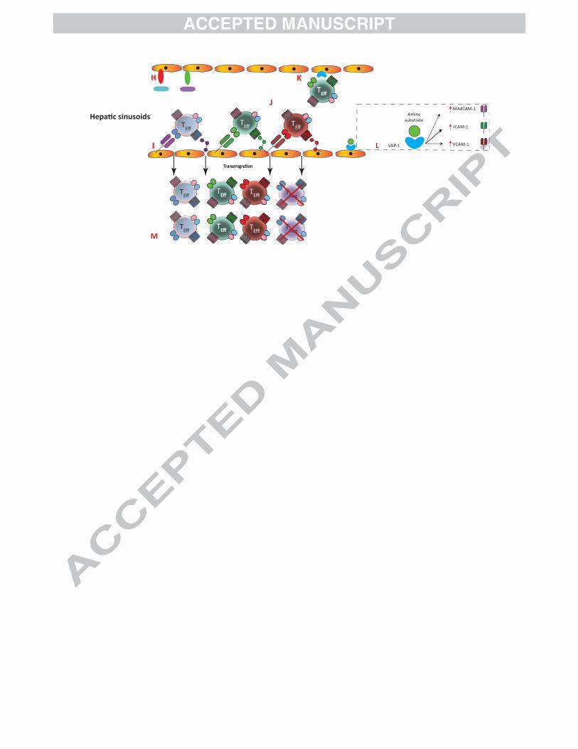
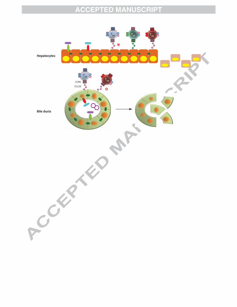
Figure 1: Gut-Liver Immunity in Primary Sclerosing Cholangitis (PSC)
[Top Panel] In a genetically predisposed individual, alterations in the gut
microbiome [A], or abnormal handling of commensal species through epithelial
pattern recognition receptor (PRR) defects [B] may result in heightened innate
immune activation as well as toxic bile acid transformations [C]. Naïve lymphocytes,
imprinted with gut-tropism by intestinal dendritic cells (DC) [D], localise within the
intestinal mucosa via MAdCAM-1/α4β7 and CCL25/CCR9 dependent mechanisms.
Effector (as opposed to regulatory) T-cell responses predominate in IBD [E] driving
intestinal inflammation leading to a defective epithelial barrier [F], exacerbated by the
loss of protective macrophage populations [G].
[Middle Panel] As a consequence of intestinal inflammation enteric pathogens
translocate beyond the mucosal barrier to the portal circulation and liver where they
can drive local inflammation via PPR activation [H]. Mucosal effector lymphocytes
bearing a 'gut-tropic' phenotype are recruited in response to hepatic endothelial
expression of CCL25 and MAdCAM-1 [I] together with effector cells primed locally
[J]. The adhesion molecule and ectoenzyme VAP-1 is upregulated during chronic
inflammation and supports both lymphocyte adhesion directly [K] and catabolises
amine substrates secreted by gut bacteria resulting in upregulation of several
endothelial adhesion molecules, including MAdCAM-1, on sinusoidal endothelium
[L]. Recruited effector cells overwhelm local regulatory networks (M).
[Bottom Panel] After entering the liver, effector cells use chemokine receptors such
as CCR6 to respond to chemokines secreted by epithelial target cells (hepatocytes [N]
or biliary epithelium [O]) resulting in cell-mediated immunological attack and bile
duct destruction. Hepatobiliary damage is likely to be enhanced through the action of
toxic bile acids and heightened PRR activation.

7
References
[1] Balmer ML, Slack E, de Gottardi A, Lawson MAE, Hapfelmeier S, Miele L, et
al. The liver may act as a firewall mediating mutualism between the host and its
gut commensal microbiota. Sci Transl Med 2014;6:237ra66.
doi:10.1126/scitranslmed.3008618.
[2] Moriwaki K, Balaji S, McQuade T, Malhotra N, Kang J, Chan FK-M. The
Necroptosis Adaptor RIPK3 Promotes Injury-Induced Cytokine Expression and
Tissue Repair. Immunity 2014;41:567–78. doi:10.1016/j.immuni.2014.09.016.
[3] Schneider KM, Bieghs V, Heymann F, Hu W, Dreymueller D, Liao L, et al.
CX3CR1 is a gatekeeper for intestinal barrier integrity in mice: Limiting
steatohepatitis by maintaining intestinal homeostasis. Hepatology 2015:n/a – n/a.
doi:10.1002/hep.27982.
[4] Trivedi PJ, Adams DH. Mucosal immunity in liver autoimmunity: A
comprehensive review. J Autoimmun 2013;46:97–111.
doi:10.1016/j.jaut.2013.06.013.
[5] Henao-Mejia J, Elinav E, Jin C, Hao L, Mehal WZ, Strowig T, et al.
Inflammasome-mediated dysbiosis regulates progression of NAFLD and
obesity. Nature 2012;482:179–85. doi:10.1038/nature10809.
[6] Sebode M, Peiseler M, Franke B, Schwinge D, Schoknecht T, Wortmann F, et
al. Reduced FOXP3(+) regulatory T cells in patients with primary sclerosing
8
cholangitis are associated with IL2RA gene polymorphisms. J Hepatol
2014;60:1010–6. doi:10.1016/j.jhep.2013.12.027.
[7] Ohnmacht C, Park J-H, Cording S, Wing JB, Atarashi K, Obata Y, et al. The
microbiota regulates type 2 immunity through RORγt+ T cells. Science
2015;349:989–93. doi:10.1126/science.aac4263.
[8] Sano T, Huang W, Hall JA, Yang Y, Chen A, Gavzy SJ, et al. An IL-23R/IL-22
Circuit Regulates Epithelial Serum Amyloid A to Promote Local Effector Th17
Responses. Cell 2015. doi:10.1016/j.cell.2015.08.061.
[9] Fergusson JR, Hühn MH, Swadling L, Walker LJ, Kurioka A, Llibre A, et al.
CD161(int)CD8+ T cells: a novel population of highly functional, memory
CD8+ T cells enriched within the gut. Mucosal Immunol 2015.
doi:10.1038/mi.2015.69.
[10] Esplugues E, Huber S, Gagliani N, Hauser AE, Town T, Wan YY, et al. Control
of TH17 cells occurs in the small intestine. Nature 2011;475:514–8.
doi:10.1038/nature10228.
[11] Cordell HJ, Han Y, Mells GF, Li Y, Hirschfield GM, Greene CS, et al.
International genome-wide meta-analysis identifies new primary biliary cirrhosis
risk loci and targetable pathogenic pathways. Nat Commun 2015.

TReg
Gut Lumen
Commensal
flora
Pathogenic
bacteria
Bacterial amines
PRR
Bile acid altera ons
E-CadherinGut epithelium
Mesenteric lymph
node
A B
D
C
E
F
G
DC
MAdCAM-1 CCL25
Transmigra on
Mucosal vessel
Mac
CCR9L-selec n
TN
TReg
CCR9
CCR6 α4β1
α4β7
TEff
TEff
TEff
TReg
H
I
K
J
Hepa c sinusoids
TReg
ICAM-1
VAP-1
Amine
substrate
VCAM-1
MAdCAM-1
Transmigra onTransmigra on
L
M
TEff
TEff
TEff
TEffTEff
TEffTEff
TEffTEff
TEffTTEffTEff
TEffTTEff
TEffTTEffTEff
TEff T
Eff
Bile ducts
Hepatocytes
TEff
TEff
TEff
TEff
CCL20
CCR6TEff
N
O


