
Hemodynamic Regulation of Occludin and
ZO-1: Effect on Barrier Function
A dissertation submitted for the degree of Ph.D
By
Nora Collins B.A. (Mod.)
Under the supervision of Prof. Paul A. Cahill
And Dr Philip M. Cummins
June 2006
Vascular Health Research Centre
Faculty of Science and Health, Dublin City University, Dublin 9,
Ireland

Declaration
I hereby certify that this material which I now submit for assessm ent on the program o f
study leading to the award o f Doctor o f Philosophy is entirely m y ow n w ork and has not
been taken from the w ork o f others save and to the extent that such w ork has been cited
and acknowledged w ithin the text o f my own work.
Date: - f / 0 ^ /o£>
II

I would like to thank Dr. Phillip Cummins for all his help and suggestions over
the past few years. A t times I ’m sure he wishes he had never heard o f occludin and ZO-
1! I would also like to thank Prof. Paul Cahill for the opportunity to spend several great
years at the ever-changing VHRC. In addition, many thanks to Yvonne, Gail and Ronan.
Also deserving o f a mention are my fellow permeability group members Olga, Nick,
Maria and Paul. I would especially like to thank Paul ‘F itz’ for all the laughter, tears, tea
and chocolate! A nother special thanks to m y family, especially m y two sisters, Mary and
Roseanna and my friends for all their support and help, needless to say m ost o f them will
still not understand w hat I do! Finally, a very special thanks to Diego, who is always
there for me.
Acknowledgments
Ill

Abstract
The vascular endothelium constitutes a highly effective fluid and solute barrier through the regulated apposition o f tight junction protein com plexes between adjacent endothelial cells. As endothelial cell-mediated functions and pathology are sensitive to hemodynamic forces (cyclic strain and shear stress), we hypothesised a dynamic regulatory link between endothelial tight junction assem bly/function and hemodynamic stimuli. We have investigated this hypothesis via exam ination o f the precise effects o f cyclic strain on the expression, modification, and function o f occludin and ZO-1, pivotal components o f the tight junction complex. Moreover, the mechanotransduction pathway by which tight junction regulation occurs was also investigated.
For these studies, cultured bovine aortic endothelial cells (BAECs) were subjected to physiological levels o f equibiaxial cyclic strain (0 or 5% strain, 60 cycles/min, 24 h).
In response to strain, protein expression o f both occludin and ZO-1 increased. Increased mRNA levels were also observed for occludin, but not Z O -1. These changes were accompanied by tyrosine dephosphorylation o f occludin and serine/threonine phosphorylation o f ZO-1, modifications that could be com pletely blocked by pharmacological inhibition w ith dephostatin (tyrosine phosphatase) and rottlerin (protein kinase C), respectively. The effects o f cyclic strain on the association and subcellular localisation o f occludin and ZO-1 was also investigated. In response to cyclic strain we observed a significant increase in endothelial occludin/ZO-1 co-association in parallel with increased localisation o f both proteins to the cell-cell border. Moreover, these events were completely blocked by dephostatin and rottlerin and w ere accompanied by a strain-dependent reduction in transendothelial permeability to FITC-dextran, an event that could also be blocked by inhibitor addition indicating a causal relationship between biochemical changes and barrier function.
We observed that these modifications o f the tight junction following strain were mediated via Gi-proteins, the small GTPase Rac-1, and the signaling molecule p38.
Overall, these findings indicate that physiological cyclic strain up-regulates vascular endothelial tight junction assembly, with subsequent consequences for barrier integrity, putatively via tyrosine phosphatase- and PKC-dependent modification o f occludin and ZO-1. The signal is transduced via G-proteins, Rac-1 and p38.
IV

Abbreviations
BAEC Bovine Aortic Endothelial Cell
BCA Bicinchoninic Acid
BSA Bovine Serum Albumin
CACO-2 Human Colorectal Adenocarcinoma Cell Line
cDNA Complimentary DNA
CVD Cardiovascular Disease
DAG Diacylglycerol
DMSO Dimethylsulfoxide
DNA Deoxyribose nucleic acid
EC Endothelial Cell
ECM Extra Cellular M atrix
EDTA Ethylenediamine Tetracetic Acid
EGF Epidermal Growth Factor
EGFR Epidermal Growth Factor Receptor
eNOS Endothelial Nitric Oxide Synthase
ERK Extra Cellular Regulated Kinase
ET -1 Endothelin-1
FAK Focal Adhesion Kinase
FBS Fetal Bovine Serum
FGF Fibroblast Growth Factor
FGFR Fibroblast Growth Factor Receptor
FD40 Fluorescein Isothiocyanate Dextran 40kda
GAPDH Glyceraldehyde Phosphate Dehydrogenase
GEF Guanine Nucleotide Exchange Factor
GFP Green Fluorescent Protein
GPCR G-Protein Coupled Receptor
Grb-2 Growth Factor Binding Protein-2
HBSS Hanks Buffered Saline Solution
HPF High Power Field
HUVEC H um an Umbilical Vein Endothelial Cells
V

IB Imm unoblot
ICAM-1 Intracellular A dhesion M olecule-1
IHF Irish Heart Foundation
ILGF Insulin Like Growth Factor
ILGFR Insulin Like Growth Factor Receptor
IP Immunoprécipitation
JAM-1 Junctional Adhesion M olecule 1
JNK C-JunN -Term inal Kinase
LDL Low Density Lipoprotein
M APK M itogen Activated Protein Kinase
MAPKK M itogen Activated Protein Kinase Kinase
M APKKK M itogen Activated Protein Kinase Kinase Kinase
MCP-1 M onocyte Chemo Attractant Protein-1
M DCK M adin-Darby Canine Kidney Cells
M EK M APK Kinase
M MP M atrix M etalloproteinase
mRNA M essenger Ribonucleic Acid
NOS N itric Oxide Synthase
PAGE Polyacrylamide Gel Electrophoresis
PBS Phosphate Buffered Saline
PCR Polymerase Chain Reaction
PDGF Platelet Derived Growth Factor
PDGFR Platelet Derived Growth Factor Receptor
PI-3 Kinase Phosphoinositide 3 Kinase
PKA Protein Kinase A
PKC Protein Kinase C
PMA Phorbol 12-Myristate 13-Acetate
PTK Protein Tyrosine Kinase
PTX Pertussis Toxin
RNA Ribonucleic Acid
ROCK Rho Kinase
VI

ROS Reactive Oxygen Species
RTK Receptor Tyrosine Kinase
RT-PCR Reverse Transcriptase Polymerase Chain Reaction
SDS Sodium Dodecyl Sulphate
She Src Homology/Collagen
siRNA Small Interfering RNA
SMC Smooth M uscle Cell
SNP Single Nucleotide Polymorphism
SSRE Shear Stress Response Element
TBS Tris Buffered Saline
TEE Transendothelial Exchange
TEER Transendothelial Electrical Resistance
TJ Tight Junction
TGF Transforming Growth Factor
T N Fa Tum our N ecrosis Factor-Alpha
VEGF Vascular Endothelial Growth Factor
ZO-1 Zonula-Occludens 1
V I I

Units
bp Base Pairs
cm Centimeters
cm2 Centim eter Squared
°C Degree Celsius
g Grams
h Hours
kDa Kilo Daltons
L Liter
M M olar
mg M iligrams
min M inute
ml M ililiter
mm M ilim eter
mM M ilimolar
ng N anogram
OD Optical Density
pM Picom olar
rpm Revolution Per Minute
sec Seconds
U Enzym e Units
Mg M icrogram
pi M icroliter
pm M icrom eter
pM M icrom olar
V/v V olum e per volume
W/v W eight per volum e
V Volts
w Watts
x g G force

Publications and Poster Presentations
Publications
N.T. Collins. O.C. Colgan, Y.A. Bim ey, G. Ferguson, R.P. M urphy, P.A. Cahill and
P.M. Cummins. Cyclic strain-mediated regulation o f vascular endothelial tight junction
assembly: putative roles for integrins, G-proteins and Rho-GTPase signaling pathways.
Arteriosclerosis, Thrombosis, & Vascular Biology. 2006; Submitted.
N.T. Collins/P.M. Cummins 0 , Y.A. Bimey, O.C. Colgan, G. Ferguson, R.P. Murphy,
G. Meade and P.A. Cahill. Cyclic strain-mediated regulation o f vascular endothelial
occludin and ZO-1: influence on intercellular tight junction assembly and function.
Arteriosclerosis, Thrombosis, & Vascular B iology. 2006; 26; 62-68. [Joint First Author]
0
O.C. Colgan, N.T. Collins. Y.A. Bim ey, G. Ferguson, P.A. Cahill and P.M. Cummins.
Polar-specific regulation o f vascular endothelial occludin and ZO-1: the influence o f
astroytes and serum. A JP -C ell Physiology. 2006; Submitted.
O.C. Colgan, N.T. Collins. Y.A. Bim ey, G. Ferguson, P.A. Cahill and P.M. Cummins.
Regulation o f m icrovascular endothelial tight junction assembly and barrier function by
laminar and pulsatile shear stress. C ardiovascular R esearch 2006; In Preparation.
D. Morrow, C.H. Sweeney, Y A . Bimey, S. Guha, P.M. Cummins, R. Murphy, N.T.
Collins. D. Walls, E.M. Redmond and P.A. Cahill. Sonic hedgehog regulates vascular
smooth muscle cell fate in vitro through VEGF activation o f notch signaling. Circulation
Research. 2006: In Revision.
IX

Poster Presentations
O.C. Colgan/P.M. Cummins, N.T. Collins. Y.A. B im ey, G. Ferguson and P.A. Cahill.
Co-culture o f bovine brain microvascular endothelial cells (BBM vEC) and c6 glioma
decreases endothelial permeability and increases tight junction formation. (A ccepted fo r
P oster Presentation, ATVB, D enver 2006).
O.C. Colgan, N.T. Collins. Y.A. Bimey, G. Ferguson, P.A. Cahill and P.M. Cummins.
Shear stress regulates microvascular endothelial occludin and ZO-1 and increases tight
junction formation. (A ccep ted fo r Poster Presentation, ATV B, D enver, 2006).
N.T. Collins. P.M. Cummins, O.C. Colgan, G. Ferguson, R.P. M urphy and P.A. Cahill.
Cyclic strain regulates occludin and ZO-1 expression, phosphorylation and localisation
in bovine aortic endothelial cells. Poster presentation at A m erican H eart Association
Annual Conference, Dallas: Circulation Supplem ent 111 (2005), 112 (17): abstract 597.
N.T. Collins. P.M. Cummins, O.C. Colgan, G. Ferguson, R.P. M urphy and P.A. Cahill.
Cyclic strain regulates occludin and ZO-1 expression, phosphorylation and localisation
in bovine aortic endothelial cells. Poster presentation at A m erican H eart Association
Annual Conference, N ew Orleans: C irculation Supplem ent 111 (2004), 110, (17): 111-
222.
N.T. Collins. P.M. Cummins, O.C. Colgan and P.A. Cahill. The effects o f mechanical
forces on endothelial cell permeability in peripheral vascular beds. 15th Annual Meeting
o f Irish Association o f Pharmacologists 2004: Dublin, Ireland.
X
Table of Contents:
Title page IDeclaration IIAcknowledgements IIIAbstract IVAbbreviations VUnits VIIIPublications IXTable of Contents XI
Chapter 1: Introduction
1.1 Cardiovascular Disease 2
1.2 The Endothelium 3
1.3 Mechanical Forces and the Endothelium 5
1.3.1 Cyclic Strain 61.3.1.1 M odeling o f Cyclic Strain 61.3.1.2 Effect o f Cyclic Strain on Endothelial Cells 7
1.3.2 Shear Stress 91.3.2.1 Effect o f Shear Stress on Endothelial Cells 9
1.4 Mechanotransduction 11
1.4.1 Integrins 121.4.1.1 Structure o f Integrins 121.4.1.2 Function o f Integrins 131.4.1.3 Integrins in M echanotransduction 141.4.1.4 Integrins and Tight Junction Regulation 17
1.4.2 Heterotrim eric G-proteins 181.4.2.1 Structure and Function o f Heterotrimeric G-proteins 181.4.2.2 G-Proteins as M echanotransducers 221.4.2.3 G-Proteins and Tight Junction Regulation 23
1.4.3 Protein Tyrosine Kinase (PTK) 241.4.3.1 Receptor Tyrosine Kinases 251.4.3.2 Non- Receptor Tyrosine Kinases 261.4.3.3 PTKs in M echanotransduction 271.4.3.4 PTKs and Tight Junction Regulation 27
1.4.4 Ion Channels 281.4.5 Intracellular Signaling M olecules 29
1.4.5.1 M itogen-Activated Protein Kinases 29
XI

1.4.5.1.1 M itogen-Activated Protein Kinasesand Tight Junction Regulation 31
1.4.5.2. GTPases 321. 4.5.2.1 Rho GTPase Family 33
1.4.5.3 Protein Kinase C 371.4.5.3.1 PKC and Tight Junction Regulation 38
1.4.5.4 Tyrosine Phosphatase 381.4.5.4.1 Tyrosine Phosphatase and Tight Junction Regulation 39
1.5 Cell-Cell Interactions in Endothelial Cells 41
1.5.1 A dherens Junctions 411.5.2 Gap Junctions 421.5.3 Desm osom es 421.5.4 Tight Junctions 43
1.5.4.1 M easurement o f TJ perm eability 44
1.6 Proteins of the Tight Junction 46
1.6.10ccludin 461.6.1.1 Discovery and Structure 461.6.1.2 Function o f Occludin 481.6.1.3 Unique Roles o f Specific Occludin Domains 49
1.6.2 ZO-1 511.6.2.1 Discovery and Structure 511.6.2.2 ZO-1 Function 52
1.6.3 Interaction o f Occludin and ZO-1 531.6.4 Claudins 541.6.5 JAM 55
1.7 Mechanical forces and Tight Junctions 56
1.7.1 M echanical Forces and Atherosclerosis 57
1.8 Tight Junctions and Disease 59
1.9 Relevance and Objectives of this Study 60
Chapter 2: Materials and Methods 63
2.1 Materials 64
2.2 Cell Culture Methods 71
2.2.1 Culture o f Bovine Aortic Endothelial Cells 712.2.2 Cell Counting 722.2.3 Cryogenic Preservation and Recovery o f Cells 72
XII

2.2.4 Cyclic Strain 732.2.5 Treatment w ith Pharmacological Inhibitors 742.2.6 Preparation o f W hole Cell Lysates 752.2.7 Bicinchoninic Acid Protein M icro Assay 76
2.3 Western Blotting Studies 76
2.3.1 W estern Blotting 762.3.2 Imm unoprécipitation 79
2.4 Measurement of Transendothelial Permeability 81
2.5 Immunocytochemistry 82
2.5.1 Standard and Confocal Imm unocytochemistry 82
2.6 RNA Preparation Methods 83
2.6.1 RNA Isolation 832.6.2 Spectrophotometric Analysis o f Nucleic Acids 842.6.3 Reverse Transcription Polymerase Chain Reaction 842.6.4 Reverse Transcription 852.6.5 Polymerase Chain Reaction 862.6.6 Agarose Gel Electrophoresis 88
2.7 Statistical Analysis 89
Chapter 3: Results Section 1 90
3.1 Introduction 91
3.2 Results 93
3.2.1. Cyclic strain regulation o f occludin and ZO-1 protein andmRNA levels. 93
3.2.2. Cyclic strain affects subcellular localisation o f occludin, ZO-1and actin. 96
3.2.3. Cyclic strain-dependent co-association o f occludin/ZO -1. 993.2.4. Effect o f cyclic strain on transendothelial permeability. 1003.2.5. Effect o f cycloheximide on cyclic strain regulation o f
occludin and Z O -1. 103
3.3 Discussion 109
3.4 Conclusion 113
XIII

Chapter 4: Results Section 2 115
4.1 Introduction 116
4.2 Results 118
4.2.1. Cyclic strain-dependent occludin and ZO-1 phosphorylationinB A E C s. 118
4.2.2. Effect o f Dephostatin on strain-induced occludin tyrosinedephosphorylation. 123
4.2.3. Effect o f Rottlerin on occludin serine/threonine phosphorylation. 1244.2.4. Effect o f Dephostatin on occludin co-association w ith Z O -1. 1264.2.5. Effect of Dephostatin and Rottlerin on cyclic strain-dependent
subcellular localisation o f occludin. 1274.2.6. Effect o f Rottlerin on strain-induced ZO-1 serine phosphorylation. 1304.2.7. Effect o f Dephostatin on ZO-1 tyrosine phosphorylation. 1314.2.8. Effect of Rottlerin on strain-induced occludin/ZO-1 co-association. 1324.2.9. Effect o f Rottlerin and Dephostatin on cyclic strain-dependent
subcellular localisation o f ZO -1. 1334.2.10. Effect o f Rottlerin and Dephostatin on cyclic strain-induced
transendothelial permeability in BAECs. 136
4.3 Discussion 138
4.4 Conclusion 142
Chapter 5: Results Section 3 144
5.1 Introduction 145
5.2 Results 147Cyclic strain regulation o f occludin and ZO-1. Effect of:
5.2.1. Genistein 1475.2.2. PTX 1535.2.3. cRGD 1595.2.4. 1RGD 1635.2.5. Rho Kinase blockade 1665.2.6. Rac-1 blockade 1725.2.7. p38 blockade 1765.2.8. M EK blockade 170
5.3 Discussion 182
5.4 Conclusion 188
XIV

Chapter 7: References 205
Chapter 6: Final Summary 189
XV

Chapter 1
Introduction
1

1.1 Cardiovascular Disease
Cardiovascular disease (CVD) refers to the class o f diseases that involve the heart
and blood vessels: arteries and veins. Over 50 m illion Am ericans have cardiovascular
problems, and m ost other W estern countries face high and increasing rates o f
cardiovascular disease. It is the number 1 cause o f death and disability in the United
States and m ost European countries. More than 8,700 people in Ireland died from heart
disease in 2004 and the number o f people diagnosed w ith the disease has increased
rapidly in recent years and now stands at 25,000 per annum [David Labanyi, Irish Times
health supplement 7/3/06]. Ireland is one o f the w orst countries in the European Union
in terms o f cardiac disease prevalence and death rates [Dr. Brian M aurer, IHF], The cost
o f cardiovascular disease in Europe is estimated to be at €169 billion per annum or
€3,724 per capita [David Labanyi, Irish Times health supplem ent 7/3/06].
It is clear to see that CVD is extremely prevalent in our society today. However
much can be done to prevent or limit the damage caused by this disease. There is
increased emphasis on preventing CVD by modifying risk factors, such as healthy
eating, exercise and not smoking. In fact w om en’s deaths from heart disease in Europe
could be halved if they stopped smoking and improved their diet, a major European
conference on w om en’s cardiovascular health was told in M arch 2006. However some
risk factors for developing the disease, such as gender, age, fam ily history and ethnicity,
cannot be modified. Hemodynam ic forces w ithin the vasculature also influence CVD.
These forces associated with the flow o f blood through the vasculature affect the
initiation and progression o f CVDs, including atherosclerosis, hypertension and

pathological vascular rem odelling [Lusis et al., 2000; Frangos et al., 2001].
1.2 The Endothelium
The vascular endothelium is a dynamic cellular interface between the vessel wall
and bloodstream where it regulates the physiological effects o f humoral and
biomechanical stimuli on vessel tone and remodelling. The endothelial monolayer forms
the lining o f the entire inner surface o f the vascular system from the heart to the
capillaries. It is therefore well located to respond to and m onitor changes in blood flow
and biochemical composition. The single layer o f cells that is the endothelium, is not
ju st an inert container for blood but a vital organ whose health is essential to normal
vascular physiology and whose dysfunction can be a critical factor in the
pathophysiology o f diseases, such as hypertension and atherosclerosis .
Blood vessels are active, integrated organs com posed o f endothelial cells (EC),
smooth muscle cells (SMC) and fibroblasts divided into structural layers (See Fig. 1.1).
The innermost layer is com posed o f a single layer o f contact inhibited ECs. The tunica
intima is a simple squamous epithelium surrounded by a connective tissue basement
membrane w ith elastic fibres. The tunica media is prim arily com prised o f SMCs, which
play a key role in maintaining vascular tone and function. The outermost layer, which
attaches the vessel to the surrounding tissue, is term ed the tunica adventitia. This is a
layer o f connective tissue, with varying amounts o f elastic and collagenous fibres.
The inner m ost layer o f cells, the endothelial cells, will form the focus o f this
3

study, and in particular the junctions that form between adjacent cells. Because o f their
structure, location and intercellular arrangement, ECs are able to respond to a wide
variety o f stimuli and are employed in a number o f unique roles within the vasculature,
such as maintenance o f a semi-permeable barrier that allows specific substances to move
between the blood and the interstitium. ECs are also involved in regulation o f
haemostasis. This involves maintaining a balance between pro- and anti-coagulant forces
in the circulation, insuring fluid movement o f blood. Another role o f the endothelium is
to carry out inflammatory responses that involve recruitment o f leukocytes, leading to a
potentiation o f the healing process. The endothelium also serves to regulate vascular
tone. The vessel has the ability to respond to changes in the microenvironment to
maintain optimal blood flow.
IBIood vesseOs:
Fig. 1.1. Structure o f Blood Vessels. Diagrammatic representation o f an artery and vein, showing different layers o f the vessel, [http://www.chemie.tu-darmstadt.de/ bt/Endothel .html].
4

1.3 Mechanical Forces and the Endothelium
Among the physiological stimuli that impact on the endothelium, mechanical or
hemodynamic forces associated with blood flow are of central importance. These
include cyclic circumferential strain, caused by a transmural force acting perpendicularly
to the vessel wall, and shear stress, the frictional force of blood dragging against cells
(See Fig. 1.2). Both of these forces are essential to maintain a healthy vessel. They also
have a profound impact on endothelial cell metabolism and can induce qualitative and
quantitative changes in endothelial gene expression leading to changes in cell fate
[Patrick et ah, 1995; Traub etal., 1998; Chien et al., 1998].
Fig. 1.2. Biomechanical stimulation of a blood vessel. Hemodynamic forces associated with blood flow play a pivotal role in the physiological control of vascular tone, remodelling and the initiation and progression of vascular pathologies. These forces include cyclic circumferential strain acting perpendicularly to the vessel wall causing outward stretching of both vascular ECs and SMCs and thus, rhythmic distension of the arterial wall, and fluid shear stress, the frictional force generated as blood drags against vascular endothelial cells.
5

1.3.1 Cyclic strain
1.3.1.1 Modeling of Cyclic Strain
All cells of the vessel wall can detect and respond to the physical force cyclic
strain, which results from the pulsitile flow of blood in the vessel. The effects of
circumferential stress on ECs have been investigated by applying cyclic stretch to
endothelial cells cultured on an elastic membrane mounted in a stretch device such as
that shown in Fig. 1.3. During this procedure, ECs are grown on a flexible membrane,
which can be precisely deformed by a microprocessor-controlled vacuum, providing
equibiaxial tension. This allows the cells to be subjected to defined levels of cyclic
strain, in a variety of wave patterns [Banes et al., 1985]. In this way, we can monitor the
effect of various levels of cyclic strain, physiological levels or detrimentally high levels
for example, on specific endothelial cell markers. Normal blood pressure is considered
to be 120/80 mm Hg, whereas blood pressures of above 140/90 mm Hg and below 90/60
mm Hg are considered high and low respectively. Factors ranging from physical
exertion to psychological stress can result in a transient rise in blood pressure, and a
consequent transient increase in cyclic stress. Genetic predisposition to hypertension can
lead to a chronic increase in cyclic stress, resulting in potentially serious clinical
manifestations. Conversely, factors such as electrolyte imbalance, ischemic heart disease
and systemic sepsis can result in transient hypotension, and a consequent transient
decrease in cyclic stress (American Heart Association).
6

B t o H e x ®
Q iliri Plato
BoF)**® Baseplate & Gasket
Fig. 1.3. In vitro cyclic strain device; The Flexercell™ Tension Plus FX-4000T™ system (Flexcell International Corp.-Hillsborough NC).
1.3.1.2 Effect of Cyclic Strain on ECs
There have been many studies investigating the effects of cyclic strain on aortic
ECs (Lehoux et al., 2005; Sipkema et al., 2003]. Both in vitro and ex vivo models reveal
that cyclic strain has a profound impact on endothelial metabolism and can induce
qualitative and quantitative changes in gene expression leading to changes in cell fate,
with consequences for endothelial phenotype and vessel wall homeostasis [Patrick et al.,
1995; Traub et al., 1998; Chien et al., 1998]. In addition to affecting the expression
and/or activation of numerous signaling molecules associated with
mechanotransduction, cyclic strain has been shown to regulate the expression and/or
activation of several classes of effector genes (and gene products) in vascular ECs,
including those regulating: (i) Vessel diameter - nitric oxide (NO), nitric oxide synthase
(NOS), cyclooxygenase-2 (COX-II), endothelin-1 (ET-1) and thimet oligopeptidase
h M »Cfctui
7

[Coen et al., 2004; Cotter et al., 2004]; (ii) Proliferation - platelet-derived growth factor
(PDGF) and vascular endothelial growth factor [Sumpio et al., 1998; Zheng et al.,
2001]; (iii) Migration - urokinase plasminogen activator (uPA), plasminogen activator-
inhibitor type-1 (PAI-1), monocyte chemotactic protein type-1 (MCP-1), MMP-2,
MMP-9 and MT1-MMP (membrane type-1 matrix metalloproteinase, MMP-14)
[Ulfhammer et al., 2005; von Offenberg Sweeney et al., 2004a; von Offenberg Sweeney
et al., 2004b; von Offenberg Sweeney et al., 2005; Wung et al., 1997]; (iv) Cell-cell
communication/barrier function - intracellular adhesion molecule type-1 (ICAM-1),
zonula occludens 1 (ZO-1) and occludin [Collins et al., 2006; Pradhan et al., 2004]; and
(v) Angiogenesis - MMP-2, MMP-9, MT1-MMP, uPA and RGD-dependent integrins
[von Offenberg Sweeney et al., 2005; Yamaguchi et al., 2002].
Vascular ECs sense and respond to cyclic strain both morphologically and
phenotypically. The influence of cyclic strain on ECs is visually apparent as early as 15
min post-strain with the formation of actin stress fibres and morphological alignment of
cells perpendicular to the force vector [Iba et al., 1991]. Alignment is subsequently
followed by phenotypic or cell fate changes, such as those mentioned above.
Physiological levels of cyclic strain on aortic ECs for example have been shown to
increase both migration [von Offenberg Sweeney et al., 2005] and proliferation [Iba et
al., 1991; Li et al., 2005], whilst concomitantly reducing apoptosis [Haga et al., 2003;
Liu et al., 2003].
8
1.3.2 Shear Stress
The endothelium is also exposed, and sensitive to, shear stress, which results
from blood flow through the vessel. The mean shear stress to which the endothelium is
subjected to is approximately 10-70 dynes/cm2. However, arteries do experience
variations in pressure and blood flow, which can be influenced by the architecture of the
vasculature. At certain positions, such as bifurcations in the vessel, or points of extreme
curvature the vessel may be exposed to turbulent flow, oscillatory shear stress and eddy
currents, all of which can abrogate the protective effects of laminar shear.
Laminar blood flow within a vessel can be described by the equation: t = 4^Q /
7tr3; where x is shear stress, |a is blood viscosity, Q is flow rate and r is the vessel radius.
The term r is raised to the third power thus where Q is constant, therefore a small change
in r will result in a large change in r [Lehoux et ah, 2003].
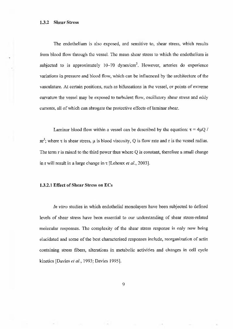
1.3.2.1 Effect of Shear Stress on ECs
In vitro studies in which endothelial monolayers have been subjected to defined
levels of shear stress have been essential to our understanding of shear stress-related
molecular responses. The complexity of the shear stress response is only now being
elucidated and some of the best characterised responses include, reorganization of actin
containing stress fibers, alterations in metabolic activities and changes in cell cycle
kinetics [Davies et al., 1993; Davies 1995].
9

Shear stress induces extensive changes in endothelial cell behaviour and has been
implicated in vasculogenesis, re-endothelialisation of vascular grafts, atherosclerosis,
and angiogenesis [Urbich et al., 2002]. Shear stress stimulates several signaling cascades
in endothelial cells including: potassium channel activation (the earliest), elevation of
inositol trisphosphate and diacylglycerol, an increase in intracellular calcium levels, G
protein activation, M APK signalling, and activation of transcription factors, such as
nuclear factor kB (Davies, 1997; Azuma et al., 2001]. The early signaling events are
followed by changes in gene expression and alignment o f actin filaments and
microtubules within the flow direction, resulting in changes in cell shape and directional
migration [Braddock et a l, 1998; Hsu et al., 20011.
Recent studies have also shown that laminar shear stress modulates many genes
which regulate cell fate (i.e. cell cycle progression and apoptosis). Physiological levels
o f laminar shear stress prevent apoptosis in ECs in response to a variety o f stimuli,
including tum our necrosis factor-a, oxidized LDL and angiotensin 11 [Dimmeler et al.,
1999]. Shear stress also upregulates gene products involved in actin remodelling (e.g. fi-
actin and m yosin heavy chain). Fluid shear stresses generated by blood flow in the
vasculature can also profoundly influence the phenotype o f the endothelium by
regulating the activity o f certain flow-sensitive proteins (for example, eNos) [Topper et
al., 1999]. Flow-dependent activation o f eNOS has been observed both in vitro and in
vivo, and the resultant release o f NO has been related to SMC relaxation as a response to
increases in flow. In conjunction with flow-mediated increases in vasodilators such as
NO, levels o f vasoconstrictors including ET-1 have been found to be decreased. A
10
number o f genes involved in thrombosis, homeostasis, and inflam m ation such as
thrombomodulin, tissue plasminogen activator (t-PA) and vascular cell adhesion
molecule-1 (VCAM -1), have all been identified as being shear responsive. Thus, shear
stress profoundly affects the health and functions o f the endothelium.
1.4 Mechanotransduction
Vascular cells have the ability to respond to the m echanical forces cyclic strain
and shear stress. The cells must be able to detect this hem odynam ic stimulus in order to
respond to it. This is achieved by several mechanically sensitive receptors/detector
molecules present in vascular cells. These receptors can then elicit a signaling pathway,
which culminates in the recruitment/activation o f an effector m olecule(s) to mediate the
cellular response. This process is referred to as mechanotransduction. M echanical forces
initiate complex signal transduction cascades that lead to functional changes in the cell.
The main classes o f mechanotransduction receptors present on EC are integrins, G-
proteins, protein tyrosine kinases (PTKs) and ion channels, each o f which will be
discussed in greater detail.
There has been very little investigative work in determining the
mechanotransduction pathway by which cyclic strain and shear stress illicit their effects
on tight junction permeability (See section 1.5.4 for inform ation on tight junctions). In
fact, to the best o f our knowledge, this study is the first definitive attempt to determine
the pathway from onset o f cyclic strain to tightening o f the EC barrier.
11

Essential to the coordination o f cellular functions in response to hemodynamic
stimuli is the ability o f cells to communicate w ith each other. The process o f
intercellular signaling is achieved through numerous molecules, which interact
specifically w ith specialized docking sites on the cell surface, called receptor proteins.
The source o f these molecules may be; i) autocrine secretion o f a signaling molecule,
that targets the secretory cell itself, ii) paracrine secretion o f a signaling molecule that
targets a cell close to the signal releasing cell or iii) endocrine secretion o f a signaling
molecule from a gland, that targets a cell distant from itself. Intracellular signaling,
results in the coordination and synchronization o f cell function w ithin a given tissue (e.g.
the vessel wall). Following the binding o f a ligand to a receptor, intracellular effector
molecules are activated leading to alterations in cell structure and/or function [Stone
1998]. As the small GTPases RhoA and Rac-1 have been im plicated as key permeability
regulators, especially w ith regards to barrier dysfunction, it was decided to look at the
effect o f these intracellular signaling molecules on the cyclic strain-regulation o f tight
junctions in ECs. We have also elected to examine the signaling molecules p38 and
MEK, as the M APK pathway can be activated in response to mechanical forces.
Furthermore, several studies have linked M APK pathways to TJ protein regulation (See
section 1.4.5.1.1).
1.4.1 Integrins
1.4.1.1 Structure of Integrins
Integrins are integral membrane proteins that are found in the basolateral plasma
12

membrane o f cells. They are obligate heterodimers comprised o f a and p subunits (See
Fig. 1.4). There are eighteen a and eight (3 subunits. In addition, variants o f some o f the
subunits are formed by differential splicing. Each possible combination o f subunits has
its own binding specificity and signaling properties [Giancotti et al., 1999]. These
subunits can form twenty-four different integrins, sixteen o f which are reportedly
involved in the vasculature with seven expressed in endothelial cells [Rupp et al, 2001].
Each subunit consists o f a large NH 2-terminal extracellular domain, a single
membrane-spanning domain and a short cytoplasmic domain [Shyy et al, 2002].
integritiheterodimer
ligand-binding site binds extracellular matrix
a p
cytosolic domain binds cytoskeletor
Fig. 1.4. The basic structure o f the integrin, a cell surface protein receptor.
[http://www.bioteach.ubc.ca].
1.4.1.2 Function of Integrins
The first main function o f integrins is attachment o f the cell to the extracellular
matrix (ECM). The extracellular domain binds directly to ECM proteins such as

fibronectin, vitronectin, collagen, laminin, fibrinogen and osteopontin. The connection
between the cell and the ECM anchors the cell and enables it'to endure physical forces.
The second integrin function is signal transduction from the ECM to the cell. The
cytoplasmic tail o f integrins are generally devoid o f enzym atic activity. As a result o f
this, integrins transduce signals via adaptor proteins such as She, which connect the
integrin to the cytoskeleton, cytoplasmic kinases and transm em brane growth factors
[Giancotti et al., 1999]. It is the role integrins play in cell signaling, where
hemodynamic forces are concerned that we are m ost interested in.
1.4.1.3 Integrins in Mechanotransduction
Integrin signaling is a crucial component in development, maintenance and
function o f the vascular system [Ruoslahti et al., 1997]. Integrins have the unique
characteristic that they can signal through the cell m em brane in either direction,
essentially forming a bridge between the ECM and the cytoskeleton. Shear stress and
cyclic strain activates integrins by switching them into an active conformation, which
increases the extracellular domains affinity and avidity for the ECM proteins. The
cytoplasmic domains o f both the a and (3 subunits interact w ith intracellular signaling
molecules and cytoskeletal proteins to regulate cellular events, such as signal
transduction, cytoskeletal reorganization and cell motility [Shyy et al., 2002].
Evidence for mechanical activation o f integrins is provided by both direct
assessment o f integrin conformational changes in response to these forces and blockade
o f the induced responses by antibodies or blocking peptides such as the Arg-Gly-Asp
14

(RGD) peptide [Lehoux et al., 1998]. In this way it has been show n that shear stress and
cyclic strain causes an increase in integrin activity in ECs.
Integrin activation is directly associated w ith m em bers o f the Rho small
GTPase family (see section 1.4.5.2.1), including RhoA, Cdc42 and Rac-1 [Shyy et al.,
2002]. Other families associated w ith integrin activation include m ultiple kinases (FAK,
and c-Src), adaptor molecules (p l3 0 Gas and She), and guanine nucleotide exchange
factors (C3G and SOS). W hen these signaling molecules are activated by integrins they
can then go on to activate M APKs such as ERK, which can then illicit cellular responses
to shear stress or cyclic strain. See Fig. 1.5 for an exam ple o f integrin-mediated
mechanotransduction.
15

6
nriDol
Fig. 1.5. Schematic diagram of how forces applied through the ECM (A) or directly to the cell surface (B) travel to integrin-anchored focal adhesions through matrix attachments or cytoskeletal filaments, respectively. Internally generated tension and forces transmitted through cell-cell contact similarly reach focal adhesions through the cytoskeleton. Forces concentrated within the focal adhesion (magnified at bottom of figure) can stimulate integrin clustering and induce recruitment o f additional cytoskeletal linker proteins (Vin, vinculin; Pax, paxillin; Tal, talin) that connect directly to microfilaments and indirectly to microtubules. Forces applied to this specialised cytoskeletal adhesion complex also activate integrin-associated signal cascades. Focal adhesion kinase (FAK) may be involved in She recruitment as well as modulation o f Rho activity which, in turn, can regulate the force response through m D ial. Caveolin-1 (cav-1) can also recruit She to integrins to activate the ERK cascade. CD47 associates with the integrin heterodimer to form a protein complex with seven transmembrane segments that mimics the action o f G protein-coupled receptors. In the case shown, when integrins are mechanically stressed, the complex stimulates Gs-mediated up- regulation o f the cAMP cascade through adenylate cyclase (AC), resulting in nuclear translocation o f the catalytic subunits o f protein kinase A, PKA-c. MEK, mitogen- activated protein kinase or extracellular signal- regulated kinase kinase; ERK, extracellular signal-regulated kinase; GDP, guanosine diphosphate; ATP, adenosine triphosphate [Alenghat et al., 2002],
16

1.4.1.4 Integrins and Tight Junction Regulation
There has been very little research looking at the direct effect o f integrin
activation by mechanical forces on tight junction perm eability. However, integrins and
Rho family GTPases are intimately connected at multiple levels and control many o f the
same cellular events. Furthermore, it appears that integrins regulate Rho family GTPases
and Rho family GTPases regulate integrins [Schwartz et al., 2000]. Integrins and Rho
family GTPases function coordinate to mediate adhesion-dependent events in cells.
Integrins and GTPases m ight therefore be organized into com plex signaling cascades
that regulate cell behavior.
Therefore, integrins regulate multiple signaling pathways. In fact, dynamic
coupling o f Rho family GTPase activation to the hem odynam ically sensitive integrin
signaling system is now well documented [Grande-García et al., 2005]. In this study,
integrins were found to independently control the translocation o f GTP-bound Rac-1 to
the plasm a membrane in m ouse fibroblast cells. This step is essential for Rac-1 binding
to effectors and eliciting its effect within the cell such as stabalising the tight junction
(See section 1.4.5.2.1). Integrins control Rac-1 signaling by preventing the
internalization o f its binding sites. Therefore, it is possible that hemodynamic forces
elicit changes in endothelial permeability by engaging an integrin/RhoA/Rac-1-GTPase
signaling axis, leading to modulation o f actin cytoskeletal organisation with direct
consequences for tight junction assembly and function. In fact, Rac-1 downstream
signaling is strictly dependent on integrins [Grande-García et al., 2005]. Pak, a Rac-1
effector that is activated by serum in attached cells is not activated in suspended cells
17

after serum stimulation, indicating that adhesion to the ECM couples Rac-1 with its
effector Pak.
A direct link between integrins and tight junction function was discussed in a
study by Tafazoli el al. It was found that Yersinia bacteria attach to beta 1-integrins at the
tight junctions o f M DCK cells. Here the bacteria perturb the F-actin structure and
distribution o f occludin and ZO-1, thereby prom oting paracellular translocation o f
bacteria and soluble compounds [Tafazoli et al., 2000].
1.4.2 Heterotrimeric G-proteins
1.4.2.1 Structure and Function of Heterotrimeric G-proteins
G-proteins discovered by Alfred Gilman and M artin Rodbell (Nobel Prize for
Physiology or Medicine, 1994), are pivotal membrane signaling systems. They are a
vital intermediary between the activation o f receptors on the cell membrane and signal
transduction within the cell. Rodbell demonstrated in the 1960s that GTP was involved
in cell signaling, whilst Gilman discovered the proteins that interacted w ith the GTP to
initiate cell signaling cascades w ithin the cell.
Receptor activated G-proteins are bound to the inside surface o f the cell
membrane. They are com prised o f a G-protein coupled receptor (GPCR) and the
heterotrimeric G-protein complex in addition to the more recently identified regulators
o f G protein signaling (RGS-proteins) and activators o f G-protein signaling (AGS-
18

proteins) [Offermanns 2003]. All o f these receptors have seven membrane spanning
elements that use intracellular loops and their C-term inal tails for interaction with
heterotrimeric G proteins. They consist o f the G a and the tightly associated G(3y
subunits (See Fig. 1.6).
Fig. 1.6. Shared features o f receptors coupled to G proteins. The model illustrates some o f the features predicted to be shared by all receptors that interact directly with G proteins [Taylor et al., 1990].
When G-proteins are activated, ligand receptors catalyses the GDP/GTP
exchange at the a subunit o f the coupled G-protein and prom otes dissociation o f the a
and (3y components, which subsequently activate their effectors, for example adenylyl
cyclase. The duration o f G-protein activation is controlled by the intrinsic GTPase
19

activity o f G a. Following GTP hydrolysis the G a subunit returns to the GDP-bound
conformation and re-associates with the G(3y subunit (See Fig. 1.7).
Fig. 1.7. Following binding o f a ligand, the activated receptor catalyses GDP/GTP exchange at the a subunit which promotes dissociation o f the G-protein complex, the a and py subunits, which subsequently activate their effectors. Following hydrolysis o f GTP by the a subunit the heterotrim er re-associates.
More than twenty G-protein a-subunits have been described which have been
divided into four families based on structural and functional homologies, a s a,j/0, a q, and
0.12. The m ajority o f GPCRs are capable o f activating more than one G-protein subtype,
which leads to initiation o f various signaling cascades. There are some characteristic
patterns o f G-protein activation by specific receptors; the cellular or physiological effect
20

o f a receptor is dependent on which G-protein sub-type it is coupled to.
The a subunits:
• G a s - family: There are two members o f this family, a s and cx0if, as well as four known
splice variants. U pon activation, this group stimulates adenylyl cyclase to increase levels
o f intracellular cAM P and to activate calcium channels. a s is ubiquitously expressed
while a.oif is restricted to neuronal cells specifically olfactory sensory neurons.
• Gal/,, family: This family consists o f an, a l2, a l3, a 0), cc02, cct.rod, c t t-COne, « g u s t and az, all
o f which are highly homologous and have the ability to inhibit adenylyl cyclase, in
addition to activation o f potassium channels. The high degree o f homology between sub-
types may suggest partially redundant functions. Expression o f the various a , sub-types
is very diverse depending on tissue examine (e.g. a ¡2 is predom inant in the mammalian
heart). A defining characteristic o f the Gaj/0 family is sensitivity to pertussis toxin.
Pertussis toxin is produced by Bordetella pertussis and catalyzes the adenosine
diphosphate (ADP)-ribosylation o f a i and a 0 subunits at a cysteine residue near the C-
terminus resulting in uncoupling o f receptor and G-protein. a z unlike the other members
o f this family has been found to be pertussis toxin insensitive and is expressed in various
tissues. It can inhibit adenylyl cyclase but its physiological function is somewhat
ambiguous, although a z-deficient mice point to roles in platelet activation.
• Gaq family: This family stimulates phospholipase C in a pertussis toxin-insensitive
manner. a q and a n are ubiquitously expressed. M oreover receptors activating a q family
members do not discriminate between a q and a n [W ange et al., 1991]. CX15/16 are only
expressed in hem atopoietic cells and is restricted to the kidney, lungs and testis.
21

• G al2 family: a n and a u constitute the members o f this fam ily and appear to be
widely expressed. The function o f these proteins is som ew hat unclear. One recently
discovered function is the interaction o f these proteins w ith cadherins causing the release
o f transcriptional activator p-catenin [Meigs et al., 2001].
The GPy subunit:
This complex is assembled from a repertoire o f five P subunits and twelve y subunits.
The sequence similarity is higher between p subunits (79-90% homology) than y
subunits and it is not yet clear how many combinations w ill actually form stable dimers
[Clapham et al., 1993]. P subunits are believed to posses a propeller structure formed by
seven p sheets. The y subunit is located at one end o f the propeller and associates with
the P subunit by a coiled structure [Bohm et al., 1997], py-sensitive effectors include
adenylyl cyclase, phospholipase C, phospholipase A, potassium channels, calcium
pumps, and phosphoinositide 3 kinase (PI-3 kinase) [Exton 1996; Yamada et al, 1989;
Lotersztajn et al., 1992]. W ith a few exceptions there appears to be no major differences
between different Py-subunit combinations in their ability to activate effector enzymes.
1.4.2.2 G-Proteins as Mechanotransducers
M embrane localisation and rapid activation strongly implicate G-proteins as a
primary sensor o f hem odynamic forces [Gudi et al., 1996], In fact, G-protein activation
by mechanical forces represents one o f the earliest mechanotransduction events reported.
Both PTX-insensitive (G aq) and PTX-sensitive (G ai) subunits are involved in this rapid
22

response [Gudi et al, 1996; Clark et al., 2002]. G-proteins m ay detect mechanical forces
in one o f two ways, either via GPCR or they may be stim ulated directly by the
deformation o f either the actin cytoskeleton or the m em brane phospholipid bilayer
during exposure to cyclic strain or shear stress.
Shear stress and cyclic strain-induced activation o f G-proteins results in several
responses which function in the regulation o f vascular tone, including release o f
vasodilators or vasoconstrictors such as ET-1 [Liu et al., 2003; Pirotton et al., 1987].
Changes in G-protein expression have been observed w ithin the physiological range o f
cyclic strain and shear stress. These changes have been correlated with enhanced NO
and PGI2 release as well as increased G-protein functionality [Redmond et al., 1998].
1.4.2.3 G-Proteins and Tight Junction Regulation
Although there has been very little research looking at the role o f G-proteins as
mechanotransducers in regulation o f occludin and ZO-1, there have been several studies
looking at the expanding role o f G-proteins in tight junction regulation.
Saha et al. observed that a fraction o f G a s was co-localised with ZO-1 in the TJ
o f M DCK cells, supporting the model o f multiple Got subunits interacting with TJ
proteins to regulate the assembly and maintenance o f the TJ [Saha et al., 2001]. Dodane
et al. speculated that the formation and permeability o f tight junctions are actively
regulated by second-messenger-generating systems involving G-proteins. They reported
23

that G a i2 was present at the cell borders along the areas o f lateral cell-cell contact in
MDCK and Caco-2 epithelial cells. This isoform formed a pattern o f distribution very
similar to ZO-1, indicating that it may be part o f the zonula occludens complex and may
locally regulate form ation and permeability o f tight junctions [Dodane et al. 1996],
Experiments by Baida et al. in 1991 observed that pertussis toxin increased
TEER in epithelial cells, indicating that junction form ation may be controlled by a
network o f reactions including G-protein activation [Baida et al., 1991], A lthough this
finding differs from ours, it indicates that G-proteins may be involved in TJ regulation.
Studies by Denker et al. also alluded to G-protein involvem ent in TJ biogenesis. They
observed that the heterotrim eric G-proteins, Gaj-2, and the Got family member, Go. 12
may participate in the maintenance and/or regulation o f TJ assembly in M DCK cells
[Denker et al. 1998].
1.4.3 Protein Tyrosine Kinase (PTK)
Tyrosine phosphorylation o f various proteins is a very important step in the
regulation o f normal cell proliferation, migration, differentiation and survival. PTKs are
integral com ponents o f signal transduction cascades that involve tyrosine
phosphorylation. They can be separated into two distinct categories: receptor and non
receptor tyrosine kinases.
24

1.4.3.1 Receptor Tyrosine Kinases
Receptor Tyrosine Kinases are enzym e-linked membrane receptors displaying
intrinsic tyrosine kinase activity within the receptor itself. Exam ples o f receptor tyrosine
kinases include the insulin-like growth factor receptor (ILGFR), the epidermal growth
factor receptor (EGFR) and the platelet-derived growth factor receptor (PDGFR). These
receptors have an extracellular domain responsible for the binding o f a ligand, for
example, a circulating hormonal stimuli, a transm embrane domain and an intracellular
domain with tyrosine kinase activity.
W hen an agonist binds to the extracellular domain o f a receptor tyrosine kinase,
it leads to the dim erisation o f the receptor i f it is made up o f two chains, such as the
insulin receptor, or in the case o f the EGF receptor, which is a monomer, binding o f a
ligand to the receptor leads to dim erisation o f two receptors. This leads to a
conformational change that causes auto-phosphorylation o f the cytoplasmic domain o f
the receptor (See Fig. 1.8). This subsequently causes the activation o f the tyrosine kinase
activity and can also create a binding site for intracellular adaptor molecules such as Sos
and Grb-2. These adaptor molecules bring other signaling molecules such as the small
GTPase Ras, into close proximity to the receptors’ intracellular domain, thereby eliciting
a signaling cascade leading to a functional cellular response [Stone 1998].
25

Ligand
ExtracellularDomain \
IntracellularDomain
TTtTHTT
Fig. 1.8. Diagrammatic representation of the initial events in receptor tyrosine kinase (RTK) signaling. Extracellular ligand (yellow) binds resulting in oligomerisation. The intracellular tyrosine kinase domains phosphorylate each other, generating a binding site for SH2-containing adaptor proteins such as Grb2. (Y is phosphorylated tyrosine amino acid) [httpi//www.n imr.mrc.ac.uk).
1.4.3.2 Non- Receptor Tyrosine Kinases
Non-receptor tyrosine kinases differ from RTKs as they do not have an
extracellular domain. They are intracellular enzymes that also possess an intrinsic
tyrosine kinase activity. Examples o f non-receptor tyrosine kinases molecules include
the FAK, c-Src and Jak families. These molecules may phosphorylate receptors, which
lack intrinsic tyrosine kinase activity or they may recruit other downstream signaling
molecules such as PI-3 kinase [Stone 1998].
26

1.4.3.3 PTKs in M echanotransduction
Protein tyrosine kinases play an important role in transducing the mechanical
signal. In fact, the activities of PTKs in cardiac myocytes, platelets, and ECs are
increased by mechanical stimuli such as cyclic stretch and shear stress [Sadoshima et a l,
1993; Ishida et a l, 1996]. Focal adhesion-associated tyrosine kinases, e.g., FAK and c-
Src, are rapidly activated in ECs by shear stress [Li et a l, 1997; Jalali et a l, 1998],
PTKs play an important role in activating MAPKs, as genistein, a PTK inhibitor
attenuates the shear stress-induced activation by ERK and JNK. PTKs are also crucial in
the shear stress regulation of cell shape and stress fibers.
PTK-mediated mechanotransduction often involves the participation o f other
receptors such as integrins and G-proteins [Soldi et a l, 1999; Linseman et a l, 1995;
Eguchi et al., 1998; Zwick et a l, 1997].
1.4.3.4 PTKs and Tight Junction Regulation
There has been quite a lot o f research focusing on the role o f non-receptor tyrosine
kinases in TJ regulation. In fact the activity o f Src kinases appears to play a role in both
assembly and disassembly of tight junctions. A study by Busaroy et al. demonstrated
that oxidative stress-induced disruption of tight junctions is mediated by the activation
of c-Src in Caco-2 cells [Busaroy el al., 2003]. In another study by Kale et al. it was
reported that c-Src binds to occludin and phosphorylates it on tyrosine residues leading
to disruption o f the tight junction in Caco-2 cells [Kale et al., 2003]. Occludin has also
27

been found to coloealise with the non-receptor tyrosine kinase, c-Yes, at cell junction
areas and to form an immunoprecipitable complex with c-Yes in vivo [Chen et al.,
2002]. This study provided strong evidence that occludin tyrosine phosphorylation is
tightly linked to tight junction formation in MDCK cells, and that the non-receptor
tyrosine kinase c-Yes is involved in the regulation o f this process.
There has been very little research regarding receptor tyrosine kinase-mediated
mechanotransduction in TJ regulation. However a study by Buchert et al. showed that
the junction-associated protein AF- 6 interacts and clusters with specific Eph receptors,
which are a subfamily o f receptor tyrosine kinases, at specialized sites of cell-cell
contact in the adult rat brain, indicating that TJ proteins may directly associate with
RTKs [Buchert et al., 1999].
1.4.4 Ion Channels
Some ion channels function as mechanotransducers and therefore play a role in
cell signaling. Two different mechano-sensitive channels have been identified in
vascular cells: shear stress activated potassium channels and stretch activated cationic
channels. The exact mechanisms by which mechanical forces regulate ion channel
conformation remains indistinct, though deformation of the cytoskeleton is thought to be
an important contributor. This premise is supported by a number o f studies, including a
study that demonstrates cytoskeleton G-protein coupling in shear-induced potassium
channel opening and integrin-cytoskeleton activation o f ion channels [Lehoux et al.,
2003].
28

There has been very little research investigating the role o f ion channels in
mechanotransduction in relation to TJ formation. However ion channels have been
implicated in TJ function in a study by Broughman and co-workers. In this study it was
reported that NC-1059, a synthetic channel-forming peptide, transiently increased trans-
epithelial electrical conductance across MDCK cell monolayers in a time- and
concentration-dependent manner. However, concomitant alterations in junctional protein
localisation (zonula occludens-1 , occludin) and cellular morphology were not observed
[Broughman et a l 2004], Calcium channels also play an important role in tight junction
function. Indeed, calcium is critical for the maintenance of cell-cell junctions in various
cell types. A study by Chen et al. showed that when Ca2+ was depleted from MDCK cell
culture medium, there was a significant reduction in TEER, indicating a global loss of
the tight junction barrier function. Reconstitution of Ca2+ resulted in an increase in
TEER [Chen et al., 2002], In another study in primary pulmonary endothelial cultures,
chelation o f extracellular calcium increased albumin transfer by 125%, decreased TEER,
and caused retraction of adjacent cells. Restoration o f extracellular calcium restored
normal barrier function [Shasby et al., 1986].
1.4.5 Intracellular signaling molecules
1.4.5.1 Mitogen-activated Protein Kinases
The mitogen-activated protein kinase (MAPK) signaling cascade is an important
29

pathway whose activation can lead to, or stimulate, gene transcription and/or protein
synthesis. The MAPK super-family is comprised o f three main signaling pathways:
• the extracellular signal-regulated protein kinase (ERK),
• the c-jun N-terminal kinases or stress-activated protein kinases (JNK/SAPK),
• the p38 family o f kinases.
Each of the MAPK modules operates as a three-tier system. The MAPK module
is activated by a MAPK kinase (MAPKK), which is a dual-specific kinase, which
phosphorylates ERK, JNK and p38 at both Ser/Thr and Tyr sites. The MAPKK is
activated by a MAPKK kinase (MAPKKK), which receives its stimulus from receptors
on the cell surface. MAPK have a key role in the regulation o f many genes because the
end targets o f these cascades are often nuclear proteins or transcription factors [Cowan
etal., 2003].
Activation o f the MAPK pathway in response to mechanical stimuli may occur
by various means including G-proteins, integrins, receptor tyrosine kinases and
cytoskeleton-associated non-receptor tyrosine kinases. Phosphorylation o f the a and (3y
subunit of a G-protein can lead to MAPK activation in kidney fibroblast cells [Crespo et
al, 1994]. Shear stress has been found to activate ERK1/2 via a G ia/Ras pathway and
JNK via a G(3y/Ras tyrosine kinase pathway in endothelial cells [Jo et al., 1997].
Similarly, small GTPases such as Ras or RhoA are stimulated by mechanical strain and
may regulate ERK1/2 or JNK activation in endothelial and smooth muscle cells [Wung
et a l, 1999; Numaguchi et a l, 1999]. Similarly integrins have been shown to be
30

involved in the activation o f members o f the MAPK family. Chen et al. observed an
increase in the association of a vß3 integrin with the adapter protein She and subsequent
activation of ERK1/2 and JNK under shear conditions in endothelial cells [Chen et al.,
1999],
1. 4.5.1.1 Mitogen-Activated Protein Kinases and Tight Junction Regulation
Several studies have linked MAPK pathways to tight junction protein regulation.
A study by Basuroy et al. shows that ERK interacts with the C-terminal region of
occludin and mediates the prevention of I-^CVinduced disruption o f TJs by epithelial
growth factor in Caco-2 cells [Basuroy et al., 2006]. Another study by Pedram et al.
reported that vascular permeability factor (VEGF) significantly enhances permeability in
aortic endothelial cells via a linked signaling pathway, sequentially involving Src, ERK,
JNK, and phosphatidylinositol 3-kinase/AKT [Pedram et al., 2002]. This led to the
serine/threonine phosphorylation and redistribution o f actin and the tight junction
proteins, zonula occludens- 1 and occludin, and the loss o f the endothelial cell barrier
architecture, thus indicating that MAPK pathways can play a very important role in TJ
regulation.
P38 has also previously been implicated in tight junction regulation. Kelvil et al.
observed that inhibition o f p38 MAP kinase during oxidant challenge in HUVECs
significantly attenuated actin stress fiber formation and prevented gap formation, thus
attenuating the increase in permeability following addition o f hydrogen peroxide [Kelvil
31

et al., 2001]. Another study using Sertoli cells has shown that TJ dynamics are
regulated, at least in part, by TGF-beta3 via the p38 mitogen activated protein (MAP)
kinase pathway. This in turn regulates the production of occludin, by Sertoli cells and a
specific p38 MAP kinase inhibitor, could block occludin loss from the blood testis
barrier [Lui et al., 2003].
I.4.5.2. GTPases
GTPases are a large family of enzymes that can bind and hydrolyze GTP. The
GTP binding and hydrolysis takes place in the highly conserved G domain common to
all GTPases. GTPases play an important role in:
• Signal transduction at the intracellular domain o f trans-membrane receptors,
including recognition of taste, smell and light.
• Protein biosynthesis (translation) at the ribosome.
• Control and differentiation during cell division.
• Translocation o f proteins through membranes.
• Transport o f vesicles within the cell. (GTPases control assembly o f vesicle coats).
One important function o f this sub-set of proteins, are their involvement in signal
transduction. The small GTPases cycle between inactive (GDP-bound) and active (GTP-
bound) states (See Fig. 1.9). Under basal conditions, GTPases are GDP-bound and
inactive. Upon stimulation, for example by cyclic strain, GTPases release GDP and bind
to GTP, a reaction accomplished by guanine nucleotide exchange factors (GEFs). In
32

inactive. Upon stimulation, for example by cyclic strain, GTPases release GDP and bind
to GTP, a reaction accomplished by guanine nucleotide exchange factors (GEFs). In
their active GTP-bound state, GTPases interact with a variety of effector proteins to
promote cellular responses. The active state of GTPases is transient because o f their
intrinsic GTPase activity, which is stimulated further by GTPase activating proteins
(GAPs).
Small ftoirasB
Small GTPtsa
Fig. 1.9. Signal Transduction and Small GTPases. [http://www.piercenet.com]
1.4.5.2.1 Rho GTPase Family
The Rho family o f small GTPases consists of at least 20 members, and three sub
families, Rho (RhoA, RhoB and RhoC), Rac (Rac-1, Rac-2 and Rac-3) and Cdc42
(Cdc42Hs and G25K). Cdc42, Rac-1 and RhoA are the most extensively characterized
members of the Rho family [Mackay et ah, 1998],
33

al., 1998]. The Rho GTPase family o f proteins control the organization o f the actin
cytoskeleton in all eukaryotic cells and also playing a major role in controlling the
stability and integrity o f the tight junction [Kozma et al., 1995]. The Rho GTPase family
of proteins also mediates regulation o f transcription, membrane trafficking and
apoptosis. Rho GTPases are activated by many different stimuli including G-protein-
coupled receptors, tyrosine kinase receptors, integrin clustering or engagement, cell-cell
adhesion and cytokine receptors [Sah et a l, 2000].
Several targets have been identified for Rho A, including p i 60 Rho kinase
(ROCK) and Rhotekin. The activity o f ROCK is enhanced upon binding to GTP-Rho
and leads to phosphorylation o f the regulatory part o f the myosin molecule, myosin light
chain, which enables the myosin molecule to change conformation, interact with actin to
cause F-actin bundling, thereby leading to stress fiber formation [Mackay et al., 1998],
This is a key event in various models of barrier dysfunction [Dudek et al., 2001;
Wojciak-Stothard et a l, 2002]. Therefore, increased Rho kinase activity is detrimental to
barrier function (See Fig. 1.10). As RhoA and ROCK are central to actin cytoskeletal
rearrangement it is thought they may play a pivotal role in junctional disassembly when
stimulated, as the tight junction proteins are intimately linked to the actin cytoskeleton.
34

Thrombin
Shear «trenÜIVK
Caz+
C «M R h o k in a s e
Junctional disassembly
P P IMMLCK
M LC -Pcontractility
♦ BARRIER FUNCTION
Sl-P $2 adrenergic agonists ▼ serotonin .
H O P
P I3K cAMP cOMP+
IQOAPf-----
]V .MHC-P j
I 1
LIM kinase
«m o actility
Fig. 1.10. Barrier Function: Rac and Rho [Wojciak-Stothard et al., 2002]
Conversely Rae-1 activation leads to an increase in barrier function (See Fig.
1.10). In fact, Rac-1 is required for the assembly and maturation o f epithelial and
endothelial junctions and its activity increases during junction formation [Noren et al.,
2001]. Its activation induces actin polymerization to form lamellipodia (broad web-like
extensions), [Kozina et al., 1995], Sphingosine 1-phosphate (S l-P) has been proposed to
act via the activation o f Rac-1. S l-P is a lysophospholipid stored and released by
activated blood platelets that plays a role in improving endothelial barrier function. Sl-P
induces enhancement o f the cortical F-actin ring, which is usually associated with
stabilization o f intercellular endothelial junctions [Garcia et al., 2001]. One o f the target
proteins for Rac-1 is p21-activated kinase (Pak). It is necessary for Sl-P-mediated

enhancement o f endothelial barrier function and also plays a key role in the linkage of
Rac-1 signaling pathways to Ras signaling pathways through the phosphorylation of Raf
and MEK (See Fig. 1.11). LIM kinase is a Pak target, phosphorylating and inactivating
cofilin, an actin severing protein, which could explain the observed accumulation of
cortical F-actin. Rac-1 therefore stabilises tight junctions and counteracts the effects o f
Rho [Dudek et al., 2001; Wojciak-Stothard et al., 2002].
RhoA and Rac-1 have emerged as key permeability regulators acting
antagonistically to regulate endothelial barrier function. Since paracellular transport
through the epithelial and endothelial tight junction is suspected to be controlled by a
‘purse-string’ contraction o f perijunctional F-actin [Madara et al., 1987; Bernent et al.,
1993], RhoA is strongly implicated as it is a potential mediator o f actin. It increases
stress fiber formation and actomyosin contractility to facilitate breakdown of
intercellular junctions and loss of barrier function, whereas Rac-1 stabilises tight
junctions through its effect on cortical F-actin [Dudek et al., 2001; Wojciak-Stothard et
al., 2002]. The ability o f this family of proteins to control the dynamics of cellular actin
polymerisation and reorganisation has fundamental implications for the control of tight
junction permeability [Hopkins et al., 2000],
36

ßmwIhfNfiLirtlrqpWt .taclrns „ Cjtakwes llrteflrjtB Growth ferctora
Wwörem»
- P ''DjrlosBl
« a n i
$tM£S m I m'I !■
w
^ 2 ®
r S EB1/2 1
■ ■(M
-Z Z M CeJJ i|»fajjlyftftsrrifcran* protrusion
ConUrncärjTi lf«8 ) üdtiföitiriStress tTlterlsrmallaii
Grollt]ßiHTeremiJilimi st»B nspntras, aimptois
Fig. 1.11. Signaling pathways mediated by Ras, Racl/Cdc42 or Rho GTPase. [http://www.piercenet.com].
1.4.5.3 Protein Kinase C
The protein kinase C (PKC) family consists of 11 members of serine/threonine
kinases that are activated by diverse mechanisms including receptor tyrosine kinases,
non-receptor tyrosine kinases, and G protein-coupled receptors [Newton, 1995]. PKC
isoforms are classified into three groups based on their structure and particular substrate
requirements: conventional (a, p i, P2, y), novel (5, e, r\, 0, ^), and atypical (¿¡, X, t)
[Newton, 1995].
Conventional PKC isoforms require diacylglycerol (DAG), calcium, and
phosphatidylserine for their activation, whereas the novel isoforms lack the requirement
for calcium, and atypical PKC isoforms require only phosphatidylserine. Treatment of
37

cells with phorbol esters such as PMA, activates PKC by mimicking DAG. PKC activity
is regulated by its subcellular distribution as well as its phosphorylation state. Upon
ligand binding and recruitment to the membrane, PKC becomes a substrate for kinases
resulting in auto-phosphorylation and enhanced catalytic activity [Parekh et al., 2000].
1.4.5.3.1 PKC and Tight Junction Regulation
Activation o f PKC is directly linked to tight junction assembly [Stuart et al.,
1995]. Activation o f PKC by 1,2-dioctanoylglycerol (diC 8 ) stimulated the partial
assembly of tight junctions in MDCK cells grown in low calcium conditions [Baida et
al., 1991]. It has also become apparent that multiple classes o f PKC isoforms contribute
to formation of the tight junction. The signaling events downstream o f PKC that regulate
cell permeability have not been completely elucidated; however, components of the tight
junction complex may be directly phosphorylated by PKC. For instance, ZO-2 was
phosphorylated in vitro by several PKC isoforms [Avila-Flores et al., 2001].
Furthermore, a C-terminal region o f mouse occludin was phosphorylated in vitro by a
purified mixture o f PKC isoforms [Andreeva et a l, 2001]. Therefore, PKC may directly
modify components o f the tight junction complex to regulate permeability.
1.4.5.4 Tyrosine Phosphatase
Protein tyrosine phosphatases are involved in a variety of cellular processes such
as signal transduction, cell cycle regulation, and differentiation. This enzyme is a low
38
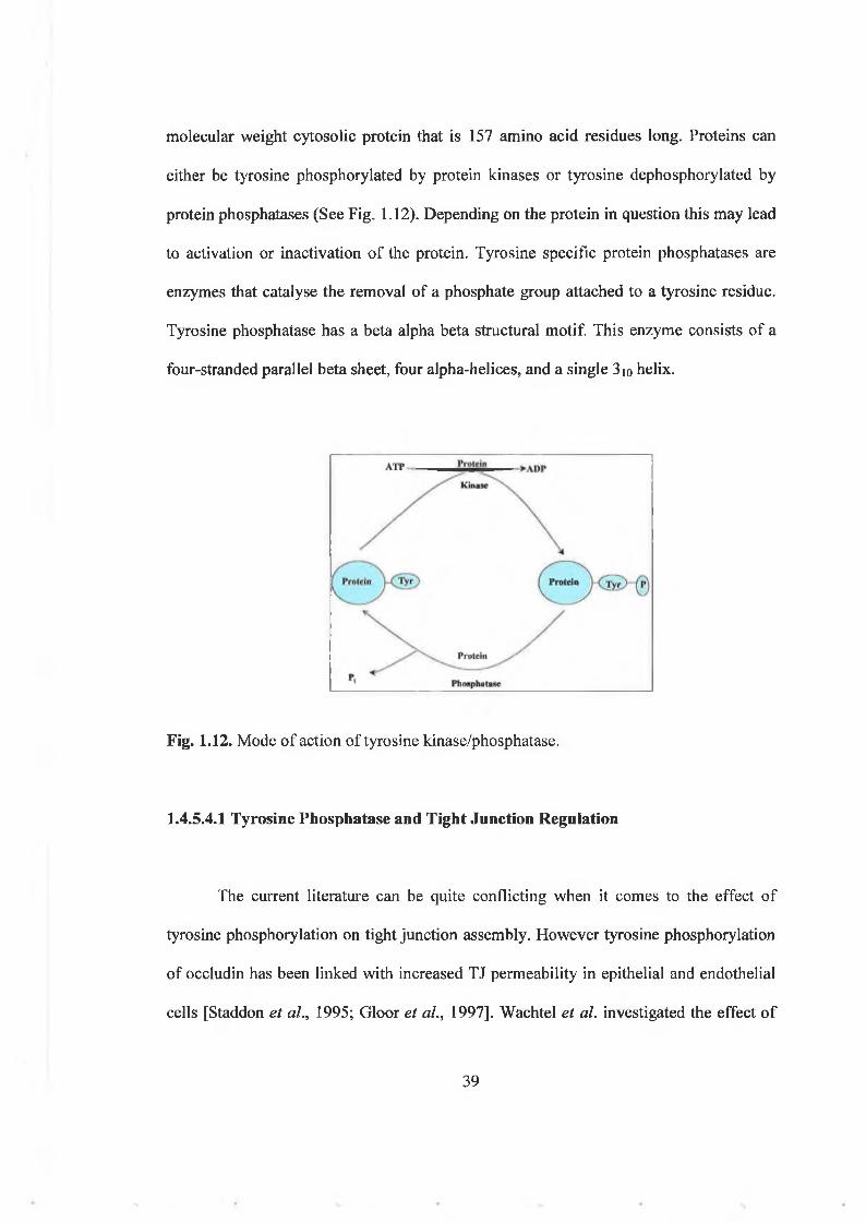
molecular weight cytosolic protein that is 157 amino acid residues long. Proteins can
either be tyrosine phosphorylated by protein kinases or tyrosine dephosphorylated by
protein phosphatases (See Fig. 1.12). Depending on the protein in question this may lead
to activation or inactivation of the protein. Tyrosine specific protein phosphatases are
enzymes that catalyse the removal of a phosphate group attached to a tyrosine residue.
Tyrosine phosphatase has a beta alpha beta structural motif. This enzyme consists of a
four-stranded parallel beta sheet, four alpha-helices, and a single 3io helix.
Fig. 1.12. Mode of action of tyrosine kinase/phosphatase.
1.4.5.4.1 Tyrosine Phosphatase and Tight Junction Regulation
The current literature can be quite conflicting when it comes to the effect of
tyrosine phosphorylation on tight junction assembly. However tyrosine phosphorylation
of occludin has been linked with increased TJ permeability in epithelial and endothelial
cells [Staddon et ah, 1995; Gloor et al., 1997]. Wachtel et al. investigated the effect o f
39

inhibiting protein tyrosine phosphatases on the composition o f endothelial TJs. In this
study it was found that inhibition of protein tyrosine phosphatase resulted in the
cleavage of occludin and subsequent loss o f occludin localisation at the TJs via an
MMP-dependent step [Wachtel et al., 1999].
The tyrosine phosphorylation level o f the intracellular C-terminal tail of
occludin, C-occludin, has been described to be critical in determining the binding ability
of occludin to other TJ proteins such as ZO-1, ZO-2, and ZO-3 [Kale et al., 2003]. In
this study, using Caco-2 cells, it was shown that the amounts o f ZO-1, ZO-2, and ZO-3
bound to tyrosine phosphorylated C-occludin were several fold less than the amounts of
these proteins bound to non-phosphorylated C-occludin. Thus, ample evidence in the
literature exists to suggest that tyrosine phosphorylation o f occludin can lead to a
decrease in barrier integrity and conversely that activation o f tyrosine phosphatases may
lead to an increase in barrier function.
40

1.5 Cell-Cell Interactions in Endothelial Cells
There are four main types of intercellular junctional complexes found in ECs;
adherens junctions, gap junctions, desmosomes and tight junctions (See Fig.. 1.13).
{iijJ959 Jt+’ n VWey s c * Sons. Inc »ghB re s id e d
Fig. 1.13. Diagrammatic representation of the types of junctions found in ECs.
1.5.1 Adherens Junctions
Adherens junctions, located below the region of tight junctions, contain the trans
membrane proteins cadherins, whose extracellular segments bind to each other and their
cytoskeletal linkers, catenins [Daniel et al., 1997; Kaibuchi et al., 1999], They provide
strong mechanical attachments between adjacent cells. For example, they hold cardiac
muscle cells together as the heart expands and contracts. Although spatially and
biochemically distinct, functional interaction between adherens junctions and tight
junctions has been demonstrated [Rubin et al., 1999].
41

1.5.2 Gap Junctions
Gap junctions, seen in virtually all cells that contact other cells in tissues, provide
open channels through the plasma membrane, allowing ions and small molecules (less
than approximately 1 kDa) to diffuse freely between neighboring cells. The gap
junction’s major physiological role therefore, is to couple both the metabolic activities
and the electric responses o f the cells they connect. An example o f their importance in
physiology is their role in electrical coupling. Gap junctions are abundant in cardiac and
smooth muscle cells and depolarization of one group of muscle cells rapidly spreads to
adjacent cells, leading to well-coordinated contractions o f those muscles [Sohl et al.,
2005]. Gap junctions are constructed o f trans-membrane proteins called connexins. Four
or sometimes six connexins assemble to form a cylinder with an open aqueous pore in its
center. Such an assembly o f connexins in the plasma membrane o f one cell then aligns
with the connexins o f an adjacent cell, forming an open channel between the two
cytoplasmic compartments. The plasma membranes o f the two cells are separated by a
gap corresponding to the space occupied by the connexin extracellular domains—hence
the term “gap junction,” which was coined by electron microscopists [Sohl et al., 2005].
1.5.3 Desmosomes
Desmosomes, a further junctional complex found in endothelial cells, and in
epithelial cells such as skin cells, are specialized for cell-cell adhesions. Like adherens
junctions, they are also cadherin-based. They anchor a second cytoskeletal filament
network, intermediate filaments, which are strong elastic polymers, to the plasma
42

membrane. Coupled to the cytoplasmic surface of the desmosome, they form a
supracellular network that strengthens tissues, protecting them against mechanical
damage.
1.5.4 Tight Junctions
Tight junctions, or zonula occludens, are the most apical component of the
intercellular junctional complex [Harhaj et al., 2004]. Farquhar and Palade originally
identified the tight junction by electron microscopy in 1963 [Farquhar et al., 1963].
Tight junctions form a barrier to diffusion o f ions and small molecules from the lumen to
the tissue parenchyma (barrier function) and restrict the diffusion of lipids and proteins
between the apical and basolateral plasma membranes (fence function) [Farquhar et al.,
1963; Van Meer et al., 1986]. However, in a relatively short time, our knowledge of the
tight junction has evolved from this relatively simple view o f it being a permeability
barrier in the paracellular space and a fence in the plane o f the plasma membrane, to one
of it acting as a multi-component, multi-functional complex that is involved in
regulating numerous and diverse cell functions [Schneeberger et al., 2004].
The tight junction is composed of anastamosing strands o f 10 nm fibrils that
completely encircle the apical region o f the cell. These fibrils are composed of trans
membrane proteins that interact with proteins on adjacent cells [Staehelin 1973]. Tight
junctions form in polarized epithelial and endothelial cells as well as in a variety of
specialized epithelial cell types, including those o f oligodendrocytes of the central
nervous system, Sertoli cells in the testis, the choroid plexus, the stria vascularis in the
43

organ of Corti, and the collecting tubules of the kidney in some cases [Gow et al., 1999].
Tight junctions are composed o f multiple trans-membrane, scaffolding, and
signaling proteins, including occludin, claudin family members, junctional adhesion
molecules 1 to 3, cingulin, 7H6, spectrin, and linker proteins, such as the zonula
occludens family members (ZO-1/2/3), the latter linking tight junction proteins to each
other and the actin cytoskeleton [Furuse et al., 1993; Furuse et al., 1994; Gardner et al.,
1996; Hirase et al., 1997]. (See Fig. 1.14).
01 H5T?153j!S
[ST
ParaceliularS p a c e
Fig. 1.14. The arrangement o f tight junction proteins in endothelial cells [Feldman et al.,
2005].
1.5.4.1 Measurement of TJ permeability
Tight junction permeability in cell culture is generally measured using either of
two techniques: trans-epithelial/trans-endothelial electrical resistance (TEER) or
44

paracellular tracer flux (See Fig. 1.15). TEER measurements are commonly used to
assess the integrily of tight junctions. Electrical resistance across a monolayer represents
the sum of the paracellular resistance, which consists o f the resistance of the junction
and the intercellular space, and the transcellular resistance, which consists of the
resistances of the apical and basolateral cell membranes [Claude, 1978]. Paracellular
tracer flux may also be used to measure tight junction integrity. In this method,
permeability to hydrophilic, uncharged paracellular tracers is measured using radioactive
or fluorescently conjugated tracers such as mannitol or dextran. Tight junction
permeability to particular solutes depends on the size, shape, and charge of the solute
[Dvorak, et al„ 1999]. Paracellular tracer flux measures permeability of a monolayer
over a time course of several hours, whereas TER represents an instantaneous
measurement.
FD40
A pical su rfa c eC u ltu re d ish
if1'
: ■ ¡Apical m e d iu m. j! j u j : . vn,T
• • - « a t* s
i ;B asa l m e d iu m
Basal Porous lamina filter M o n o la y e r o f
B A E C s
TF.F.RIII
Fig. 1.15. Diagrammatic representation o f measurement o f TEER and paracellular tracer flux in BAECs.
45

1.6.1 Occludin
1.6.1.1 Discovery and Structure
Occludin was the first tight junction integral membrane protein to be identified
and was originally isolated from chick livers [Furuse et al., 1993]. The nucleotide
sequence o f chicken occludin encodes a 504 amino acid polypeptide with a predicted
molecular mass o f 55.9 kDa. The amino acid sequence predicted four putative
membrane-spanning segments, two 44-amino acid extracellular loops and two
intracellular domains (See Fig. 1.16). Furuse et al. established that the amino acid
sequence o f the first extracellular loop is highly concentrated with tyrosine and glycine
residues (-60% ) [Furuse et al., 1993]. Further cDNA cloning and sequencing disclosed
more definitive information about the structure of occludin [Furuse et al., 1994]. It was
determined that there were four transmembrane domains in the NH 2 -terminal half of
occludin, which divide the protein into five separate domains and were referred to as
domains A -E . The COOH-terminal half (domain E) is comprised o f -250 amino acid
residues and is located in the cytoplasm. These early observations also revealed that
charged amino acids were strongly concentrated in COOH-terminal cytoplasmic domain
(domain E) and there was a very high content o f tyrosine and glycine residues in the
extracellular domains (domains B and D) [Furuse et al., 1994],
It wasn’t confirmed that occludin was present in mammals until work in 1996 by
Ando-Akatsuka et al. Chicken occludin knowledge was limiting when it came to gene
1.6 Proteins o f the Tight Junction
46

targeting experiments and therefore the identification o f mammalian occludin genes was
another important discovery. Ando-Akatsuka first reported the nucleotide sequences o f
cDNAs encoding rat-kangaroo (potoroo), human, mouse, and dog occludin. As found in
chicken occludin, all o f the occludins expressed in these species contained four trans
membrane domains. The amino acid sequences o f human, murine, and canine occludin
are approximately 90% homologous but have significantly deviated from the avian and
marsupial homologues. Each o f the species is capable o f forming a common a-helical
coiled structure. It was postulated that this segment o f the amino acid sequence was
retained in order to interact with occludin’s binding partner ZO-1 [Ando-Akatsuka el al.,
Fig. 1.16. Schematic representation of occludin structure. Adapted from Feldman et a!.,
1996],
Ifig h m c h B g e d ia la s t 150 a r■ binds to ZO -1 a u iZ O -2 *N B p n d U a pomeahalitv
2005.
47

1.6.1.2 Function o f Occludin
The function o f occludin was initially investigated by introducing chick occludin
cDNA into MDCK cells in a Lac-inducible vector [McCarthy et al., 1996]. In this study
occludin expression was induced by the addition of isopropyl-h-d-thiogalactoside
(IPTG) to the medium. 5 h after addition of IPTG, the TER across the MDCK
monolayer increased progressively, relative to control level. The TER level across the
transfected cells was 30-40% higher than the TER across the control monolayer at 31-h
post-induction, indicating that the expression of occludin correlates with barrier
properties. This can also be observed in various other tissues. For example, arterial
endothelial cells express 18-fold greater occludin protein levels than venous endothelial
cells and form a tighter solute barrier [Kevil et al., 1998]. Similarly, occludin is highly
expressed in brain endothelium which forms a very tight barrier, but occludin is
expressed at much lower levels in endothelial cells o f non-neuronal tissue, which have
lower barrier properties than brain endothelium [Hirase et al., 1997], Clearly, occludin is
a key tight junction protein whose expression levels dictate tissue barrier properties.
However, somewhat surprisingly, occludin is not required to maintain the structural
integrity of tight junctions, since structurally intact tight junctions were formed in the
absence of occludin [Saitou et al., 1998]. These findings indicate that there are as yet
unidentified TJ integral membrane proteins which can form strand structures, recruit
ZO-1, and function as a barrier without occludin.
48

1.6.1.3 Unique Roles o f Specific Occludin Domains (See Fig. 1.16)
COOH-terminus
The COOH-terminus o f occludin is necessary for correct TJ assembly and
function [Chen et al., 1997]. In this study, Xenopus embryo were used to show that full-
length and COOH-terminally truncated occludin localize at the TJ. However, it was
reported that the TJs containing the mutant occludin proteins were leaky and this leakage
induced by the COOH-terminally-truncated occludin could be corrected by transfection
with full-length occludin mRNA. The COOH-terminus o f occludin binds to ZO-1 (See
section 1.6.3).
N-terminus
A study by Bamforth et al. investigated the localisation o f N-terminally truncated
occludin in the murine epithelial cell line CSG 120/7. An N-terminally truncated
occludin construct, was correctly targeted to the TJ and co-localised with ZO-1.
However, the truncated construct generated a detrimental effect on the barrier function
o f the TJs, as demonstrated by electrophysiological measurements [Bamforth et al.,
1999]. Overall, the result o f removing the N-terminus was a disruption of the TJ’s
sealing/barrier properties.
49
Extracellular and Transm em brane Domains
Wong and Gumbiner have previously employed synthetic peptides (Occ-1 and
Occ-2) corresponding to the two putative extracellular domains of occludin and studied
their ability to modulate TJs in A 6 cells (Xenopus kidney epithelial cell line). The
second extracellular domain peptide (Occ-2) reversibly disrupted the integrity o f the TJ
barrier. A subsequent study discovered that the second extracellular loop was also
required for occludin localisation at the TJ [Medina el al., 2000].
It has also been reported in a recent study that multiple extracellular and trans
membrane domains o f occludin are implicated in the regulation o f the TJ barrier [Baida
et al., 2000]. It was observed that the extracellular domains and at least one of the
transmembrane domains were crucial in maintaining selective paracellular permeability.
Coiled-coil domain of COOH-terminal region
Studies by Ando-Akatsuka et al. showed that occludin’s coiled-coil domain
appears to function as a site for specific interactions o f potential regulatory proteins
[Ando-Akatsuka et al., 1996]. Photoactivation studies demonstrated that c-Yes, the
regulatory (p85) subunit o f PI 3-kinase, PKC-t, and the gap junction component,
connexin 26, interacted with this domain. These findings provide the first evidence to
support a highly specific interaction between occludin and regulatory proteins that had
been previously implicated in TJ modulation.
50

1.6.2 ZO-1
The first protein identified as a TJ constituent was ZO-1 [Stevenson et al., 1989;
Anderson et al., 1988]. ZO-2 and ZO-3 were later isolated as proteins that co-associate
with ZO-1. ZO-1 (210-225 kDa), ZO-2 (180 kDa), and ZO-3 (130 kDa) are peripheral
membrane-associated components o f the cytoplasmic plaque o f tight junctions and are
found ubiquitously within tight junctions of epithelial and endothelial cells [Harhaj et
al., 2003]. In addition, ZO-1 and ZO-2 are also localised to adherens junctions in cells
that do not form tight junctions, such as fibroblasts and cardiac myocytes [Itoh, et al.,
1999]. ZO proteins are members of the membrane-associated guanylate kinase
(MAGuK) homologue family, containing three PDZ domains, an SH3 domain, and a
non-catalytic guanylate kinase (GuK) homology domain [Woods et al., 1993]. ZO
family members also contain an acidic domain, a basic domain, a leucine zipper
dimerisation motif, and a proline-rich C-terminus [Haskins et al., 1998; Jesaitis et al.,
1994; Willott et al., 1993; Beatch, et al., 1996]. ZO-1 expression may be regulated at the
post-transcriptional level by alternative splicing. Specifically, ZO-1 contains an
alternatively spliced 80 amino acid domain within its C-terminus, the a-m otif [Willott et
al., 1993], likely explaining why researchers often see two bands when monitoring ZO-1
by Western blotting.
1.6.2.1 Discovery and Structure
51

1.6.2.2 ZO-1 Function
Because ZO-1 is found in the TJ in all epithelial and endothelial cells, its
proximity to the membrane bilayer as determined by immuogold electron microscopy
[Stevenson et al., 1986], and its close stoichiometry to junction fibril particles [Anderson
et al., 1988], suggests ZO-1 is an important component o f the tight junction.
One o f its most important functions arises form the fact that ZO-1 has binding
sites for many cellular proteins via multiple protein-binding domains. As previously
mentioned, occludin co-associates with ZO-1. This co-association occurs at specific
domains within the N-terminal (MAGUK-like) half o f ZO-1. The PDZ-1 domain of ZO
family members also binds to the C-terminal regions o f claudins-1 to - 8 [Itoh, et al.,
1999]. ZO-1 binds to JAM in vitro and in vivo [Ebnet et al., 2000]. ZO-1 also interacts
with cingulin in vitro. [D’Atri, et al., 2002], ). In addition to tight junction proteins, ZO-
1 also binds to the adherens junction protein, ß-catenin, and the gap junction protein,
connexin-43 [Itoh, et al., 1999].
In addition to binding to many tight and adherens junction proteins, the SH3
domain of ZO-1 may also mediate binding to G-proteins such as Gai2 [Meyer, et al.,
2002]. Furthermore, ZO-1 interacts with the Ras target, AF-6 , and its binding was
disrupted by activated Ras in vitro [Yamamoto et al., 1997].
O f utmost importance is the fact that the distinctive proline-rich C-terminal half
o f ZO-1 co-sediments with F-actin. These findings clarify that ZO-1 establishes a link
52

between occludin and the actin cytoskeleton [Fanning et al., 1998]. One can therefore
hypothesise a dynamic regulatory association between endothelial permeability and
hemodynamic stimuli, as tight junction components are intimately coupled to the
hemodynamically responsive actin cytoskeleton [McCue et al., 2004], It is plausible
therefore that force-dependent modulation of tight junction assembly is a highly likely
process, albeit very poorly understood.
It is clear that ZO-1 plays a pivotal role in organising and promoting interactions
in the tight junction. This is evident from the large array o f cellular proteins that it
interacts with, through multiple binding sites and interaction domains.
1.6.3 Interaction of Occludin and ZO-1
As previously mentioned, the tight junction in endothelial cells consists of an
intimate gathering of various proteins and signaling molecules which help control
paracellular transport and apical and basolateral separation. It has been well documented
that two important proteins, occludin and ZO-1, and more specifically their interaction,
plays a crucial role in tight junction function. In fact, increased co-association of both
proteins, as investigated via immunoprécipitation, can be used as a measure of tight
junction stability. A study by Rao et al. showed that oxidative stress disrupts the tight
junction in part by causing disassociation o f the occludin-ZO-1 complex [Rao et al.,
2002],
To find out more about the interaction of the two proteins, occludin’s Domain E
53

(the COOH-terminal cytoplasmic domain) was expressed in Escherichia coli fused with
glutathione-S-transferase and this COOH-terminal fusion protein specifically bound to a
complex o f ZO-1, ZO-2 and a number of other peripheral membrane proteins (see Fig.
1.17). Subsequent in vitro binding experiments using glutathione-S-transferase fusion
proteins with a range o f deletion mutants o f Domain E narrowed down the sequence
necessary for the ZO-l/ZO-2 association [Furuse et al., 1994]. It was established that the
same sequence is required for occludin to localise at the TJ and to bind to the ZO-l/ZO-
2 complex and is therefore central to tight junction functioning. Furuse’s 1994 study
concluded that ZO-1 is directly bound to the Domain E o f occludin, and that ZO-2 may
be associated with occludin through ZO-1.
E x t r a c e l lu la r
In t r a c e l lu la r
Fig. 1.17. Schematic drawing of the possible molecular architecture o f tight junctions; occludin interaction with ZO-1 and ZO-2 [Furuse et al., 1994].
1.6.4. C laudins
Similar to occludin, the claudins are a family o f membrane spanning proteins
comprised o f four trans-membrane domains, two extracellular loops and cytoplasmic N-
54

and C-termini (see Fig. 1.18). They are also involved in the formation o f tight junctions
in epithelial and endothelial cells. There are 24 members o f the claudin family, all of
which share a YV m otif in the final two amino acids o f the C-terminal that is required
for binding to the ZO family [Furuse et al., 2001]. Claudins are the primary regulators of
the formation of tight junctions [Furuse et al., 1998], They also regulate permeability
and barrier function o f tight junctions, for example, claudin-1 over-expression in MDCK
cells increased TER by four-fold and reduced flux of 4 and 40 kDa dextran compared
with control cells [Inai, et al., 1999],
1.6.5 JAM
Another well documented TJ protein is JAM or Junctional Adhesion Molecule. It
is a 36-41 kDa, single pass trans-membrane protein found in epithelial and endothelial
tight junctions [Martin-Padura et ah, 1998] (see Fig. 1.18). It is thought to play an
important part o f TJ permeability as over-expression of JAM in CHO cells reduced
paracellular permeability to 40 kDa dextran by 50% in a calcium-dependent manner
[Martin-Padura et al., 1998]. JAM has been shown to co-precipitate with ZO-1, which
suggests that JAM may indirectly mediate the recruitment o f occludin to the TJ via ZO-1
[Feldman et al., 2005].
55

65kDa 22-24 kDa2 isoforms 24 isoforms
C lanrlin -I TAM-1
40 kDa 4 isoforms
Fig. 1.18. Integral membrane TJ proteins [Schneeberger et al., 2004],
1.7 Mechanical Forces and Tight Junctions
Because vascular pathologies exhibiting altered vessel hemodynamic loading with
associated remodeling (e.g., atherosclerosis, restenosis, retinopathy, inflammatory lung
disease, sepsis, edema, and systemic carcinomas) frequently correlate with compromised
endothelial barrier integrity [Harhaj et al., 2004; Van Nieuw-Armerongen et al., 2002;
Tinsley et al., 2004; Koschinsky 2004], we hypothesise a direct link between mechanical
forces and EC permeability.
In fact, mechanoregulation o f vascular endothelial tight junction protein expression
has also been confirmed in 2 recent, albeit contrasting, studies. In a report by DeMaio et
al. exposure of BAECs to shear stress reduced occludin mRNA and protein expression
in parallel with an increase in tyrosine phosphorylation and endothelial permeability
(monitored as hydraulic conductivity) [DeMaio et al., 2001]. In a more recent article,
56

Conklin et al. demonstrated shear stress-induced up regulation o f occludin mRNA
[Conklin et al., 2002]. The contrast between these studies may reflect differences in the
shearing paradigms used. Overall, however, these results show us that tight junction
protein expression (and, therefore, permeability) in vascular endothelial cells are subject
to regulation by hemodynamic forces. Consistent with this, a recent study by Shin et al.
demonstrated reduced transendothelial permeability to albumin after exposure o f human
umbilical vein endothelial cells (HUVECs) to chronic pulse pressure (cyclic pressure),
an important hemodynamic component o f pulsatile blood flow (i.e., in addition to, but
distinct from, cyclic strain) [Shin et al., 2003].
1.7.1 Mechanical Forces and Atherosclerosis
The accumulation o f atherogenic substances such as low density lipoprotein
(LDL), growth factors, and fibrinogen in the intima o f arteries is thought to be one of the
initiating factors o f atherosclerosis [Fry 1987; Curmi et al., 1990; Nielson et al., 1992].
It has been shown experimentally that the endothelium in regions o f the arterial system
that tend to collect LDL and other macromolecules may have compromised barrier
function compared to areas which do not [Bell et al., 1974; Kao et al., 1995; Stemerman
et al., 1986]. Furthermore, these areas have been found to correspond in vivo to
branches, bifurcations, and curves in the arterial system that may have certain common
hemodynamic factors such as low shear stress and boundary layer separation [Kao et al.,
1995; Stemerman et al., 1986; Herrmann et al., 1994], Data such as these lead us to
assume that the endothelium is rendered dysfunctional by the aberrant hemodynamic
stimuli in these areas, leading to increased permeability and the build up of atherogenic

assume that the endothelium is rendered dysfunctional by the aberrant hemodynamic
stimuli in these areas, leading to increased permeability and the build up o f atherogenic
particles in the intima [Kao et al., 1995; Penn etal., 1992; Fry 1987].
Fig. 1.19 readily illustrates the fact that atherosclerotic plaques do not occur at
random locations in the vasculature but rather at areas where there are unstable
hemodynamic features. In this diagram the aorta has been stained with Oil-Red-O,
which shows lipid-rich atherosclerotic lesions in the arterial wall. Despite the systemic
nature o f the hyperlipidemia, the lesions in this animal are largely confined to areas
around curvatures and branch points, suggesting that patterns o f blood flow are
important in localising this disease [Topper et al., 1999].
Fig. 1.19. Non-random localisation o f early atherosclerotic lesions in a Watanabe heritable hyperlipidemic (WHHL) rabbit. The upper arrow indicates the arch o f the thoracic aorta, which has been opened up to display the intimal lining en face. The lower arrow indicates the localised nature o f the atherosclerotic lesions adjacent to the paired
*
58

1.8. Tight Junctions and Disease
An intact tight junction is central to normal cell function and disruption of this
junction can lead to many pathophysiological conditions such as the development of
atherosclerosis which has been previously discussed. Enteropathogenic E. coli, an
important cause o f third world infant mortality, produces a protein, EspF, that can
increase TJ permeability and redistribute occludin [McNamara et al., 2001], V. cholera
has also been found to produce a cytotoxin, which can function as a protease and has
been shown to digest occludin bands.
Decreased occludin expression is observed in parallel with diminished barrier
function, in a wide variety o f tissues and inflammatory conditions. Collagenous colitis
occurs with decreased trans-epithelial resistance and decreased expression o f occludin
and claudin-4 in the colon [Burgel et al., 2002]. The best investigated inflammatory
model with regard to epithelial barrier function is however inflammatory bowel disease
(IBD), a condition that has long been associated with altered epithelial barrier function
[Hollander 1988]. The actively inflamed tissue component is observed to be leaky.
Occludin has been observed to be down regulated even in non-actively inflamed tissue
in ulcerative colitis [Kucharzik et al., 2001].
Another disease state that sees changes to tight junctions is diabetes. Occludin
expression is significantly reduced in ECs of the Blood Retinal Barrier (BRB) of
diabetic rats. This occurred simultaneously with increased permeability to
macromolecules [Antonetti et al., 1998; Barber et al., 2003], Elevated VEGF is thought
59

to be responsible for these changes as well as for altered occludin phosphorylation state.
Similarly, occludin content of brain capillaries was sharply reduced in diabetic rats, even
though ZO-1 content was unchanged [Antonetti et al., 1998].
Tight junction integrity is also compromised in epithelial and endothelial cancers.
Although the undermining of TJ barriers occurs early in the process of cancer, it is
unclear yet whether the process is in any way causal or more precisely, promotional, to
the process o f cancer [Feldman et al., 2005]. Processes and agents known to augment the
promotional stage o f cancer induce TJ leakiness [Mullin et al., 1997]. Oncogenes are
likewise known to induce TJ leakiness [Li et al., 2000]. Occludin expression has been
shown to decrease progressively with the carcinoma grade in human endometrial cancer
[Tobiok el al., 2004], moreover occludin distribution and expression are similarly
altered in increasing Gleason grades o f prostate cancer [Busch et al., 2002], In addition,
microvessels o f human brain tumors, whose TJ leaks are responsible for cerebral edema
in certain types o f brain cancer, are another example of downregulation o f occludin in
cancer, a phenomenon perhaps attributable to increased VEGF secretion [Davies 2002],
1.9 Relevance and Objectives of this Study
Loss o f tight junction function has been implicated in many pathophysiological
conditions such as atherosclerosis. The areas where permeability has been compromised
are not random and frequently correlate to areas o f unstable hemodynamic stimulation
and endothelial dysfunction. It is therefore the aim o f this study to examine how
hemodynamic force, in particular cyclic strain, regulates vascular endothelial tight
60

junction assembly and barrier function at the molecular and cellular level.
To this end the tight junction proteins occludin and ZO-1 have been investigated. In
particular the effect o f physiological levels of cyclic strain on occludin and ZO-1 protein
and mRNA expression, subcellular localisation and co-association has been examined.
Phosphorylation o f these proteins is known to be a key event in TJ regulation and as
such has been examined in-depth. These changes have also been correlated to changes in
TJ function by means o f a permeability assay. The mechanotransduction pathway by
which the TJ proteins detect and respond to cyclic strain has also been investigated.
To our knowledge, this is the first in-depth investigation o f this physiologically
significant phenomenon, and as such, it enhances our overall understanding of how
hemodynamic forces regulate tight junctions within the vascular endothelium.
61

The main aim of this thesis therefore is to examine the effect of cyclic strain on
the TJ proteins occludin and ZO-1. The findings of this research project have been
divided into three results chapters with the following objectives:
Chapter 3:
The aim o f this chapter is firstly to examine the regulatory effects o f cyclic strain
on occludin and ZO-1 protein expression, mRNA levels, subcellular localisation and co
association. The effect on actin organisation is also examined, as is the effect o f strain
on transendothelial permeability.
Chapter 4:
The aim o f this chapter is firstly to determine any changes in the
phosphorylation states o f occludin and ZO-1 following strain. Secondly, the pathways by
which occludin and ZO-1 are phosphorylated will be examined using pharmacological
inhibitors. Thirdly the relationship between these phosphorylation events and the
downstream changes reported in the previous chapter will be examined.
Chapter 5:
The specific aim o f this chapter is to investigate the signaling mechanisms
involved in cyclic strain-regulation o f occludin and ZO-1 in vascular ECs, with specific
emphasis on G-proteins, integrins and PTK-mediated pathways.
62

C h a p t e r 2
M a t e r i a l s a n d M e t h o d s
63

All reagents used in this study were o f the highest purity commercially available and
were o f cell culture standard when applicable.
2.1 Materials
AGB Scientific (Dublin, Ireland)
Whatmann chromatography paper
Amersham Pharmacia Biotech (Buckinghamshire, UK)
Anti-mouse 2° antibody, HRP-conjugated
Anti-rabbit 2° antibody, HRP-conjugated
ECL Hybond nitrocellulose membrane
ECL Hyperfilm
Rainbow molecular weight marker, broad range (6-175kDa)
Protein-A Sepharose beads
Protein-G Sepharose beads
Bachem UK Ltd. (St. Helens, UK)
Linear RGD peptide
Cyclic RGD peptide
Bio Sciences Ltd (Dun Laoghaire, Ireland)
DMEM
dNTP’s

DEPC-treated water
Trizol® reagent
Calbiochem (San Diego, CA)
Dephostatin-Tyrosine phosphatase inhibitor
Genistein-Protein tyrosine kinase inhibitor
N SC 23766-Rac-l inhibitor
Pertussis toxin- Gi-protein inhibitor
PD98059-M EK inhibitor
PD169316-p38 inhibitor
PMA-PKC activator
ROCK (Y-27632)-Rho kinase inhibitor
Rottlerin-PKC inhibitor
Cornell Cell Repository (NJ, USA)
Bovine Aortic Endothelial Cells
Cells derived from the thoracic aorta o f a one-year-old male Hereford cow
Passages 7-15 used
Costar (Buckinghamshire, UK)
Transwell®-Clear plates (6 well format, 0.4 |am pore size, 24 m m filter diameter)
Dako Cytomation (UK)
Dako mounting media
65

Dunn Labortechnik GmBH (Asbach, Germany)
6 -well Bioflex® plates
Flexcell International Corp. (Hillsborough, NC)
Flexercell® Tension Plus™ FX-4000T™ system
Scientific Imaging Systems (Eastman Kodak Group, Rochester, NY)
Kodak ID image analysis software
Molecular Probes (Oregon, USA)
Alexia-conjugated anti-rabbit IgG
Alexia-conjugated anti-mouse IgG
Alexia-Phalloidin 568
MWG Biotech (Milton Keynes, UK)
Occludin primer set
ZO-1 primer set
GAPDH primer set
PALL Corporation (Dun Laoghaire, Ireland)
Biotrace nitrocellulose membrane
Pierce Chemicals (Cheshire, UK)
BCA protein assay kit
66

Supersignal W est Pico chem ilum escent substrate
Promega (Madison, WI)
Taq DNA Polymerase
MLV-RT
R N aseH
Oligo-dT
Santa Cruz Biotechnology (CA, USA)
Anti-occludin rabbit polyclonal IgG
Sarstedt (Drinagh, Wexford, Ireland)
T25 tissue culture flasks
T75 tissue culture flasks
T175 tissue culture flasks
6 -well tissue culture plates
5,10 and 25 ml serological pipettes
15 and 50 m l falcon tubes
Sigma Chemical Company (Poole, Dorset, England)
2 -mercaptoethanol
Acetic Acid
Acetone
Agarose
67

Bisacrylamide
Bovine Serum Albumin
Brightline H aemocytometer
Bromophenol blue
Chloroform
Dapi Nuclear Stain
EDTA
EGT A
Ethidium Bromide
Fibronectin
Foetal C alf Serum
FITC-Dextran 40kD a
Tris Acetate
Glycerol
Glycine
Hanks Balanced Salt Solution
Hydrochloric acid
Isopropanol
Leupeptin
M ethanol
Penicillin-Streptom ycin (lOOx)
Ponceau S
Potassium Chloride
Ammonium Persulphate

Potassium Phosphate (Dibasic)
Phosphatase Inhibitor Cocktail
Protease Inhibitor Cocktail
RPMI-1640
Sodium Chloride
Sodium Hydroxide
Sodium Orthovanadate
Sodium Phosphate
Sodium Pyrophosphate
SYBER green Jum p Start Taq Ready M ix
SDS
TEMED
Tris Base
Tris Cl
Triton X-I00
Trypsin-EDTA solution (lOx)
Tween 20
Qiagen (West Sussex, U.K.)
SYBR Green® PCR Kit
Potassium Iodide
69

Anti-ZO-1 rabbit polyclonal IgG
Anti-occludin rabbit polyclonal IgG
Anti-ZO-1 m ouse monoclonal IgG
Anti-occludin m ouse monoclonal IgG
Phosphoprotein Antibody Sampler Pack: phospho-serine, threonine and tyrosine
antibodies
Zymed Laboratories Inc., (San Francisco)
70

2.2 Cell Culture M ethods
All cell culture techniques were carried out in a clean and sterile environment
using a Bio Air 2000 MAC laminar flow cabinet. Cells were visualized using an
Olympus CK30 phase contrast microscope unless otherwise stated.
2.2.1 Culture of Bovine Aortic Endothelial Cells_
D ifferentiated BAECs were obtained from Coriell Cell Repository, N ew Jersey,
USA. (CAT NO: AG08500). The cells w ere derived from a one-year-old male Hereford
cow. The thoracic aorta was removed immediately post-m ortem on 22/10/85. Cells were
maintained in RPM I-1640 supplemented w ith 10% (v/v) fetal bovine serum (FBS), 100
2 2U/ml penicillin and 100 (ig/ml streptomycin. Cells were cultured in T175 cm , T75 cm ,
T25 cm2 flasks and 6 well plates. In the case o f cyclic strain experiments, cells were
grown on Bioflex® series culture plates which have a flexible, pronectin bonded growth
surface. In the case o f dextran permeability studies cells were grown on 24 m m round
polyester filters, (0.4 |im pore size, 6 well format). Cells between passages 7-15 were
used in these experiments.
BAECs are a strongly adherent cell line forming a confluent contact-inhibited
monolayer, w ith a distinct cobblestone morphology. As such, trypsinisation was
necessary for sub-culturing or harvesting o f cells. For trypsinisation, growth media was
removed from the flask and the cells were gently washed two times in Hanks buffered
saline solution (HBSS) to remove a-m acroglobulin, a trypsin inhibitor present in FBS. A
71

s u ita b le v o lu m e o f t r y p s in / e th y le n e d ia m in e te tr a c e t ic a c id ( E D T A ) ( 1 0 % v / v t r y p s in
E D T A in H B S S ) w a s a d d e d to th e f la s k a n d in c u b a te d f o r 5 m in o r u n t i l a l l th e c e lls
w e re r e m o v e d f r o m th e f la s k s u r fa c e . T r y p s in w a s in a c t iv a te d b y th e a d d it io n o f g r o w th
m e d iu m a n d th e c e lls w e r e r e m o v e d f r o m s u s p e n s io n b y c e n t r i fu g a t io n a t 2 5 0 0 x g f o r 6
m in ( C e n t r i f u g e E B A 8 [H e t t ic h , G e r m a n y ] ) . C e l ls w e r e th e n re s u s p e n d e d in c u ltu re
m e d iu m a n d t y p ic a l ly d i lu t e d 1 :5 in to c u ltu re f la s k s , o r c r y o g e n ic a l ly p re s e rv e d . C e lls
w e re in c u b a te d in a h u m id i f ie d a tm o s p h e re 5 % v / v C O 2 a t 3 7 ° C .
2.2.2 Cell counting
C e lls c o u n ts w e r e p e r fo r m e d u s in g a S ig m a b r ig h t l in e h e m o c y to m e te r s lid e .
T r y p a n b lu e e x c lu s io n d y e w a s r o u t in e ly u s e d to d e te r m in e c e l l v ia b i l i t y . 2 0 | i l o f t r y p a n
b lu e w a s a d d e d to 10 0 jj.1 o f c e l l s u s p e n s io n , th e m ix tu r e w a s le f t to in c u b a te f o r tw o
m in . 2 0 (j.1 o f th is m ix tu r e w a s lo a d e d to th e c o u n t in g c h a m b e r o f th e h e m o c y to m e te r
a n d c e lls v is u a l iz e d b y l ig h t m ic r o s c o p y . V ia b le c e lls e x c lu d e d th e d y e w h i ls t d e a d c e lls
s ta in e d b lu e . T h e n u m b e r o f c e lls w a s c a lc u la te d u s in g th e f o l lo w in g e q u a tio n :
A v e r a g e C e l l N o . x d i lu t io n fa c to r x l x l O 4 (a r e a u n d e r c o v e r s l ip m m 3) = V ia b le c e l ls /m l
2.2.3 Cryogenic preservation and recovery of cells
F o r lo n g - t e r m s to ra g e o f c e lls , B A E C s w e r e m a in ta in e d in l iq u id n it r o g e n in a
c r y o fre e z e r u n it . C e l ls to b e s to re d w e r e c e n tr ifu g e d f o l lo w in g t r y p s in iz a t io n a n d th e
re s u lta n t p e l le t w a s re s u s p e n d e d in 2 0 % ( v / v ) F B S c o n ta in in g d im e th y ls u lp h o x id e
72

( D M S O ) a t a f in a l c o n c e n tra t io n o f 1 0 % ( v /v ) . 1 m l a l iq u o ts w e r e t ra n s fe r re d to s te r ile
c r y o v ia ls a n d f r o z e n in a - 8 0 ° C f r e e z e r a t a ra te o f - l ° C / m i n u t e u s in g a N a lg e n e c ry o -
f r e e z in g c o n ta in e r . F o l lo w in g o v e r n ig h t f r e e z in g a t - 8 0 ° C , th e c r y o v ia ls w e re
tr a n s fe r re d to a c y r o f r e e z e u n i t ( T h e r m o y le n lo c a to r j r . c ry o s to r a g e s y s te m ). C e lls w e re
re c o v e re d f r o m lo n g - te r m s to ra g e b y r a p id th a w in g a t 3 7 ° C a n d re s u s p e n d e d in 1 0 m l o f
g r o w th m e d iu m f o l lo w e d b y c e n tr ifu g a t io n a t 3 5 0 0 r p m f o r 5 m in . T h e re s u lta n t c e ll
p e l le t w a s re s u s p e n d e d in f r e s h m e d iu m a n d tra n s fe r re d to a c u ltu re f la s k . T h e f o l lo w in g
d a y , m e d ia w a s r e m o v e d a n d c e lls w e r e w a s h e d in H B S S a n d f r e s h c u ltu re m e d ia a d d e d .
2.2.4 Cyclic Strain
F o r c y c l ic s tra in s tu d ie s , B A E C s w e re s e e d e d in to 6 - w e l l B io f le x ® p la te s (D u n n
L a b o r te c h n ik G m B H - A s b a c h , G e r m a n y ) a t a d e n s ity o f a p p r o x im a te ly 6 x 1 0 5
c e l ls /w e l l . B io f le x ® p la te s c o n ta in a p ro n e c t in -c o a te d s i l ic o n m e m b r a n e b o t to m w h ic h
e n a b le s p re c is e d e fo r m a t io n o f c u ltu re d c e lls b y m ic r o p r o c e s s o r -c o n tr o lle d v a c u u m
(B a n e s et al., 1 9 8 5 ) . W h e n c e lls w e r e tw o d a y s p o s t -c o n f lu e n t , a F le x e r c e l l® T e n s io n
P lu s ™ F X - 4 0 0 0 T ™ s y s te m ( F le x c e l l In te r n a t io n a l C o r p . - H i l ls b o r o u g h , N C ) w a s
s u b s e q u e n tly e m p lo y e d to a p p ly a p h y s io lo g ic a l le v e l o f c y c l ic s tra in to e a c h p la te ( 0 -
5 % s tra in , 6 0 c y c le s /m in , 0 - 2 4 h ) . C o n t r o l p la te s w i t h s ta t ic e n d o th e l ia l c e l l c u ltu re s
w e re p la c e d in th e s a m e in c u b a to r .
F o l lo w in g 2 4 h o f c y c l ic s tra in , c e lls w e r e e ith e r h a rv e s te d f o r ( i ) W e s te rn
b lo t t in g a n d Im m u n o p r é c ip i ta t io n ( I P ) p u rp o s e s ; ( i i ) f o r a n a ly s is o f m R N A e x p re s s io n
b y R e a l - T im e P C R ; ( i i i ) f o r m e a s u re m e n t o f t r a n s e n d o th e lia l p e r m e a b i l i t y ; ( i v ) o r f ix e d
73

i n s i t u for im m unocytochem ical analysis.
2.2.5 Treatment with pharmacological inhibitors:
C e lls w e r e r o u t in e ly c u ltu re d fo r a t le a s t 2 p a s s a g e s p r io r to t r e a tm e n t w i t h
p h a r m a c o lo g ic a l in h ib ito rs . F o r th e s e e x p e r im e n ts , B A E C s w e r e g r o w n u n t i l 2 d a y s p o s t
c o n f lu e n t , a f te r w h ic h th e g r o w th m e d ia w a s r e m o v e d a n d c e lls w e r e w a s h e d 3 t im e s in
H B S S . In h ib ito r s w e r e d i lu te d in R P M I - 1 6 4 0 s u p p le m e n te d w i t h a n t ib io t ic s . F o r
D M S O - s o lu b le in h ib ito r s , a s u ita b le s to c k c o n c e n tra t io n w a s p re p a re d so th a t th e f in a l
c o n c e n tra t io n o f D M S O in w o r k in g s o lu t io n s w a s less th a n 0 .5 % .
F o r p h a r m a c o lo g ic a l in h ib i t io n s tu d ie s w i t h P K C a n d T y r o s in e P h o s p h a ta s e
in h ib ito r s , c e lls w e r e in c u b a te d in c o m p le te m e d ia c o n ta in in g R o t t le r in ( 2 0 | i M ) , P M A
( 1 0 0 n M ) o r D e p h o s ta t in ( 2 0 ( i M ) a p p r o x im a t e ly 1 h o u r p r io r to in i t ia t io n o f c y c lic
s tra in (a n d s u b s e q u e n tly f o r th e d u r a t io n o f th e e x p e r im e n t ) . F o r c y c lo h e x im id e s tu d ie s ,
c e lls w e r e in c u b a te d in c o m p le te m e d ia c o n ta in in g 2 0 fa g /m l c y c lo h e x im id e .
F o r p h a r m a c o lo g ic a l in h ib i t io n o f s ig n a l t r a n s d u c t io n p a th w a y s , th e fo l lo w in g
in h ib ito rs w e r e u s e d ; p e r tu s s is t o x in ( 1 0 0 n g /m l ) , w h ic h in h ib its G i-p r o te in s , l in e a r R G D
p e p t id e ( 0 .5 m M ) a n d c y c l ic R G D (0 .1 | i M ) , th a t in h ib i t in te g r in s , g e n is te in ( 5 0 (a M ) ,
w h ic h in h ib its p r o te in ty r o s in e k in a s e s , R O C K in h ib i t o r Y - 2 7 6 3 2 ( 1 0 f - iM ) , w h ic h
in h ib its R h o k in a s e , N S C 2 3 7 6 6 ( 5 0 ( i M ) , w h ic h in h ib its R a c G T P a s e , P D 9 8 0 5 9 ( 1 0
| i M ) , w h ic h in h ib its M E K a n d P D 1 6 9 3 1 6 ( 1 0 |a M ) , w h ic h in h ib its p 3 8 . In h ib ito r s w e re
a p p lie d f o r a t le a s t 1 h p r io r to in i t ia t io n o f c y c l ic s tra in (a n d s u b s e q u e n tly f o r th e
7 4

d u r a t io n o f th e e x p e r im e n t ) w i t h th e e x c e p t io n o f p e r tu s s is t o x in , w h ic h w a s a p p lie d f o r
4 h b e fo re c o m m e n c e m e n t o f c y c l ic s tra in to in s u re f u l l in h ib i t io n o f G i-p r o te in s .
C o n c e n tr a t io n s u s e d w e r e ta k e n f r o m c u r r e n t l i te r a tu r e a n d b a s e d o n
m a n u fa c tu r e rs re c o m m e n d a t io n s in ta n d e m w i t h a n in h ib i t o r c o n c e n tr a t io n g ra d ie n t.
2.2.6 Preparation of whole cell lysates
F o r p u rp o s e s o f W e s te r n b lo t t in g a n d I P , c e lls w e r e w a s h e d t w ic e in p h o s p h a te
b u f fe r e d s a lin e ( P B S ) b e fo re b e in g h a rv e s te d u s in g a c e l l s c ra p e r. P e l le te d c e lls w e re
s u b s e q u e n tly ly s e d in a m o d i f ie d R I P A b u f f e r ( 5 0 m M H E P E S , 1 5 0 m M N a C l , 1 0 m M
E D T A , 1 0 m M s o d iu m p y ro p h o s p h a te , 1 m M s o d iu m o r th o v a n a d a te , 1 0 0 m M N a F a n d
1 % T r i t o n X - 1 0 0 ) , s u p p le m e n te d w i t h p ro te a s e /p h o s p h a ta s e in h ib i t o r c o c k ta ils ( 1 /1 0 0
d i lu t io n o f s to c k , S ig m a - A ld r ic h ) , F o l lo w in g g e n t le r o ta t io n a t 4 ° C fo r 6 0 m in , ly s a te s
w e re c e n tr ifu g e d a t 1 3 ,0 0 0 r p m f o r 2 0 m in a t 4 ° C to s e d im e n t a n y t r i t o n s o lu b le m a te r ia l
a n d g e n e ra te a s u p e rn a ta n t f r a c t io n . P e lle te d t r i to n - in s o lu b le m a te r ia l w a s s u b s e q u e n tly
re s u s p e n d e d , a c c o r d in g to th e m e th o d o f S a k a k ib a r a el al. in S D S - IP b u f f e r ( 2 5 m M
H E P E S , 4 m M E D T A , 2 5 m M N a F , 1 % S D S a n d 1 m M s o d iu m o r th o v a n a d a te ) a n d
h o m o g e n is e d u s in g a K o n te s h o m o g e n is e r b e fo re b e in g p a s s e d 1 0 t im e s th r o u g h a 2 7 -
g u a g e n e e d le [S a k a k ib a r a et al., 1 9 9 7 ] . F o l lo w in g g e n t le r o ta t io n a t 4 ° C f o r 3 0 m in ,
ly s a te s w e re c e n t r i fu g e d a t 1 3 ,0 0 0 r p m f o r 3 0 m in a t 4 ° C to g e n e ra te a n S D S -s o lu b le
s u p e rn a ta n t f r a c t io n w h ic h w a s c o m b in e d w i t h th e t r i to n -s o lu b le f r a c t io n to y ie ld a to ta l
B A E C ly s a te (u s e d f o r a l l W e s te r n B lo t t in g a n d IP s tu d ie s ).
75

s to ra g e .
2.2.7 Bicinchoninic Acid (BCA) protein microassay
I n th is ass a y C u 2+ re a c ts w i t h th e p r o te in u n d e r a lk a l in e c o n d it io n s to p ro d u c e
C u +, w h ic h in tu r n re a c ts w i t h B C A to p ro d u c e a c o lo u r e d p r o d u c t [P ie r c e , 1 9 9 7 ] . T w o
s e p a ra te re a g e n ts w e r e s u p p lie d in th is c o m m e r c ia l ly a v a i la b le a s s a y k i t (P ie rc e
C h e m ic a ls ) , A ; a n a lk a l in e b ic a rb o n a te s o lu t io n a n d B; a c o p p e r s u lp h a te s o lu t io n . 1 p a r t
s o lu t io n B is m ix e d w i t h 5 0 p a r ts s o lu t io n A ; 2 0 0 ^il o f th is m ix t u r e is a d d e d to 1 0 \x\ o f
p r o te in ly s a te o r B S A s ta n d a rd s (s ta n d a rd c u rv e in th e ra n g e 0 - 2 m g /m l ) . T h e 9 6 - w e l l
p la te is in c u b a te d a t 3 7 ° C f o r 3 0 m in a n d th e a b s o rb a n c e re a d a t 5 6 0 n m u s in g a
m ic r o t i t r e p la te re a d e r.
2.3 Western Blotting Studies
2.3.1 Western Blots
S D S - P A G E w a s c o n d u c te d o n B A E C ly s a te s ( 8 jxg { Z O - 1 } o r 1 0 (xg { o c c lu d in }
o f p r o te in ) a c c o r d in g to th e m e th o d o f L a e m m li et al. u n d e r r e d u c in g c o n d it io n s u s in g
6 % ( Z O - 1 ) o r 1 2 % (o c c lu d in ) p o ly a c r y la m id e g e ls [ L a e m m l i et al., 1 9 7 0 ] . S a m p le s w e re
m ix e d w i t h 4 X lo a d in g b u f f e r ( 8 % S D S , 2 0 % (3 -m e rc a to e th a n o l, 4 0 % g ly c e r o l, B r i l l i a n t
B lu e R in 0 . 3 2 M T r is p H 6 . 8 ) a n d b o i le d a t 9 5 ° C f o r 5 m in u te s , th e n im m e d ia te ly p la c e d
o n ic e . T h e g e l w a s e le c tro p h o re s e d in re s e v io r b u f f e r ( 0 . 0 2 5 M T r is p H 8 .3 ; 0 . 1 9 2 M
Sam ples w ere stored at -2 0 ° C for short-term storage or -8 0 ° C for long-term
76
G ly c in e ; 0 .1 % ( w / v ) S D S ) a t 4 0 m il l ia m p s ( m A ) p e r g e l u s in g a n A t t o v e r t ic a l m in i
e le c tro p h o re s is s y s te m u n t i l th e d y e f r o n t re a c h e d th e b o t t o m o f th e g e l.
6 % a n d 1 2 % r e s o lv in g g e ls a n d 5 % a n d 3 % s ta c k in g g e ls w e r e p r e p a re d as
fo l lo w s :
Occludin Resolving Gel (12%):
3 .9 4 m l B u f f e r A ( 1 . 5 M T r is p H 8 .8 ) 4 .7 3 m l 4 0 % a c r y la m id e s to c k 6 .9 2 m l d is t i l le d w a te r1 5 7 n l 1 0 % ( w / v ) S D S7 9 p i 1 0 % ( w / v ) a m m o n iu m p e rs u lp h a te2 4 ( i l T E M E D
ZO-1 Resolving Gel (6%):
3 .9 4 m l B u f f e r A ( 1 . 5 M T r is p H 8 .8 ) 2 .3 6 m l 4 0 % a c r y la m id e s to c k9 .3 m l d is t i l le d w a te r1 5 7 H i 1 0 % ( w / v ) S D S7 9 (j.1 1 0 % ( w / v ) a m m o n iu m p e rs u lp h a te2 4 j i l T E M E D
Occludin Stacking Gel (5%):
1 m l B u f f e r B ( 0 . 5 M T r is p H 6 .8 )0 .5 m l 4 0 % a c r y la m id e s to c k2 .4 6 m l d is t i l le d w a te r4 0 H i 1 0 % ( w / v ) S D S2 0 h 1 1 0 % ( w / v ) a m m o n iu m p e rs u lp h a te6 H i T E M E D
ZO-1 Stacking Gel (3%):
1 m l B u f f e r B ( 0 . 5 M T r is p H 6 .8 )3 0 0 h ! 4 0 % a c r y la m id e s to c k2 .6 6 m l d is t i l le d w a te r4 0 h 1 1 0 % ( w / v ) S D S2 0 h 1 1 0 % ( w / v ) a m m o n iu m p e rs u lp h a te6 H i T E M E D
77
T h e r e s o lv e d p r o te in s w e r e e le c tr o b lo t te d o n to a n it r o c e llu lo s e m e m b ra n e
(A m e r s h a m P h a r m a c ia B io t e c h - B u c k , U K ) in a t r is /g ly c in e b u f f e r s y s te m ( 2 5 m M tr is ,
0 .1 9 2 m M g ly c in e , 1 0 % m e th a n o l p H 8 .3 ) u s in g a w e t b lo t te r ( Z O - 1 ) o r s e m i-d r y b lo t te r
( O c c lu d in ) , ( H o r i z B lo t , A t t o C o rp o r a t io n - T o k y o , J a p a n ). M e m b r a n e s w e r e
s u b s e q u e n tly b lo c k e d f o r 1 .5 h a t ro o m te m p e ra tu r e in P B S - T (p h o s p h a te b u f fe r e d
s a lin e , 0 .1 % T w e e n - 2 0 ) c o n ta in in g 5 % w /v B S A , b e fo r e b e in g im m u n o s ta in e d w i t h a
s p e c if ic p r im a r y a n t ib o d y o v e r n ig h t a t 4 ° C . P r im a r y a n t ib o d ie s u s e d w e r e a n ti Z O - 1 a n d
a n t i -o c c lu d in r a b b i t p o ly c lo n a l I g G ( 1 : 1 5 0 0 a n d 1 : 5 0 0 ) ( Z y m e d L a b o ra to r ie s In c , S a n
F r a n c is c o ) o r a n t i - o c c lu d in r a b b it p o ly c lo n a l I g G ( 1 ; 1 0 0 0 ) (S a n ta C r u z B io te c h n o lo g y
C A , U S A ) . M e m b r a n e s w e r e s u b s e q u e n tly g iv e n 3 w a s h e s w i t h P B S - T p r io r to a 1 .5 h
in c u b a t io n w i t h a h o rs e ra d is h p e r o x id a s e -c o n ju g a te d g o a t a n t i - r a b b i t s e c o n d a ry I g G
( 1 : 3 0 0 0 a n d 1 : 2 0 0 0 ) (A m e r s h a m P h a r m a c ia B io t e c h - B u c k , U K ) .
A n t i - o c c lu d in r a b b it p o ly c lo n a l Ig G is h ig h ly p u r i f ie d f r o m ra b b it a n t is e ru m b y
e p ito p e c h r o m a to g r a p h y u s in g a G S T - o c c lu d in c o u p le d g e l. T h e r e a c t iv i ty o f th is
a n t ib o d y w i t h th e o c c lu d in p r o te in h as b e e n c o n f ir m e d b y W e s te r n b lo t t in g u s in g to ta l
c e ll ly s a te d e r iv e d f r o m M D C K c e lls , h u m a n T 8 4 c e lls , a n d r a t f ib r o b la s ts b y th e
m a n u fa c tu re rs . W i t h re g a rd s to A n t i - Z O - 1 r a b b i t p o ly c lo n a l Ig G , th is a n t ib o d y w a s
p u r i f ie d f r o m r a b b i t a n t is e ru m b y a n t ig e n - a f f in i t y c h ro m a to g r a p h y . I n a d d it io n , its
re a c t iv i ty w i t h Z O - 1 p r o te in h as a ls o b e e n c o n f ir m e d b y th e m a n u fa c tu r e rs .
W e s te rn im m u n o b lo ts w e re d e v e lo p e d w i t h W e s t P ic o S u p e rS ig n a l re a g e n t
(P ie rc e - C h e s h ire , U K ) w i t h s u b s e q u e n t v is u a l iz a t io n b y e x p o s u re to X - r a y f i l m
( H y p e r f i lm ™ E C L ™ , A m e r s h a m P h a r m a c ia B io te c h - B u c k , U K ) . F o r s e m i-
78

q u a n t ita t iv e c o m p a r is o n s b e tw e e n b a n d s , s c a n n in g d e n s ito m e tr y w a s p e r fo r m e d u s in g
N I H Im a g e v l . 6 1 s o ftw a re . M e m b r a n e s w e r e r o u t in e ly s ta in e d w i t h P o n c e a u S to
n o r m a liz e f o r p r o te in lo a d in g /t ra n s fe r .
In tr a -a s s a y W e s t e r n b lo t t in g v a r ia b i l i t y v a lu e s ra n g e d f r o m 5 % to 2 5 % . W i t h
re g a rd s to in te r -a s s a y v a r ia b i l i t y , d a ta is n o r m a l iz e d f o r b a s e lin e v a r ia b i l i t y w h ic h c a n
re s u lt f r o m c h a n g e s in p r o te in e x p re s s io n a n d p h o s p h o r y la t io n le v e ls a c c o r d in g to
p ass a g e c e l l n u m b e r e tc . I t a ls o a l lo w s u s to n o r m a l iz e f o r v a r ia b i l i t y in a b s o lu te v a lu e s
o r re a d in g s b e tw e e n e x p e r im e n ts , w h ic h c a n a r is e f r o m th e d e te c t io n m e th o d . T h is is
p a r t ic u la r ly p e r t in e n t w i t h g e l d e n s ito m e try . V a r ia t io n s in p r o te in t ra n s fe r ,
c h e m ilu m in e s c e n c e e f f ic ie n c y , d e v e lo p m e n t c o n d it io n s a n d f i l m q u a l i ty c a n a l l
c o n tr ib u te s ig n i f ic a n t ly to th e d e n s ito m e tr ic v a lu e a s s ig n e d b y N I H Im a g e to a n y g iv e n
p r o te in b a n d . A s s u c h , o n e f r e q u e n t ly o b s e rv e s d i f f e r e n t a b s o lu te v a lu e s ( r a w n u m b e rs )
‘ b e tw e e n ’ s e p a ra te e x p e r im e n ts , w h i ls t o b s e rv in g th e s a m e d e n s ito m e tr ic t r e n d b e tw e e n
b a n d s ‘ w i t h in ’ e a c h e x p e r im e n t .
2.3.2 Immunoprécipitation (IP)
F o l lo w in g s tra in e x p e r im e n ts , to ta l B A E C ly s a te (c o m b in e d t r i to n - a n d S D S -
s o lu b le m a te r ia l ) w e r e p r e p a re d as d e s c r ib e d in s e c t io n 2 .2 .6 a n d m o n ito r e d b y IP
a n a ly s is fo r fo r c e -m e d ia te d c h a n g e s in b o th o c c lu d in /Z O - 1 p h o s p h o r y la t io n s ta te a n d
o c c lu d in /Z O -1 c o -a s s o c ia t io n . I P w a s p e r fo r m e d a c c o r d in g to th e m e th o d o f F e rg u s o n et
al. w i t h m in o r m o d if ic a t io n s [F e rg u s o n et al., 2 0 0 0 ] . C e l l ly s a te c o n ta in in g 2 0 0 j ig
p r o te in w a s in c u b a te d w i t h 5 \xg o f “ p u l l d o w n ” a n t ib o d y ( a n t i -o c c lu d in / Z O - 1 ra b b it
79

p o ly c lo n a l I g G - Z y m e d L a b o ra to r ie s In c . ) , 2 0 |j.L o f 1 0 % B S A a n d 2 0 ( iL o f p r o t e in - A
S e p h a ro s e b e a d s ( Z O - 1 ) o r p r o te in -G S e p h a ro s e b e a d s (o c c lu d in ) (A m e r s h a m
B io s c ie n c e s ) to g iv e a f in a l r e a c t io n v o lu m e o f 5 0 0 ( iL . In c u b a t io n p ro c e e d e d o v e r n ig h t
a t 4 ° C o r f o r 2 h a t r o o m te m p e ra tu re w i t h c o n t in u o u s e p p e n d o r f r o ta t io n . F o l lo w in g
in c u b a t io n , b e a d s w e r e w a s h e d 4 t im e s , t w ic e w i t h h o m o g e n iz a t io n b u f f e r c o n ta in in g
1 % t r i to n X - 1 0 0 a n d t w ic e w i t h h o m o g e n iz a t io n b u f f e r a lo n e . B e a d s w e r e p e lle te d a n d
re s u s p e n d e d in 5 0 [J.L o f S D S - P A G E s a m p le s o lu b i l is a t io n b u f f e r a n d h e a te d f o r 1 0 m in
a t 9 0 ° C . B e a d s w e r e a g a in p e lle te d [C a p s u le fu g e ( T o m y , F r e e m o n t , C A ) ] a n d th e
re s u lt in g s o lu b il is e d p ro te in s (s u p e rn a ta n t) r e s o lv e d b y S D S - P A G E w i t h W e s te rn
b lo t t in g as d e s c r ib e d in s e c t io n 2 .3 .1 . F o r m o n ito r in g o c c lu d in /Z O - 1 p h o s p h o r y la t io n
state , im m u n o b lo ts w e r e p r o b e d a n d v is u a l iz e d in th e n o r m a l m a n n e r w i t h p h o s p h o -
s e rin e , - th r e o n in e a n d - t y r o s in e a n tib o d ie s (P h o s p h o p ro te in A n t ib o d y S a m p le r P a c k ,
Z y m e d L a b o ra to r ie s In c . ) a n d d e n s ito m e tr ic d a ta n o r m a lis e d to c h a n g e s in o c c lu d in /Z O -
1 p r o te in e x p re s s io n . F o r m o n ito r in g o c c lu d in /Z O -1 c o -a s s o c ia t io n , im m u n o b lo ts w e re
p ro b e d a n d v is u a l iz e d i n th e n o r m a l m a n n e r w i t h p o ly c lo n a l a n t i - Z O - 1 r a b b it I g G as th e
‘p u l l d o w n ’ a n t ib o d y a n d p o ly c lo n a l a n t i -o c c lu d in r a b b it I g G as th e p r im a r y a n t ib o d y
fo r W e s te r n B lo t .
O c c lu d in a n d Z O - 1 W e s te r n b lo ts re s p e c t iv e ly r e v e a l d o u b le ts w h e n th e p r o te in
in q u e s tio n w a s im m u n o p r e c ip ita te d f r o m u n s t im u la te d e n d o th e l ia l c e l l ly s a te s a n d
re s o lv e d b y S D S - P A G E . W h e n g e ls w e r e p r o b e d f o r p h o s p h o - T y r , -S e r a n d - T h r , o n ly
o n e b a n d w a s re v e a le d . I n e a c h in s ta n c e , th e lo w e r o f th e t w o Z O - 1 b a n d s , a n d th e lo w e r
o f th e tw o o c c lu d in b a n d s w a s s e e n to b e p h o s p h o r y la te d . M o r e o v e r , in te r e s t in g ly w e
o n ly o b ta in e d a s in g le o c c lu d in b a n d w h e n w e I P a n d im m u n o b lo t f o r o c c lu d in , d e s p ite
80

th e o b s e rv a t io n o f tw o b a n d s w h e n w e s im p ly W e s t e r n b lo t f o r o c c lu d in . T h e m o le c u la r
w e ig h t o f th is s in g le b a n d ( 5 0 - 5 5 k D a ) a p p e a rs to c o n c u r w i t h th e m o le c u la r w e ig h t o f
th e lo w e r b a n d in th e o c c lu d in W e s te rn b lo t . M o r e o v e r , i t a ls o a p p e a rs to b e th e
p h o s p h o r y la te d s p e c ie s . T h e re a s o n fo r th is is u n k n o w n a t p re s e n t, a l th o u g h i t l i k e ly
s tem s f r o m te c h n ic a l c h a ra c te r is t ic s o f th e a n t is e ra a n d /o r I P p r o to c o l e m p lo y e d .
2.4 Measurement of Transendothelial Permeability
F o l lo w in g s tra in e x p e r im e n ts , c e lls in B i o f l e x ® w e l ls w e re t r y p s in iz e d a n d r e
p la te d a t h ig h d e n s ity ( 5 x l 0 5 c e l ls /w e l l ) in to T r a n s w e l l® - C le a r p la te s w i t h p o ly e s te r
m e m b ra n e in s e rts ( 6 - w e l l fo r m a t , 0 .4 pim p o re s iz e , 2 4 m m f i l t e r d ia m e te r - C o s ta r ,
B u c k in g h a m s h ire , U K ) . W h e n c e lls h a d re a c h e d c o n f lu e n c y ( w i t h in 2 4 h ) ,
t r a n s e n d o th e lia l p e r m e a b i l i t y w a s m o n ito r e d as p r e v io u s ly d e s c r ib e d [ Z in k et al., 1 9 9 5 ] ,
B r ie f ly , R P M I 1 6 4 0 c o m p le te m e d ia (s u p p le m e n te d w i t h 1 0 % s e ru m a n d a n t ib io t ic s )
w a s a d d e d to th e u p p e r a n d lo w e r c h a m b e rs o f th e t r a n s -w e l l p la te (u p p e r : 1 .5 m l, lo w e r :
2 .6 m l) . A t t = 0 , F I T C - la b e l l e d d e x tra n [ F D 4 0 ] ( 4 0 k D a , S ig m a - A ld r ic h ) w a s a d d e d to
th e a b lu m in a l c h a m b e r ( to g iv e a f in a l c o n c e n tra t io n o f 2 5 0 □ g / m l ) a n d d if fu s io n o f
d e x tra n acro s s th e m o n o la y e r a l lo w e d to p ro c e e d a t 3 7 ° C f o r 2 h. M e d ia s a m p le s (7 ji\)
w e re c o lle c te d e v e r y 15 m in f r o m th e s u b lu m in a l c o m p a r tm e n t a n d m o n ito r e d f o r F I T C -
d e x tra n f lu o re s c e n c e a t e x c ita t io n a n d e m is s io n w a v e le n g th s o f 4 9 0 a n d 5 2 0 n m ,
re s p e c t iv e ly ( P e r k in - E lm e r L u m in e s c e n c e S p e c tr o m e te r L S 5 0 B w i t h m ic r o -p la te re a d e r
a tta c h m e n t) . R e s u lts a re e x p re s s e d as to ta l s u b lu m in a l f lu o re s c e n c e a t a g iv e n t im e p o in t
81

( f r o m 0 - 1 2 0 m in ) e x p re s s e d as a p e rc e n ta g e o f to ta l a b lu m in a l f lu o re s c e n c e a t t = 0 m in
( i .e . % T E E o f F D 4 0 o r % T ra n s E n d o th e l ia l E x c h a n g e ) .
2.5 Immunocytochemistry
2.5.1 Standard and Confocal Immunocytochemistry
F o l lo w in g s tra in e x p e r im e n ts , c e lls w e r e p re p a re d f o r im m u n o c y to c h e m ic a l
a n a ly s is a c c o r d in g to th e m e th o d o f G r o a r k e et al. w i t h m in o r m o d if ic a t io n s [G r o a r k e et
al., 2 0 0 1 ] . C e l ls in B i o f l e x ® w e l ls w e r e w a s h e d t w ic e w i t h P B S a n d f ix e d w i t h 3 %
fo r m a ld e h y d e f o r 1 5 m in . C e lls w e re th e n p e r m e a b il is e d w i t h 0 .2 % T r i t o n X - 1 0 0 f o r 5
m in , b lo c k e d in 5 % B S A f o r 3 0 m in a n d in c u b a te d w i t h p r im a r y a n t ib o d y ( 8 / ig /m l
p o ly c lo n a l a n t i - o c c lu d in r a b b it I g G o r 1 ¿ig/ml p o ly c lo n a l a n t i - Z O - 1 r a b b it Ig G - Z y m e d
L a b o ra to r ie s I n c . ) , o r a n t i -o c c lu d in r a b b i t p o ly c lo n a l I g G ( 1 : 1 0 0 ) (S a n ta C r u z
B io te c h n o lo g y C A , U S A ) fo r 2 h . T h is w a s f o l lo w e d b y in c u b a t io n fo r 1 h w i t h
s e c o n d a ry a n t ib o d y ( 1 : 4 0 0 A le x ia -c o n ju g a te d g o a t a n t i - r a b b it I g G - M o le c u la r P ro b e s ,
E u g e n e , O R ) . B i o f l e x ® w e l l m e m b ra n e s w e r e s u b s e q u e n tly e x c is e d b y s c a lp e l, m o u n te d
o n to s lid e s a n d s e a le d u s in g D a k o m o u n t in g m e d ia ( D a k o C y to m a t io n , C a m b r id g e s h ir e ,
U K ) a n d c o v e r s lip s fo r v is u a l is a t io n b y b o th s ta n d a rd f lu o re s c e n t m ic r o s c o p y (O ly m p u s
B X 5 0 ) a n d c o n fo c a l m ic r o s c o p y (Z e is s L S M - 5 1 0 M E T A A x ip la n 2 U p r ig h t C o n fo c a l
M ic r o s c o p e ) . S u ita b le c o n tro ls w e r e in c lu d e d in a l l e x p e r im e n ts . T h e s e in c lu d e d
e x c lu s io n o f p r im a r y a n t ib o d y a n d s ta in in g o f c e l l n u c le i w i t h D A P I (S ig m a - A ld r ic h ) .
82

2.6 RNA Preparation M ethods
2.6.1 RNA isolation
T r iz o l® is a r e a d y - to -u s e re a g e n t f o r th e is o la t io n o f to ta l R N A , D N A a n d /o r
p r o te in f r o m c e lls a n d tis s u e s . T r iz o l® re a g e n t m a in ta in s th e in t e g r i t y o f th e R N A w h i le
d is r u p t in g th e c e lls a n d d is s o lv in g th e c e l l c o m p o n e n ts . C e l ls w e r e ly s e d d ir e c t ly in
c u ltu re p la te s b y th e a d d it io n o f 1 m l o f T r iz o l® p e r 1 0 c m 2. A v o lu m e le s s th a n th is c a n
re s u lt in c o n ta m in a t io n o f th e R N A w i t h D N A . T o e n s u re c o m p le te h o m o g e n is a t io n ,
c e lls w e re ly s e d b y p a s s in g th r o u g h a p ip e t te a n u m b e r o f t im e s . T h e s a m p le s w e re th e n
in c u b a te d f o r 5 m in a t r o o m te m p e r a tu r e to a l lo w c o m p le te d is s o c ia t io n o f n u c le o -
p r o te in c o m p le x e s . 0 .2 m l o f c h lo r o fo r m w a s a d d e d p e r m l o f T r iz o l® re a g e n t u s e d a n d
w a s th e n m ix e d v ig o r o u s ly f o r 1 5 sec b e fo re b e in g in c u b a te d f o r 5 m in a t ro o m
te m p e ra tu re . S a m p le s w e r e th e n c e n tr ifu g e d a t 1 2 ,0 0 0 x g f o r 1 5 m in a t 4 ° C . T h e
m ix tu r e s e p a ra te d in to a lo w e r re d , p h e n o l-c h lo r o fo r m p h a s e , a n in te rp h a s e a n d a n u p p e r
c o lo u r le s s a q u e o u s p h a s e . R N A re m a in s e x c lu s iv e ly in th e a q u e o u s p h a s e . T h e a q u e o u s
p h a s e w a s c a r e f u l ly r e m o v e d a n d t r a n s fe r r e d to a fre s h , s te r i le tu b e . T h e R N A w a s
p r e c ip ita te d o u t o f s o lu t io n b y th e a d d it io n o f 0 .5 m l o f is o p r o p a n o l p e r 1 m l o f T r iz o l®
u s e d . S a m p le s w e r e in c u b a te d f o r 15 m in a t r o o m te m p e ra tu r e a n d th e n c e n tr ifu g e d a t
1 2 ,0 0 0 x g f o r 1 0 m in a t 4 ° C . T h e R N A p r e c ip ita te fo r m s a g e l - l i k e p e l le t o n th e s id e o f
th e tu b e . T h e s u p e rn a ta n t w a s r e m o v e d a n d th e p e l le t w a s h e d in 1 m l o f 7 5 % e th a n o l p e r
m l o f T r iz o l® u s e d f o l lo w e d b y c e n t r i fu g a t io n a t 7 ,5 0 0 x g f o r 5 m in a t 4 ° C . T h e
re s u lta n t p e l le t w a s a ir -d r ie d fo r 5 - 1 0 m in b e fo re b e in g re s u s p e n d e d in 5 0 f-iL D E P C -
tre a te d w a te r . T h e s a m p le w a s th e n s to re d a t - 8 0 ° C u n t i l u s e d

2.6.2 Spectrophotom etric analysis o f nucleic acids:
D N A o r R N A c o n c e n tra t io n s w e r e d e te r m in e d b y m e a s u r in g th e a b s o rb a n c e
(u s in g a q u a r tz c u v e t te ) a t 2 6 0 n m , th e w a v e le n g th a t w h ic h n u c le ic a c id s a b s o rb l ig h t
m a x im a l ly . A 5 0 f ig /m l s o lu t io n o f D N A o r 4 0 | ig /m l s o lu t io n o f R N A (d i lu te d w i t h T E
b u f f e r - b u f f e r - l O m M T r i s - H C L , I m M E D T A , p H 8 . 0 ) h a s a n a b s o rb a n c e re a d in g o f 1 .0
a t th is w a v e le n g th . I n o r d e r to c a lc u la te th e c o n c e n tra t io n o f D N A / R N A i n s a m p le s th e
fo l lo w in g c a lc u la t io n s w e r e u s e d ;
F o r D N A : A b s @ 2 6 0 n m x 5 0 x d i lu t io n fa c to r = | ig /m l
F o r R N A : A b s @ 2 6 0 n m x 4 0 x d i lu t io n fa c to r = (J.g /m l
T h e p u r i t y o f th e D N A o r R N A s a m p le s w a s e s ta b lis h e d b y re a d in g th e
a b s o rb a n c e a t 2 6 0 n m a n d th e a b s o rb a n c e a t 2 8 0 n m a n d th e n d e te r m in in g th e ra t io
b e tw e e n th e tw o ( A 260/ A 280) . P u re D N A w h ic h h as n o p r o te in im p u r i t ie s h a s a ra t io o f
1 .8 w h e re a s p u r e R N A h a s a r a t io o f 2 .0 . L o w e r ra t io s in d ic a te th e p re s e n c e o f p ro te in s ,
h ig h e r ra t io s im p ly th e p re s e n c e o f o rg a n ic re a g e n ts .
2.6.3 Reverse Transcription Polymerase Chain reaction (RT-PCR)
P C R h as o v e r th e la s t 2 0 y e a rs p r o v e d to b e a n im p o r ta n t a n d p o w e r f u l to o l fo r
a m p l i f y in g s m a ll q u a n t it ie s o f D N A f o r a n a ly s is . R T - P C R is a m o d if ic a t io n o f th is
te c h n iq u e in w h ic h s m a ll q u a n t it ie s o f s p e c if ic m e s s e n g e r R N A ( m R N A ) a re a n a ly s e d .
T o t a l R N A is is o la te d u s in g o l ig o - d T p r im e r s , w h ic h is s u b s e q u e n tly c o n v e rte d
84

to c o p y D N A ( c D N A ) u s in g th e e n z y m e re v e rs e t ra n s c r ip ta s e . c D N A o f in te re s t w a s
a m p l i f ie d b y P C R u s in g g e n e -s p e c if ic p r im e rs . E x p re s s io n o f G A P D H ( a h o u s e -k e e p in g
g e n e ) w a s m o n ito r e d in ta n d e m w i t h th e g e n e o f in te re s t , to s e rv e as a lo a d in g c o n tro l
a n d to fa c i l i t a te s e m i-q u a n t i ta t iv e a n a ly s is o f e x p re s s io n o f g e n e s o f in te re s t . T h e r a t io o f
g e n e X to G A P D H t y p ic a l ly s e rv e d as a m e a n s o f d e te r m in in g r e la t iv e a m o u n ts o f th e
ta rg e t g e n e s in e a c h re a c t io n .
2.6.4 Reverse Transcription
R e v e rs e t r a n s c r ip t io n w a s p r e fo r m e d u s in g M o lo n e y M u r in e L e u k e m ia V i r u s
R e v e rs e T ra n s c r ip ta s e ( M - M L V R T ) in a c c o rd a n c e w i t h m a n u fa c tu r e r s s p e c if ic a t io n s
w it h s o m e m in o r m o d if ic a t io n s [R o th et al., 1 9 8 5 ; S a m b r o o k et al., 1 9 8 9 ] . 0 .5 j-ig o f
to ta l R N A ( is o la te d as d e s c r ib e d in s e c t io n 2 .6 .1 ) w a s m ix e d w i t h 0 .1 2 5 ( ig o l ig o - d T
p r im e rs a n d th e r e a c t io n m ix t u r e b r o u g h t to a f in a l v o lu m e o f 1 2 (J.1 w i t h D E P C w a te r .
T h is m ix tu r e w a s h e a te d f o r 1 0 m in a t 7 0 ° C to a l lo w a n n e a l in g o f o l ig o - d T p r im e rs to
th e p o ly A t a i l o f m R N A . F o l lo w in g th is , P C R tu b e s w e r e im m e d ia t e ly c o o le d o n ic e a n d
th e r e m a in in g c o m p o n e n ts o f th e r e a c t io n w e r e a d d e d as fo l lo w s :
M L V 5 X R e a c t io n B u f f e r 5 (al
1 0 m M d N T P 3 (al
M L V - R T 2 0 0 u n its
T h e m ix t u r e w a s th e n m a d e u p to a f in a l v o lu m e o f 2 5 | i l u s in g D E P C w a te r a n d
in c u b a te d f o r 6 0 m in a t 4 2 ° C . C o n ta m in a t in g R N A w a s s u b s e q u e n tly re m o v e d b y th e
85
a d d it io n o f 1 \x\ o f R N a s e H ( 2 u n its /|a l) a t 3 7 ° C f o r 2 0 m in . c D N A s a m p le s w e r e th e n
e ith e r u s e d im m e d ia t e ly o r s to re d a t - 8 0 ° C u n t i l re q u ire d .
2.6.5 Polymerase Chain Reaction
A. Standard PCR
A 5 0 p i P C R re a c t io n m ix tu r e w a s p r e p a re d as fo l lo w s ;
R N a s e f r e e w a te r 3 6 .5 |al
1 0 X re a c t io n b u f f e r 5 |j,l
l O m M d N T P 1 | i l
2 5 m M M g C l 3 | i l
1 0 | i M F o r w a r d p r im e r 1 | i l
1 0 (J.M R e v e rs e p r im e r l | i l
T a q P o ly m e ra s e 0 .5 [ i l
c D N A s a m p le 2 |^1
T h e m ix tu r e w a s o v e r la id w i t h 50(0.1 o f m in e r a l o i l a n d th e n p la c e d in a H y b a id
P C R T h e r m o c y c le r ( S P R T 0 0 1 ) . S a m p le s w e r e s u b je c te d to a n in i t ia l in c u b a t io n o f 9 2 ° C
f o r 2 m in f o l lo w e d b y 3 0 c y c le s c o m p r is in g o f th e f o l lo w in g s teps : 9 2 ° C fo r 1 m in ,
a n n e a lin g te m p e r a tu r e f o r 2 m in a n d 7 2 ° C f o r 3 m in . P C R p ro d u c ts w e r e re m o v e d f r o m
b e n e a th th e m in e r a l o i l a n d p la c e d in fre s h tu b e s b e fo re b e in g s u b je c te d to a g a ro s e g e l
e le c tro p h o re s is (s e e s e c t io n 2 .6 .6 ) .
86

Bovine Primers:
5 ’ -a g g tg g a ta t tc c c tg a tc c a g tc g tc g tc -3 ’ ( R e v )
G e n e B a n k a c c e s s io n n u m b e r : H u m a n , N M 0 0 2 5 3 8 [ A n d o - A k a t s u k a et al., 1 9 9 6 ] .
ZO-1 (2 8 1 b p ) : 5 ’ -a g c c tc a tc tc c a g g c c c t ta c c t t -3 ’ ( F o r )
5 ’ -c g tg g ttc tg tc tc a tc a tc tc c tc -3 ’ ( R e v )
G e n e B a n k a c c e s s io n n u m b e r : H u m a n , N M 0 0 3 2 5 7 [ W i l l o t t et al., 1 9 9 3 ]
G APDH (3 3 7 b p ) 5 ’ a g g tc a tc c a tg a c c a c ttt 3 ’ ( F o r )
5 ’ ttg a a g tc g c a g g a g a c a a 3 ’ ( R e v )
Table 1 Primers used with standard and Real Time PCR
B. Real-Time PCR
Q u a n t i t a t iv e P C R w a s a ls o c a r r ie d o u t u s in g a R e a l - T im e R o t o r - G e n e R G -
3 0 0 0 T M l ig h t c y c le r (C o r b e t t R e s e a rc h ) . T h e p r in c ip le o f R e a l - T im e a m p l i f ic a t io n
d e te c t io n is th a t th e a m o u n t o f f lu o re s c e n c e is p r o p o r t io n a l to th e c o n c e n tra t io n o f
p r o d u c t in a re a c t io n . H ig h e r f lu o re s c e n c e in d ic a te s a h ig h e r c o n c e n tr a t io n o f a p ro d u c t.
R e a c t io n s w e r e se t u p in R e a l - T im e P C R re a c t io n tu b e s o n ic e . A m a s te r m ix c o n ta in in g
th e f o l lo w in g f o r e a c h tu b e w a s p re p a re d :
Occludin (220bp): 5 ’-agtggctcaggagctgccattgacttcacc-3’ (For)
87

2 n l c D N A
1 2 .5 (¿1 S Y B R g re e n
8 .5 | i l D n a s e - f r e e w a t e r
2 3 |-il o f m a s te r m ix w a s a d d e d to e a c h tu b e a lo n g w i t h 1 ( i l fo r w a r d a n d re v e rs e
p r im e rs (s e e ta b le 1 ). T u b e s w e r e s p u n d o w n f o r 6 sec a n d p la c e d in R e a lT im e P C R
m a c h in e S e le c t R o to r G e n e -5 p r o g ra m .
E a c h s a m p le w a s a s s a y e d in t r ip l ic a te , a n d th e p r o g r a m u s e d f o r th e d if fe r e n t
p r im e r sets w a s as fo l lo w s ;
D e n a tu r in g P h a s e : 9 5 ° C ;1 5 m in
A n n e a l in g P h a s e : 5 5 ° C ; 3 0 sec
E lo n g a t io n P h a s e : 7 2 ° C ; 3 0 sec
4 5 c y c le s
W h e n th e r u n h a d f in is h e d , a c o n t ro l G A P D H s a m p le v a lu e w a s s e t to 1 a n d re s u lts
g ra p h e d .
2.6.6 Agarose gel electrophoresis
A g a ro s e g e ls w e r e p re p a re d b y b o i l in g th e a p p r o p r ia te q u a n t i ty o f a g a ro s e in 1 0 0
m l o f I X T A E b u f f e r ( 4 0 m M T r is -A c e ta te p H 8 .2 , 1 m M E D T A ) , g e ls w e re g e n e ra lly
1 - 2 % ( w / v ) d e p e n d in g o n th e s iz e o f th e D N A b e in g v is u a l is e d . G e ls c o n ta in e d 0 .5 (j.g
e th id iu m b r o m id e p e r 1 m l o f a g a ro s e f o r v is u a l iz a t io n o f D N A w i t h in th e g e l. W h e n th e
88
g e l w a s h a n d -h o t th e g e l w a s c a s t in a G ib c o B R L H o r iz o n g e l e le c tro p h o re s is a p p a ra tu s .
S a m p le s w e r e m ix e d w i t h 6X g e l lo a d in g b u f f e r ( 4 0 % w / v s u c ro s e , 0 .2 5 % w / v
b r o m o p h e n o l b lu e ) . 1 2 .5 ( i l o f P C R p r o d u c t w a s m ix e d w i t h 3 |iil o f lo a d in g b u f f e r a n d
s u b s e q u e n tly lo a d e d . T h e g e l w a s ru n a t 1 0 0 V in I X T A E b u f f e r u n t i l th e b lu e d y e f r o n t
w a s a p p r o x im a te ly 0 .5 c m f r o m th e e n d o f th e g e l. D N A w a s v is u a l iz e d o n a
t r a n s i l lu m in a to r a n d p h o to g ra p h e d fo r d e n s ito m e tr ic a n a ly s is u s in g th e K o d a k ID g e l
d o c u m e n ta t io n s y s te m ( S c ie n t i f ic Im a g in g S y s te m s , E a s tm a n K o d a k G r o u p , R o c h e s te r ,
N Y ) . S ta n d a rd P C R a n d th e s u b s e q u e n t s e p a ra t io n o f p ro d u c ts u s in g a g a ro s e g e l
e le c tro p h o re s is w e r e im p le m e n te d as a m e th o d to e n s u re p r im e r s p e c if ic i ty a n d to ru le
o u t th e p o s s ib i l i ty o f n o n -s p e c if ic b in d in g a n d p r im e r d im e r is a t io n . P C R p ro d u c ts th a t
y ie ld e d a s in g le b a n d c o n f ir m e d th e s u ita b i l i ty o f p r im e r s sets f o r u s e w i t h th e m o r e
s e n s it iv e m e th o d o f R e a l - T im e P C R .
2.7 Statistical analysis
R e s u lts a re e x p re s s e d as m e a n ± S E M o f a m in im u m o f th re e in d e p e n d e n t
e x p e r im e n ts ( n = 3 ) u n le s s o th e rw is e s ta te d . S ta t is t ic a l c o m p a r is o n s b e tw e e n g ro u p s o f
n o rm a lis e d d e n s ito m e tr ic d a ta w e r e p e r fo r m e d u s in g u n p a ir e d S t u d e n t ’ s M e s t . F o r
p e r m e a b i l i t y a s s a y s , T w o - W a y A N O V A w a s u s e d fo r c o m p a r is o n o f s a m p le s w i t h
c o n tro l (u n s tra in e d ) g ro u p s . A v a lu e o f P £ 0 .0 5 w a s c o n s id e re d s ig n if ic a n t .
89

C h a p t e r 3
Examination of the effect of cyclic strain on the tight junction proteins occludin and
ZO-1 and subsequent effects on transendothelial permeability in BAECs.
3 .1 In t r o d u c t io n
3 .2 R e s u lts
3 .3 D is c u s s io n
3 .4 C o n c lu s io n
90

3.1 Introduction
A m o n g th e p h y s io lo g ic a l s t im u l i th a t im p a c t u p o n th e e n d o th e l iu m , m e c h a n ic a l
o r h e m o d y n a m ic fo rc e s a s s o c ia te d w i t h b lo o d f l o w a re o f c e n tr a l im p o r ta n c e . T h e s e
in c lu d e c y c l ic c ir c u m fe r e n t ia l s tra in , c a u s e d b y a t r a n s m u r a l fo r c e a c t in g p e r p e n d ic u la r ly
to th e v e s s e l w a l l , a n d s h e a r s tress , th e f r ic t io n a l fo r c e o f b lo o d d ra g g in g a g a in s t c e lls .
(S e e s e c tio n 1 .3 ) . T h e s e fo rc e s c a n h a v e a p r o fo u n d im p a c t o n e n d o th e lia l c e lls a n d in
p a r t ic u la r E C b a r r ie r fu n c t io n . T h e v a s c u la r e n d o th e l iu m c o n s titu te s a h ig h ly e f fe c t iv e
b a r r ie r th a t r e g u la te s f lu id a n d s o lu te b a la n c e in a d d i t io n to m o v e m e n t o f
m o le c u la r /c e l lu la r c o m p o n e n ts b e tw e e n b lo o d s tre a m a n d tis s u e s [ A le x a n d e r et al., 2 0 0 2 ;
R u b in et al., 1 9 9 9 ] . A s s u c h , r e g u la t io n o f e n d o th e l ia l b a r r ie r in te g r i ty ( o r p e r m e a b i l i t y )
is c ru c ia l f o r v a s c u la r h o m e o s ta s is .
B a r r ie r f u n c t io n is m a in ta in e d b y th e r e g u la te d a p p o s it io n o f t ig h t ju n c t io n s ,
c o m p o s e d o f o c c lu d in , Z O - 1 a n d a c o m p le x n e tw o r k o f a d d it io n a l p ro te in s , in a d d it io n
to a d h e re n s p r o te in c o m p le x e s , b e tw e e n a d ja c e n t e n d o th e l ia l c e lls . T h e o rg a n is a t io n o f
th e s e p r o te in c o m p le x e s is c o n t r o lle d b y a n u m b e r o f p h y s io lo g ic a l /p h a r m a c o lo g ic a l
m e d ia to rs . T h e l i n k b e tw e e n b a r r ie r fu n c t io n a n d h e m o d y n a m ic fo rc e s h a s b e e n
ta n ta l is in g d u e to th e e v id e n c e o f c o m p ro m is e d b a r r ie r f u n c t io n in m a n y d is e a s e s tates
w it h a lte re d h e m o d y n a m ic p r o f i le . I n s u p p o rt o f th is , tw o e a r l ie r s tu d ie s h a v e in d ic a te d
th a t s h e a r s tress m a y p u ta t iv e ly re g u la te e n d o th e l ia l o c c lu d in e x p re s s io n a n d
p h o s p h o r y la t io n [ D e M a io et al., 2 0 0 1 ; C o n k l in et al., 2 0 0 2 ] , th e r e b y im p l ic a t in g
h e m o d y n a m ic fo r c e as a p u ta t iv e p h y s io lo g ic a l (a n d p a th o lo g ic a l) r e g u la to r o f v a s c u la r
e n d o th e lia l p e r m e a b i l i t y .

The aim o f this chapter is to examine the regulatory effects o f cyclic strain on
occludin and ZO-1 protein expression, mRNA levels, subcellular localisation and co
association. The effect on actin organisation is also examined, as is the effect o f strain
on transendothelial permeability. The effect o f cycloheximide is also examined in
order to more thoroughly define the role ofprotein expression in barrier function.
92
3.2 Results
T h e r e g u la to r y e f fe c t o f 5 % c y c l ic s tra in o n o c c lu d in a n d Z O - 1 p r o te in
e x p re s s io n in B A E C s w a s d e te r m in e d b y m e a s u r in g p r o te in le v e ls in c e l l ly s a te .
F o l lo w in g e x p o s u re o f B A E C s to c y c lic s tra in , ( 5 % , 2 4 h ) , o c c lu d in p r o te in e x p re s s io n
in c re a s e d s ig n i f ic a n t ly b y 2 .3 ± 0 . 1 f o ld c o m p a re d to u n s t ra in e d c o n t ro l (F ig . 3 .1 A ) .
Im m u n o b lo ts in d ic a te th a t o c c lu d in m ig r a te s as t w o m a jo r b a n d s ( 5 0 a n d 6 5 k D a ) , a
f in d in g re f le c te d in o th e r s tu d ie s [ D e M a io et al., 2 0 0 1 ] . M o r e o v e r , e x p re s s io n o f b o th
b a n d s in c re a s e d in re s p o n s e to s tra in . F o l lo w in g e x p o s u re o f B A E C s to c y c l ic s tra in ,
( 5 % , 2 4 h ) , Z O - 1 p r o te in e x p re s s io n in c re a s e d s ig n i f ic a n t ly b y 2 .0 ± 0 .3 f o ld c o m p a re d
to u n s tra in e d c o n t ro l (F ig . 3 . I B ) . Im m u n o b lo ts in d ic a te th a t Z O - 1 a ls o m ig ra te s as tw o
m a jo r b a n d s ( 2 1 0 a n d 2 2 5 k D a ) , a f in d in g a ls o r e f le c te d in o th e r s tu d ie s [ D e M a io et al.,
2 0 0 1 ] . M o r e o v e r , e x p re s s io n o f b o th b a n d s in c re a s e d in re s p o n s e to s tra in .
F o l lo w in g th e o b s e rv a t io n th a t c y c l ic s tra in in c re a s e d o c c lu d in a n d Z O - 1 p r o te in
le v e ls , th e r e g u la to r y e f fe c t o f c y c l ic s tra in w a s e x a m in e d a t th e le v e l o f m R N A . R e a l -
T im e P C R w a s u s e d to d e te rm in e m R N A le v e ls . G A P D H , a h o u s e k e e p in g g e n e w a s
u sed as a n in te r n a l c o n t ro l f o r n o r m a lis in g p u rp o s e s . F o l lo w in g e x p o s u re o f B A E C s to
c y c l ic s tra in ( 5 % , 2 4 h ) , o c c lu d in m R N A le v e ls in c re a s e d s ig n i f ic a n t ly b y 2 . 6 ± 0 .4 f o ld
c o m p a re d to u n s tra in e d c o n tro l ( F ig . 3 .2 A ) . N o s ig n i f ic a n t c h a n g e in m R N A le v e ls f o r
Z O - 1 w a s e v id e n t f o l lo w in g s tra in (F ig . 3 .2 B ) .
3.2.1. Cyclic strain regulation o f occludin and ZO-1 protein and mRNA levels.
93

C o n tro l 5% S tra inA --------------------- --------------------
C o n tro l 5% S train
Control 5% Strain
Fig. 3.1. Increased protein expression of occludin and ZO-1 following 5% strain for24 h. B A E C s w e r e e x p o s e d to c y c lic s tra in ( 5 % , 2 4 h ) a n d m o n ito re d f o r ( A ) o c c lu d in o r ( B ) Z O -1 p ro te in e x p re s s io n b y W e s te rn b lo t t in g . R e p re s e n ta t iv e b lo ts a re s h o w n a b o v e each g ra p h . D e n s ito m e tr ic in te n s ity o f b o th b a n d s h as b e e n c o m b in e d . H is to g ra m s re p re s e n t fo ld c h a n g e in b a n d in te n s ity re la t iv e to u n s tra in e d c o n tro ls a n d a re a v e ra g e d f ro m th re e in d e p e n d e n t e x p e r im e n ts ± S E M ; * P < 0 .0 5 v e rs u s u n s tra in e d c o n tro ls .
94

A
Control 5% Strain
Fig. 3.2. Change in mRNA expression of occludin and ZO-1 following 5% Strainfor 24 h. B A E C s w e r e e x p o s e d to c y c l ic s t r a in (5%, 2 4 h ) a n d m o n ito r e d f o r ( A ) o c c lu d in o r ( B ) Z O - 1 m R N A u s in g R e a l - T im e P C R . H is to g r a m s re p re s e n t f o ld c h a n g e m R N A in s t r a in e d s a m p le s to u n s t r a in e d c o n t r o ls a n d a r e a v e r a g e d f r o m th r e e in d e p e n d e n t e x p e r im e n ts ± S E M ; * P ^ 0 . 0 5 v e rs u s u n s tra in e d c o n tro ls .
9S

3.2.2. Cyclic strain affects subcellular localisation o f occludin, ZO-1 and actin.
S u b c e l lu la r lo c a l is a t io n o f o c c lu d in a n d Z O - 1 w e r e m o n ito r e d b y
im m u n o c y to c h e m is tr y . O c c lu d in : ( i ) c o n t ro l v e rs u s ( i i ) s tra in . Z O - 1 : ( i i i ) c o n tro l v e rs u s
( i v ) s tra in . B o t h p r o te in s w e r e m o n ito r e d u s in g s ta n d a rd f lu o re s c e n c e m ic ro s c o p y
( lO O O x ). Im a g e s a re re p re s e n ta t iv e o f a t le a s t th r e e in d iv id u a l sets o f e x p e r im e n ts (F ig .
3 .3 ) . O c c lu d in im m u n o r e a c t iv i ty w a s o b s e rv e d w i t h in th e c e l l n u c le u s a n d c y to s o l ( i ) b u t
b e c a m e m o r e c o n c e n tra te d a lo n g th e c e l l b o r d e r in re s p o n s e to c h r o n ic s tra in ( i i ) .
M o r e o v e r , Z O - 1 im m u n o r e a c t iv i ty , w h ic h e x h ib ite d a d is c o n t in u o u s a n d ja g g e d
lo c a l is a t io n p a t te rn a t th e c e l l - c e l l b o r d e r in u n s t ra in e d c e lls ( i i i ) , b e c a m e m o re
c o n t in u o u s a n d w e l l d e f in e d f o l lo w in g s tra in ( i v ) . W h i t e a r r o w s in d ic a te c e l l - c e l l b o rd e r
lo c a lis a t io n .
F o l lo w in g e x p o s u re o f B A E C s to c y c l ic s tra in ( 5 % , 2 4 h ) , s u b c e llu la r
a rr a n g e m e n t o f a c t in w a s a ls o m o n ito r e d b y im m u n o c y to c h e m is t r y . A l e x i a P h a l lo id in
w a s u s e d to c o u n te rs tr a in F -a c t in f i la m e n ts ( F ig . 3 .4 ) . A c t in : ( i ) c o n t ro l v e rs u s ( i i ) s tra in
( 2 0 0 x ) a n d ( i i i ) c o n t r o l v e rs u s i v ) s tra in ( lO O O x ). A c t in w a s m o n ito r e d u s in g c o n fo c a l
f lu o re s c e n c e m ic r o s c o p y . C o n t r o l c e lls e x p re s s e d a c t in m a in ly in a n u n o rg a n is e d
fa s h io n . H o w e v e r a f te r e x p o s u re o f c e lls to c y c l ic s tra in , a c t in lo c a lis e d a t a re a s o f c e l l
c e ll c o n ta c ts a n d w e o b s e rv e a c t in c o r t ic a l r in g fo r m a t io n a ro u n d th e c e l l p e r ip h e r y .
96

O c c l u d i t i Z O - 1
0 % S <----- ►
5 % S « - »
Fig. 3.3. Effect of cyclic strain on occludin and ZO-1 subcellular localization inBAECs. F o l lo w in g e x p o s u re o f B A E C s to c y c l ic s tra in ( 5 % , 2 4 h ) , s u b c e llu la r lo c a l is a t io n o f o c c lu d in a n d Z O - 1 w e r e m o n ito r e d b y im m u n o c y to c h e m is t r y . O c c lu d in :( i ) c o n t r o l v e rs u s ( i i ) s tra in . Z O - 1 : ( i i i ) c o n t r o l v e rs u s ( i v ) s tra in . B o th p ro te in s w e r e m o n ito r e d u s in g s ta n d a rd f lu o re s c e n c e m ic r o s c o p y ( lO O O x ). W h i t e a r r o w s in d ic a te c e l l c e l l b o r d e r lo c a l iz a t io n . Im a g e s a re re p re s e n ta t iv e o f a t le a s t th re e in d iv id u a l sets o f e x p e r im e n ts .
97

Actin
0%S <---►
Fig. 3.4. Effect of cyclic strain on actin arrangement in BAECs. Following exposure o f BAECs to cyclic strain (5%, 24 h), subcellular arrangement o f actin was monitored by immunocytochemistry. Actin: (i) control 200x versus (ii) strain 200x (iii) control lOOOx and iv) strain lOOOx. Actin was monitored using confocal fluorescence microscopy. Images are representative o f at least three individual sets o f experiments W hite arrows indicate cell-cell border localisation.
98

3.2.3. Cyclic strain-dependent co-association o f occludin/ZO-1.
Following exposure o f BAECs to cyclic strain (5%, 24 h), co-association o f
occludin and ZO-1 was monitored in total BAEC lysates by IP. In response to strain, the
level o f occludin detected in anti-ZO-1 immunoprecipitates was seen to increase
significantly by 2.0 ± 0.1 fold compared to unstrained control (Fig. 3.5).
50kDa
2.5so
a ©cj u u CCm O . _± U 1-5 U 5cu0 ■£ iS3 a. 2■d1 0.5Oo
Control 5% Strain
Control 5% Strain
IP ZO-1IB Occludin
IP ZO-1IB ZO-1
Control 5% Strain
Fig. 3.5. Effect of cyclic strain on occludin and ZO-1 co-association in BAECs.Following exposure o f BAECs to cyclic strain (5%, 24 h), co-association o f occludin and ZO-1 was monitored by IP and Western blotting. Representative blot is shown above graph. Also included is a control blot measuring strain-dependent change in total ZO-1 protein i.e. lower blot. Histogram represents fold change in band intensity relative to unstrained control and is averaged from three independent experiments ±SEM; *P<0.05 versus unstrained controls.
99

3.2.4. Effect o f cyclic strain on transendothelial perm eability.
As transendothelial permeability could not be directly m onitored in Bioflex®
plates post-strain, both control and “strain-conditioned (5%, 24 h)” BAECs were
trypsinized and re-plated into Transwell®-Clear plates at a density sufficient to reach
confluency w ithin 24 h. BAECs were subsequently im munocytochemically monitored
for localization o f occludin and ZO-1 after 24 h (Fig. 3.6). M embrane localization o f
occludin in (i) unstrained versus (ii) strained BAECs, and o f ZO-1 in (iii) unstrained
versus (iv) strained BAEC is shown 24 h after re-plating. B oth proteins were monitored
using standard fluorescence microscopy (lOOOx). W hite arrows indicate cell-cell border
localisation, confirming that strain-induced changes in ZO-1 and Occludin localisation
fully persist 24 h after passage from Bioflex® plates into Transw ell® -Clear plates.
BAEC m onolayer permeability to 40 kD a FITC-dextran was subsequently
monitored as described in section 2.4. Briefly, m edia samples (7 pi) were collected
every 15 min from the subluminal com partment and m onitored for FITC-dextran
fluorescence at excitation and emission wavelengths o f 490 and 520 nm, respectively.
Cyclic strain significantly reduces BAEC permeability to FITC-dextran, with control
cells showing a 3.5 ± 1.0 fold higher level o f FITC-dextran in the subluminal chamber
after 2 h relative to strained BAEC (Fig. 3.7).
100

O c c l u d i l i Z O - 1
» 1
0 % s < ------------------ »
( l i ) ( i \ )
/
Gfl ▲
£
N
F i g . 3 .6 . E f f e c t o f c y c l ic s t r a i n o n s u b c e l lu la r lo c a l is a t io n o f o c c lu d in a n d Z O - 1 2 4 h p o s t - s t r a i n . Membrane localization o f occludin in (i) unstrained and (ii) strained BAECs and of ZO-1 in (iii) unstrained and (iv) strained BAECs is shown 24 h after replating. Both proteins were monitored using standard fluorescence microscopy (lOOOx). White arrows indicate cell-cell border localisation.
101

Fig. 3.7. Effect of cyclic strain on BAEC transendothelial permeability. Following exposure of BAECs to cyclic strain (5%, 24 h), control and “strain-conditioned” BAECs were trypsinized and re-plated into Transwell®-Clear plates and monitored for permeability to 40 kDa FITC-dextran. Data points are shown as total subluminal fluorescence at a given time point (from 0-120 min) expressed as a percentage of total abluminal fluorescence at t=0 min (i.e. %TEE of FD40 or % Trans Endothelial Exchange).
102

3.2.5. Effect o f cyclohexim ide on cyclic strain regulation o f occludin and ZO-1
The effect of cycloheximide (20 pg/ml) on cyclic strain-induced changes to occludin and ZO-1 protein expression in BAECs was determined by measuring protein levels in cell lysates. Addition of cycloheximide is known to halt new protein production and as such was employed to look at the proportion of strain-induced changes that were due to new protein synthesis. Following exposure of BAECs to cyclic strain (5%, 24 h), occludin protein expression increased significantly by 1.5 ± 0.3 fold compared to unstrained control. However, following strain in the presence of cycloheximide, occludin protein expression fell significantly to 0.8 ±0.1 (Fig. 3.8A). Following cyclic strain, ZO-1 protein expression also increased significantly compared to unstrained controls (2.0 ± 0.2 fold), and fell significantly in the presence of cycloheximide to 0.75 ± 0.1 (Fig. 3.8B). Thus cycloheximide can block new protein synthesis and reduce occludin / ZO-1 protein levels to below pre-strain levels by 20-25%
Following exposure of BAECs to cyclic strain (5%, 24 h), co-association of occludin and ZO-1 was monitored in total BAEC lysates by IP. In response to strain, the level of occludin detected in anti-ZO-1 immunoprecipitates was seen to increase significantly by 2.0 ±0.1 fold compared to unstrained control. However, following strain in the presence of cycloheximide the level of occludin detected in anti-ZO-1 immunoprecipitates fell significantly to 1.6 ± 0.1 (Fig. 3.9). Following BAEC exposure to cyclic strain (5%, 24 h) in the absence or presence of cycloheximide subcellular
103

localisation of occludin and ZO-1 was monitored by immunocytochemistry. Results indicated that cyclic strain-induced localisation of either occludin (Fig. 3.10) or ZO-1 (Fig. 3.11) to the cell-cell border was not effected by cycloheximide treatment
104

EO
2.5
M.So 2 .0-s*I Sa U 1.5'O ®9 Sl l 1 0
.sSbJDB68xsV
2'oto
0.5
0 .0
Con
I
Con + Cyc Strain
Strain+ Cyc
Control Control + Cyc Strain
Occludin
IStrain + Cyc
B225 kDa 210 kD a----------^
a>2.5
2 .0oa t« a© ° IS
i-H
S i i-o. S |&P5 0.5
S 0 .0oto11
ZO -1
Control Control + Cyc Strain
IStrain + Cyc
Fig. 3.8. Increased protein expression of occludin and ZO-1 following 5% Strain for24 h can be attenuated by addition of cycloheximide. BAECs were exposed to cyclic strain (5%, 24 h) ± cycloheximide and monitored for (A) occludin or (B) ZO-1 protein expression by Western blotting. Representative blots are shown above each graph. Densitometric intensity of both bands has been combined. Histograms represent fold change in band intensity relative to unstrained controls and are averaged from three independent experiments ±SEM; *P<0.05 versus unstrained controls. ,5P<0.05 versus untreated strain.
105

ConCon + Cyc Strain
Strain +Cyc
<------ IP ZO-1 IB Occludin
|0.0 2.0
1 y 1 .6
O -32 > 12 s ia I 0 -8
g 2.4
Uuo
0.4
0 . 0IControl Control + Cyc Strain Strain + Cyc
Fig. 3.9. Effect of 5% Strain + cycloheximide for 24 h on occludin and ZO-1 coassociation in BAECs. Following exposure of BAECs to cyclic strain (5%, 24 h), coassociation of occludin and ZO-1 were monitored by IP and Western blotting. Histogram represents fold change in band intensity relative to unstrained control and is averaged from three independent experiments ±SEM; *P<0.05 versus unstrained controls. ^P<0.05 versus untreated strain.
106
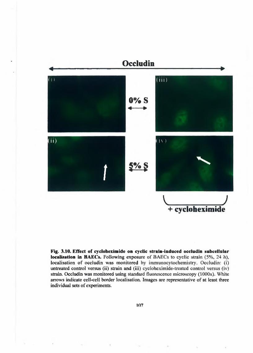
O c c l u d i l i*
(i )
0 % S
( i i i )
( i i )
t
( l \ )
N
V___________ )+ c y c l o h e x i m i d e
F i g . 3 .1 0 . E f f e c t o f c y c lo h e x im id e o n c y c l ic s t r a i n - i n d u c e d o c c lu d in s u b c e l l u l a r lo c a l is a t io n i n B A E C s . F o l lo w i n g e x p o s u r e o f B A G C s to c y c l ic s t r a in ( 5 % , 2 4 h ) , lo c a l is a t io n o f o c c lu d in w a s m o n i t o r e d b y im m u n o c y t o c h e m is t r y . O c c lu d in : ( i ) u n tr e a te d c o n t r o l v e rs u s ( i i ) s t r a in a n d ( i i i ) c y c lo h e x im id e - t r e a t e d c o n t r o l v e rs u s ( i v ) s tra in . O c c lu d in w a s m o n ito r e d u s in g s ta n d a rd f lu o re s c e n c e m ic r o s c o p y ( lO O O x ) . W h i t e a r r o w s in d ic a te c e l l - c e l l b o r d e r lo c a l is a t io n . Im a g e s a re r e p re s e n ta t iv e o f a t le a s t th re e in d iv id u a l sets o f e x p e r im e n ts .
107

Z O - 1
( i )
0 % S <— ►
( i i i )
(I I ! (IN )
NN
V. J
+ c y c l o h e x i m i d e
F i g . 3 . 1 1 . E f f e c t o f c y c l o h e x i m i d e o n c y c l i c s t r a i n - i n d u c e d Z O - 1 s u b c e l l u l a r lo c a l i s a t io n i n B A E C s . F o l lo w i n g e x p o s u re o f B A E C s to c y c l ic s t r a in ( 5 % , 2 4 h ) , lo c a l is a t io n o f Z O - 1 w a s m o n i t o r e d b y im m u n o c y t o c h e m is t r y . Z O - 1 : ( i ) u n t r e a te d c o n t r o l v e rs u s ( i i ) s tra in a n d ( i i i ) c y c lo h e x im id e - t r e a te d c o n t ro l v e rs u s ( i v ) s tra in . Z O - 1 w a s m o n ito r e d u s in g s ta n d a rd f lu o r e s c e n c e m ic r o s c o p y ( lO O O x ) . W h i t e a r r o w s in d ic a te c e l l - c e l l b o r d e r lo c a lis a t io n . Im a g e s a re r e p re s e n ta t iv e o f a t le a s t th r e e in d iv id u a l sets o f e x p e r im e n ts .
IftR

3.3 Discussion:
This chapter deals with the effect o f cyclic strain on the tight junction proteins
occludin and ZO-1. Given the importance o f mechanical forces in regulating vascular
hemeostasis, the ability o f cyclic strain to alter the permeability o f endothelial cells was
investigated.
Firstly, the effect o f cyclic strain on TJ protein expression and mRNA levels was
investigated. Mechanoregulation o f vascular endothelial tight junction protein
expression has also been confirmed in recent studies [Demaio et al., 2001: Conklin et
al., 2002]. Our investigations clearly demonstrate that chronic cyclic strain (5%, 24 h) of
BAECs significantly up-regulates protein expression of occludin and ZO-1 in parallel
with an increase in mRNA levels for occludin (but not ZO-1). This latter result suggests
that the strain-dependent increase in ZO-1 protein levels is most likely due to increased
protein translation and/or decreased ZO-1 protein turnover. It may also allude to the
importance o f post-translational modification o f ZO-1 in the tight junction process (see
Chapter 4). Conklin et al. also demonstrated that mechanical forces, in this case shear
stress, induced up regulation of occludin mRNA [Conklin et a l , 2002].
In conjunction with the changes in protein and mRNA we then examined the
location o f both proteins in the cell before and after exposure to cyclic strain using
standard fluorescent microscopy. A considerable change in the localisation of both
proteins within the cell following strain can be seen. Occludin is mainly distributed in
the nucleus and cytoplasm of the static cell and ZO-1 can be observed in a discontinuous
109

and jagged fashion along the cell border, both patterns o f staining indicative o f a poorly
formed junction. In response to strain both proteins relocate. Occludin distribution
becomes more focused at the cell-cell border and ZO-1 transforms into a more linear
distribution at the cell-cell junction. A similar change in ZO-1 localisation was observed
by Shin et al. in HUVECs exposed to chronic pulse pressure [Shin et al., 2003]. This
change in subcellular localisation indicates that both proteins are now in a more
favourable location to function as a barrier to paracellular permeability.
ZO-1 contains many binding sites and is known to link tight junction proteins
such as occludin to each other and the actin cytoskeleton [Alexander et al., 2002; Furuse
et al., 1993; Furuse et al., 1994; Gardner et al., 1996; Hirase et al., 1997]. Therefore
actin staining was examined in cells exposed to hemodynamic forces as it is entirely
plausible to link the changes seen in actin realignment with the localisation o f occludin
and ZO-1 following hemodynamic stimulation [McCue et al., 2004]. From Fig. 3.4 we
can see that there are changes in the distribution o f actin following strain. The actin
cytoskeleton becomes more localised to areas of cell-cell contacts, presumably with
increased binding to ZO-1. This is consistent with the important role the cytoskeleton
plays in modulating inter-endothelial junctions and endothelial permeability.
The immunocytochemical evidence putatively indicates that occludin and ZO-1
are localised together at the cell-cell border following exposure to strain. Use of IP
techniques has proved conclusively that following strain there is significantly more
occludin bound to ZO-1, another index o f tight junction upregulation. Therefore, in
conjunction with the increase in occludin and ZO-1 protein expression following strain,
110

we can see an increase in binding o f both proteins to each other. As mentioned
previously, disruption o f tight junctions can occur in part by dis-association of the
occludin -ZO-1 complex, and conversely increased occludin/ZO-1 co-association leads
to an increase in endothelial barrier integrity.
Junctional integrity is regulated by cytoskeletal tension, alterations in junctional
protein co-association, and linkage between junctional proteins and the actin
cytoskeleton, all of which help to govern intercellular cleft size and degree of
fluid/solute permeability [Rubin et al., 1999; Baida et al., 1998; Rubin et al., 1999]. One
would therefore expect changes in subcellular localisation o f occludin and ZO-1 at the
cell-cell border where tight junctions form, coupled with parallel changes in
occludin/ZO-1 association (i.e. co-IP), to accompany a change in endothelial barrier
function, as previously evidenced by numerous studies [Furuse et al., 1994; Rao et a l,
2002; Wang et al., 2002; Lee et al., 2004; Fischer et al., 2004; Sheth et al., 2003]. The
changes discussed so far have all indicated that cyclic strain induces barrier properties in
BAECs. However it is the transendothelial permeability assay that provides the most
compelling evidence. Cyclic strain significantly reduces BAEC transendothelial
permeability to FITC-dextran (i.e. a measurable index of barrier function).
Immunocytochemistry carried out in tandem confirms that strain-induced changes in
ZO-1 and occludin localisation fully persist 24 h after passage from Bioflex® plates into
Transwell®-Clear plates, indicating that the changes in transendothelial permeability are
critically linked to the effect o f strain on occludin and ZO-1. When viewed collectively,
these data lead us to conclude that cyclic strain up-regulates endothelial occludin/ZO-1
expression and tight junction assembly, putatively leading to increased barrier integrity.

Consistent with this conclusion, a recent study by Shin et al. demonstrated reduced
transendothelial permeability to albumin following exposure o f HUVECs to chronic
pulse pressure (cyclic pressure), an important hemodynamic component of pulsatile
blood flow (i.e. in addition to, but distinct from, cyclic strain) [Shin et a l, 2003]. To our
knowledge, this is the only other existing study reporting on the putative role of pulsatile
force in the modulation o f endothelial barrier function.
In order to determine if the strain-dependent increases in occludin and ZO-1
protein expression were causative o f increases in co-association and subcellular
localisation o f both proteins, cycloheximide was employed. Cycloheximide is an
inhibitor of protein biosynthesis in eukaryotic organisms, produced by the bacterium
Streptomyces griseus. It exerts its effect on protein synthesis by blocking translational
elongation. The addition o f cycloheximide successfully blocked the increases in
occludin and ZO-1 protein expression that were observed following strain. However,
although protein levels are reduced to control levels with the addition o f cycloheximide,
there is still a significant increase in occludin/ZO-1 co-association, suggesting that only
approximately 40% of the co-association is a direct consequence of new protein
synthesis and that other regulatory mechanisms are occurring in these events. These may
include phosphorylation and post-translational modifications o f occludin and ZO-1, each
of which will be discussed in the following chapters. Moreover, the increases in occludin
and ZO-1 protein levels following strain are not necessary for the subcellular
localisation o f occludin and ZO-1. This is evident because even with the addition of
cycloheximide, the changes in localisation o f both proteins following strain still exists.
This adds further evidence to the case for post-translational modifications o f occludin
112

and ZO-1 fo llo w in g strain.
3 .4 C o n c lu s io n
It has been demonstrated that physiological levels o f cyclic strain act to promote
a healthy and functioning tight junction in BAECs. We have clearly shown that in
BAECs, occludin and ZO-1 protein expression are up-regulated following strain, in
addition to an up-regulation o f occludin but not ZO-1 mRNA. We have also
demonstrated that cyclic strain leads to increased localisation o f occludin, ZO-1 and
actin to the cell-cell border, which is indicative o f a functioning tight junction. In
addition to this, we see an increase in the amount o f occludin bound to ZO-1 following
strain. These changes are very importantly linked to a functionally relevant model that
demonstrates a decrease in transendothelial permeability following strain (See Fig.
3.12). We have also demonstrated that the increase in the amount o f occludin bound to
ZO-1 following strain is only partially reliant on new protein synthesis, and the
increased localisation o f occludin and ZO-1 to the cell-cell border is not dependent on
the increase in protein levels observed, suggesting post-translational regulation o f tight
junction proteins following strain.
Thusfar, our data suggests that hemodynamic forces play an important role in
dictating the permeability o f BAECs via its effect on occludin and ZO -1, two proteins of
pivotal importance in the TJ. This relationship is to be investigated in more detail in the
following chapters.
113

O v e rv ie w o f R esults P resen ted in C h a p te r 3
Cyclic Strain
Occludin protein levels f Occludin mRNA levels ^Localisation of Occludin to cell mem brane f Association with ZO-1
Decreasedpermeability to
FD40
f ZO-1 protein levels M ore continuous distribution of ZO-1 at cell-cell border f Association with Occludin
ZO-1
Fig. 3.12. The effect of cyclic strain (5%, 24h) on occludin and ZO-1 in BAECs.
114

C h a p t e r 4
Examination of the phosphorylation changes in occludin and ZO-1 following strain
and their subsequent effects on TJ permeability in BAECs.
4 .1 In t r o d u c t io n
4 .2 R e s u lts
4 .3 D is c u s s io n
4 .4 C o n c lu s io n
115

4.1 Introduction
As the increases in co-association and sub-cellular localisation o f occludin and
ZO-1 observed following strain are likely due, in part, to post translational modifications
of both proteins, it was decided to look at phosphorylation changes in occludin and ZO-1
following exposure o f BAECs to cyclic strain.
In this regard, much attention has focused on the role o f phosphorylation in the
assembly and function o f tight junctions. Numerous studies have identified
kinase/phosphatase-dependent mechanisms leading to modulation o f junctional protein
association and subcellular distribution [Alexander et al., 2002; Rubin et al., 1999;
Baida et al., 1998]. Recent papers by Rao et al. and Sheth et al. for example,
demonstrate that oxidative stress-induced disruption o f tight junctions results from
increased tyrosine phosphorylation of occludin and ZO-1 leading to their subsequent
dissociation from the actin cytoskeleton, reduction in occludin/ZO-1 co-association, and
cellular redistribution o f both proteins [Rao et al., 2002; Sheth et al., 2003]. However
the processes by which these changes take place are poorly understood, although they
are believed to be central to tight junction regulation and therefore highly worthy of
investigation.
116

The aim o f this chapter is firstly to determine any changes in the phosphorylation
states o f occludin and ZO-1 following strain. Secondly, the pathways by which
occludin and ZO-1 are phosphorylated will be examined using pharmacological
inhibitors. Thirdly the relationship between these phosphorylation events and the
downstream changes reported in the previous chapter will be examined.
117

4.2 Results
F o l lo w in g e x p o s u re o f B A E C s to c y c l ic s tra in ( 5 % , 2 4 h ) , p h o s p h o r y la t io n o f
o c c lu d in w a s m o n ito r e d i n to ta l ly s a te b y I P u s in g p h o s p h o -s p e c if ic a n t ib o d ie s . I n
re s p o n s e to s tra in , ty r o s in e p h o s p h o r y la t io n o f o c c lu d in d e c re a s e d s ig n i f ic a n t ly b y 7 5 .7
± 8 % c o m p a re d to u n s t ra in e d c o n tro l (F ig . 4 .1 A ) . T h is is in d is t in c t c o n tra s t to r e la t iv e ly
s m a ll d e c re a s e s i n th r e o n in e ( F ig . 4 . I B ) a n d s e r in e ( F ig . 4 . 1 C ) p h o s p h o r y la t io n o f
o c c lu d in o b s e rv e d i n re s p o n s e to s tra in ( 1 6 .3 ± 6 % a n d 3 0 ± 5 % , r e s p e c t iv e ly ) . R e s u lts
w e re n o r m a lis e d to c h a n g e s in o c c lu d in p r o te in e x p re s s io n .
F o l lo w in g e x p o s u re o f B A E C s to c y c l ic s tra in ( 5 % , 2 4 h ) , p h o s p h o r y la t io n o f
Z O - 1 w a s a ls o m o n ito r e d in to ta l ly s a te b y IP u s in g p h o s p h o -s p e c if ic a n t ib o d ie s . I n
re s p o n s e to s tra in , th r e o n in e (F ig . 4 . 2 A ) a n d s e r in e ( F ig . 4 . 2 B ) p h o s p h o r y la t io n o f Z O - 1
in c re a s e d s ig n i f ic a n t ly ( 5 1 .7 ± 9% a n d 8 2 .7 ± 2 5 % , r e s p e c t iv e ly ) c o m p a re d to c o n tro l.
B y c o n tra s t, ty r o s in e p h o s p h o r y la t io n o f Z O - 1 r e m a in e d u n c h a n g e d ( F ig . 4 .2 C ) in
re s p o n s e to s tra in . R e s u lts w e r e n o r m a lis e d to c h a n g e s in Z O - 1 p r o te in e x p re s s io n .
4.2.1. Cyclic strain-dependent occludin and ZO-1 phosphorylation in BAECs.
118

Control 5% Strain
50 kDa-------- ►
50 kDa------ ► IP Ocdudin 111 pTyr
IP Occludin IB Ocdudin
Control 5% Strain
50 kDa--------►
Control 5% Strain
Control 5% Strainv i S
I ° L 5
-S £O £ ,.a £o> a W) oI I S F 5
o p.nr, 6«« o 43
50 kDa--------► M M IP Occludin1B pScr
Control 5% Strain
Fig. 4.1. Effect of cyclic strain on occludin phosphorylation in BAECs. BAECs were exposed to cyclic strain (5%, 24 h) and monitored for phosphorylation o f occludin in control and strained BAECs by IP and Western blotting using (A) phospho-tyrosine, (B) -threonine and (C)-serine -specific antibodies. Representative blots are shown above
119

e a c h g ra p h . H is to g r a m s re p re s e n t fo ld c h a n g e in b a n d in te n s ity r e la t iv e to u n s tra in e d c o n tro ls a n d a re a v e ra g e d fr o m th re e in d e p e n d e n t e x p e r im e n ts ± S E M ; * P < 0 .0 5 v e rs u s u n s tra in e d c o n tro ls . W i t h re s p e c t to ( A ) , ( B ) , a n d ( C ) , h is to g ra m s are n o r m a lis e d to ch an g es in o c c lu d in p ro te in e x p re s s io n (S e e F ig . 4 .1 .A lo w e r b lo t ) .
120
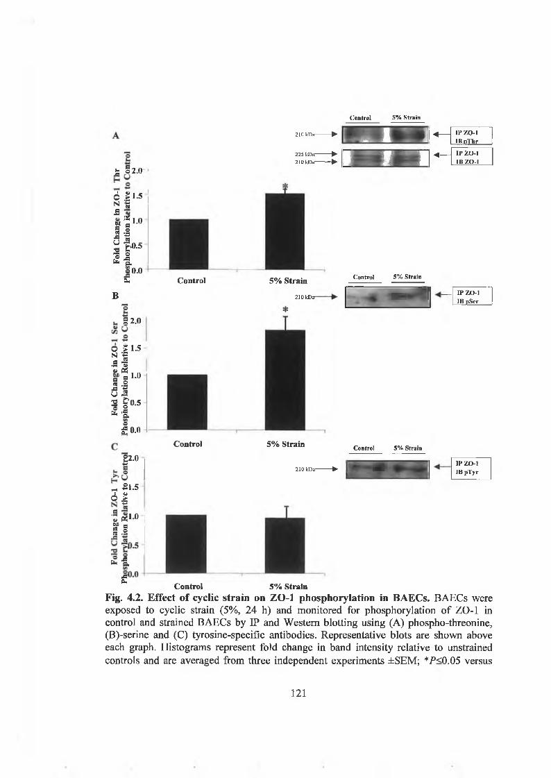
Control 5% Strain
S §2.0
Iosi.am « § § 5 -J3AUg£
B
S i 1 ' 5
I Ss i 1 ' 043 -PG -2S £“0.5ta -S* a-
I 0 .0
| 2 .0
- 21 .5 Ô |
Î tfi on
¿3a.ofc*H
sPm
210 kDa--------►J 1A —
IPZO-l II» n 'llir
225 kDa--------►210 kDa--------► 0 - “ | « -
IP ZO-1IB ZO-1
1 1.5 ee*5 i.o
u f>0.5
« 1 o 0 .0
ta Control 5% Strain
210 kDff
Control 5% Strain
- 11 ----IP ZO-1
1 IB pScr
9 2 .0
Control 5% Strain Control 5% Strain
IP ZO-1210 kDa----— ► 1 * ^ IB pTyr
Control 5% StrainFig. 4.2. Effect of cyclic strain on ZO-1 phosphorylation in BAECs. BAECs were exposed to cyclic strain (5%, 24 h) and monitored for phosphorylation o f ZO-1 in control and strained BAECs by IP and Western blotting using (A) phospho-threonine, (B)-serine and (C) tyrosine-specific antibodies. Representative blots are shown above each graph. Histograms represent fold change in band intensity relative to unstrained controls and are averaged from three independent experiments ±SEM; *P<0.05 versus
121

u n s tra in e d c o n tro ls . W i t h re s p e c t to ( A ) , ( B ) , a n d ( C ) , h is to g ra m s a re n o rm a lis e d to ch an g es in Z O - 1 p ro te in e x p re s s io n (S e e F ig . 4 .2 .A lo w e r b lo t ) .
122

4.2.2. Effect of Dephostatin on strain-induced occludin tyrosine
dephosphorylation.
Investigation confirmed that strain-induced decreases in occludin tyrosine
phosphorylation could be completely blocked by treatment of BAECs with dephostatin
(tyrosine phosphatase inhibitor), (Fig. 4.3). Tyrosine phosphorylation of occludin
decreased significantly to 0.57 ± 0.2 fold when cells were strained. Following strain +
dephostatin, tyrosine phosphorylation of occludin increased significantly to 1.17 ± 0.1
fold. Therefore dephostatin prevents tyrosine dephosphorylation o f occludin.
Dephostatin addition had no effect on unstrained samples.
50 kDa—
- 1 .6
H ya s i21 gI Ia.5 Pi 0.8gW) o« n
« ® 0 42 -sô e.S S
0 . 0
Con + Strain +Con Strain
IP Occludin IB pTyr
Control Control + DP Strain Strain + DP
Fig. 4.3. Effect of Dephostatin on Strain-induced occludin tyrosine phosphorylation. Graph illustrates the ability of DP to ablate strain-induced occludin tyrosine dephosphorylation. Representative blot is shown above the graph. Histogram represent fold change in band intensity relative to unstrained control is averaged from three independent experiments ±SEM; *P<0.05 versus unstrained controls. 5P<0.05 versus untreated strain.
123
4.2.3. Effect of Rottlerin on occludin serine/threonine phosphorylation.
The effect of rottlerin on occludin serine/threonine phosphorylation was
monitored to show that the inhibitory effects o f rottlerin were due specifically to PKC
inhibition and subsequent blockade of ZO-1 ser/thre phosphorylation and therefore to
rule out PKC as a modulator o f occludin phosphorylation state changes in response to
strain (Fig. 4.4). PMA was included as a positive control because it activates PKC by
mimicking DAG. It was found that the addition o f rottlerin had no effect on occludin
threonine phosphorylation. However addition of PMA, caused threonine
phosphorylation to increase significantly by 2.6 ± 0.2 fold, compared to control. It was
also found that the addition of rottlerin had no effect on occludin serine phosphorylation.
However addition of PMA, caused serine phosphorylation to increase significantly by
2 . 1 ± 0 . 1 fold, compared to control.
124

■a I 3.0 H U B ° , ,
1 I8 1 2 .« ° 3 ■s * I s « s 1,5w> .2 c -*-*a 5 i.ou I*2 1 0.5 £ »
£ o.o
•3 3.5
CooCon + ROT
Con + PMA Strain
Strain + ROT
IP O C CH H It (M W IB p T h r
I
IP O C CIB O C C
C ontrol C ontro l + R O T
C ontrol + P M A
Strain Strain + R O T
Con + Con + Strain +Con ROT PMA strain r o t
B
o 2.5
fa I^ rj.5 © 20 -a ^9 aj u« 1 cO -2 1,3a £ «5 ^2 «U ^2 o o.5
£ &O£ o.o
E P O C C
IB pSer
i i S11C ontrol C ontro l + C ontrol + S train S train -
R O T P M A R O T
Fig. 4.4. Effect of Rottlerin on strain-induced occludin threonine and serine phosphorylation. Graph A illustrates the effect of ROT on threonine phosphorylation, of occludin. Graph B illustrates the effect of ROT on occludin serine phosphorylation, Representative blots are shown above the graph. Histograms represent fold change in band intensity relative to unstrained control and are averaged from three independent experiments ±SEM; *P<0.05 versus unstrained controls. (ROT, rottlerin, PMA phorbol 12-myristate 13-acetate). With respect to (A) and (B) histogram is normalised to changes in occludin protein expression (See Fig. 4.A. lower blot).
125

4.2.4 Effect of Dephostatin on occludin co-association with ZO-1.
BAECs were exposed to cyclic strain (5%, 24 h) in the absence or presence of
dephostatin and occludin/ZO-1 co-association monitored in total BAEC lysates by IP as
described. Results indicated that the significant increase in strain-induced co-association
of occludin/ZO-1 could be blocked significantly following treatment with dephostatin,
compared with unstrained control (Fig. 4.5). However, it is of interest to note that co
association following treatment with dephostatin in unstrained cells is also significantly
increased from unstrained control.
Con + Strain +Con Strain
50 kDa—
2 .8s©« 2.4ts
©« u 2 0GPh o ¿ « 1 .6 W 2V g 1 2 O £N «e ^ 0 .8 ■a "| 0.4O
0 .0
-4----- IP ZO-1a------► IB Occludin
1 1 * 1
IControl Control + DP Strain Strain + DP
Fig. 4.5. Effect of Dephostatin on cyclic strain-induced occludin/ZO-1 coassociation. Following exposure of BAECs to cyclic strain (5%, 24 h), co-association of occludin was monitored by IP and Western blotting. The effect of dephostatin on strain- induced occludin/ZO-1 co-association is shown. Representative blots are shown above the graphs. Histograms represent fold change in band intensity relative to unstrained control and are averaged from three independent experiments ±SEM; *P<0.05 versus unstrained controls. rfP<0.05 versus untreated strain.
126

4.2.5. Effect of Dephostatin and Rottlerin on cyclic strain-dependent subcellular
localisation of occludin.
F o l lo w in g B A E C e x p o s u re to c y c lic s tra in ( 5 % , 2 4 h ) in th e a b s e n c e o r p re s e n c e
o f d e p h o s ta tin , s u b c e llu la r lo c a lis a t io n o f o c c lu d in w a s m o n ito re d b y
im m u n o c y to c h e m is try . R e s u lts in d ic a te d th a t c y c lic s tra in - in d u c e d lo c a lis a t io n o f
o c c lu d in to th e c e l l - c e l l b o rd e r w a s c o m p le te ly a b a te d b y d e p h o s ta tin tr e a tm e n t (F ig . 4 .6
i - iv ) .
F o l lo w in g B A E C e x p o s u re to c y c lic s tra in ( 5 % , 2 4 h ) in th e ab se n c e o r p res e n c e
o f r o t t le r in , a P K C in h ib ito r , s u b c e llu la r lo c a lis a t io n o f o c c lu d in w a s a ls o m o n ito re d b y
im m u n o c y to c h e m is try . R e s u lts in d ic a te d th a t c y c lic s tra in - in d u c e d lo c a lis a t io n o f
o c c lu d in to th e c e l l -c e l l b o rd e r w a s n o t e ffe c te d b y r o t t le r in tre a tm e n t, in c o r re la t io n
w it h th e fa c t th a t r o t t le r in h a d n o e f fe c t o n o c c lu d in s e r in e /th re o n in e p h o s p h o ry la t io n
(F ig . 4 .7 i - i v ) .
127

O c c l u d i n
+ D e p h o s t a t i nJ
t t l e r iF o l io
i w a s( i i ) stiuSj n g ; F i g . 4 . 6 . E f f e c t o f D e p h o s t a t i n o n c y c l ic s t r a i n - i n d u c e d o c c lu d in s u b c e l l u l a rP | o c a ] lo c a l is a t io n in B A E C s . F o l lo w i n g e x p o s u re o f B A E C s to c y c l ic s t r a in ( 5 % , 2 4 h ) ,ients lo c a l is a t io n o f o c c lu d in w a s m o n i t o r e d b y im m u n o c y t o c h e m is t r y . O c c lu d in : ( i )
u n tre a te d c o n tro l v e rs u s ( i i ) s tra in a n d ( i i i ) d e p h o s ta t in - t re a te d c o n t ro l v e rs u s ( i v ) s tra in . O c c lu d in w a s m o n ito re d u s in g c o n fo c a l f lu o re s c e n c e m ic r o s c o p y ( 1 2 0 0 x ) . W h i t e a r r o w s in d ic a t e c e l l - c e l l b o r d e r lo c a l is a t io n . Im a g e s a re r e p r e s e n t a t iv e o f a t le a s t th r e e in d iv id u a l sets o f e x p e r im e n ts .
178

4.2.6 Effect of Rottlerin on strain-induced ZO-1 serine phosphorylation.
Investigation confirmed that strain-induced increases in ZO-1 serine
phosphorylation could be completely blocked by treatment o f BAECs with rottlerin
(PKC inhibitor). Serine phosphorylation of ZO-1 increased significantly following
strain, compared to control, by 1.83 ± 0.2 fold. However on addition of rottlerin, strain-
induced serine phosphorylation o f ZO-1 was significantly reduced to 0.5 ±0 . 1 fold
compared to strained samples (Fig. 4.8). Serine phosphorylation o f ZO-1 was also
reduced in non-strained samples following addition of rottlerin.
210 kDa
l Ï 1 0
S .!
■O t ‘0.5 Ô 2 t. n
Con
C on + S train +
R O T S train R O T
Con Con + RO T Strain Strain+ R O T
Fig. 4.8. Effect of Rottlerin on strain-induced serine ZO-1 phosphorylation. Graph illustrates the ability of ROT to ablate strain-induced serine phosphorylation, of ZO-1. Representative blot is shown above the graph. Histogram represent fold change in band intensity relative to unstrained control is averaged from three independent experiments ±SEM; *P<0.05 versus unstrained controls. ^<0.05 versus untreated strain.
130

4.2.7 Effect of Dephostatin on ZO-1 tyrosine phosphorylation.
Results showed that there was no change in ZO-1 tyrosine phosphorylation
following strain. However changes in ZO-1 tyrosine phosphorylation were also
monitored in the presence o f dephostatin to illustrate that the inhibitory effects of
dephostatin were due specifically to tyrosine phosphatase inhibition and subsequent
blockade of ZO-1 tyrosine dephosphorylation. It was found that the addition of
dephostatin had no effect on ZO-1 tyrosine phosphorylation in control or strained
samples (Fig. 4.9).
210 kDa
Strain + DP
IP ZO-1IB pTyr
Control Control + D P Strain Strain + DP
mo iStomjlBidl Jpfiinii.iimr#. IP ZO-1210 kDa-------- p. 4HBK33j|P
-4 -------IB ZO-1
Fig. 4.9. Effect of Dephostatin on ZO-1 tyrosine phosphorylation. Graph illustrates the effect of DP on ZO-1 tyrosine phosphorylation. Representative blot is shown above the graph. Histogram represent fold change in band intensity relative to unstrained control and is averaged from three independent experiments ±SEM. With respect to (A) histogram is normalised to changes in ZO-1 protein expression (See Fig. 4.9 lower blot).
131

4.2.8 Effect of Rottlerin on strain-induced occludin/ZO-1 co-association.
BAECs were exposed to cyclic strain (5%, 24 h) in the absence or presence of
rottlerin and occludin/ZO-1 co-association was monitored in total BAEC lysates by IP as
described. Results indicated that strain-induced co-association o f occludin/ZO-1 could
be blocked significantly by 87.7 ± 30% following treatment with rottlerin (Fig. 4.10). Is
it of interest to note that co-association increased significantly in non-strained samples
following treatment with rottlerin.
5.0aoS3M 4.0.& o w u a> +*u aa, o •in¿U a o
0 £ 2 0N &a v
12 1 0
1 0 . 0
Con + Strain +
Con * Strain
L ■*- ■ - IP Z O -lm * - IB Occludin
iControl Control + ROT Strain Strain + ROT
Fig. 4.10. Effect of ROT on cyclic strain-induced occludin/ZO-1 co-association.Following exposure of BAECs to cyclic strain (5%, 24 h), co-association o f occludin was monitored by IP and Western blotting. The effect o f rottlerin on strain-induced occludin/ZO-1 co-association is shown. Representative blots are shown above the graphs. Histograms represent fold change in band intensity relative to unstrained control and are averaged from three independent experiments ±SEM; */*<0.05 versus unstrained controls. ^<0.05 versus untreated strain.
132

4.2.9. Effect of Rottlerin and Dephostatin on cyclic strain-dependent subcellular
localisation of ZO-1.
Following BAECs exposure to cyclic strain (5%, 24 h) subcellular localisation of
ZO-1 was monitored in the absence or presence of rottlerin. Results indicated that the
continuous and well-defined organisation of ZO-1 immunoreactivity initially observed
along the plasma membrane in response to cyclic strain was completely abated by
rottlerin treatment, reverting to a discontinuous and jagged localisation pattern along the
cell-cell border (Fig. 4.11 i-iv).
Following BAECs exposure to cyclic strain (5%, 24 h) in the absence or
presence of dephostatin, subcellular localisation o f ZO-1 was monitored by
immunocytochemistry. Results indicated that cyclic strain-induced localisation of ZO-1
to the cell-cell border in a more continuous fashion was not effected by dephostatin
treatment (Fig. 4.12 i-iv).
133

I 1 \ »
R o t t l e r i n
F i g . 4 .1 1 . E f f e c t o f P K C i n h i b i t i o n o n c y c l ic s t r a i n - i n d u c e d Z O - 1 s u b c e l l u l a rlo c a l i s a t io n i n B A E C s . F o l lo w i n g e x p o s u r e o f B A E C s to c y c l ic s t r a in ( 5 % , 2 4 h ) , lo c a l is a t io n o f Z O - 1 w a s m o n i t o r e d b y im m u n o c y t o c h e m is t r y Z O - 1 : ( i ) u n t r e a te d c o n t r o l v e rs u s ( i i ) s tra in a n d ( i i i ) r o t t le r in - t r e a t e d c o n t r o l v e rs u s ( i v ) s tra in . Z O - 1 w a s m o n ito r e d u s in g c o n fo c a l f lu o re s c e n c e m ic r o s c o p y ( 1 2 0 0 x ) . W h i t e a r r o w s in d ic a te c e l l c e l l b o r d e r lo c a l is a t io n . Im a g e s a re re p r e s e n ta t iv e o f a t le a s t th r e e in d iv id u a l sets o f e x p e r im e n ts
m

4.2.10. Effect of Rottlerin and Dephostatin on cyclic strain-induced
transendothelial permeability in BAECs.
Following exposure of BAECs to cyclic strain (5%, 24 h), control and strain-
conditioned BAECs ± ROT were trypsinized and re-plated into Transwell®-Clear plates
and monitored for permeability to 40 kDa FITC-dextran. As can be seen from Fig.
4.13.A, ROT-treated strain-conditioned cells had increased permeability to 40 kDa
FITC-dextran compared with strain-conditioned cells.
A
Time (mins)
Fig. 4.13A. Effect o f ROT on cyclic strain-induced transendothelial permeability in BAECs. Following exposure of BAECs to cyclic strain (5%, 24 h), control and strain- conditioned BAECs ± ROT (unstrained + ROT omitted from graph for clarity) were trypsinized and re-plated into Transwell®-Clear plates and monitored for permeability to 40 kDa FITC-dextran. Data points are shown as total subluminal fluorescence at a given time point (from 0-120 min) expressed as a percentage of total abluminal fluorescence at t=0 min (i.e. %TEE of FD40 or % Trans Endothelial Exchange). Representative graph shown averaged from two experiments.
136

Following exposure of BAECs to cyclic strain (5%, 24 h), control and strain-
conditioned BAECs ± DP were trypsinized and re-plated into Transwell®-Clear plates
and monitored for permeability to 40 kDa FITC-dextran. As can be seen from Fig.
4.13.B, DP-treated strain-conditioned cells had increased permeability to 40 kDa FITC-
dextran compared with strain-conditioned cells.
B
Time (mins)
Fig. 4.13.B. Effect of DP on cyclic strain-induced transendothelial permeability inBAECs. Following exposure of BAECs to cyclic strain (5%, 24 h), control and strain- conditioned BAECs ± DP (unstrained + DP omitted from graph for clarity) were trypsinized and re-plated into Transwell®-Clear plates and monitored for permeability to 40 kDa FITC-dextran. Data points are shown as total subluminal fluorescence at a given time point (from 0-120 min) expressed as a percentage of total abluminal fluorescence at t=0 min (i.e. %TEE of FD40 or % Trans Endothelial Exchange). Representative graph shown averaged from two experiments.
137

4.3 Discussion
It has become clear that post-translational-modification of tight junction proteins
via their phosphorylation, may be a pivotal step in the formation of functioning tight
junctions. We therefore decided to reconcile the cyclic strain-dependent phosphorylation
of occludin and ZO-1 observed in the present study with the strain-dependent
modulation of occludin/ZO-1 co-association and plasma membrane localisation
discussed in the previous chapter.
We first set out to examine baseline phosphorylation events in occludin and ZO-
1 following strain. Experiments showed that a tyrosine dephosphorylation event was
occurring in occludin following strain, in conjunction with increased serine/threonine
phosphorylation o f ZO-1.
We went on to confirm the role o f tyrosine phosphatases in occludin tyrosine
dephosphorylation and also to examine the consequences of this event in relation to tight
junction assembly. Addition of dephostatin, a pharmacological inhibitor of tyrosine
phosphatase, to the cells prior to strain prevents the tyrosine dephosphorylation of
occludin. Moreover, when dephostatin was added to the cells prior to strain, the increase
in occludin/ZO-1 co-association observed was attenuated, thereby linking the tyrosine
dephosphorylation event in occludin to its ability to bind to ZO-1 This finding has been
observed in other studies. A recent report by Kale et al. has shown that tyrosine
phosphorylation of occludin reduces its co-association with ZO-1, implying that tyrosine
dephosphorylation of occludin leads to an increase in co-association with ZO-1 [Kale et
138

al., 2003]. With respect to dephostatin addition, occludin/ZO-1 co-association was
significantly elevated above baseline in unstrained cells following inhibitor treatment.
This may reflect an indirect, albeit uncharacterized, increase in ZO-1 serine/threonine
phosphorylation, which could putatively lead to an elevation in protein-protein co
association above untreated baseline. Irrespective of this, dephostatin treatment led to a
clear reduction in strain-induced occludin/ZO-1 co-association to unstrained inhibitor-
treated levels.
We next decided to investigate the effects of the inhibitor on occludin
localisation. When tyrosine dephosphorylation of occludin is inhibited its ability to
relocate to the cell membrane following strain is ablated. Therefore, tyrosine
dephosphorylation of occludin, in addition to being necessary for increased
occludin/ZO-1 co-association following strain, is also essential for its strain-dependent
subcellular localisation.
The effect of rottlerin on occludin serine/threonine phosphorylation and sub-
cellular localisation was also investigated to rule out any influence of PKC on occludin
phosphorylation or localisation. Addition of rottlerin had no significant effect on
occludin serine/threonine phosphorylation when levels were normalised to occludin
protein. Moreover, addition of rottlerin also had no effect on the ability of occludin to
localise to the cell membrane following strain, thereby excluding PKC from this process.
Conversely, we also began the process to confirm the role of PKC in the serine
139

/threonine phosphorylation of ZO-1 and also to examine the consequences of this event
in relation to tight junction formation. Addition of rottlerin, a pharmacological inhibitor
of PKC, (highest specificity for PKCô [Gschwendt et al., 1994]), prevents the serine
phosphorylation of ZO-1 observed following exposure of BAECs to cyclic strain. In
addition, rottlerin also decreased serine phosphorylation of ZO-1 in non-strained cells.
The addition of rottlerin therefore, also provided a valuable tool in determining the
function of serine/threonine phosphorylation events in tight junction assembly. In
tandem with the occludin dephostatin results discussed previously, when rottlerin was
added to the cells the increase in occludin/ZO-1 co-association observed following
cyclic strain was attenuated, thereby linking the serine/threonine phosphorylation of ZO-
1 to its ability to bind to occludin at the tight junction. With respect to rottlerin addition,
occludin/ZO-1 co-association was significantly elevated above baseline in unstrained
cells following inhibitor treatment. This may reflect an indirect, albeit uncharacterized,
increase in occludin tyrosine dephosphorylation, which could putatively lead to an
elevation in protein-protein co-association above untreated baseline. Irrespective of this,
rottlerin treatment led to a clear reduction in strain-induced occludin/ZO-1 co
association to unstrained inhibitor-treated levels.
Due to the changes observed in occludin/ZO-1 co-association following strain
with the addition of rottlerin we next sought to investigate the effects o f the inhibitor on
ZO-1 localisation. When serine/threonine phosphorylation of ZO-1 is inhibited, its
ability to localise to the cell membrane in a continuous fashion in response to strain is
abrogated. Therefore the serine/threonine phosphorylation of ZO-1 is necessary for its
subcellular localisation following strain.
140

In view of the inhibitory effects of rottlerin, PKC likely plays a very important
role in determining ZO-1 function at the tight junction in ECs in response to
physiological levels of cyclic strain, In a contrasting study, PKC phosphorylation of ZO-
1 was shown to increase paracellular permeability in response to poly-l-arginine
stimulation [Ohtake et al., 2003]. The difference observed in the role of ZO-1
phosphorylation in this study may be due to the fact that the stimulation was
biochemical and also took place in different cell types (rabbit nasal epithelium cells),
however it does emphasise the fact the PKC regulation of ZO-1 is important in TJ
formation.
The effect of dephostatin on ZO-1 localisation and tyrosine phosphorylation were
also monitored to illustrate that the inhibitory effects of dephostatin were due
specifically to tyrosine phosphatase inhibition and subsequent blockade of occludin
tyrosine dephosphorylation. Addition of dephostatin had no significant effect on ZO-1
tyrosine phosphorylation when levels were normalised to ZO-1 protein. Moreover,
addition of dephostatin has no effect on the ability of ZO-1 to localise to the cell
membrane in a continuous fashion following strain, thereby excluding tyrosine
phosphatase from this process.
Thus far we have linked the occludin tyrosine dephosphorylation event and the
ZO-1 serine/threonine phosphorylation event to an increase in the co-association of the
two proteins and to changes in their subcellular localisation within the cell following
strain. We next sought to determine if post translation modifications have any effect on
the tightness of the paracellular barrier. We have demonstrated that addition of either
141

rottlerin or dephostatin, respectively, cause the decrease seen in transendothelial
permeability following strain to be attenuated, clearly suggesting that there is a causal
relationship between changes in occludin and ZO-1 biochemical properties and
endothelial barrier function in response to strain. No statistical analysis was carried out
on this data as each experiment was only repeated twice. However, as the two inhibitors
respectively exhibit a similar effect on transendothelial permeability, and correlate to the
biochemical data observed, there is a strong likelihood that the results are significant. A
similar finding was reported in a study by DeMaio et al. which demonstrated that shear
stress-induced increases in transendothelial permeability correlated with increased
tyrosine phosphorylation of occludin [DeMaio et al., 2001], However, to the best of our
knowledge, this is the first instance in which a direct link has been established.
4.4 Conclusion
In this chapter we have shown that phosphorylation changes in occludin and ZO-
1 following strain contribute to increased barrier integrity in BAECs. More specifically
we have observed a tyrosine dephosphorylation event in occludin that can be blocked by
dephostatin and a serine/threonine phosphorylation of ZO-1 that can be attenuated by
rottlerin suggesting involvement of tyrosine phosphatase and PKCô, respectively in
these events. Moreover the changes in phosphorylation of both proteins can be linked to
the subcellular localisation of occludin and ZO-1 and also to the increase in their co
association following strain. Furthermore the phosphorylation changes can also be
linked to the decrease in endothelial permeability seen following strain, as treating the
cells with dephostatin and rottlerin respectively caused the junction to become more
142

permeable (See Fig. 4.14). In conclusion therefore, post-translation modification of
occludin and ZO-1 following strain by tyrosine phosphatase and PKC leads to increased
barrier integrity.
O v erv iew o f R esu lts P re se n te d in C h a p te r 4
Cyclic Strain
Fig. 4.14 Cyclic strain-induced phosphorylation events in BAECs
1 4 ^

Chapter 5
Examination of the mechanotransduction pathway by which cyclic strain regulates
tight junction permeability in BAECs.
5.1 Introduction
5.2 Results
5.3 Discussion
5.4 Conclusion
1 4 4

5.1 Introduction
Vascular cells have the ability to respond to mechanical stimuli, namely cyclic
strain and shear stress. The ability to respond to these mechanical forces is facilitated by
mechanically sensitive receptors or “mechanoreceptors” present in vascular cells. This
process by which mechanical forces are detected and converted into a cell signal to elicit
a response is referred to as mechanotransduction [Lehoux et al, 1998], This process
allows changes in cellular phenotype and function in response to hemodynamic
stimulation. Mechanotransduction requires the activation of mechanosensitive receptors,
which may be activated directly by force, disruption of the ECM, or distortion of the cell
membrane and cytoskeleton.
G-proteins, integrins and PTKs have all demonstrated mechano-sensitivity. As
such they have formed the focus of this study as a means of elucidating the signaling
pathway(s) involved in cyclic strain-mediated regulation of occludin and ZO-1. A
number of studies have also demonstrated the downstream activation of a Rac-l/RhoA
signaling cascade by a number of receptors in response to mechanical stimuli. This
family of small GTPases are known to play a role in permeability and inflammation
[Dudek et al., 2001; Wojciak-Stothard et al., 2002], Thus, we have also examined the
possible activation/inhibition of Rac-l/RhoA signaling in transducing the cyclic strain
signal to ECs. We have also elected to examine the signaling molecules p38 and MEK,
as the MAPK pathway is known to be activated in response to mechanical stimuli and
have been linked to tight junction protein regulation.
145

To date there is very little reported about the mechanotransduction pathway that
leads to activation of occludin and ZO-1 and ultimately to a tighter endothelial cell
barrier.
The specific aim o f this chapter is to investigate the signaling mechanisms involved in
cyclic strain-regulation o f occludin and ZO-1 in vascular ECs, with specific emphasis
on G-proteins, integrins and PTK-mediatedpathways.
146

5.2 Results
The regulatory effect of genistein (50 |iM), a receptor tyrosine kinase inhibitor,
on cyclic strain-induced changes of occludin and ZO-1 protein expression in BAECs
was determined by measuring total protein levels in cell lysates (by IP). Following
exposure of BAECs to cyclic strain, (5%, 24 h), occludin protein expression increased
significantly by 2.0 ± 0 .1 fold compared to unstrained control. Following strain +
genistein, occludin protein expression levels did not alter significantly from strain
values, 2.0 ±0.1 fold (Fig. 5.1 A). Following exposure of BAEC to cyclic strain, (5%,
24 h), ZO-1 protein expression increased significantly by 2.0 ± 0.4 fold compared to
unstrained control. Following strain + genistein, ZO-1 protein expression was reduced
slightly although not significantly from strain values 1.7 ± 0.1 (Fig. 5.IB).
BAECs were exposed to cyclic strain (5%, 24 h) in the absence or presence of
genistein and occludin/ZO-1 co-association monitored in total BAEC lysates by IP as
described. Results indicated that the strain-induced increase in co-association of
occludin/ZO-1 was not significantly effected following treatment with genistein (Fig.
5.2).
Following BAEC exposure to cyclic strain (5%, 24 h) subcellular localisation of
occludin and ZO-1 was monitored in the absence or presence of genistein. Results
5.2.1. Effect of Genistein on the cyclic strain regulation of occludin and ZO-1.
147

indicated that cyclic strain-induced localisation of occludin to the cell-cell border was
not effected by genistein treatment (Fig. 5.3 i-iv). Furthermore, cyclic strain-induced
localisation of ZO-1 to the cell-cell border in a more continuous fashion was not effected
by genistein treatment (Fig. 5.4 i-iv).
148

Con
Con +
Gen Strain
S tra in + Gen
m 1______ IP O cc lud in50 kDa — ^ IB O cc lu d in
£ 2.5 a.Xw. 5 - 2 . 0a 2o. o bU1.S3 8 73 > o l L0 .3«1 0.5A£u2 0.0 "oLi. C o n tro l C o n tro l + Gen S tra in S tra in + G en
B
6 2,5 otri& 2.0 w_.5 £2 C| c © © 1.3
S i 1-0s “— CJ<u <v ^ 0.5 «
1 0.0 fctl
Con
Con + S tra in +Gen S tra in Gen
IP ZO -1
IB Z O -12 2 5 k D a -------------- ► r f t— «É
C o n tro l C o n tro l + Gen S tra in S tra in + Gen
Fig. 5.1. The effect o f Genistein on protein expression o f occludin and ZO-1 following 5% Strain for 24h. BAECs were exposed to cyclic strain (5%, 24 h) ± genistein and monitored for (A) occludin or (B) ZO-1 protein expression by Western blotting. Representative blots are shown above each graph. Histograms represent fold change in band intensity relative to unstrained controls and are averaged from three independent experiments ±SEM; *P<0.05 versus unstrained controls. Gen, Genistein.
1 4 9

C o ® + S t r a i n +
C o n G e n S t r a i n G e n
50 kDit-
EO
3.0
2.5
S i 2-0■u aQ* OAW
<J 5 1 .5
O I ^ ■s 1. 0I *
2 0.5
0.0
IP ZO -1IB O cc lud in
C o n t r o l C o n t r o l + G e n S t r a i n S t r a i n + G e n
Fig. 5.2. Effect of Genistein on strain-induced occludin and ZO-1 co-association.Following exposure of BAECs to cyclic strain (5%, 24 h), co-association of occludin and ZO-1 were monitored by IP and Western blotting. Histogram represents fold change in band intensity relative to unstrained control and is averaged from three independent experiments ±SEM; *7^0.05 versus unstrained controls.
150

O ccludili*►
(i)
0 % s lb
(iii)
W
5% S <---- ►
(ii) <iv)
v
V______ J+ Genistein
Fig. 5.3. Effect of Genistein on cyclic strain-induced occludin subcellular localisation in BAECs. Following exposure of BAECs to cyclic strain (5%, 24 h), localisation of occludin was monitored by immunocytochemistry. Occludin: (i) untreated control versus (ii) strain and (iii) Genistein-treated control versus (iv) strain. Occludin was monitored using standard fluorescence microscopy (lOOOx). White arrows indicate cell-cell border localisation. Images are representative of at least three individual sets of experiments.
151

ZO-1►
(i) (iii)
0 % S<---- ►
(ii) (iv)
/ /
V_______ J
+ Genistein
Fig. 5.4. Effect of Genistein on cyclic strain-induced ZO-1 subcellular localisationin BAECs. Following exposure of BAECs to cyclic strain (5%, 24 h), localisation of ZO-1 was monitored by immunocytochemistry. ZO-1: (i) untreated control versus (ii) strain and (iii) Genistein -treated control versus (iv) strain. ZO-1 was monitored using standard fluorescence microscopy (lOOOx). White arrows indicate cell-cell border localisation. Images are representative of at least three individual sets of experiments.
152

5.2.2 Effect of PTX on the cyclic strain regulation of occludin and ZO-1.
The regulatory effect of PTX (50 pM), a Goij protein inhibitor, on cyclic strain-
induced changes of occludin and ZO-1 protein expression in BAECs was determined by
measuring total protein levels in cell lysates. Following exposure of BAECs to cyclic
strain (5%, 24 h), occludin protein expression increased significantly by 2.1 ± 0.4 fold
compared to unstrained control. However, addition of PTX reduced occludin protein
expression to 1.2 ± 0.3 (Fig. 5.5A). Following exposure of BAECs to cyclic strain, ZO-1
protein expression increased significantly by 1.85 ±0.1 fold. However addition of PTX
reduced ZO-1 protein expression to 1.1 ± 0.1 (Fig. 5.5B) compared to unstrained
control.
BAECs were exposed to cyclic strain (5%, 24 h) in the absence or presence of
PTX and occludin/ZO-1 co-association monitored in total BAEC lysates by IP as
described. Results indicated that the strain-induced increase in co-association of
occludin/ZO-1 could be reduced from a 1.8 ± 0.1 fold increase following strain to a 1.2
± 0.2 fold increase following treatment with PTX (Fig. 5.6).
Following BAEC exposure to cyclic strain (5%, 24 h) subcellular localisation of
occludin and ZO-1 was monitored in the absence or presence of PTX. Results indicated
that the localisation of occludin along the cell membrane observed in response to cyclic
strain was completely ablated by PTX treatment (Fig. 5.7 i-iv). Furthermore, the
continuous and well-defined organization of ZO-1 immunoreactivity initially observed
153

along the plasma membrane in response to cyclic strain was also completely ablated by
PTX treatment, reverting to a discontinuous and jagged localisation pattern along the
cell-cell border (Fig. 5.8 i-iv).
1 5 4

ConC o n +
P T X StrainS t r a i n +
P T X
I P O c c l u d i n50 kDa---- — ► m m Mpvf I B O c c l u d i n
•§ g o 5L. S
3.0
0H °•SU'd -2 3 «o ts
2.0
O »5-aS
1.5a
a l« fg1.0
U & 2 * o0.5
to 0.0C o n t r o l C o n t r o l + P T X S t r a i n S t r a i n + P T X
B
C o n t r o l C o n t r o l + P T X S t r a i n S t r a i n + P T X
Fig. 5.5. The effect of PTX on occludin and ZO-1 protein expression following 5% strain for 24 h. BAECs were exposed to cyclic strain (5%, 24 h) ± PTX and monitored for (A) occludin or (B) ZO-1 protein expression by Western blotting. Representative blots are shown above each graph. Densitometric intensity of both bands has been combined. Histograms represent fold change in band intensity relative to unstrained controls and are averaged from three independent experiments ±SEM; *Ps0.05 versus unstrained controls. 8Ps0.05 versus untreated strain.
1S5
C o n + S t r a i n +
C o n P T X S t r a i n P T X
225
210

ao2.0
C o n + S t r a i n +
C o n P T X S t r a i n P T X
I P Z O - 150 kDa----------^ m m r n 4 M C i
I B O c c l u d i n
f t
1.5o uu S3 Ph o¿V^ a>o £N 2S 5 s *uso
0.5
0.0
*
C o n t r o l C o n t r o l + P T X S t r a i n S t r a i n + P T X
Fig. 5.6. Effect of 5% Strain + PTX on occludin and ZO-1 co-association in BAECs.Following exposure of BAECs to cyclic strain (5%, 24 h), co-association of occludin and ZO-1 were monitored by IP and Western blotting. Histogram represents fold change in band intensity relative to unstrained control and is averaged from three independent experiments ±SEM; *P<0.05 versus unstrained controls. &P<,0.05 versus untreated strain.

Occludili
(i)
¡1
(iii)
(ii)
N
N .
5% S <----►
(iv)
v______;+ PTX
Fig. 5.7. Effect of FTX on cyclic strain-induced occludin subcellular localisation in BAECs. Following exposure of BAECs to cyclic strain (5%, 24 h), localisation of occludin was monitored by immunocytochemistry. Occludin: (i) untreated control versus (ii) strain and (iii) PTX -treated control versus (iv) strain. Occludin was monitored using standard fluorescence microscopy (lOOOx). White arrows indicate cell-cell border localisation. Images are representative of at least three individual sets of experiments.
157

ZO-1►
(i) ( i n )
( i i )
/
(iv)
0 % s<------ ►
5% S<------ ►
v______ J+ PTX
Fig. 5.8. Effect of PTX on cyclic strain-induced ZO-1 subcellular localisation in BAECs. Following exposure of BAECs to cyclic strain (5%, 24 h), localisation of ZO-1 was monitored by immunocytochemistry. ZO-1: (i) untreated control versus (ii) strain and (iii) PTX -treated control versus (iv) strain. ZO-1 was monitored using standard fluorescence microscopy (lOOOx). White arrows indicate cell-cell border localisation. Images are representative of at least three individual sets of experiments.
158

5.2.3. Effect of cyclic RGD on the cyclic strain regulation of occludin and ZO-1.
BAECs were exposed to cyclic strain (5%, 24 h) in the absence or presence of
cRGD (0.1 /¿M), an integrin inhibitor, and occludin/ZO-1 co-association monitored (Fig.
5.9) in total BAEC lysates by IP as described. Results indicated that the strain-induced
increase in co-association of occludin/ZO-1 was not significantly effected following
treatment with cRGD, (1.9 ±0.1 fold following strain and 2.1 ±0.1 fold following strain
and addition of cRGD).
Following BAEC exposure to cyclic strain (5%, 24 h) subcellular localisation of
occludin and ZO-1 was monitored in the absence or presence of cRGD. Results
indicated that cyclic strain-induced localisation of occludin to the cell-cell border was
not effected by cRGD treatment (Fig. 5.10 i-iv). Furthermore, the cyclic strain-induced
localisation of ZO-1 to the cell-cell border in a more continuous fashion was also not
effected by cRGD treatment (Fig. 5.11 i-iv).
159

50 kDa—
2 .5
2.0
<u +- U 0Oh O t c-¿Ul-S U *
g | 10"a a»■3 c£■a wwuO
0 .5
0.0
Con + Con cRGD
Strain + Strain cRGD
IP ZO-1IB Occludin
Control Control + cRGD
S t r a i n Strain + cRGD
Fig. 5.9. Effect o f 5% Strain + cRGD on occludin and ZO-1 co-association inBAECs. Following exposure of BAECs to cyclic strain (5%, 24 h), co-association of occludin and ZO-1 were monitored by IP and Western blotting. Histogram represents fold change in band intensity relative to unstrained control and is averaged from three independent experiments ±SEM; *Ps0.05 versus unstrained controls.
1 6 0

Occludin
(0
0%SM--- ►
(iii)
(io m a o
® >NI \
(iv)
V_______)
+ cyclic RGD peptide
Fig. 5.10. Effect of cyclic RGD peptide on cyclic strain-induced occludin subcellular localisation in BAECs. Following exposure of BAEC to cyclic strain (5%, 24 h), localisation of Occludin was monitored by immunocytochemistry. Occludin : (i) untreated control versus (ii) strain and (iii) cyclic RGD peptide-treated control versus (iv) strain, occludin was monitored using standard fluorescence microscopy (lOOOx). White arrows indicate cell-cell border localisation. Images are representative of at least three individual sets of experiments.
161

ZO-1►
(i) (iii)
0 % S<---- ►
(ii) (iv)
\
v_______)
+ cyclic RGD peptide
Fig. 5.11. Effect of cyclic RGD peptide on cyclic strain-induced ZO-1 subcellular localisation in BAECs. Following exposure of BAECs to cyclic strain (5%, 24 h), localisation of ZO-1 was monitored by immunocytochemistry. ZO-1: (i) untreated control versus (ii) strain and (iii) cyclic RGD peptide-treated control versus (iv) strain. ZO-1 was monitored using standard fluorescence microscopy (lOOOx). White arrows indicate cell-cell border localisation. Images are representative of at least three individual sets of experiments.
162

Occludili►
(i) (Ìii)
(ii)
\\
9 %
V______ )
+ linear RGD peptide
Fig. 5.12. Effect of linear RGD peptide on cyclic strain-induced occludin subcellular localisation in BAECs. Following exposure of BAECs to cyclic strain (5%, 24 h), localisation of occludin was monitored by immunocytochemistry. Occludin: (i) untreated control versus (ii) strain and (iii) linear RGD peptide -treated control versus (iv) strain. Occludin was monitored using standard fluorescence microscopy (lOOOx). White arrows indicate cell-cell border localisation. Images are representative of at least three individual sets of experiments.
164

ZO-1►
(T)
0 % S<------- ►
(ii) (iv)
5% S“ w
\v______ ;
+ linear RGD peptide
Fig. 5.13. Effect of linear RGD peptide on cyclic strain-induced ZO-1 subcellular localisation in BAECs. Following exposure of BAECs to cyclic strain (5%, 24 h), localisation of ZO-1 was monitored by immunocytochemistry. ZO-1: (i) untreated control versus (ii) strain and (iii) linear RGD peptide -treated control versus (iv) strain. ZO-1 was monitored using standard fluorescence microscopy (lOOOx). White arrows indicate cell-cell border localisation. Images are representative of at least three individual sets of experiments.
165

5.2.5. Effect of Rho Kinase inhibition on the cyclic strain regulation of occludin and
ZO-1.
The regulatory effect of Y-27632 (20 (J.M), a Rho kinase inhibitor, on cyclic
strain-induced changes of occludin and ZO-1 protein expression in BAECs was
determined by measuring total protein levels in cell lysates. Following exposure of
BAECs to cyclic strain, (5%, 24 h), occludin protein expression increased by 1.6 ± 0.2
fold compared with control. Following strain and addition of Y-27632, occludin protein
expression increased significantly to 2.2 ± 0.2, compared to strain levels. (Fig. 5.14A).
Following exposure of BAEC to cyclic strain, (5%, 24 h), ZO-1 protein expression
increased by 1.7 ± 0.2 fold compared with control. Following strain and addition of Y-
27632, ZO-1 protein expression increased to 2.3 ± 0.5 fold compared to strain levels
(Fig. 5.14B).
BAECs were exposed to cyclic strain (5%, 24 h) in the absence or presence of Y-
27632 and occludin/ZO-1 co-association monitored in total BAEC lysates by IP as
described. Results indicated that strain-induced co-association of occludin/ZO-1 was
increased significantly following treatment with Y-27632, (2.0 ± 0.1 fold following
strain and 2.3 ±0.1 fold following strain + Y-27632). (Fig. 5.15).
Following BAEC exposure to cyclic strain (5%, 24 h), in the absence or presence
of Y-27632, subcellular localisation of occludin was monitored. Results indicated that
cyclic strain-induced localisation of occludin to the cell-cell border was not affected by
Y-27632 treatment (Fig. 5.16 i-iv).
166

Following BAEC exposure to cyclic strain (5%, 24 h) in the absence or presence
of Y-27632, subcellular localisation of ZO-1 was also monitored by
immunocytochemistry. Results indicated that cyclic strain-induced localisation of ZO-1
to the cell-cell border was not affected by Y-27632 treatment (Fig. 5.17 i-iv).
Furthermore non-strained cells that were incubated with Y-27632 were observed to have
a more continuous distribution of ZO-1 at the cell membrane (Fig. 5.17 iii).
167

50 kDa-
O-Xs ^i 2 £ s
Pm 9
.2 « V .>« -C O J2
oD£Bu2©to
2 .5
2.0
1.5
1.0
0 .5
0.0
C o n + S t r a i n +
C o n Y - 2 7 6 3 2 S t r a i n Y - 2 7 6 3 2
*
IC o n C o n + Y - 2 7 6 3 2 S t r a i n S t r a i n + Y - 2 7 6 3 2
B2 2 5 k D a -
2 1 0 k D a -
a©
aHW-
£ ° — OiO £ N-B
.s —<u6D
u2’©to
C o n
C o n + S t r a i n +
Y - 2 7 6 3 2 S t r a i n Y - 2 7 6 3 2
• * mI P Z O - l
I B Z O - l
C o n C o n + Y - 2 7 6 3 2 S t r a i n S t r a i n + Y - 2 7 6 3 2
Fig. 5.14. The effect of Rho Kinase inhibition (Y-27632) on protein expression of occludin and ZO -l following 5% Strain for 24 h. BAECs were exposed to cyclic strain (5%, 24 h) ± Y-27632 and monitored for (A) occludin or (B) ZO-l protein expression by Western blotting. Representative blots are shown above each graph. Densitometric intensity of both bands has been combined. Histograms represent fold change in band intensity relative to unstrained controls and are averaged from three independent experiments ±SEM; *7^ 0.05 versus unstrained controls. 6P^0.05 versus untreated strain.
16R

C o n + S t r a i n +
C o n Y - 2 7 6 3 2 S t r a i n Y - 2 7 6 3 2
C o n C o n + Y - 2 7 6 3 2 S t r a i n S t r a i n + Y - 2 7 6 3 2
Fig. 5.15. Effect of 5% Strain + Rho Kinase inhibition (Y-27632) for 24hrs on occludin and ZO-1 co-association in BAECs. Following exposure of BAECs to cyclic strain (5%, 24 h), co-association of occludin and ZO-1 were monitored by IP and Western blotting. Representative blot is shown above graph. Histogram represents fold change in band intensity relative to unstrained control and is averaged from three independent experiments ±SEM; *P<,0.05 versus unstrained controls. 6P^0.05 versus untreated strain.
1 6 9

O ccluditi
(1)
0 % S
<-------►
(ii) (iv)
/ *
5% S•4-------►
NV J
+ Y-27632
Fig. 5.16. Effect of Rho Kinase inhibition on cyclic strain-induced occludin subcellular localisation in BAECs. Following exposure of BAECs to cyclic strain (5%, 24 h), localisation of occludin was monitored by immunocytochemistry. Occludin: (i) untreated control versus (ii) strain and (iii) Y-27632-treated control versus (iv) strain. Occludin was monitored using standard fluorescence microscopy (lOOOx). White arrows indicate cell-cell border localisation. Images are representative of at least three individual sets of experiments.
170

ZO-1►
(i)
0%S <--- ►
(iii)
(ii)
\ 5% S <--- ►
(iv)
\v______ ;
+ Y-27632
Fig. 5.17. Effect of Rho Kinase inhibition on cyclic strain-induced ZO-1 subcellular localisation in BAECs. Following exposure of BAECs to cyclic strain (5%, 24 h), localisation of ZO-1 was monitored by immunocytochemistry. ZO-1: (i) untreated control versus (ii) strain and (iii) Y-27632-treated control versus (iv) strain. ZO-1 was monitored using standard fluorescence microscopy (lOOOx). White arrows indicate cellcell border localisation. Images are representative of at least three individual sets of experiments.
171

BAEC were exposed to cyclic strain (5%, 24 h) in the absence or presence of
NSC23766, (50 |iM), a Rac-1 inhibitor, and occludin/ZO-1 co-association monitored in
total BAEC lysates by IP as described. Results indicated that strain-induced co
association of occludin/ZO-1 could be significantly attenuated from an increase of 1.8
fold ± 0.2 fold following strain to 1.2 ± 0.2 fold following treatment with NSC23766
(Fig. 5.18).
Following BAEC exposure to cyclic strain (5%, 24 h) subcellular localisation of
occludin was monitored in the absence or presence of NSC23766. Results indicated that
the localisation of occludin along the cell membrane observed in response to cyclic
strain was completely ablated by NSC23766 treatment (Fig. 5.19 i-iv).
Following BAEC exposure to cyclic strain (5%, 24 h) subcellular localisation of
ZO-1 was also monitored in the absence or presence of NSC23766. Results indicated
that the continuous and well-defined organization of ZO-1 immunoreactivity initially
observed along the plasma membrane in response to cyclic strain was completely
ablated by NSC23766 treatment, reverting to a discontinuous and jagged localisation
pattern along the cell-cell border (Fig. 5.20 i-iv).
5.2.6. Effect of Rac-1 inhibition on the cyclic strain regulation of occludin and ZO-
1.
172

Con + Strain +NSC NSC
Con 23766 Strain 23766
2.5
Con Con + Strain Strain +NSC23766 NSC23766
Fig. 5.18. Effect of 5 % Strain + NSC23766 for 24 h on occludin and ZO-1 coassociation in BAECs. Following exposure of BAECs to cyclic strain (5%, 24 h), coassociation of occludin and ZO-1 was monitored by IP and Western blotting. Representative blot is shown above graph. Histogram represents fold change in band intensity relative to unstrained control and is averaged from three independent experiments ±SEM; *P<0.05 versus unstrained controls. 6P<0.05 versus untreated strain.
m

Occludin<-----------------------------------------------------------------------------------------------------►
(i)
40 % s <--- ►
(iii)r ' w
• M
(ii)
5% S <--- ►
(W)
• ■:*a .
v______ J
+ NSC23766
Fig. 5.19. Effect of NSC23766 on cyclic strain-induced occludin subcellular localisation in BAECs. Following exposure of BAECs to cyclic strain (5%, 24 h), localisation of occludin was monitored by immunocytochemistry. Occludin: (i) untreated control versus (ii) strain and (iii) NSC23766-treated control versus (iv) strain. Occludin was monitored using standard fluorescence microscopy (lOOOx). White arrows indicate cell-cell border localisation. Images are representative of at least three individual sets of experiments.
174

ZO-1►
(i)
0% S <--- ►
(iii)
(ii)
5% S <--- ►
(iv)• *
v______ J
+ NSC23766
Fig. 5.20. Effect of NSC23766 on cyclic strain-induced ZO-1 subcellular localisation in BAECs. Following exposure of BAECs to cyclic strain (5%, 24 h), localisation of ZO-1 was monitored by immunocytochemistry. ZO-1: (i) untreated control versus (ii) strain and (iii) NSC23766-treated control versus (iv) strain. ZO-1 was monitored using standard fluorescence microscopy (lOOOx). White arrows indicate cell-cell border localisation. Images are representative of at least three individual sets of experiments.
175

5.2.7. Effect of p38 on the cyclic strain regulation of occludin and ZO-1 localisation.
Following BAEC exposure to cyclic strain (5%, 24 h) subcellular localisation of
occludin was monitored in the absence or presence of PD169316 (10 |jM), a p38
pharmacological inhibitor. Results indicated that the localisation of occludin along the
cell membrane observed in response to cyclic strain was completely ablated by
PD 169316 treatment (Fig. 5.21 i-iv).
Following BAEC exposure to cyclic strain (5%, 24 h) subcellular localisation of
ZO-1 was also monitored in the absence or presence of PD 169316. Results indicated
that the continuous and well-defined organization of ZO-1 immunoreactivity initially
observed along the plasma membrane in response to cyclic strain was completely
ablated by PD 169316 treatment, reverting to a discontinuous and jagged localisation
pattern along the cell-cell border (Fig. 5.22 i-iv).
176

Occludin<------------------------------------------------------------------------------------------------ ►
(i)
0 % s <— ►
(iii)
(ii)
/
-
5% S <---►
(iv)
v______ ;+ PD169316
Fig. 5.21. Effect of PD169316 on cyclic strain-induced occludin subcellular localisation in BAECs. Following exposure of BAECs to cyclic strain (5%, 24 h), localisation of occludin was monitored by immunocytochemistry. Occludin: (i) untreated control versus (ii) strain and (iii) PD169316-treated control versus (iv) strain. Occludin was monitored using standard fluorescence microscopy (lOOOx). White arrows indicate cell-cell border localisation. Images are representative of at least three individual sets of experiments.
177

ZO-1<------------------------------------------------------------------------------------------------ ►
(i)
0 % s <---►
(iii)
(ii) y/
S '5% S <----►
(iv)
v______ )+ PD169316
Fig. 5.22. Effect of PD169316 on cyclic strain-induced ZO-1 subcellular localisation in BAECs. Following exposure of BAECs to cyclic strain (5%, 24 h), localisation of ZO-1 was monitored by immunocytochemistry. ZO-1: (i) untreated control versus (ii) strain and (iii) PD169316-treated control versus (iv) strain. ZO-1 was monitored using standard fluorescence microscopy (lOOOx). White arrows indicate cell-cell border localisation. Images are representative of at least three individual sets of experiments.
178

localisation.
Following BAEC exposure to cyclic strain (5%, 24 h) subcellular localisation of
occludin was monitored in the absence or presence of PD98059 (10 jiM), a MEK
pharmacological inhibitor. Results indicated that cyclic strain-induced localisation of
occludin to the cell-cell border was not effected by PD98059 treatment (Fig. 5.23 i-iv).
Following BAEC exposure to cyclic strain (5%, 24 h) in the absence or presence
of PD98059, subcellular localisation o f ZO-1 was also monitored by
immunocytochemistry. Results indicated that cyclic strain-induced localisation of ZO-1
to the cell-cell border in a more continuous fashion was not effected by PD98059
treatment (Fig. 5.24 i-iv).
5.2.8. Effect of MEK on the cyclic strain regulation of occludin and ZO-1
179

-4Occludili
(i)
0 % sM-----------►
(iii)
i i . ^ v * • «\ « i V
(ii) \\
A * 1 .. Sf/t
5% SM -----------►
(iv)V/ • y
\
v______ ;+ PD98059
Fig. 5.23. Effect of PD98059 on cyclic strain-induced occludin subcellular localisation in BAECs. Following exposure of BAECs to cyclic strain (5%, 24 h), localisation of occludin was monitored by immunocytochemistry. Occludin: (i) untreated control versus (ii) strain and (iii) PD98059-treated control versus (iv) strain. Occludin was monitored using standard fluorescence microscopy (lOOOx). White arrows indicate cell-cell border localisation. Images are representative of at least three individual sets of experiments.
180

ZO-1
0 % S<------ ►
(ii) S ' (iv)
5% S \ \ —w
v______ )+ PD98059
Fig. 5.24. Effect of PD98059 on cyclic strain-induced ZO-1 subcellular localisation in BAECs. Following exposure of BAECs to cyclic strain (5%, 24 h), localisation of ZO-1 was monitored by immunocytochemistry. ZO-1: (i) untreated control versus (ii) strain and (iii) PD98059-treated control versus (iv) strain. ZO-1 was monitored using standard fluorescence microscopy (lOOOx). White arrows indicate cell-cell border localisation. Images are representative of at least three individual sets of experiments.
181

5.3 Discussion
Structural adaptation of the vasculature occurs in response to both physiological
and pathological changes in blood pressure and flow [Schwartz et al., 1995; Libby et al,
2003]. The effect of cyclic strain on the expression, localisation, co-association and
phosphorylation of occludin and ZO-1 has been discussed in great detail in the previous
two chapters. We have clearly demonstrated that physiological levels of cyclic strain
increase endothelial cell tight junction function. In this chapter we have investigated
PTKs, G-proteins, integrins, small GTPase (RhoA and Rac-1), p38 and MEK as possible
components of the mechanical signaling cascade involved in the mechano-regulation of
occludin and ZO-1 in BAECs.
Tyrosine kinases have been implicated in hemodynamic force-induced changes
in EC function [Labrador et al., 2003]. Therefore we examined the effects of inhibiting
PTK using the pharmacological inhibitor Genistein, on tight junction assembly.
Genistein had no effect on the strain-induced increase of occludin and ZO-1 protein
expression nor any effect on the strain-induced co-association of both proteins.
Moreover localisation of the proteins to the cell membrane following strain was not
ablated following Genistein addition. Collectively, these results indicate that PTKs do
not mediate biochemical changes to tight junction proteins in response to cyclic strain.
However, to fully elucidate the role to PTKs in TJ regulation, it would be
interesting and necessary to repeat these experiments with a variety of inhibitors capable
of inhibiting non-receptor TKs also. This would be especially relevant as the activity of
182

Src kinases and c-Yes appear to play a role in both assembly and disassembly of tight
junctions [Kale et al., 2003]. In fact, c-Yes is tightly linked to tight junction function due
to its ability to bind to and phosphorylate occludin [Kale et al., 2003].
We next turned our attention to G-proteins as their cellular localisation and rapid
activation strongly implicate them as a primary sensor of hemodynamic forces [Gudi et
al., 1996], In the current study, we inhibited G-protein signaling with the
pharmacological inhibitor PTX. This inhibitor is known to block Ga, signaling [Gudi et
al., 1996]. Results indicated that PTX significantly inhibits the increase in occludin and
ZO-1 protein expression observed following strain, suggesting that the changes are
transduced via a Ga,j protein pathway. Cyclic strain-induced increases in occludin and
ZO-1 co-association were also significantly attenuated following addition of PTX and
localisation of both proteins to the cell membrane following strain was also attenuated
following inhibition of Gcij proteins. Thus, we have implicated Gccj signaling in
transducing cyclic strain-induced changes to tight junction protein turnover and
assembly. G-proteins have previously been reported to play a role a tight junction
protein regulation. Saha et al. observed that treatment of MDCK cells with cholera
toxin, a G as activator, accelerated TJ assembly as measured by transepithelial resistance
[Saha et al., 2001]. They also reported that G as was predominantly localised in the
lateral membrane, but a fraction co-localises with ZO-1 in the TJ. Studies by Denker et
al. also alluded to G-protein involvement in TJ biogenesis, indicating that G-proteins
may participate in the maintenance and/or regulation of TJ assembly in MDCK cells
[Denker et al. 1998]. In another study by Baida et al. in 1991 pertussis toxin increased
183

TEER in epithelial cells, indicating that junction formation may be controlled by a
network of reactions including G-protein activation [Baida et al, 1991]. Although this
finding differs from ours, it suggests that G-proteins are be involved in TJ regulation
Integrins have also been linked to changes observed following mechanical
stimulation [Shyy et al., 2002] and therefore were examined in this study to ascertain
their role in tight junction protein regulation following strain. To this end, synthetic
cyclic RGD and synthetic linear RGD were applied to BAECs prior to and for the
duration of strain. Results indicate that cRGD had no significant effect on strain-induced
co-association of occludin and ZO-1 or on localisation of the proteins to the cell
membrane following strain. The latter was also observed when the cells were treated
with 1RGD. Taken together these results indicate that integrins are not involved in tight
junction regulation in response to strain.
With G-proteins implicated as the mechanotransducers of cyclic-strain-mediated
changes in occludin and ZO-1, we then set out to examine the possible downstream
signaling molecules that may be recruited in further transducing the mechanical signal.
An obvious choice were the small GTPases RhoA and Rac-1 as they are known to play a
major role in inflammation, permeability and cytoskeletal regulation [Dudek et al.,
2001], Rho GTPases have now been implicated in signaling by many barrier-modifying
substances including thrombin, vascular endothelial growth factor (VEGF) and
histamine [Wojciak-Stothard et al., 2002]. In fact, RhoA and Rac-1 have emerged as key
permeability regulators acting antagonistically to regulate barrier function.
184

Results indicate that RhoA, investigated in the form of Rho Kinase, acts to
weaken and destabilise the tight junction. We have ascertained this because when we
inhibit Rho Kinase the junction becomes tighter, as indicated by the indices of
permeability; co- association and subcellular localisation of TJ proteins. Rho Kinase
blockade leads to an increase in occludin and ZO-1 protein levels following strain, in
parallel with a significant increase in their co-association. Moreover, from our
immunocytochemical evidence we can see that inhibiting Rho Kinase does not effect
cyclic-strain induced localisation of occludin or ZO-1. Interestingly, under static, non
strained conditions, Rho Kinase blockade actually induced localisation of ZO-1
suggesting strengthening of the tight junction. This supports our hypothesis that RhoA
and thus Rho Kinase, is inhibited during strain and thus further inhibition does not block
strain-induced upregulation of barrier function. Moreover, inhibition of Rho Kinase
would also be expected to increase barrier function under static non-strain conditions as
seen here. Future studies involving a Rho Kinase activity kit would further strengthen
this idea. Interestingly RhoA inhibition has been found to lead to Rac-1 activation
[Rottner and Small, 1999], and this could also be responsible for the increase in barrier
function observed in the static non-strained cells. In a study by Carbajal and Schaeffer,
inhibition of RhoA with C3 transferase or expression of dominant negative RhoA as
well as inhibition of Rho kinase with Y-27632 in endothelial cells prevents thrombin-
induced permeability [Carbajal and Schaeffer, 1999]. It is plausible that Rho Kinase
inhibition prevents the F-actin bundling caused by RhoA activation and hence precludes
disruption of the tight junction. In fact, actomyosin contractility induced by RhoA is
usually required for increased endothelial permeability [Wojciak-Stothard et al., 2002].
A similar finding was reported in brain endothelial cells, with observations that ICAM 1

cross-linking induces Rho-dependent cytoskeletal rearrangements, which may facilitate
transmigration and barrier disruption [Etienne et al., 1998; Adamson et al., 1999].
We then set about determining the role of Rac-1, another small GTPase that is
known to work in tandem with RhoA. When we inhibit Rac-1, with the pharmacological
inhibitor NSC23766, the increase in occludin/ZO-1 co-association is attenuated. This
indicates that Rac-1 activation is involved in the increased binding of the two proteins
following strain and therefore also involved in promoting a tight, functioning junction.
In support of this, Rac-1 inhibition was also seen to inhibit the strain-induced
localisation of occludin and ZO-1. Therefore Rac-1 is also responsible for the
subcellular localisation of tight junction proteins, conceivably due to its effect on actin
localisation. As previously mentioned, Rac-1 is required for the assembly and
maturation of epithelial and endothelial junctions and its activity increases during
junction formation [Noren et a l, 2001]. Moreover, in the absence of vasoactive stimuli,
dominant negative Rac increases endothelial permeability and affects adherens and tight
junctions, consistent with Rac being important for maintaining cell-cell junctions
[Wojciak-Stothard et al., 2001]. In fact, Yersinia bacteria decreases endothelial barrier
function by injecting effector proteins into the cell cytoplasm. One of these proteins,
YopE, is a GAP that has been shown to inhibit Rac-1 responses in cells and thereby
induce breakdown in intercellular junctions in HUVECs [von Pawel-Rammingen et al,
2000]
Collectively therefore, these findings suggest a synergistic regulation of the tight
junction by RhoA/Rho kinase and Rac-1. RhoA and Rac-1 generally have opposing
186

effects on junctional integrity and activation of one GTPase coincides with inactivation
of the other [Sander et al., 1999; Noren et al., 2001]. Thus, in a simplified model,
improvement of endothelial barrier function may be achieved by increasing the Rac-
1/RhoA activity ratio either by inactivation of RhoA or activation of Rac-1. Conversely,
increased permeability may be achieved by activating RhoA and/or inactivating Rac-1.
However, the results of many experiments suggest that junctional stability requires a
very fine balance between Rac-1 and RhoA activity [Wojciak-Stothard et al., 2002].
Several studies have linked MAPK pathways to tight junction protein regulation
[Basuroy et al., 2006: Pedram et al., 2002] and therefore, finally, we looked at the
signaling molecules p38 and MEK. MEK activity has been found to be required for
claudin-mediated formation of tight junctions in human intestinal epithelial cells
[Kinugasa et al., 2000]. However, in another study by Chen et al. after treatment of Ras-
transformed MDCK cells with the mitogen-activated protein kinase inhibitor PD98059,
occludin, claudin-1, and ZO-1 were recruited to the cell membrane, tight junctions were
assembled, and E-cadherin protein expression was induced [Chen et al., 2000]. In our
study, MEK, a MAP kinase inhibitor had no effect on occludin or ZO-1 localisation
following strain and therefore is not involved in the cyclic strain-induced modification
of TJ proteins. As previously discussed, P38 has been implicated in tight junction
regulation. Inhibition of p38 during oxidant challenge in HUVECs was found to
attenuate the increase in permeability following addition of hydrogen peroxide [Kelvil et
al., 2001], indicating that the increase in TJ permeability is mediated by p38 in this
instance. In our study, inhibition of p38, leads to an attenuation of the subcellular
localisation of both proteins to the cell membrane, indicating that the decrease in TJ
187

permeability, following strain is mediated by p38. The conflicting findings in these
studies may be explained by the fact that different cell types and stimuli are involved.
However, it is likely that p38 plays a signaling role in transducing the cyclic strain
stimuli to the tight junction proteins in this instance, although its sequence in these
events would require further investigation.
5.4 Conclusion
In conclusion, our data demonstrates that cyclic strain-mediated changes in tight
junction function are transduced in a G-protein-dependent but integrin- and PTK-
independent fashion. Inhibition of the small GTPase Rac-1 attenuates the strain-induced
changes in occludin and ZO-1 and therefore also plays a pivotal role in cyclic strain
signal transduction to ECs. Moreover, the MAPK, p38 is also involved in transducing
the cyclic-strain mediated response to TJ proteins.
188

Chapter 6
Final Summary
189

Hemodynamic forces, such as shear stress and cyclic strain, are potent mediators
of endothelial cell remodelling, angiogenesis and maintenance of vascular tone.
Common hemodynamic factors, such as low or oscillatory shear stress, can be found at
areas of compromised permeability in the vascular tree, implying that there may be a
relationship between EC permeability and physical forces. Also, as disruption of
vascular endothelial barrier integrity is a central feature of many homeostatic and
pathophysiological processes (e.g. atherosclerosis, sepsis etc.), and frequently correlates
with a perturbation in vessel hemodynamic loading and remodelling [Harhaj et al., 2004;
Van Nieuw-Armerongen et al., 2002; Tinsley et al., 2004; Koschinsky et al., 2004], we
endeavoured to determine the link between physiological levels of hemodynamic force
and tight junction protein regulation. The focus of this study was the hemodynamic force
cyclic strain.
Endothelial permeability is controlled by a series of junctional complexes. One
of the chief contributors to barrier function is the tight junction, which forms between
adjacent endothelial cells. It is comprised of several proteins, two of which; occludin and
ZO-1, formed the basis of this study. Occludin is a membrane-bound protein whose
expression can be directly linked to barrier function. ZO-1 is a transmembrane protein,
best known for its role in organising the proteins of the tight junction due to its many
binding sites.
It was our hypothesis that there exists a dynamic regulatory co-association
between vascular endothelial permeability and hemodynamic stimuli. We also sought to
190

determine the mechanotransduction pathway by which this modulation took place (See
Fig. 6.1 for diagrammatic representation of experimental approach).
Cyclic Strain Bovine Aortic5%, 24h fcnaorneiiai
Cells
•V• °,e'ns
Effect on Tight Junction Permeability
via Permeability Assay
Occludin & ZO-1•Protein levels
•mRNA levels
•Co-association
•Subcellular localisation
•Phosphorylation changes
Fig. 6.1 Diagrammatic representation of experimental approach. The regulation of occludin and ZO-1 in response to cyclic strain was investigated along with the identification of signaling pathways involved in the cyclic strain-induced increase in TJ barrier function.
Investigations clearly demonstrate that chronic cyclic strain (5%, 24h) of BAECs
up-regulates protein expression of occludin and ZO-1, in parallel with an increase in
mRNA for occludin (but not ZO-1). It is plausible that TJ proteins are increased
following strain to allow a tighter junction to form between ECs. The increase in ZO-1
protein, but not mRNA, can be reconciled by the fact there is possibly increased protein
translation and/or decreased protein turnover of ZO-1. The integrity of the TJ is
191

regulated by junctional protein interactions. As such, one would expect changes in
subcellular localisation of occludin and ZO-1 at the cell-cell border where tight junctions
form, coupled with parallel changes in occludin/ZO-1 co-association, to accompany a
change in endothelial barrier function, as previously evidenced by numerous studies
[Furuse et ah, 1994; Rao et ah, 2002; Wang et al, 2002; Lee et ah, 2004; Fischer et al.,
2004; Sheth et ah, 2003]. With this in mind, we decided to broaden our early
investigations by examining occludin/ZO-1 co-association and subcellular localisation
(i.e. measurable indices of tight junction assembly) in our cyclic strain model. Our
results clearly indicate a significant strain-dependent increase in occludin and ZO-1 co
association by way of immunoprecipitation. As mentioned previously, ZO-1 can bind
specifically to the COOH-terminal cytoplasmic tail of occludin. Therefore, we can
deduce that the two proteins are bound together at the tight junction, thus contributing to
the functional barrier. Also, following strain we observe that the proteins have re
localised within the cell, as seen by immunocytochemistry. Occludin protein moves
from the cytosol to the cell membrane and ZO-1 staining becomes more linear and
continuous at the cell-cell junction. This is also indicative of a tighter junction. We can
also observe a change in F-actin staining, as the actin cytoskeleton is also subject to
modification by cyclic strain. Actin staining becomes more cortical and intimately
linked to the proteins of the tight junction as it is directly bound to ZO-1 [Fanning et ah,
1998]. In parallel with these strain-induced changes, we observe a decreased
permeability of the endothelial monolayer to small macromolecules such as FD40, thus
indicating that cyclic strain leads to an increase in the barrier function of ECs. When
viewed collectively, these data lead us to conclude that cyclic strain up-regulates
192

endothelial occludin/ZO-1 expression and tight junction assembly, putatively leading to
increased barrier integrity. Consistent with this conclusion as previously mentioned, a
study by Shin et al. demonstrated reduced transendothelial permeability to albumin
following exposure of HUVECs to chronic pulse pressure (cyclic pressure) [Shin et al.,
2003],
Having ascertained that cyclic strain causes an increase in barrier function in
BAECs, we set about examining the pathway by which this increase took place. As
protein levels of both occludin and ZO-1 were increased following strain, we wanted to
determine the contribution of this new protein synthesis to the changes in barrier
function indices observed. To this end we utilised cycloheximide, which stops new
protein production, and observed that the new protein produced did not lead to the
changes in localisation of either protein. However, inclusion of cycloheximide was seen
to reduce the increase in co-association observed following strain, suggesting that
approximately 40% of the co-association is a direct consequence of new protein
synthesis. Therefore, the majority of strain-induced co-association of occludin and ZO-1,
along with the localisation of both proteins to the cell membrane is not dependent on
new protein synthesis but due to some other modification of TJ proteins following strain.
It was subsequently hypothesised that strain-induced phosphorylation changes in
occludin and ZO-1 were responsible for the up-regulation in tight junction function
observed. In this regard, tyrosine dephosphorylation of occludin, via tyrosine
phosphatase and serine/threonine phosphorylation of ZO-1, via PKC, were correlated to
the strain-induced localisation and co-association of both proteins in this study.
193

Treatment of BAECs with either dephostatin or rottlerin was also found to reverse the
strain-mediated reduction in transendothelial permeability as measured by the flow of
FD40 across the monolayer. This suggests a causal link between changes in
occludin/ZO-1 biochemical properties and endothelial barrier function in response to
cyclic strain. The specific mechanistic roles of these phosphorylation events in tight
junction assembly however remain unclear. Perturbation of the interaction between
proximal membrane occludin domains leading to activation of intracellular signalling
cascades and subsequent alteration in the phosphorylation state of tight junction proteins
is an attractive and highly probable model of barrier regulation by extracellular stimuli
[Alexander e t al., 2002; Baida et al., 1998]. Consistent with the current study, previous
papers have clearly demonstrated that reduction in endothelial barrier integrity is usually
accompanied by increased tyrosine phosphorylation of occludin leading to loss of
junctional protein co-association and membrane localisation [Rao e t al., 2002; Sheth et
a l, 2003; Kale e t al., 2003]. Published evidence also indicates that binding of ZO-1 (and
ZO-2/3) to the COOH-terminal cytoplasmic tail of occludin is important for targeting of
the latter to the tight junction as well as for it’s cytoskeletal tethering [Mitic et al., 1999],
a process which is almost certainly sensitive to the phosphorylation state of both
junctional and cytoskeletal components involved [Baida e t al., 1998; Sakakibara et al.,
1997; Rao e t al., 2002; Kale e t al., 2003]. Also consistent with the current study, papers
by Lohmann e t al. and Wachtel e t a l have demonstrated that pharmacological blockade
of tyrosine phosphatase significantly reduces vascular endothelial barrier integrity in
blood brain barrier and peripheral vasculature, respectively [Lohmann et al., 2004;
Wachtel et al., 1999]. Stuart e t al. observed that activation of PKC is directly linked to
tight junction assembly [Stuart e t al., 1995]. Overall however, whilst clearly central to

tight junction regulation, further investigation will be necessary to clarify the precise
mechanistic roles of these phosphorylation events in this process.
The composition and integrity of tight junctions can vary immensely depending
on environmental and humoral factors. Tight junction assembly is a highly regulated
process that involves a complex network of signalling pathways that include G-proteins,
integrins, tyrosine phosphatase, protein kinase C, phospholipase C and calmodulin
[Rosson et al., 1997; Baida et al., 1991; Denker et al., 1996]. Having demonstrated that
cyclic strain upregulates tight junction function in ECs, we next tried to ascertain if the
phenomenon was mediated by protein tyrosine kinases, G-proteins and/or integrins.
Pharmacological inhibitors were employed to determine the mechanotransduction
pathway(s) involved. Results indicated that protein tyrosine kinase and integrin
inhibition did not block strain-induced upregulation of the barrier. However, inhibition
of G-protein signaling attenuated the strain-induced modifications. More specifically,
addition of PTX resulted in attenuation of the increase in occludin and ZO-1 protein
levels observed following strain. PTX also resulted in attenuation of the increase in
occludin/ZO-1 co-association and membrane localisation. To our knowledge this is the
first time G-protein mechanotransduction has been implicated in hemodynamic
regulation of TJ protein assembly. G-proteins have however been linked to TJ function
in several other studies. For example, LPA binds to a seven transmembrane domain G
protein-coupled receptor on endothelial cells, leading to compromised junctional
permeability [Schulze et al., 1997], which can be partially attributed to dissociation of
actin from its binding proteins [Meerschaert et al., 1998]. Several other lines of evidence
suggest that regulation of TJ assembly may be mediated through the actions of multiple

Ga-subunits. Confocal studies demonstrate several G a subunits (Gc^, G a0 and G a^)
and multiple isoforms of PKC to be present in the TJ [de Almeida et al., 1994; Denker et
al., 1996; Dodane et al., 1996; Hamilton et al., 1997; Rosson et al., 1997]. In addition,
modulators of G-protein activity such as, cholera toxin and pertussis toxin have been
shown to affect TJ biogenesis [Saha et al., 1998; Baida et al., 1991].
We hypothesised that the intermediate signaling molecules recruited following
G-protein activation would be the small GTPases RhoA and Rac-1 as they are known to
play a major role in inflammation, permeability and cytoskeletal regulation [Dudek et
al., 2001]. RhoA destabilises the tight junction via the activation of its effector ROCK. It
leads to phosphorylation of myosin light chain and causes F-actin bundling, thereby
leading to stress fiber formation [Mackay et al., 1998]. This is a key event in various
models of barrier dysfunction [Dudek et al., 2001; Wojciak-Stothard et al., 2002].
Therefore inhibiting RhoA, via Rho Kinase, should have no negative effect on barrier
function. In fact, strain-induced changes in occludin and ZO-1 protein expression, co
association and localisation were not attenuated by Rho Kinase inhibition. Moreover,
under static, non-strained conditions, Rho Kinase blockade actually induced localisation
of ZO-1 to the cell-cell border, suggesting strengthening of the tight junction. In addition
to this, inhibiting Rho Kinase under static, non-strained conditions led to a significant
increase in occludin protein and occludin/ZO-1 co-association in parallel with an
increase in ZO-1 protein expression. This supports our hypothesis that RhoA, and thus
Rho Kinase is inhibited during strain. Conversely, Rac-1 activation leads to an increase
in barrier function. It leads to formation of cortical F-actin, which is usually associated
with stabilisation of intercellular endothelial junctions [Garcia et al., 2001]. Therefore

we would expect Rac-1 inhibition to lead to an increase in TJ permeability. Our results
indicate that Rac-1 inhibition lead to attenuation of the strain-induced increases in
occludin/ZO-1 co-association and localisation. This indicates that Rac activation is
involved in the increased binding of the two proteins following strain and their
subcellular localisation, and therefore also involved in promoting a tight, functioning
junction (See Fig. 6.2).
Future work should include looking at the distribution of F-actin in BAECs by
immunocytochemistry following addition of Rac-1 and RhoA inhibition to visualise the
effect on F-actin reorganisation observed following strain. Also of interest would be to
ascertain the functional consequences of RhoA and Rac-1 inhibition on TJ permeability
via a permeability assay.
It
RhoA (ROCK) f Barrier Function
Rac-1
Fig. 6.2 Tight junction barrier function is regulated by the small GTPases RhoA and
Rac-1.
As previously mentioned, inhibition of integrins, using the synthetic inhibitors
cRGD and 1RGD, had no effect on TJ junction protein co-association and localisation
following strain. However we did observe a relationship between cyclic strain and the
small GTPases, RhoA and Rac-1. It is well documented in the literature that there exists
an intimate relationship between integral stimulation and small GTPase activation [Shyy
1 0 7

et al., 2002]. In fact, integrins can lead directly to activation of RhoA, and Rac-1 and
therefore it was believed they would play a major role in transducing the cyclic strain
signal to the proteins of the TJ [Grande-García et al., 2005]. How can we reconcile the
findings in our study, with regards to integrins and small GTPases? It is possible that the
integrin synthetic inhibitors used only inhibited a sub-class of integrins present on the
EC, RGD-dependent and not RGD-independent integrins. Therefore it is plausible that
the cyclic strain-induced integrin signal was not fully inhibited. Further studies are
needed using alternative integrin inhibitors and dominant negative mutants of She,
which is known to be recruited by integrins and to cause activation/inhibition of the
small GTPases RhoA and Rac-1 [Shyy et al., 2002], to fully illicit the role of integrin
signalling in TJ protein regulation.
We also investigated the role of p38 and MEK in the cyclic strain-mediated
regulation of occludin and ZO-1 as several studies have linked MAPK pathways to tight
junction protein regulation [Basuroy et al., 2006; Pedram et al., 2002]. Our data
indicates the p38, but not MEK, also plays a role in cyclic strain-signal transduction in
BAECs. P38 has previously been implicated in tight junction regulation. Kelvil et al.
reported a contrasting role of p38 in TJ permeability. They observed that inhibition of
p38 MAP kinase during oxidant challenge in HUVECs significantly attenuated actin
stress fiber formation and prevented gap formation, thus attenuating the increase in
permeability following addition of hydrogen peroxide [Kelvil et al. 2001]. Another
study using Sertoli cells has shown that TJ dynamics are regulated, at least in part, by
TGF-beta3 via the p38 mitogen activated protein (MAP) kinase pathway. This in turn
198

regulates the production of occludin, by Sertoli cells and a specific p38 MAP kinase
inhibitor could block occludin loss from the blood testis barrier [Lui et al., 2003]. The
differences in the role of p38 reported in these studies is likely be related to different
experimental paradigms and different cell types.
G-proteins have been implicated as the mechanotransducers of cyclic strain-
mediated alterations of tight junction proteins in BAECs. Rac-1 and p38-mediation of
occludin and ZO-1 has also been reported. A direct association between G-protein
activation and Rac-1 activation has previously been shown [Mehta et al., 2005]. In this
study it was reported that Sphingosine 1-phosphate ligation of endothelial differentiation
gene-1 receptor coupled to a heterotrimeric Gi-protein, promotes endothelial barrier
strengthening via rac-dependent assembly of adherens junctions. In another study it was
reported that LPA, working through a GPCR mechanism in addition to stimulating cell
proliferation, also induced cytoskeletal changes and promoted cell migration in a RhoA-
and Rac-1-dependent manner [van Leeuwen et al., 2003]. Significantly in this study
LPA-induced Rac-1 activation is inhibited by pertussis toxin also indicating a clear
association between PTX-sensitive G-proteins and Rac-1 activation. Therefore it is
probable that G-protein activation by cyclic strain leads to Rac-1 activation in BAECs.
As previously discussed, Rac-1 acts to stabilise the TJ and promote a healthy,
functioning barrier.
In our study, p38 inhibition ablated the stain-induced sub-cellular localisation of
occludin and ZO-1, indicating that p38 is involved in the mechanotransduction process.
It is possible that p38 is activated by G-proteins and/or Rac-1. G-protein-mediated
199

activation of P38 has previously been reported in a study by Li et al. In this study, rat
SMCs exposed to cyclic strain, showed activation of p38. Inhibition of Gi-proteins with
PTX significantly inhibited p38 phosphorylation, suggesting that the p38 signal pathway
is at least partially dependent on Gi-protein in response to mechanical stress in SMCs.
Rac-1 also played a role in the strain-induced activation of p38, as dominant-negative
Rac-1 cells attenuated p38 phosphorylation following strain [Li et al., 2000], Activation
of p38 can lead to phosphorylation of p38 at both Ser/Thr and Tyr sites, which can then
lead to protein synthesis and gene transcription [Cowen et al., 2003]. It is therefore
possible that p38 is involved in new occludin and ZO-1 protein synthesis and gene
transcription, stimulated by either G-proteins and/or Rac-1, to increase barrier function
in ECs following strain.
As previously discussed, strain-induced phosphorylation changes in occludin and
ZO-1 were responsible for the up-regulation in tight junction function observed.
Furthermore, these phosphorylation/dephosphorylation events were mediated by PKC
and tyrosine phosphatase respectively. Cyclic strain-induced activation of PKC has been
previously reported in ECs [Cheng et al. 2001], Moreover, a study by Dodane et al.
suggests that the formation and permeability of tight junctions are actively regulated by
second-messenger-generating systems involving G proteins and protein kinase C in
MDCK and Caco-2 epithelial cells [Dodane et al., 1996], Another study by Denker et al.
suggests that TJ biogenesis appears to be regulated, in part, by classic signal
transduction pathways involving heterotrimeric G proteins and activation of protein
kinase C [Denker et al., 1998]. Signals that stimulate members of the large families of G
protein-coupled receptors, tyrosine kinase receptors, or non-receptor tyrosine kinases
200

can cause diacylglycerol production and PKC activation [Newton, 1995]. Therefore
PKC is necessary for cyclic strain-induced modification of TJs, possibly directly
stimulated by cyclic strain or in a G-protein dependent manner. Furthermore, evidence
exists for G-protein dependent modulation of tyrosine phosphatase. Pan and co-workers
demonstrated that protein tyrosine phosphatase activation by either guanyl-5'-yl
imidodiphosphate or somatostatin can be blocked by pertussis toxin in mouse fibroblast
cells [Pan et al., 1992]. However, the number and identity of protein tyrosine
phosphatases regulated, directly or indirectly, by G proteins is unknown. Fig. 6.3 depicts
probable signaling pathways involved in cyclic strain regulation of the tight junctions
proteins occludin and ZO-1.
-------► Diract Modification........ ► Putative Modification........► TranalocoUon■---- ► Uncharacterlxed pathwayL. p. Transcriptional Modification
Cyclic Strain
Transcriptionfactor
Basolateral PM
To tight Junction Dephostatin
Baaament mambrana/ECM
EXTRACELLULARMILIEU
ContractilityJunctionalDisassembly
Fig . 6.3 Model for signalling pathways downstream of hemodynamic stimuli in endothelial cells. Figure offers a simplified view of probable signalling pathways involved in cyclic strain regulation of the tight junctions proteins occludin and ZO-1.
201

It should be noted that all investigations were carried out using pharmacological
inhibitors, however, future investigations will use dominant-negative phosphorylation
mutants of occludin/ZO-1, in conjunction with small interfering RNA strategies to
selectively knock down certain pathways, to definitively reconfirm these findings.
This is the first conclusive study to look at the mechanotransduction signaling
pathway in relation to tight junction permeability. To the best of our knowledge, no
other study has looked in-depth at the effect of cyclic strain on EC tight junction
permeability from initiation of the stimulus to recording of the downstream effects on
tight junction proteins. Thus, the data enhances our overall understanding of how
hemodynamic forces regulate vascular endothelial functions and behavior.
Increased knowledge of how tight junctions are regulated could have important
therapeutic benefits. These would focus on developing methods to increase or decrease
tight junction permeability in specific areas of the vasculature. For example most small
drugs molecules are transported by way of the transcellular pathway by passive and
active transport, easily entering the systemic circulation and distributing to various
tissues in the bodies [Ohtake et al., 2003]. However there is no easy way to deliver large
hydrophilic molecules such as bioactive peptides, hormones, vaccines, and genes by way
of the transmucosal routes into the systemic circulations. Therefore the ability to
produce a transient reduction in barrier function of TJ to deliver large amounts of these
compounds without disruption to epithelial cellular sheets would be convenient [Ohtake
et al., 2003]. Recently it has been noted that polycationic materials such as poly-l-lysine,
ploy-l-arginine [Me Ewan et al., 1993], protamine [Hammes et al., 1999], and chitosan
202

[Ilium et al., 1994], have the potential to promote transmucosal delivery of
macromolecules due in part to alteration of the TJ proteins occludin and ZO-1 and
adherens junction protein E-cadherin. These finding could be useful in developing a
transnasal delivery system for macromolecules.
Conversely in disease states such as atherosclerosis and diabetic retinopathy we
would strive to tighten the barrier between endothelial cells. In this way we would hope
to halt the movement of molecules such as low-density lipoprotein, and as such, prevent
disease progression because it known that the accumulation of atherogenic substances
such LDL, growth factors, and fibrinogen in the intima of arteries is one of the initiating
factors of atherosclerosis [Fry et al., 1987; Curmi et al., 1990; Nielson et al., 1992]. In
fact, occludin expression had been increased experimentally in transfected cultured
epithelial cells, and this coincided with improved barrier function [McCarthy et al.,
1996]. This is the exact opposite situation that has been chronicled in so many diverse
disease states, where both occludin levels fall and TJ permeability rises eg.
inflammatory bowel disease, diabetes and cancer. Indeed in our study, decreased levels
of occludin protein were found in non-strained BAECs that also had increased
endothelial cell permeability. It is possible therefore, that approaches to prevent or
reverse the downregulation of occludin may provide a useful therapeutic strategy in the
future. Probiotics such as Lactobacillus acidophilus when incubated with epithelial cell
cultures have been shown to increase transepithelial electrical resistance while
enhancing occludin protein phosphorylation [Resta-Lenert et al., 2003]. Hydrocortisone
has likewise been observed to increase occludin content at the cell border while also
decreasing paracellular leakage in the blood-brain barrier [Antonetti et al., 2002]. These

and other possible therapeutic strategies aimed at occludin may in future provide
beneficial results in a variety of diseases.
Moreover, our results indicated that the small GTPases RhoA and Rac-1 played
an important role in endothelial cell permeability and as such may also be useful targets
for therapeutical intervention in disease states involving impaired endothelial barrier
function. In fact, the Rho kinase inhibitor, Y-27632, has been used in several animal
models mainly because it is a small cell-permeable molecule. For example, Y-27632
administered as an aerosol in guinea pigs in vivo partially reduced airway microvascular
leakage caused by leukotriene D4 and inhibited the lung resistance induced by histamine
and leukotriene D4 [Tokuyama et ah, 2002].
In conclusion, our data suggests that cyclic strain plays a key role in tight
junction regulation in BAECs, an important component of normal vascular function.
Moreover, it suggests that physiological levels of cyclic strain may be athro-protective
and lead to an intact, fully functioning endothelial barrier. Identification of the
mechanisms by which cyclic strain regulates occludin and ZO-1 may lead to possible
new drug targets that can promote/inhibit tight junctions in vivo. Similarly a more
complete understanding of how tight junctions interact in response to hemodynamic
forces may lead to a better understanding of pathological conditions such as
atherosclerosis.
204

Chapter 7
References
205

Adamson, P., Etienne, S., Couraud, P.O., Calder, V. and Green-wood, J. Lymphocyte migration through brain endothelial cell monolayers involves signalling through endothelial ICAM-1 via a rho-dependent pathway. Journal of Immunology. 1999; 162: 2964-2973.
Alenghat, F,J. and Ingbe, D.E. Mechanotransduction: all signals point to cytoskeleton, matrix, and integrins. Stroke. 2002; 119: 1-4.
Alexander, J.S. and Elrod, J.W. Extracellular matrix junctional integrity and matrix metalloproteinase interactions in endothelial permeability regulation. Journal of Anatomy. 2002; 200: 561-574.
Anderson, J.M., Stevenson, B.R., Jesaitis, L.A., Goodenough, D.A. and Mooseker, M.S. Characterization of ZO-1, a protein component of the tight junction from mouse liver and Madin-Darby canine kidney cells. Journal o f Cell Biology. 1988; 106: 1141-1149.
Ando-Akatsuka, Y., Saitou, M., Hirase, T., Kishi, M., Sakakibara, A., Itoh, M., Yonemura, S., Furuse, M. and Tsukita, S. Interspecies diversity of the occludin sequence cDNA cloning of human, mouse, dog, and rat-kangaroo homologues. Journal o f Cell Biology. 1996; 133: 43-47.
Andreeva, A., Krause, E., Müller, E.C., Blasig, I.E. and Utepbergenov, D.I. Protein kinase C regulates the phosphorylation and cellular localization of occludin. The Journal of Biological Chemistry. 2001; 276: 38480-38486.
Antonetti, D.A., Wolpert, E.B., DeMaio, L., Harhaj, N.S. and Scaduto Jr., R.C. Hydrocortisone decreases retinal endothelial cell water and solute flux coincident with increased content and decreased phosphorylation of occludin. Journal of Neurochemistry. 2002; 80: 667-677.
Antonetti, D.A., Barber, A.J., Khin, S., Lieth, E., Tarbell, J.M. and Gardner, T.W. Vascular permeability in experimental diabetes is associated with reduced endothelial occludin content: vascular endothelial growth factor decreases occludin in retinal endothelial cells. Diabetes. 1998; 47: 1953-1959.
Avila-Flores, A., Rendon-Huerta, E., Moreno, J., Islas, S., Betanzos, A., Robles-Flores, M. and Gonzalez-Mariscal, L. Tight-junction protein zonula occludens 2 is a target of phosphorylation by protein kinase C. Biochemical Journal. 2001; 360: 295-304.
Azuma, N., Akasaka, N., Kito, H., Ikeda, M., Gahtan, V., Sasajima, T. and Sumpio, B.E. Role of p38 MAP kinase in endothelial cell alignment induced by fluid shear stress. American Journal o f Physiology, Heart Circulatory Physiology. 2001; 280: 189-197.
2 06

Baida, M.S., Whitney, J.A., Flores, C., Gonzalez, S., Cereijido, M. and Matter, K. Functional dissociation of paracellular permeability and transepithelial electrical resistance and disruption of the apical-basolateral intra-membrane diffusion barrier by expression of a mutant tight junction membrane. Journal o f Membrane Biology. 2000; 178: 235-247.
Baida, M.S. and Matter, K. Tight Junctions. Journal o f Cell Science. 1998; 111: 541- 547.
Baida, M.S., Gonzales-Mariscal, L., Contreras, R.G., Macias-Silva, M., Torres-Marquez, M.E., Garcia Sainz, J.A. and Cereijido, M. Assembly and sealing of tight junctions: Possible participation of G-proteins, phospholipase C, protein kinase C and calmodulin. Journal o f Membrane Biology. 1991; 122: 193-202.
Bamforth, S.D., Kniesel, U., Wolburg, H., Engelhardt, B. and Risau. W. A Dominant mutant of occludin disrupts tight junction structure and function. Journal o f Cell Science. 1999; 112: 1879-1888.
Banes, A.J., Gilbert, J., Taylor, D. and Monbureau, O. A new vacuum-operated stress- providing instrument that applies static or variable duration cyclic tension or compression to cells in vitro. Journal of Cell Science. 1985; 75: 35-42.
Barber, A.J. and Antonetti, D.A. Mapping the blood vessels with paracellular permeability in the retinas of diabetic rats. Investigative Ophthalmology and Visual Science. 2003; 44: 5410-5416.
Basuroy, S., Seth, A., Elias, B., Naren, A.P. and Rao, R. MAPK interacts with occludin and mediates EGF-induced prevention of tight junction disruption by hydrogen peroxide. Biochemical Journal. 2006; 393: 69-77.
Beatch, M., Jesaitis, L.A., Gallin, W.J., Goodenough, D.A. and Stevenson, B.R. The tight junction protein ZO-2 contains three PDZ (PSD-95/discs-large/ZO-l) domains and an alternatively spliced region. The Journal o f Biological Chemistry. 1996; 271: 25723- 25726.
Bell, F.P., Gallus, A.S. and Schwartz, C.J. Focal and regional patterns of uptake and the transmural distribution of 131-I-fibrinogen in the pig aorta in vivo. Experimental and Moelcular Pathology. 1974; 20: 281-292.
Bement, W.M., Forscher, P. and Mooseker, M.S. A novel skeletal structure involved in purse string wound closure and cell polarity maintenance. Journal o f Cell Biology. 1993; 121: 565-578.
Bohm, A., Gaudet, R. and Sigler, P.B. Structural aspects of heterotrimeric G-protein signaling. Current Opinion in Biotechnology. 1997; 8 : 480-487.
207

Braddock, M., Schwachtgen, J.L., Houston, P., Dickson, M.C., Lee, M.J. and Campbell, C.J. Fluid shear stress modulation of gene expression in endothelial cells. News in Physiological Science. 1998; 13: 241-246.
Broughman, J.R., Brandt, R.M., Hastings, C., Iwamoto, T., Tomich, J.M. and Schultz, B.D. Channel-forming peptide modulates transepithelial electrical conductance and solute permeability. American Journal of Physiology, Cell physiology. 2004; 286: 1312- 1323.
Buchert, M., Schneider, S., Meskenaite, V., Adams, M. T., Canaani, E., Baechi, T., Moelling, K. and Hovens, C. M. The Junction-associated Protein AF- 6 Interacts and Clusters with Specific Eph Receptor Tyrosine Kinases at Specialized Sites of Cell-Cell Contact in the Brain. Cell Biology. 1999; 144: 361-371.
Burgel, N., Bojarski, C., Mankertz, J., Zeitz, M., Fromm, M. and Schulzke, J.D. Mechanisms of diarrhea in collagenous colitis. Gastroenterology. 2002; 123: 433-443.
Busch, C., Hanssen, T.A., Wagener, C. and O’Brink, B. Downregulation of CEACAM1 in human prostate cancer: correlation with loss of cell polarity, increased proliferation rate, and Gleason grade 3 to 4 transition. Human Pathology. 2002; 33: 290-298.
Carbajal, J.M. and Schaeffer Jr., R.C. RhoA inactivation enhances endothelial barrier function. American Journal of Physiology. 1999; 277: 955-964.
Chen, Y., Lu, Q., Goodenough, D. A. and Jeansonne, B. Non-receptor tyrosine kinase c- yes interacts with occludin during tight junction formation in canine kidney epithelial cells. Molecular Biology o f the Cell. 2002; 13:1227-1237.
Chen, Y., Lu, Q., Schneeberger, E.E. and Goodenough, D.A. Restoration of Tight Junction Structure and Barrier Function by Down-Regulation of the Mitogen-activated Protein Kinase Pathway in Ras-transformed Madin-Darby Canine Kidney Cells. Molecular Biology of the Cell. 2000; 11: 849-862.
Chen, K.D., Li, Y.S., Kim, M., Li, S., Yuan, S., Chien, S. and Shyy, J.Y., Mechanotransduction in response to shear stress. Roles of receptor tyrosine kinases, integrins and She. The Journal Of Biological Chemistry. 1999; 274:18393-18400.
Chen, Y., Merzdorf, C., Paul, D.L. and Goodenough, D.A. COOH terminus of occludin is required for tight junction barrier function in early Xenopus embryos. Journal of Cell Biology. 1997; 138: 891-899.
Cheng, J.J., Wung, B.S., Chao, Y.J. and Wang, D.L. Sequential activation of protein kinase C (PKC)-alpha and PKC-epsilon contributes to sustained Raf/ERKl/2 activation in endothelial cells under mechanical strain. Journal o f Biological Chemistry. 2001; 276: 31368-31375.
208

Chien, S., Li, S. and Shyy, Y.J. Effects of mechanical forces on signal transduction and gene expression in endothelial cells. Hypertension. 1998; 31:162-169.
Clapham, D.E. and Neer, E.J. New roles for G-protein ßy-dimers in transmembrane signaling. Nature. 1993; 365: 403-406.
Clark, C.B., McKnight, N.L. and Frangos, J.A., Strain and strain rate activation of G proteins in human endothelial cells. Biochemical and Biophysical Research Communications. 2002; 299:258-262.
Claude, P. Morphological factors influencing transepithelial permeability: A model for the resistance of the zonula occludens. Journal o f Membrane Biology. 1978; 39: 219— 232.
Coen, P., Cummins, P., Birney, Y., Devery, R. and Cahill, P. Modulation of PGF-la and nitric oxide production in bovine aortic endothelial cells by conjugated linoleic acid. Endothelium. 2004; 11: 211-220.
Collins, N.T., Cummins, P.M., Colgan, O.C., Ferguson, G., Bimey, Y.A., Murphy, R.P., Meade, G. and Cahill, P.A. Cyclic strain-mediated regulation of vascular endothelial occludin and ZO-1: influence on intercellular tight junction assembly and function. Arteriosclerosis, Thrombosis and Vascular Biology. 2006; 26: 62-68.
Conklin, B.S., Zhong, D.S., Zhao, W., Lin, P.H. and Chen, C. Shear stress regulates occludin and VEGF expression in porcine arterial endothelial cells. Journal of Surgical Research. 2002; 102: 13-21.
Cotter, E.J., von Offenberg Sweeney, N , Coen, P.M., Birney, Y.A., Glucksman, M.J., Cahill, P.A. and Cummins, P.M. Regulation of endopeptidases EC3.4.24.15 and EC3.4.24.16 in vascular endothelial cells by cyclic strain: Role of Gi protein signalling. Arteriosclerosis Thrombosis and Vascular Biology. 2004; 24: 457-463.
Cowan, K.J. and Storey, K.B. Mitogen-activated protein kinases: new signaling pathways functioning in cellular responses to environmental stress. Journal of Experimental Biology. 2003; 206: 1107-1115.
Crespo, P., Xu, N., Simonds, WF. and Gutkind, JS. Ras-dependent activation of MAP kinase pathway mediated by GßY protein subunits. Nature. 1994; 369: 418^420.
Curmi, P. A., Juan, L. and Tedgui, A. Effect of transmural pressure on low density lipoprotein and albumin transport and distribution across the intact arterial wall. Circulation Research. 1990; 6 6 : 1692-1702.Daniel, J.M. and Reynolds, A.B. Tyrosine phosphorylation and cadherin/catenin function. Bioessays. 1997; 19: 883-891.
209

D’Atri, F., Nadalutti, F. and Citi, S. Evidence for a functional interaction between cingulin and ZO-1 in cultured cells. The Journal o f Biological Chemistry. 2002; 277: 27757-27764.
Davies, D.C. Blood-brain barrier breakdown in septic encephalopathy and brain tumours. Journal o f Anatomy. 2002; 200: 639-646.
Davies, P.F. Overview: temporal and spatial relationships in shear stress-mediated endothelial signalling. Journal of Vascular Research. 1997; 34: 208-211.
Davies, P.F. Flow-mediated endothelial mechanotransduction. Physiological Reviews, 1995; 75: 519-560.
Davies, P.F. and Tripathi, S.C. Mechanical stress mechanisms and the cell. An endothelial paradigm. Circulation Research. 1993; 72: 239-245.
de Almeida, J. B., Holtzman, E. J., Peters, P., Ercolani, L., Ausiello, D. A. and Stow, J. L. Targeting of chimeric Gai-proteins to specific membrane domains. Journal o f Cell Science. 1994; 107: 507-515.
de Leeuw, H.P.J.C., Koster, P.M., Calafat, J., Janssen, H., van Mourik, A.-J. and Voorberg, J. Small binding proteins in human endothelial cells. British Journal of Haematology. 1998; 103: 15-19.
DeMaio, L., Chang, Y.S., Gardner, T.W., Tarbell, J.M. and Antonetti, D.A. Shear stress regulates occludin content and phosphorylation. American Journal of Physiology, Heart Circulatory physiology. 2001; 281: 105-113.
Denker, B.M. and Nigam, S.K. Molecular structure and assembly of the tight junction. American Journal o f Physiology, Renal Physiology. 1998; 274: 1-9.
Denker, B. M., Saha, C., Khawaja, S., and Nigam, S. K. Involvement of a heterotrimeric G-protein a subunit in tight junction biogenesis. The Journal o f Biological Chemistry. 1996; 271: 25750-25753.
Dimmeler, S., Fleming, I., Fisslthaler, B., Hermann, C., Busse, R. and Zeiher, A. M. Activation of nitric oxide synthase in endothelial cells by Akt-dependent phosphorylation. Nature. 1999: 399: 601-605.
Dodane, V. and Kachar, B. Identification of isoforms of G proteins and PKC that colocalize with tight junctions. The Journal o f Membrane Biology. 1996; 149: 199-209.
Dudek, S.M. and Garcia, J.G. Cytoskeletal regulation of pulmonary vascular permeability. Journal o f Applied Physiology. 2001; 91:1487-500.
210
Dvorak, H.F., Nagy, J.A., Feng, D., Brown, L.F. and Dvorak, A.M. Vascular permeability factor/vascular endothelial growth factor and the significance of microvascular hyperpermeability in angiogenesis. Current Topics In Microbiology And Immunology. 1999; 237: 97-132.
Ebnet, K., Schulz, C.U., Brickwedde, M.K.M.Z., Pendl, G.G. and Vestweber, D. Junctional adhesion molecule interacts with the PDZ domain-containing proteins AF- 6
and ZO-1. The Journal o f Biological Chemistry. 2000; 275: 27979-27988.
Eguchi, S., Numaguchi, K., Iwasaki, H., Matsumoto, T., Yamakawa, T., Utsunomiya,H., Motley, E.D., Kawakatsu, H., Owada, KM., Hirata, Y., Marumo, F. and Inagami, T., Calcium-dependent epidermal growth factor receptor trans-activation mediates the angiotensin II induced mitogen-activated protein kinase activation in vascular smooth muscle cells. The Journal o f Biological Chemistry. 1998; 273: 8890-8896.
Etienne, S., Adamson, P., Greenwood, J., Strosberg, A.D., Cazaubon, S. and Couraud, P.O. ICAM-1 signaling pathways associated with Rho activation in microvascular brain endothelial cells. Journal o f Immunology. 1998; 161: 5755-5761.
Exton, J.H. Regulation of phosphoinositide phospholipases by hormones, neurotransmitters, and other agonists linked to G-proteins. Annual Review Of Pharmacology And Toxicology. 1996; 36: 481-509.
Fanning, A.S., Jameson, B.J., Jesaitis, L.A. and Anderson, J.M. The tight junction protein ZO-1 establishes a link between the transmembrane protein occludin and the actin cytoskeleton. The Journal o f Biological Chemistry. 1998; 273: 29745-29753.
Farquhar, M,G. and Palade, G.E. Junctional complexes in various epithelia. Journal of Cell Biology. 1963; 17: 375^112.
Feldman, G, J., Mullin, J.M. and Ryan, M.P. Occludin: structure, function and regulation. Advanced Drug Delivery Reviews. 2005; 57: 883-917.
Ferguson, G., Watterson, K.R. and Palmer, T.M. Subtype-specific kinetics of inhibitory adenosine receptor internalization are determined by sensitivity to phosphorylation by g protein-coupled receptor kinases. Molecular. Pharmacology. 2000; 57: 546-552.
Fischer, S., Wiesnet, M., Marti, H.H., Renz, D. and Schaper, W. Simultaneous activation of several second messengers in hypoxia-induced hyperpermeability of brain derived endothelial cells. Journal o f Cell Physiology. 2004; 198: 359-369.
Frangos, S.G., Knox, R., Yano, Y., Chen, E., Di Luozzo, G., Chen, A.H. and Sumpio, B.E. The integrin-mediated cyclic strain-induced signaling pathway in vascular endothelial cells. Endothelium. 2001; 8 : 1-10.Fry, D. L. Mass transport, atherogenesis, and risk. Arteriosclerosis. 1987; 7: 88-100.
211

Furuse, M., Furuse, K., Sasaki, H. and Tsukita, S. Conversion of zonulae occludentes from tight to leaky strand type by introducing claudin-2 into Madin-Dardy Canine Kidney I Cells. Journal o f Cell Biology. 2001; 153: 263-272.
Furuse, M., Fujita, K., Hiiragi, T., Fujimoto, K. and Tsukita, S. Claudin-1 and -2: Novel integral membrane proteins localizing at tight junctions with no sequence similarity to occludin. Journal of Cell Biology. 1998; 141: 1539-1550.
Furuse, M., Itoh, M., Hirase, T., Nagafuchi, A., Yonemura, S. and Tsukita, S., Direct association of occludin with ZO-1 and its possible involvement in the localization of occludin at tight junctions. Journal o f Cell B io logy. 1994; 127: 1617-1626.
Furuse, M., Hirase, T., Itoh , M., Nagafuchi, A,. Yonemura, S. and Tsukita, S. Occludin: a novel integral membrane protein localising at tight junctions. Journal o f Cell Biology. 1993;123: 1777-1788.
Garcia, J.G., Liu, F., Verin, A.D., Birukova, A., Dechert, M.A., Gerthoffer, W.T., Bamberg, J.R. and English, D. Sphingosine 1-phosphate promotes endothelial cell barrier integrity by Edg-dependent cytoskeletal rearrangement. The Journal of Clinical Investigation. 2001; 108: 689-701.
Gardner, T.W., Lesher, T., Khin, S., Vu, C., Barber, A.J. and Brennan, Jr, W.A. Histamine reduces ZO-1 tight-j unction protein expression in cultured retinal microvascular endothelial cells. Biochemical Journal. 1996; 320: 717-721.
Giancotti, F.G. and Ruoslahti, E. Integrin Signaling. Science. 1999; 285: 1028-1033.
Gloor, S.M., Weber, A., Adachi, N. and Frei, K. Interleukin-1 modulates protein tyrosine phosphatase activity and permeability of brain endothelial cells. Biochemical And Biophysical Research Communications. 1997; 239: 804-809.
Gow, A., Southwood, C.M., Li, J. S., Pariali, M., Riordan, G.P., Brodie, S.E., Bronstein, J. M., Kachar, B. and Lazzarini, R.A. CNS myelin and sertoli cell tight junction strands are absent in Osp/Claudin-11 null mice. Cell. 1999; 99: 649-659.
Grande-Garcia, A., Echarri, A. and Del Pozo, M.A. Integrin regulation of membrane trafficking and Rac targeting. Biochemical Society Transactions. 2005; 33: 609-613.
Groarke, D.A., Drmota, T., Bahia, D.S., Evans, N.A., Wilson S. and Milligan, G. Analysis of the c-terminal tail of the rat thyrotropin-releasing hormone receptor-1 in interactions and co-internalisation with (3-arrestin 1 -green fluorescent protein. Molecular Pharmacology. 2001; 59: 375-385.
Gschwendt, M., Muller, H.J., Kielbassa, K., Zang, R., Kittstein, W., Rincke, G. and Marks, F. Rottlerin, a novel PKC inhibitor. Biochemical And Biophysical Research Communications. 1994; 199: 93-98.
212

Gudi, S., Nolan, J.P. and Frangos, J.A. Modulation of GTPase activity of G-proteins by fluid shear stress and phospholipid composition. PNAS. 1996; 95: 2515-2519.
Haga, M., Chen, A., Gortler, D., Dardik, A. and Sumpio. B.E. Shear stress and cyclic strain may suppress apoptosis in endothelial cells by different pathways. Endothelium. 2003; 10: 149-157.
Hamilton, S. E. and Nathanson, N. M. Differential localization of G proteins, Gao and Ga2,3 in polarized epithelial MDCK cells. Biochemical And Biophysical Research Communications. 1997; 234: 1-7.
Hammes, M. and Singh, A. Effect of polycations on permeability of glomerular epithelial cell monolayers to albumin. The Journal o f Laboratory And Clinical Medicine. 1994:123; 437^46.
Harhaj, N.S. and Antonetti, D.A. Regulation of tight junctions and loss of barrier function in pathophysiology. The International Journal o f Biochemistry & Cell Biology. 2004; 36: 1206-1237.
Haskins, J., Gu, L., Wittchen, E. S., Hibbard, J. and Stevenson, B. R. ZO-3, a novel member of the MAGUK protein family found at the tight junction, interacts with ZO-1 and occludin. Journal of Cell Biology. 1998; 141: 199-208.
Herrmann, R.A., Malinauskas, R.A. and Turskey, G.A. Characterisation of sites with elevated LDL permeability at intercostal, celiac, and iliac branches in the normal rabbit aorta .Arteriosclerosis, Thrombosis and Vascular Biology. 1994; 14: 313-323.
Hirase, T., Staddon, J.M., Saitou, M., Ando-Akatsuka, Y., Itoh, M., Furuse, M., Fujimoto, K., Tsukita, S. and Rubin, L.L. Occludin as a possible determinant of tight junction permeability in endothelial cells. Journal of Cell Science. 1997; 110: 1603— 1613.
Hollander, D. Crohn’s disease—a permeability disorder of the tight junction? Gut. 1988; 29: 1621- 1624.
Hopkins, A.M., Li, D., Mrsny, R.J., Walsh, S.V. and Nusrat, A. Modulation of tight junction function by G-protein-coupled events. Advanced Drug Delivery Reviews. 2000; 41:329-340.
Hsu, P.P., Li, S., Li, Y.S., Usami, S., Ratcliffe, A., Wang, X. and Chien, S. Effects of flow patterns on endothelial cell migration into a zone of mechanical denudation. Biochemical and Biophysical Research Communications. 2001; 285: 751-759.Iba, T. and Sumpio, B.E. Morphological response of human endothelial cells subjected to cyclic strain in vitro. Microvascular Research. 1991; 42: 245-254.
213

Ilium, L., Farrai, N. and Davis. S.S. Chitosan as a novel nasal delivery system for peptide drugs. Pharmaceutical Research. 1994; 11: 1186-1189.
Inai, T., Kobayashi, J. and Shibata, Y. Claudin-1 contributes to the epithelial barrier function in MDCK cells. European Journal o f Cell Biology. 1999; 78: 849-855.
Ishida, T., Peterson, T.E., Kovach, N.L. and Berk, B.C. MAP kinase activation by flow in endothelial cells: role of 131 integrins and tyrosine kinases. Circulation Research. 1996; 79: 310-316.
Itoh, M., Furuse, M., Morita, K., Kubota, K., Saitou, M. and Tsukita, S. Direct binding of three tight junction-associated MAGUKs, ZO-1, ZO-1, and ZO-3, with the COOH termini of claudins. Journal of Cell Biology. 1999; 147: 1351-1363.
Jalali, S., Li, YS., Sotoudeh, M., Yuan, S., Li, S., Chien, S. and Shyy, JY. Shear stress activates p60src-ras-mapk signaling pathways in vascular endothelial cells. Arteriosclerosis, Thrombosis and Vascular Biology. 1998; 18: 227-234.
Jesaitis, L.A. and Goodenough, D,A. Molecular characterization and tissue distribution of ZO-2, a tight junction protein homologous to ZO-1 and the Drosophila discs-large tumor suppressor protein. Journal o f Cell Biology. 1994; 124: 949-961.
Jo, H., Sipos, K., Go, Y.M., Law, R., Rong, J. and McDonald, J.M. Differential effect of shear stress on extracellular signal-regulated kinase and n-terminal jun kinase in endothelial cells. Gi2- and Gp/y-dependent signaling pathways. The Journal of Biological Chemistry. 1997; 272: 1395-1401.
Kaibuchi, K., Kuroda, S., Fukata, M. and Nakagawa, M. Regulation of cadherins- mediated cell-cell adhesion by the Rho family GTPases. Current Opinion in Cell Biology. 1999; 11: 591-596.
Kale, G., Naren, A.P., Sheth, P. and Rao, R.K. Tyrosine phosphorylation of occludin attenuates its interactions with ZO-1, ZO-2, and ZO-3. Biochemical And Biophysical Research Communications. 2003; 302: 324-329.
Kao, C.H., Chen, J.K., Kuo, J.S. and Yang, V.C. Visualization of the transport pathways of low density lipoproteins across the endothelial cells in the branched regions of rat arteries. Atherosclerosis. 1995; 116: 27-34.
Kevil, C.G., Alexander, J.S. and Oshima, T. The role of p38 MAP kinase in hydrogen peroxide-mediated endothelial solute permeability. Endothelium. 2001; 8 : 107-116.
Kevil, C.G., Okayama, N., Trocha, S.D., Kalogeris, T.J., Coe, L.L., Specian, R.D., Davis, C.P. and Alexander, J.S. Expression of zonula occludens and adherens junctional
214
proteins in human venous and arterial endothelial cells: Role of occludin in endothelial solute barriers. Microcirculation. 1998; 5: 197-210.
Kinugasa, T., Sakaguchi, T., Gu, X. and Reinecker, H.C. Claudins regulate the intestinal barrier in response to immune mediators. Gastroenterology. 2000; 118: 1001-1011.
Koschinsky, M.L. Lipoprotein and the link between atherosclerosis and thrombosis. The Canadian Journal o f Cardiology. 2004; 20: 37-43.
Kozma, R., Ahmed, S., Best, A. and Lim, L. The Ras-related protein Cdc42Hs and bradykinin promote formation of peripheral actin microspikes and filopodia in Swiss 3T3 fibroblasts. Molecular Cell Biology. 1995; 15: 1942-1952.
Kucharzik, T., Walsh, S.V., Chen, J., Parkos, C.A. and Nusrat, A. Neutrophil transmigration in inflammatory bowel disease is associated with differential expression of epithelial intercellular junction proteins. The American Journal o f Pathology. 2001; 159:2001-2009.
Labrador, V., Chen, K.D., Li, Y.S., Muller, S., Stoltz, J.F. and Chien, S., Interactions of mechanotransduction pathways. Biorheology. 2003; 40: 47-52.
Laemmli. Cleavage of structural proteins during assembly of the head of bacteriophage T4. Nature. 1970; 227: 680-685.
Lee, H.S., Namkoong, K., Kim, D.H., Kim, K.J., Cheong, Y.H., Kim, S.S., Lee, W.B. and Kim, K.Y. Hydrogen peroxide-induced alterations of tight junction proteins in bovine brain microvascular endothelial cells. Microvascular Research. 2004; 6 8 : 231- 238.
Lehoux, S., Esposito, B., Merval, R. and Tedgui, A. Differential regulation of vascular focal adhesion kinase by steady stretch and pulsatility. Circulation. 2005; 111: 643-649.
Lehoux, S. and Tedgui, A. Cellular mechanics and gene expression in blood vessels. Journal o f Biomechanics. 2003; 36: 631-643.
Lehoux, S. and Tedgui, A. Signal transduction of mechanical stresses in the vascular wall. Hypertension. 1998; 32: 338-345.
Li, W. and Sumpio, B.E. Strain-induced vascular endothelial cell proliferation requires PI3K-dependent mTOR-4E-BPl signal pathway. American Journal o f Physiology, Heart Circulatory Physiology. 2005; 288: 1591-1597.
Li, C., Hu, Y., Sturm, G., Wick, G. and Xu, Q. Ras/Rac-dependent activation of p38 mitogen-activated protein kinases in smooth muscle cells stimulated by cyclic strain stress. Arteriosclerosis, Thrombosis and Vascular Biology. 2000; 20: 1-9.Li, D. and Mrsny, R.J. Oncogenic Raf-1 disrupts epithelial tight junctions via downregulation of occludin. Journal of Cell Biology. 2000; 148: 791-800.
215

Li, S., Kim, M., Hu, YL., Jalali, S., Schlaepfer, DD., Hunter, T., Chien, S. and Shyy, J. Y . Fluid shear stress activation of focal adhesion kinase. Linking to mitogen-activated protein kinases. The Journal of Biological Chemistry. 1997; 272: 30455-30462.
Libby, P. Vascular biology of atherosclerosis: overview and state of the art. American Journal o f Cardiology. 2003; 91: 3-6.
Linseman, D.A., Benjamin, C.W. and Jones, D.A. Convergence of angiotensin II and platelet-derived growth factor receptor signaling cascades in vascular smooth muscle Cells. The Journal o f Biological Chemistry. 1995; 270: 12563-12568.
Liu, X.M., Ensenat, D., Wang, H., Schafer, A.I. and Durante, W. Physiologic cyclic stretch inhibits apoptosis in vascular endothelium. FEBS Letters. 2003a; 541: 52-56.
Liu, S., Premont, R.T., Kontos, C.D., Huang, J. and Rockey, D.C., Endothelin-1 activates endothelial cell nitric-oxide synthase via heterotrimeric G-protein beta gamma subunit signalling to protein kinase B/Akt. The Journal o f Biological Chemistry. 2003b; 278: 49929-35.
Lohmann, C., Krischke, M., Wegener, J. and Galla, H. Tyrosine phosphatase inhibition induces loss of blood-brain barrier integrity by matrix metalloproteinase-dependent and - independent pathways. Brain Research. 2004; 995: 184-196.
Lotersztajn, S., Pavoine, C., Deterre, P., Capeau, J., Mallat, A., LeNguyen, D., Dufour, M., Rouot, B., Bataille, D. and Peeke, F. Role of G protein beta gamma subunits in the regulation of the plasma membrane Ca2+ pump. The Journal o f Biological Chemistry. 1992; 267: 2375-2379.
Lui, W.Y., Wong, C.H., Mruk, D.D. and Cheng, C.Y. TGF-beta3 regulates the blood- testis barrier dynamics via the p38 mitogen activated protein (MAP) kinase pathway: an in vivo study. Endocrinology. 2003; 144:1139-42.
Lusis, A.J. Atherosclerosis. Nature. 2000; 407: 233-241.
Mackay, D.J.G. and Hall, A. Rho GTPases. The Journal o f Biological Chemistry. 1998; 273: 20685-20688.
Madara, J.L. Intestinal absorptive cell tight junctions are linked to the cytoskeleton American Journal o f Physiology. 1987; 253: 171-175.
Martin-Padura, I., Lostaglio, S., Schneemann, M., Williams, L., Romano, M., Fruscella, P., Panzeri, C., Stoppacciaro, A., Ruco, L., Villa, A., Simmons, D. and Dejana, E.
216

Junctional adhesion molecule, a novel member of the immunoglobulin superfamily that distributes at intercellular junctions and modulates monocyte transmigration. Journal of Cell Biology. 1998; 142: 117-127.
McCarthy, K.M., Skare, I.B., Stankewich, M.C., Furuse, M., Tsukita, S., Rogers, R.A., Lynch, R.D. and Schneeberger, E.E. Occludin is a functional component of the tight junction. Journal o f Cell Science. 1996; 109: 2287-2298.
McCue, S., Noria, S. and Langille, B.L. Shear-induced reorganization of endothelial cell cytoskeleton and adhesion complexes. Trends in Cardiovascular Medicine. 2004; 14: 143-151.
McEwan, G. T. A., Jepson, M. A., Hirst, B. H. and Simmons, N. L. Polycation-induced enhancement of epithelial paracellular permeability is independent of tight junctional characteristics. Biochimica et Biophysica Acta. 1993; 1148: 51-60.
McNamara, B.P., Koutsouris, A., O’Connell, C.B., Nougayrede, J.P., Donnenberg, M.S., and Hecht, G. Translocated EspF protein from enteropathogenic Escherichia coli disrupts host intestinal barrier function. Journal of. Clinical Investigation. 2001; 107: 621-629.
Medina, R., Rahner, C., Mitic, L.L., Anderson, J.M. and Van Itallie, C.M. Occludin localization at the tight junction requires the second extracellular loop. Journal of Membrane Biology. 2000; 178: 235-247.
Meerschaert, K., De Corte, V., De Ville, Y., Vandekerckhove, J. and Gettemans, J. Gelsolin and functionally similar actin-binding proteins are regulated by lysophosphatidic acid. EMBO Journal. 1998; 17: 5923-5932.
Mehta, D., Konstantoulaki, M., Ahmmed, G.U. and Mali A.B. Sphingosine 1- phosphate-induced mobilization o f intracellular calcium mediates rac activation and adherens junction assembly in endothelial cells. The Journal o f Biological Chemistry. 2005; 280: 17320-17328
Meigs, T.E., Fields, T.A., McKee, D.D. and Casey, P.J. Interaction of Galphal2 and Galphal3 with the cytoplasmic domain of cadherin provides a mechanism for beta- catenin release. PNAS. 2001; 98: 519-524.
Meyer, T.N., Schwesinger, C. and Denker, B.M. Zonula occludins-1 is a scaffolding protein for signaling molecules: Ga^ directly binds to the src homology 3 domain and regulates paracellular permeability in MDCK cells. The Journal o f Biological Chemistry. 2002; 277: 24855-24858.
Mitic, L.L., Schneeberger, E.E., Fanning, A.S. and Anderson, J.M. Connexin-occludin chimeras containing the ZO-binding domain of occludin localise at MDCK tight
217

junctions and NRK cell contacts. Journal of Cell Biology. 1999; 146: 683-693.
Mullin, J.M., Kampherstein, J.A., Laughlin, K.Y., Saladik, D.T. and Soler, A.P. Transepithelial paracellular leakiness induced by chronic phorbol ester exposure correlates with polyp-like foci and redistribution of protein kinase C-alpha. Carcinogenesis. 1997; 18: 2339-2345.
Newton, A. C. Protein kinase C: Structure, function, and regulation. The Journal of Biological Chemistry. 1995; 270: 28495-28498.
Nielson, L. B., Nordestgaard, B. G., Stender, S. and Kjeldsen, K. Aortic permeability to LDL as a predictor of aortic cholesterol accumulation in cholesterol fed rabbit. Arteriosclerosis, Thrombosis and Vascular Biology. 1992; 12: 1402-1409.
Noren, N.K., Niessen, C.M., Gumbiner, B.M. and Burridge, K. Cadherin engagement regulates Rho family GTPases. The Journal of Biological Chemistry. 2001; 276: 33305- 33308.
Numaguchi, K., Eguchi, S., Yamakawa, T., Motley, E.D. and Inagami, T. Mechanotransduction of Rat Aortic Vascular Smooth Muscle Cells requires Rho A and intact actin filaments. Circulation Research. 1999; 85: 5-11.
Offermanns, S. G-proteins as transducers in transmembrane signaling. Progress in Biophysics & Molecular Biology. 2003; 83: 101-130.
Outtake, O., Mean, T., Ueda, Ghana, M., Natsume, H. and Morimoto, Y. Poly-l-arginine enhances paracellular permeability via serine/threonine phosphorylation of ZO-1 and tyrosine dephosphorylation of occludin in rabbit nasal epithelium. Pharmaceutical Research. 2003; 20: 1838-1845.
Pan, M.G., Florio, T. and Stork, P.J. G protein activation of a hormone-stimulated phosphatase in human tumor cells. Science. 1992:256; 1215-1217.
Parekh, D. B., Ziegler, W. and Parker, P. J. Multiple pathways control protein kinase C phosphorylation. EMBO Journal. 2000; 19: 496-503.
Patrick, C.W., Jr. and Mclntire, L.V. Shear stress and cyclic strain modulation of gene expression in vascular endothelial cells. Blood Purification. 1995; 13: 112-124.
Pedram, A., Razandi, M. and Levin, E.R. Deciphering vascular endothelial cell growth factor/vascular permeability factor signaling to vascular permeability. The Journal of Biological Chemistry. 2002; 277: 44385-44398.
Penn, M.S., Saidel, G.M. and Chisolm, G.M. Vascular injury by endotoxin: Changes in macromolecular transport parameters in rat aortas in vivo. American Journal of Physiology. 1992; 262: 563-571.
218

Pirotton, S., Emeux, C. and Boeynaems, J.M. Dual role of GTP-binding proteins in the control of endothelial prostacyclin. Biochemical and Biophysical Research Communications. 1987; 147:1113-1120.
Pradhan, S. and Sumpio, B. Molecular and biological effects of hemodynamics on vascular cells. Frontiers in Bioscience. 2004; 9: 3276-3285.
Rao, R.K., Basuroy, S., Rao, V.U., Karnaky, Jr K.J. and Gupta, A. Tyrosine phosphorylation and dissociation of occludin-ZO-1 and E-cadherin-|3-catenin complexes from the cytoskeleton by oxidative stress. Biochemical Journal. 2002; 368: 471-481.
Redmond, E.M., Cahill, P.A. and Sitzmann,. J.V. Flow-mediated regulation of g-protein expression in co-cultured vascular smooth muscle and endothelial cells. Arteriosclerosis, Thrombosis and Vascular Biology. 1998; 18: 75-83.
Resta-Lenert, S. and Barrett, K.E. Live probiotics protect intestinal epithelial cells from the effects of infection with entero-invasive Escherichia coli. Gut. 2003; 52: 988-997.
Rosson, D., O’Brien, T.G., Kampherstein, J.A., Szallasi, Z., Bogi, K., Blumberg, P.M. and Mullin, J.M. Protein kinase C-a activity modulates transepithelial permeability and cell junctions in the LLC-PK1 epithelial cell line. The Journal of Biological Chemistry 1997; 272: 14950-14953.
Roth, M.J., Tanese, N. and Goff, S.P. Purification and characterization of murine retroviral reverse transcriptase expressed in Escherichia coli. The Journal o f Biological Chemistry. 1985; 260: 9326-9335.
Rottner, K., Hall, A. and Small, J.V. Interplay between Rac and Rho in the control of substrate contact dynamics. Current Biology. 1999; 17: 640-648.
Rubin, L.L. and Staddon, J.M. The cell biology of the blood brain barrier. Annual Review ofNeuroscencei. 1999; 22:11-28.
Ruoslahti, E. and Engvall, E. Integrins and vascular extracellular matrix assembly. Journal o f Clinical Investigation. 1997; 99: 1149-1152.
Rupp, P.A. and Little, C.D. Integrins in vascular development. Circulation Research. 2001; 89:566-572.
Sadoshima, J. and Izumo, S. Mechanical stretch rapidly activates multiple signal transduction pathways in cardiac myocytes: potential involvement of anautocrine/paracrine mechanism. EMBO Journal. 1993; 12: 1681-1692.Sah, V.P., Seasholtz, T.M., Sagi, S.A. and Brown, J.H. The role of Rho in G-protein- coupled receptor signal transduction. Annual Review of Pharmacology and Toxicology. 2000; 40: 459-489.
219

Saha, C., Nigam, S.K. and Denker, B.M. Expanding role of G-proteins in tight junction regulation: Gas stimulates TJ assembly. Biochemical And Biophysical Research Communications. 2001: 13; 250-256.
Saha, C., Nigam, S. K. and Denker, B. M. Involvement of Gai2 in the maintenance and biogenesis of epithelial cell tight junctions. The Journal o f Biological Chemistry. 1998; 273:21629-21633.
Saitou, M., Fujimoto, K., Doi, Y., Itoh, M., Fujimoto, T., Furuse, M., Takano, H., Noda, T. and Tsukita, S. Occludin-deficient embryonic stem cells can differentiate into polarized epithelial cells bearing tight junctions. Journal o f Cell Biology. 1998; 141: 397-408.
Sakakibara, A., Furuse, M., Saitou, M. and Ando-Akatsuka, Y. J. Possible involvement of phosphorylation of occludin in tight junction formation. Journal o f Cell Biology. 1997; 137:1393-1401.
Sambrook, J., Fritsch, E.F. and Maniatis, T. Molecular Cloning: A Laboratory Manual, Cold Spring Harbor Laboratory, Cold Spring Harbor, New York. 1989; 8.64.
Sander, E.E., ten Klooster, J.P., van Delft, S., van der Kammen, R.A. and Collard, J.G. Rac down-regulates Rho activity: reciprocal balance between both GTPases determines cellular morphology and migratory behaviour. Journal of Cell Biology. 1999; 147: 1009-1022.
Schneeberger, E.E. and Lynch, R.D. The tight junction: a multifunctional complex. American Journal o f Cell Physiology. 2004; 286: 1213-1228.
Schulze, C., Smales, C., Rubin, L.L. and Staddon, J.M. Lysophosphatidic acid increases tight junction permeability in cultured brain endothelial cells. Journal of. Neurochemistry. 1997;68:991-1000.
Schwartz, M.A. and Shattil, S.J. Signaling networks linking integrins and Rho family GTPases. Trends in Biochemical Science. 2000; 25: 388-391.
Schwartz, S.M., deBlois, D. and O'Brien, E.R. The intima. Soil for atherosclerosis and restenosis. Circulation Research. 1995; 77: 445-465.
Shasby, D.M. and Shasby, S.S. Effects of calcium on transendothelial albumin transfer and electrical resistance. Journal o f Applied Physiology. 1986; 60: 71-79.
Sheth, P., Basuroy, S., Li, C., Naren, A.P. and Rao, R.K. Role of phosphatidylinositol 3- kinase in oxidative stress-induced disruption of tight junctions. The Journal of Biological Chemistry. 2003; 278: 49239-49245.
220

Shin, H.Y., Bizios, R. and Gerritsen, M.E. Cyclic pressure modulates endothelial barrier function. Endothelium. 2003; 10: 179-187.
Shyy, JY. and Chien, S. Role of integrins in endothelial mechanosensing of shear stress. Circulation Research. 2002; 91: 769-775.
Sipkema, P., van der Linden, P.J., Westerhof, N. and Yin, F.C. Effect of cyclic axial stretch of rat arteries on endothelial cytoskeletal morphology and vascular reactivity. Journal o f Biomechanics. 2003; 36: 653-659.
Sohl, G., Maxeiner, S. and Willecke, K. Expression and functions of neuronal gap junctions. Nature Reviews Neuroscience. 2005; 6 : 191-200.
Soldi, R., Mitola, S., Strasly, M., Defilippi, P., Tarone, G. and Bussolino, F. Role of av|33 integrin in the activation of vascular endothelial growth factor receptor-2. EMBO Journal. 1999; 18: 882-892.
Staddon, J.M., Herrenknecht, K., Smales, C. and Rubin, L.L. Evidence that tyrosine phosphorylation may increase tight junction permeability. Journal o f Cell Science. 1995; 108: 609-619.
Staehelin, L.A. Further observations of the fine structure of freeze-cleaved tight junctions. Journal o f Cell Science. 1973; 13: 763-786.
Stemerman, M.B., Morrel, E.M., Burke, K.R., Colton, C.K., Smith, K.A. and Lees, R. S. Local variation in arterial wall permeability to low density lipoprotein in normal rabbit aorta .Arteriosclerosis. 1986; 6 : 64-69.
Stevenson, B.R., Heintzeiman, M.B., Anderson, J.M., Citi, S. and Mooseker M.S. ZO-I and cingulin: tight junction proteins with distinct identities and localisations. American Journal o f Physiology. 1989; 257: 621-628.
Stevenson, B.R., Siliciano, J.D. and Mooseker, M.S. Identification of ZO-1: a high molecular weight polypeptide associated with the tight junction (zonula occludens) in a variety of epithelia. Journal o f Cell Biology. 1986; 103: 755-766.
Stone, D.K. Receptors: structure and function. American Journal of Medicine. 1998; 105: 244-250.
Stuart, R.O. and Nigam, S.K. Regulated assembly of tight junctions by protein kinase C.Proceedings of the National Academy o f Science USA. 1995; 92: 6072-6076.
Sumpio, B.E., Du, W., Galagher, G., Wang, X.J., Khachigian, L.M., Collins, T., Gimbrone, M.A. and Resnick, N. Regulation of PDGF-|3 in endothelial cells exposed to cyclic strain. Arteriosclerosis, Thrombosis and Vascular Biology. 1998; 18: 349-355.
221

Tafazoli, F., Holmstrom, A., Forsberg, A. and Magnusson, K.E. Apically exposed, tight junction-associated betal-integrins allow binding and YopE-mediated perturbation of epithelial barriers by wild-type Yersinia bacteria. Infection and Immunity. 2000; 6 8 : 5335-5343.
Taylor, C. The role of G-proteins in transmembrane signaling. Biochemical Journal. 1990; 272: 1-13.
Tinsley, J.H., Yuan, S.Y. and Wilson, E. Isoform-specific knockout of endothelial myosin light chain kinase: closing the gap on inflammatory lung disease. Trends in Pharmacological Sciences. 2004; 25: 64—66.
Tobioka, H., Isomura, H., Kokai, Y., Tokunaga, Y., Yamaguchi, J. and Sawada, N. Occludin expression decreases with the progression of human endometrial carcinoma. Human Pathology. 2004; 35: 159-164.
Tokuyama, K., Nishimura, H., Iizuka, K., Kato, M., Arakawa, H., Saga, R., Mochizuki,H. and Morikawa, A. Effects of Y-27632, a Rho/Rho kinase inhibitor, on leukotriene D(4)- and histamine-induced airflow obstruction and airway microvascular leakage in guinea pigs in vivo. Pharmacology. 2002; 64: 189-195.
Topper, J. and Gimbrone, A. Blood flow and vascular gene expression: fluid shear stress as a modulator of endothelial phenotype. Molecular Medicine Today. 1999; 5: 40-46.
Traub, O. and Berk, B.C. Laminar shear stress: mechanisms by which endothelial cells transduce an atheroprotective force. Arteriosclerosis, Thrombosis and Vascular Biology. 1998;18:677-685.
Ulfhammer, E., Ridderstrale, W., Andersson, M., Karlsson, L., Hrafnkelsdottir, T. and Jem, S. Prolonged cyclic strain impairs the fibrinolytic system in cultured vascular endothelial cells. American Journal o f Hypertension. 2005; 23: 1551-1557.
Urbich, C., Dernbach, E., Reissner, A., Vasa, M., Zeiher, A.M. and Dimmeler, S. Shear stress-induced endothelial cell migration involves integrin signaling via the fibronectin receptor subunits alpha(5) and beta(l). Arteriosclerosis, Thrombosis and Vascular Biology. 2002; 22: 69-75.
van Leeuwen, F.N., Olivo, C., Grivell, S., Giepmans, B.N.G., Collard, J.G. and Moolenaar, W.H. Rac activation by lysophosphatidic acid lpal receptors through the guanine nucleotide exchange factor tiaml. The Journal o f Biological Chemistry. 2003; 278: 400-406.
van Meer, G. and Simons, K. The function of tight junctions in maintaining differences in lipid composition between the apical and basolateral cell surface domains of MDCK cells. EMBO Journal. 1986; 5: 1454-1455.
222

van Nieuw-Armerongen, G.P. and Van Hinsbergh, V.W. Targets for pharmacological intervention of endothelial hyperpermeability and barrier function. Vascular Pharmacology. 2002; 39: 257-272.
von Offenberg Sweeney, N., Cummins, P.M., Cotter, E.J., Fitzpatrick, P.A., Bimey, Y.A., Redmond, E.M. and Cahill, P.A. Cyclic strain-mediated regulation of vascular endothelial cell migration and tube formation. Biochemical And Biophysical Research Communications. 2005; 329: 573-582.
von Offenberg Sweeney, N., Cummins, P.M., Birney, Y.A., Cullen, J.P., Redmond, E.M. and Cahill, P.A. Cyclic strain-mediated regulation of endothelial matrix metalloproteinase-2 expression and activity. Cardiovascular Research. 2004a; 63: 625- 634.
von Offenberg Sweeney, N., Cummins, P.M., Bimey, Y.A., Redmond, E.M. and Cahill, P.A. Cyclic strain-induced endothelial MMP-2: role in vascular smooth muscle cell migration. Biochemical And Biophysical Research Communications. 2004b; 320: 325- 333.
von Pawel-Rammingen, U., Telepnev, M.V., Schmidt, G., Aktories, K., Wolf-Watz, H. and Rosqvist, R. GAP activity of the Yersinia YopE cytotoxin specifically targets the Rho pathway: a mechanism for disruption of actin microfilament structure. Molecualar Microbiology. 2000; 36: 737-748.
Wachtel, M., Frei, K., Ehler, E., Fontana, A., Winterhalter, K. and Gloor, S.M. Occludin proteolysis and increased permeability in endothelial cells through tyrosine phosphatase inhibition. Journal o f Cell Science. 1999; 112: 4347-4356.
Wang, Y., Gu, Y., Granger, D.N., Roberts, J.M. and Alexander, J.S. Endothelial junctional protein redistribution and increased monolayer permeability in human umbilical vein endothelial cells isolated during pre-eclampsia. American Journal Of Obstetrics And Gynecology. 2002; 186: 214-220.
Wange, R.L,. Smrcka, A.V., Stemweis, P.C. and Exton, J.H. Photoaffmity labelling of two rat liver plasma membrane proteins with [32P]gamma-azidoanilido GTP in response to vasopressin. Immunologic identification as alpha subunits of the Gq class of G- proteins. The Journal o f Biological Chemistry. 1991; 266: 11409-11412.
Willott, E., Baida, M.S., Fanning, A.S., Jameson, B., Van Itallie, C. and Anderson, J.M. The tight junction protein ZO-1 is homologous to the Drosophila discs-large tumor suppressor protein of septate junctions. Proceedings of the National Academy of Science USA. 1993; 90: 7834-7838.
Wojciak-Stothard, B. and Ridley, A.J. Rho GTPases and the regulation of endothelial permeability. Vascular Pharmacology. 2002; 39: 187-99.
223

Wojciak-Stothard, B., Potempa, S., Eichholtz, T. and Ridley, A.J. Rho and Rac but not Cdc42 regulate endothelial cell permeability. Journal of Cell Science. 2001; 114: 1343- 1355.
Wong, V. and Gumbiner, B.M. A synthetic peptide corresponding to the extracellular domain of occludin perturbs the tight junction permeability barrier. Journal o f Cell Biology. 1997; 136: 399^09.
Woods, D.F. and Bryant, P.J. ZO-1, DlgA, and PSD-95/SAP90: Homologous proteins in tight, septate, and synaptic cell junctions. Mechanical Development. 1993; 45: 155-162.
Wung, B.S., Cheng, J.J., Chao, Y.J., Hsieh, H.J. and Wang, D.L. Modulation of Ras/Raf/Extracellular Signal-Regulated Kinase Pathway by Reactive Oxygen Species Is Involved in Cyclic Strain-Induced Early Growth Response-1 Gene Expression in Endothelial Cells. Circulation Research. 1999; 84: 804-812.
Wung, B.S., Cheng, J.J., Hsieh, H.J., Shyy, Y.J. and Wang, D.L. Cyclic strain-induced monocyte chemotactic protein- 1 gene expression in endothelial cells involves reactive oxygen species activation of activator protein 1. Circulation Research. 1997; 81: 1-7.
Yamada, M., Inanobe, A. and Kurachi, Y. G-protein regulation of potassium ion channels. Pharmacological Reviews. 1989; 50: 723-760.
Yamaguchi, S., Yamaguchi, M., Yatsuyanagi, E., Yun, S.S., Nakajima, N., Madri, J.A. and Sumpio, B.E. Cyclic strain stimulates early growth response gene product 1- mediated expression of membrane-type 1 matrix metalloproteinase in endothelium. Laboratory Investigation. 2002; 82: 949-956.
Yamamoto, T., Harada, N., Kano, K., Taya, S., Canaani, E., Matsuura, Y., Mizoguchi, A., Ide, C. and Kaibuchi, K. The Ras target AF- 6 interacts with ZO-1 and serves as a peripheral component of tight junctions in epithelial cells. Journal of Cell Biology. 1997; 139: 785-795.
Zheng, W., Seftor, E.A., Meininger, C.J., Hendrix, M.J. and Tomanek, R.J. Mechanisms of coronary angiogenesis in response to stretch: role of VEGF and TGF-beta. American Journal of Physiology, Heart Circulation Physiology. 2001; 280: 909-917.
Zink, S., Rosen, P. and Lemoyne, H. Micro- and macro-vascular endothelial cells in B- adrenergic regulation of trans-endothelial permeability. The American Physiology Society. 1995; 269: 1209-1218.
Zwick, E., Daub, H., Aoki, N., Yamaguchi-Aoki, Y., Tinhofer, I., Maly, K. and Ullrich,A. Critical role of calcium-dependent epidermal growth factor receptor trans-activation in pci2 cell membrane depolarization and bradykinin signaling. The Journal of Biological Chemistry. 1997; 272: 24767-24770.
224

Pierce Products Catalog. 1997. Pierce Chemical Company, Rockford, Illinois.
Web sites
http://www.bioteach.ubc.ca [Accessed April 2006].
http://www.chemie.tu-darmstadt.de/ bt/Endothel.html [Accessed April 2006].
http://www.nimr.mrc.ac.uk [Accessed April 2006].
http://www.piercenet.com [Accessed April 2006],
Newspapers
Labanyi, D. Irish Times, 2006. EU conference to target women’s heart disease. Irish Times health supplement. 7 March, pi.
Catalogues
225


