Download - Human Anatomy & Physiology Seventh Edition Edited by Dr. Ryan Lambert-Bellacov Special Senses
Human Anatomy & Physiology
Seventh Edition
Edited by Dr. Ryan Lambert-Bellacov
Special Senses
The SensesThe Senses
Slide 8.1
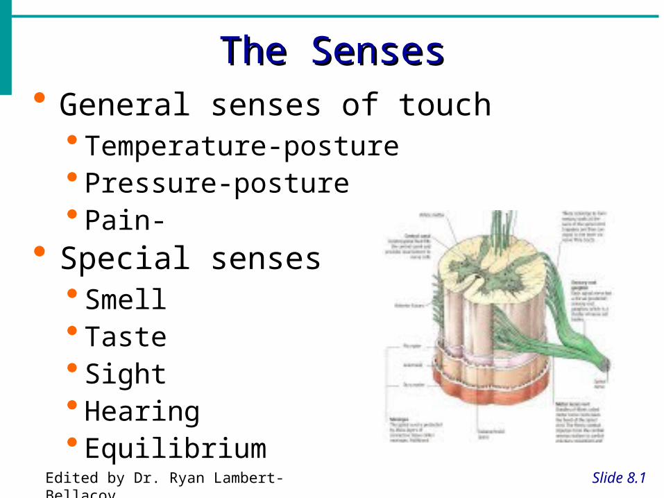
General senses of touch Temperature-posture Pressure-posture Pain-
Special senses Smell Taste Sight Hearing Equilibrium
Edited by Dr. Ryan Lambert-Bellacov

The Eye and VisionThe Eye and Vision
Slide 8.2
70 percent of all sensory receptors are in the eyes,
Each eye has over a million nerve fibers
Protection for the eye
Most of the eye is enclosed in a bony orbit
A cushion of fat surrounds most of the eye
Edited by Dr. Ryan Lambert-Bellacov
Accessory Structures of the EyeAccessory Structures of the Eye
Slide 8.3aCopyright © 2003 Pearson Education, Inc. publishing as Benjamin Cummings
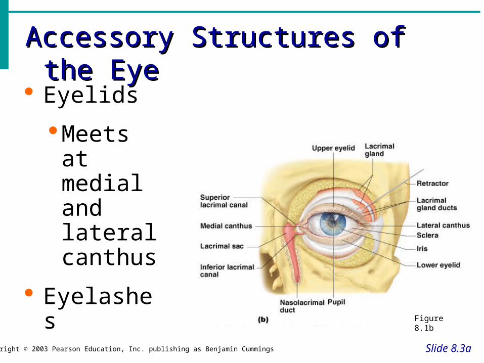
Eyelids
Meets at medial and lateral canthus
Eyelashes
Figure 8.1b
Accessory Structures of the EyeAccessory Structures of the Eye
Slide 8.3b
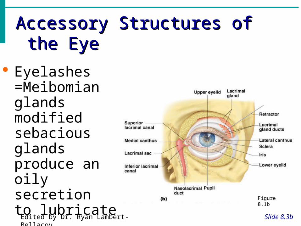
Eyelashes =Meibomian glands modified sebacious glands produce an oily secretion to lubricate the eye Figure 8.1b
Edited by Dr. Ryan Lambert-Bellacov
Accessory Structures of the EyeAccessory Structures of the Eye
Slide 8.3c
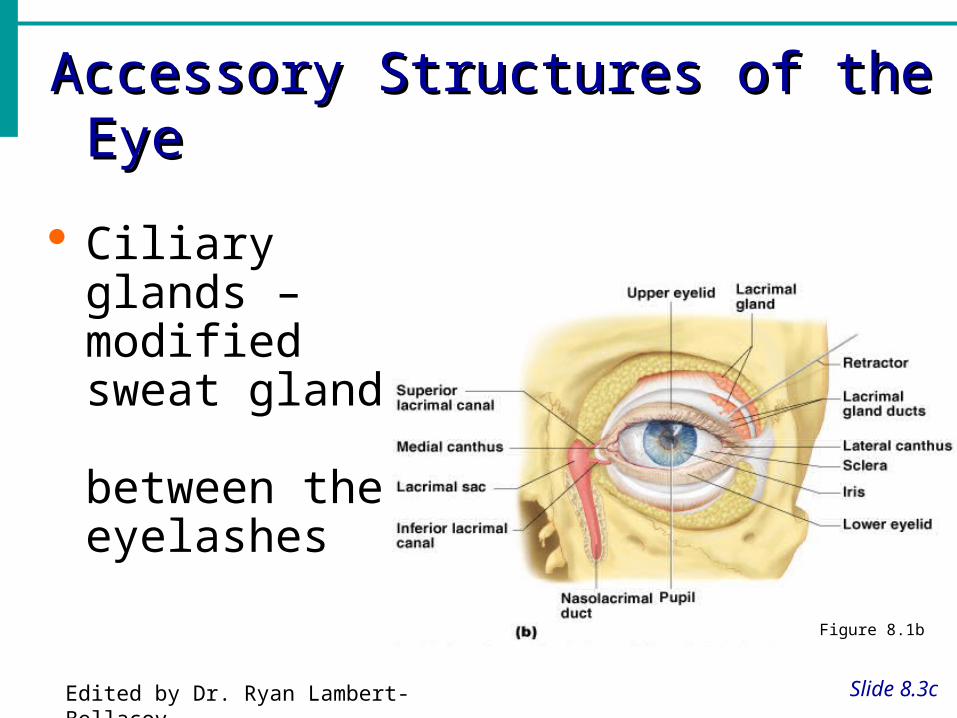
Ciliary glands – modified sweat glands between the eyelashes
Figure 8.1b
Edited by Dr. Ryan Lambert-Bellacov
Accessory Structures of the EyeAccessory Structures of the Eye
Slide 8.4a
Conjunctiva Membrane that lines the eyelids
Connects to the surface of the eye
Secretes mucus to lubricate the eye
Edited by Dr. Ryan Lambert-Bellacov
Accessory Structures of the EyeAccessory Structures of the Eye
Slide 8.4bCopyright © 2003 Pearson Education, Inc. publishing as Benjamin Cummings
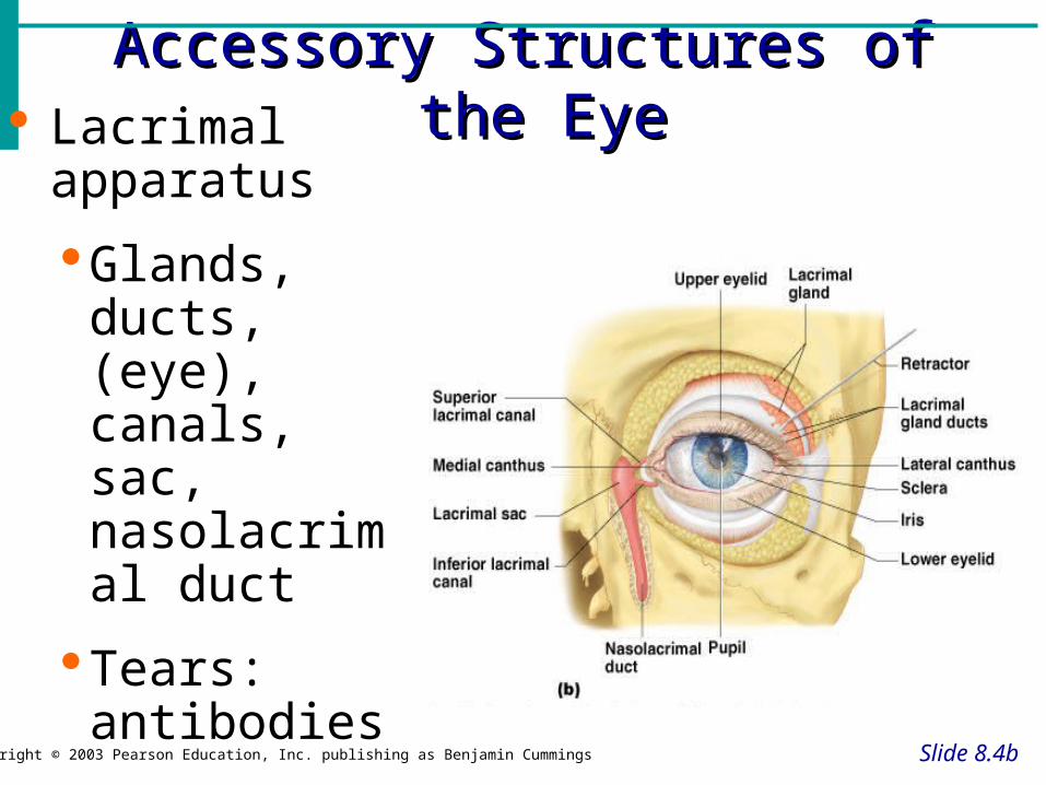
Lacrimal apparatus
Glands, ducts, (eye), canals, sac, nasolacrimal duct
Tears: antibodies, lysozymes, stress?
Figure 8.1a

Extrinsic Eye MusclesExtrinsic Eye Muscles
Slide 8.6
Muscles attach to the outer surface of the eye
Produce eye movements
Figure 8.2
Edited by Dr. Ryan Lambert-Bellacov
Structure of the EyeStructure of the Eye
Slide 8.7Copyright © 2003 Pearson Education, Inc. publishing as Benjamin Cummings
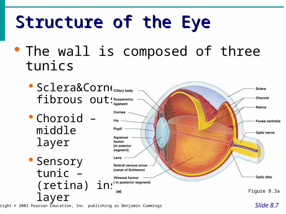
The wall is composed of three tunics
Sclera&Cornea fibrous outside layer
Choroid – middle layer
Sensory tunic – (retina) inside layer
Figure 8.3a
The Fibrous TunicThe Fibrous Tunic
Slide 8.8
Sclera White connective tissue layer
Seen anteriorly as the “white of the eye”
Cornea Transparent, central anterior portion
Allows for light to pass through
Repairs itself easily
The only human tissue that can be transplanted without fear of rejection
Edited by Dr. Ryan Lambert-Bellacov
Choroid LayerChoroid Layer
Slide 8.9
Blood-rich nutritive tunic
Pigment prevents light from scattering
Modified interiorly into two structures Cilliary body – smooth muscle
Iris
Pigmented layer that gives eye color
Pupil – rounded opening in the iris
Edited by Dr. Ryan Lambert-Bellacov

Sensory Tunic (Retina)Sensory Tunic (Retina)
Slide 8.10Copyright © 2003 Pearson Education, Inc. publishing as Benjamin Cummings
Contains receptor cells (photoreceptors) Rods
Cones
Signals pass from photoreceptors and leave the retina toward the brain through the optic nerve

Neurons of the RetinaNeurons of the Retina
Slide 8.11
Figure 8.4
Edited by Dr. Ryan Lambert-Bellacov

Neurons of the Retina and VisionNeurons of the Retina and Vision
Slide 8.12a
Copyright © 2003 Pearson Education, Inc. publishing as Benjamin Cummings
Rods
Most are found towards the edges of the retina
Allow dim light vision and peripheral vision
Perception is all in gray tones

Neurons of the Retina and VisionNeurons of the Retina and Vision
Slide 8.12b
Cones – 3 types detect different colors
Densest in the center of the retina
Fovea centralis – area of the retina with only cones
Lack of one type = color blindness
No photoreceptor cells are at the optic disk, or blind spot
Edited by Dr. Ryan Lambert-Bellacov

LensLens
Slide 8.14
Biconvex crystal-like structure
Held in place by a suspensory ligament attached to the ciliary body
Figure 8.3aEdited by Dr. Ryan Lambert-Bellacov
Internal Eye Chamber FluidsInternal Eye Chamber Fluids
Slide 8.15a
Aqueous humor in Anterior Segment
Watery fluid found in chamber between the lens and cornea
Similar to blood plasma
Helps maintain intraocular pressure
Provides nutrients for the lens and cornea
Reabsorbed into venous blood
Blocked drainage = glaucomaEdited by Dr. Ryan Lambert-Bellacov
Internal Eye Chamber FluidsInternal Eye Chamber Fluids
Slide 8.15b
Vitreous humor in Posterior Segment
Gel-like substance behind the lens
Keeps the eye from collapsing
Lasts a lifetime and is not replaced
Edited by Dr. Ryan Lambert-Bellacov
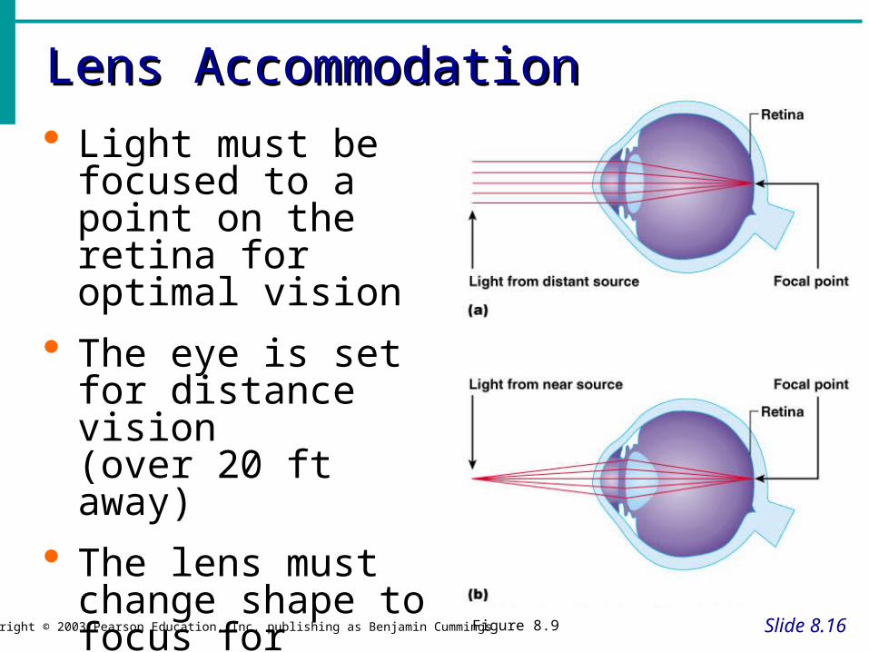
Lens AccommodationLens Accommodation
Slide 8.16Copyright © 2003 Pearson Education, Inc. publishing as Benjamin Cummings
Light must be focused to a point on the retina for optimal vision
The eye is set for distance vision (over 20 ft away)
The lens must change shape to focus for closer objects
Figure 8.9

Correcting the Eye
• Correct Focus = emmetropia• Nearsightedness = myopia
– Focus of light in front of retina– Eyeball too long or lens too strong– Distant objects are blurry
• Farsightedness = hyperopia– Focus of light beyond the retina– Short eyeball or lazy lens– Near objects are blurry.
Edited by Dr. Ryan Lambert-Bellacov
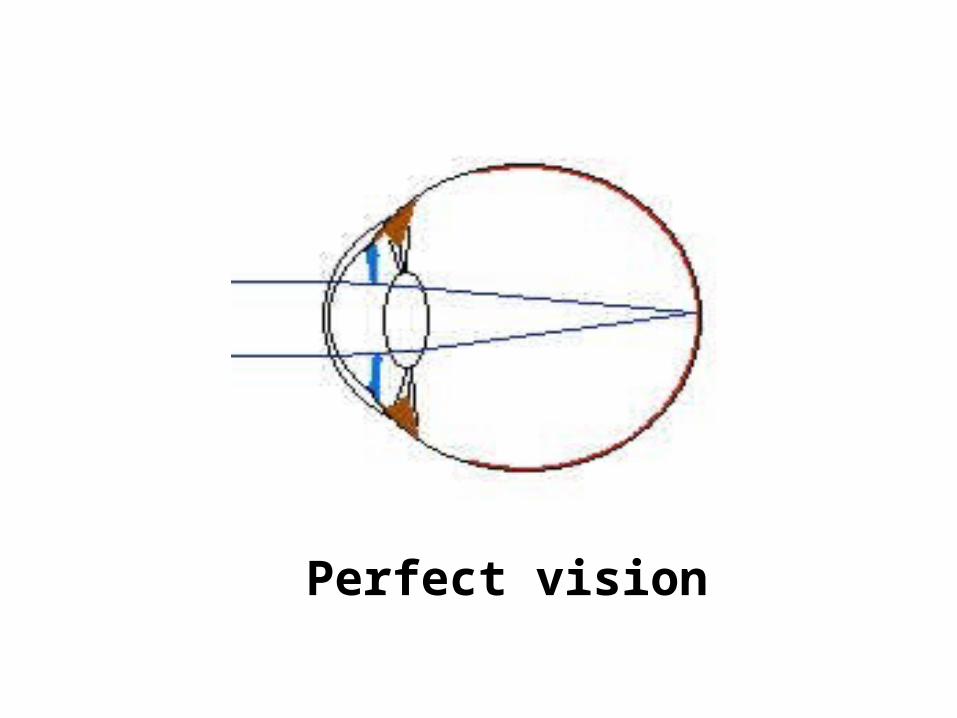
Perfect vision
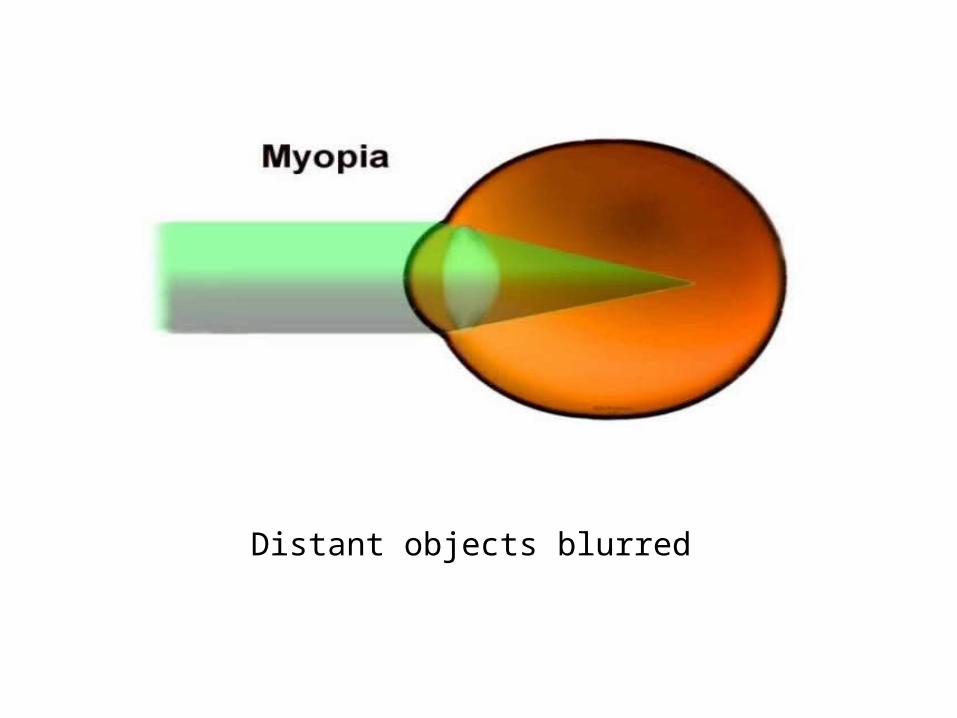
Distant objects blurred
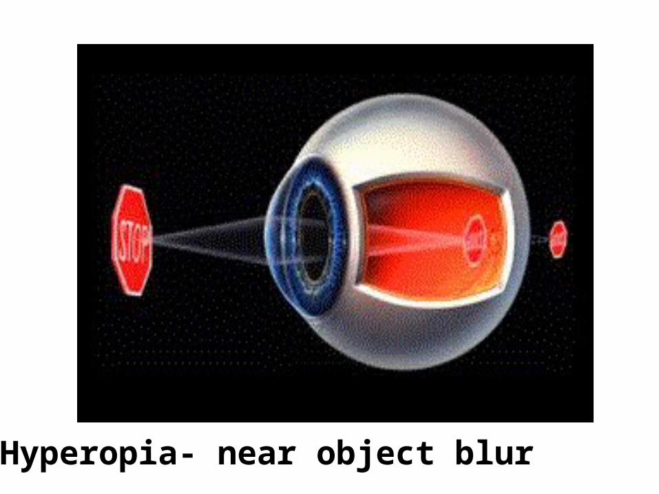
Hyperopia- near object blur

Astigmatism
• Unequal curvatures in cornea & lens

The EarThe Ear
Slide 8.20
Houses two senses
Hearing
Equilibrium (balance)
Receptors are mechanoreceptors
Anatomy of the EarAnatomy of the Ear
Slide 8.21Copyright © 2003 Pearson Education, Inc. publishing as Benjamin Cummings
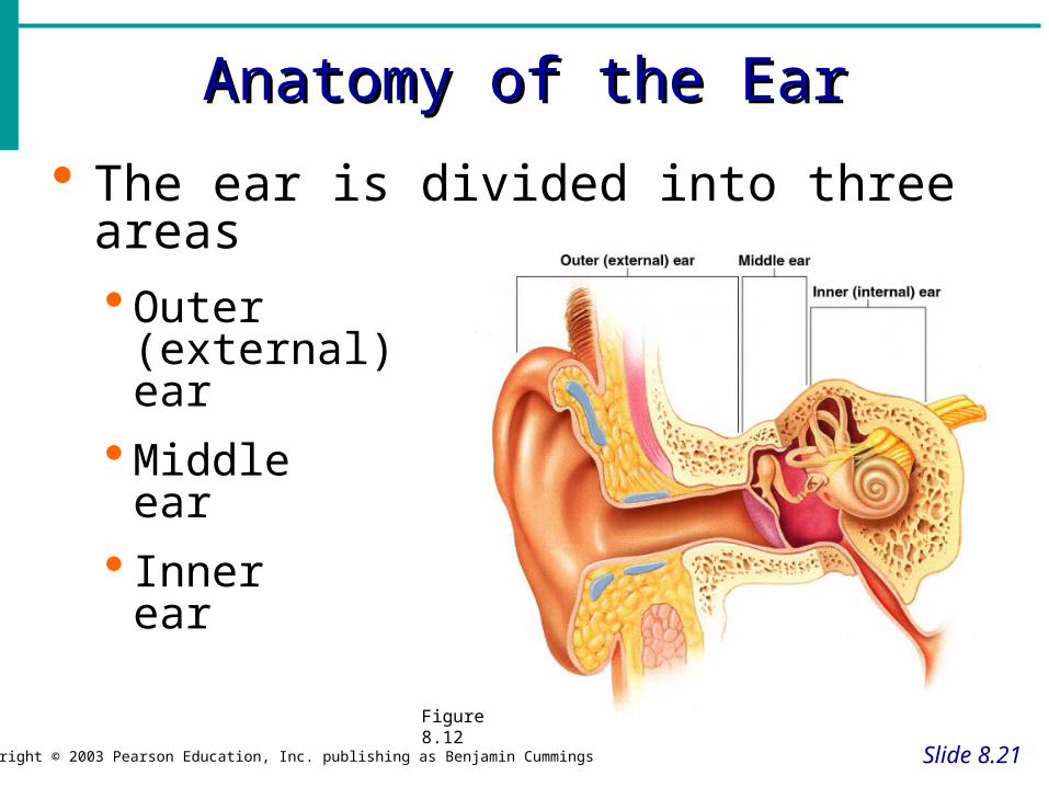
The ear is divided into three areas Outer
(external) ear
Middle ear
Inner ear
Figure 8.12
The External EarThe External Ear
Slide 8.22Copyright © 2003 Pearson Education, Inc. publishing as Benjamin Cummings
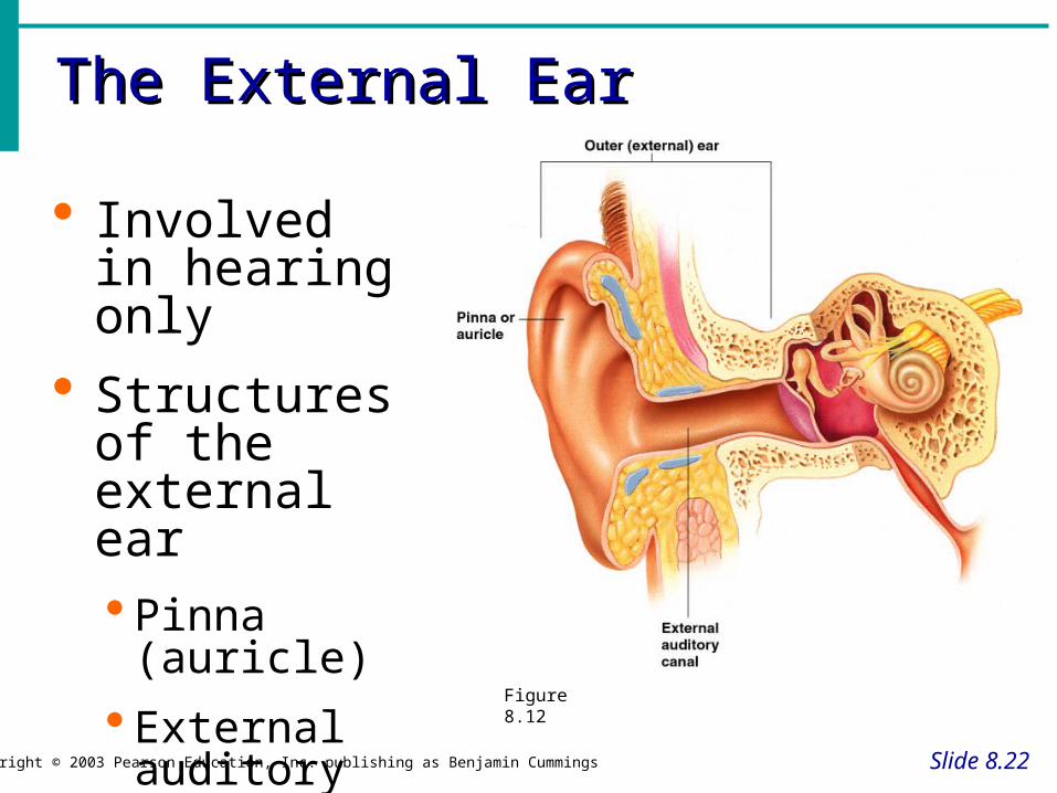
Involved in hearing only
Structures of the external ear Pinna
(auricle)
External auditory canal
Figure 8.12

The External Auditory CanalThe External Auditory Canal
Slide 8.23Copyright © 2003 Pearson Education, Inc. publishing as Benjamin Cummings
Narrow chamber in the temporal bone
Lined with skin
Ceruminous (wax) glands are present
Ends at the tympanic membrane
The Middle Ear or Tympanic CavityThe Middle Ear or Tympanic Cavity
Slide 8.24a
Air-filled cavity within the temporal bone
Only involved in the sense of hearing

The Middle Ear or Tympanic CavityThe Middle Ear or Tympanic Cavity
Slide 8.24b
Copyright © 2003 Pearson Education, Inc. publishing as Benjamin Cummings
Two tubes are associated with the inner ear
The opening from the auditory canal is covered by the tympanic membrane
The auditory tube connecting the middle ear with the throat
Allows for equalizing pressure during yawning or swallowing
This tube is otherwise collapsed

Bones of the Tympanic CavityBones of the Tympanic Cavity
Slide 8.25a
Copyright © 2003 Pearson Education, Inc. publishing as Benjamin Cummings
Three bones span the cavity
Malleus (hammer)
Incus (anvil)
Stapes (stirrip)
Figure 8.12

Bones of the Tympanic CavityBones of the Tympanic Cavity
Slide 8.25b
Copyright © 2003 Pearson Education, Inc. publishing as Benjamin Cummings
Vibrations from eardrum move the malleus
These bones transfer sound to the inner ear
Figure 8.12
Inner Ear or Bony LabyrinthInner Ear or Bony Labyrinth
Slide 8.26a
Copyright © 2003 Pearson Education, Inc. publishing as Benjamin Cummings
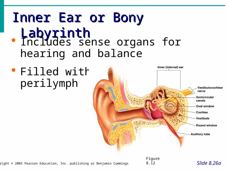
Includes sense organs for hearing and balance
Filled with perilymph
Figure 8.12

Inner Ear or Bony LabrynthInner Ear or Bony Labrynth
Slide 8.26b
Copyright © 2003 Pearson Education, Inc. publishing as Benjamin Cummings
A maze of bony chambers within the temporal bone
Cochlea
Vestibule
Semicircular canals
Figure 8.12

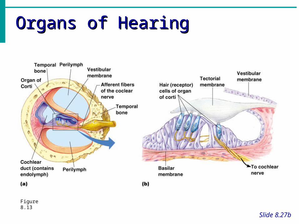
Organs of HearingOrgans of Hearing
Slide 8.27a
Organ of Corti
Located within the cochlea
Receptors = hair cells on the basilar membrane
Gel-like tectorial membrane is capable of bending hair cells
Cochlear nerve attached to hair cells transmits nerve impulses to auditory cortex on temporal lobe
Organs of HearingOrgans of Hearing
Slide 8.27b
Figure 8.13
Mechanisms of HearingMechanisms of Hearing
Slide 8.28Copyright © 2003 Pearson Education, Inc. publishing as Benjamin Cummings
Vibrations from sound waves move tectorial membrane
Hair cells are bent by the membrane
An action potential starts in the cochlear nerve
Continued stimulation can lead to adaptation
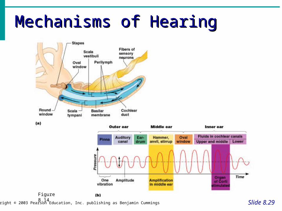
Mechanisms of HearingMechanisms of Hearing
Slide 8.29Copyright © 2003 Pearson Education, Inc. publishing as Benjamin Cummings
Figure 8.14

Organs of EquilibriumOrgans of Equilibrium
Slide 8.30a
Copyright © 2003 Pearson Education, Inc. publishing as Benjamin Cummings
Receptor cells are in two structures
Vestibule
Semicircular canals
Figure 8.16a, b

Organs of EquilibriumOrgans of Equilibrium
Slide 8.30b
Copyright © 2003 Pearson Education, Inc. publishing as Benjamin Cummings
Equilibrium has two functional parts
Static equilibrium – sense of gravity at rest
Dynamic equilibrium – angular and rotary head movements
Figure 8.16a, b
Static Equilibrium - RestStatic Equilibrium - Rest
Slide 8.31Copyright © 2003 Pearson Education, Inc. publishing as Benjamin Cummings
Maculae – receptors in the vestibule Report on the position of the head
Send information via the vestibular nerve
Anatomy of the maculae Hair cells are embedded in the otolithic
membrane
Otoliths (tiny stones) float in a gel around the hair cells
Movements cause otoliths to bend the hair cells
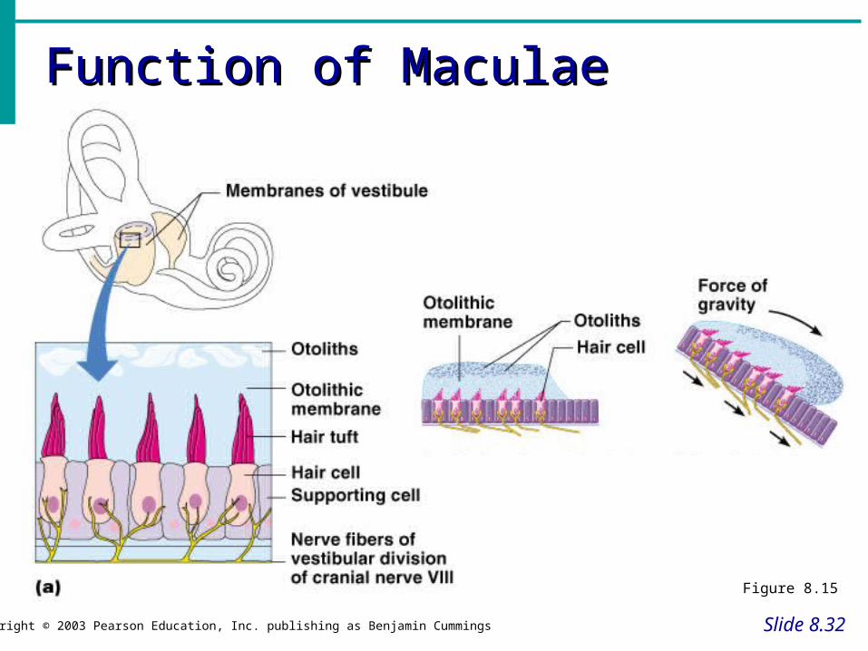
Function of MaculaeFunction of Maculae
Slide 8.32Copyright © 2003 Pearson Education, Inc. publishing as Benjamin Cummings
Figure 8.15

Dynamic Equilibrium - MovementDynamic Equilibrium - Movement
Slide 8.33a
Copyright © 2003 Pearson Education, Inc. publishing as Benjamin Cummings
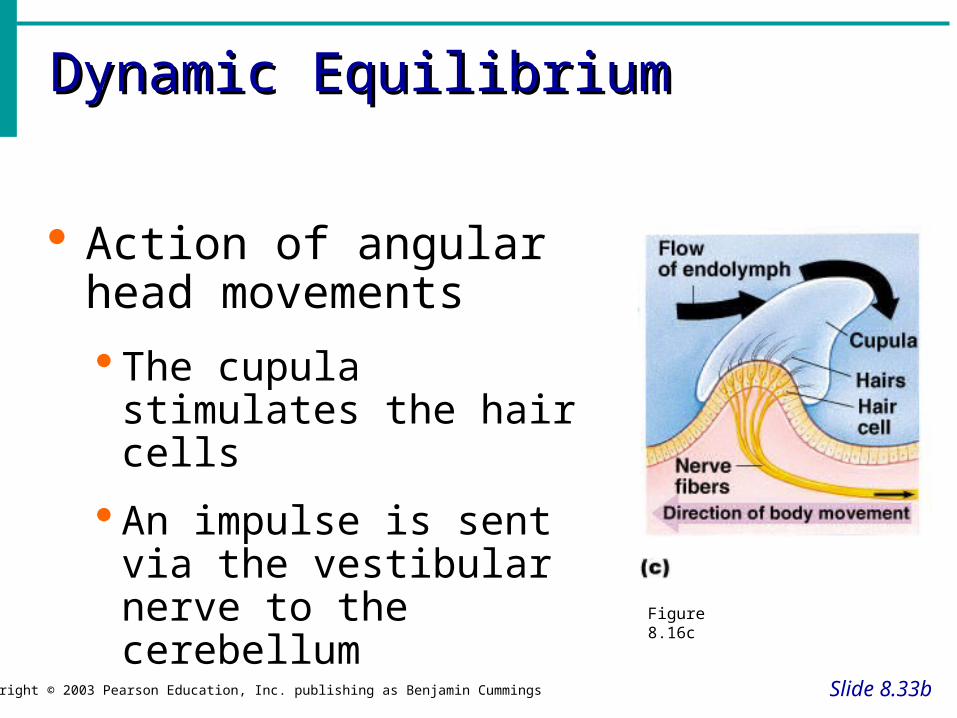
Crista ampullaris – receptors in the semicircular canals
Tuft of hair cells
Cupula (gelatinous cap) covers the hair cells
Figure 8.16c
Dynamic EquilibriumDynamic Equilibrium
Slide 8.33b
Copyright © 2003 Pearson Education, Inc. publishing as Benjamin Cummings
Action of angular head movements
The cupula stimulates the hair cells
An impulse is sent via the vestibular nerve to the cerebellum
Figure 8.16c

Chemical Senses – Taste and Chemical Senses – Taste and SmellSmell
Slide 8.34
Both senses use chemoreceptors
Stimulated by chemicals in solution
Taste has four types of receptors
Smell can differentiate a large range of chemicals
Both senses complement each other and respond to many of the same stimuli
Olfaction – The Sense of SmellOlfaction – The Sense of Smell
Slide 8.35
Olfactory receptors are in the roof of the nasal cavity
Neurons with long cilia
Chemicals must be dissolved in mucus for detection
Impulses are transmitted via the olfactory nerve
Interpretation of smells is made in the cortex

Olfactory EpitheliumOlfactory Epithelium
Slide 8.36Copyright © 2003 Pearson Education, Inc. publishing as Benjamin Cummings
Figure 8.17
The Sense of TasteThe Sense of Taste
Slide 8.37Copyright © 2003 Pearson Education, Inc. publishing as Benjamin Cummings
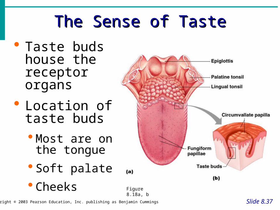
Taste buds house the receptor organs
Location of taste buds Most are on
the tongue
Soft palate
Cheeks Figure 8.18a, b

The Tongue and TasteThe Tongue and Taste
Slide 8.38Copyright © 2003 Pearson Education, Inc. publishing as Benjamin Cummings
The tongue is covered with projections called papillae
Filiform papillae – sharp with no taste buds
Fungifiorm papillae – rounded with taste buds
Circumvallate papillae – large papillae with taste buds
Taste buds are found on the sides of papillae
Structure of Taste BudsStructure of Taste Buds
Slide 8.39a
Copyright © 2003 Pearson Education, Inc. publishing as Benjamin Cummings
Gustatory cells are the receptors
Have gustatory hairs (long microvilli)
Hairs are stimulated by chemicals dissolved in saliva

Structure of Taste BudsStructure of Taste Buds
Slide 8.39b
Copyright © 2003 Pearson Education, Inc. publishing as Benjamin Cummings
Impulses are carried to the gustatory complex by several cranial nerves because taste buds are found in different areas
Facial nerve
Glossopharyngeal nerve
Vagus nerve

Anatomy of Taste BudsAnatomy of Taste Buds
Slide 8.40Copyright © 2003 Pearson Education, Inc. publishing as Benjamin Cummings
Figure 8.18
Taste SensationsTaste Sensations
Slide 8.41
Sweet receptors Sugars Saccharine Some amino acids
Sour receptors Acids
Bitter receptors Alkaloids
Salty receptors Metal ions
Developmental Aspects of the Developmental Aspects of the Special SensesSpecial Senses
Slide 8.42
Formed early in embryonic development
Eyes are outgrowths of the brain
All special senses are functional at birth


