Incomplete Ossification of the
Humeral Condyle (IOHC) in Dogs
John Ferguson BVM&S CertSAO MRCVS
Alasdair Renwick BVMS DSAS (Ortho) MRCVS
____________________________________________________________
What is IOHC?
The elbow joint in dogs is formed by three
bones: the radius, ulna and the humerus. These
bones rotate around a cylinder of bone attached
to the humerus called the humeral condyle
giving a “hinge” type of arrangement.
The cylinder of the humeral condyle develops
from two separate parts which “fuse” usually
between 8 and 12 weeks of age. This process is
called ossification. However, in some pups this
process fails to take place leaving an area of
cartilage in the middle of the condyle which
weakens this region significantly (so called
incomplete ossification of the humeral condyle or
IOHC).
However, recent work suggests that in some
dogs, a stress fracture occurs through previously
solid bone of the condyle. This situation is more
common in mature dogs over 18 months old.
Spaniel breeds, especially Springers, are
particularly prone to IOHC/fissure fracture and
the condition is likely to have a strong hereditary
basis.
What are the signs of IOHC?
Most dogs suffering from IOHC/fissure fracture
exhibit persistent or intermittent forelimb
lameness or stiffness particularly after rising.
This problem usually responds poorly to
standard painkillers and some patients will be
reluctant to jump from heights (e.g. from
furniture or out the car).
Some dogs suffering IOHC will not exhibit any
signs of an issue. In the majority of cases, a
pain response will be elicited on manipulation
of the affected elbow joint.
How is IOHC diagnosed?
In many cases IOHC
can be diagnosed by
conventional X-rays of
the elbow joint. The
red arrow on the X-ray
opposite points to the
fissure (IOHC) seen as
a dark line. However,
in many cases, the
IOHC will not be visible
on conventional X-rays
particularly if the fissure
or area of IOHC is very
small or incomplete
The best way of diagnosing IOHC is to obtain a
CT scan of the elbow. This will show whether or
not the humeral condyle is normal.
Why is IOHC important?
Incomplete ossification of the humeral condyle
can cause significant pain and lameness in dogs
and limit the affected individual’s ability to
exercise. Furthermore, the weakness present
within the humerus can result in a complete
spontaneous fracture during regular activity,
such as running in a field or jumping from a low
wall. The resulting fracture can be in the form of
a lateral condylar (two piece) or bi-condylar “Y”
(three piece) fracture.
CT scan showing IOHC (arrow)
Normal CT scan
X-ray of a 7 wk old pup
showing the two parts of
the humeral condyle (1 & 2) separated by the
growth centre (arrowed)
Model of humerus
showing the two parts of
the condyle to be completely “fused” and
the growth centre closed
1 2
These types of fractures can be difficult to repair
and occasionally treatment is not possible
because the break is so severe. The only option
in this situation being
amputation or sadly
even euthanasia.
How is IOHC treated?
If the patient is not lame or suffering problems
related to the IOHC, treatment is not necessary.
However, in the majority of cases, we would
recommend surgery to resolve pain caused by
the abnormality, to restore the patient to
soundness and remove the possibility of a
complete fracture occurring in the future. Our
current protocol is to place a large screw
across the condyle to stabilise the IOHC and to
strengthen the area. Other types of treatment
have been tried such as placing bone grafts into
the region but these have not be proven to give
a better outcome than traditional screw
placement.
What are the risks of surgery? The risks of surgery includes anaesthetising the
patient to facilitate surgery. However, with
modern drugs and techniques and close
monitoring of the patient anaesthetic
complications thankfully are very rare. Post
operative infection and soft tissue swelling were
previously common after surgery to treat IOHC.
However, at the East Neuk Vet Clinic we have
pioneered the placement of “in to out” direction
of the screw position (instead of the traditional
“out to in”) and we have since seen the incidence
of post- operative issues to have reduced
dramatically. We have published this work
recently.
Since we do not expect
the area of incomplete
ossification or fissure
fracture to fuse or
“heal”, the screw is
constantly subjected to
stress and loading.
This can lead to metal
fatigue and ultimately
breakage and the
patient will become
lame again (or in the
worst case the
humeral condyle could
fracture completely).
This complication can occur month to years after
the original surgery. A revision operation is
usually necessary to repair the fracture or to
replace the broken screw. To reduce the slim
chance of screw breakage, we try and place as
large a diameter of screw as possible.
What is the prognosis after surgery?
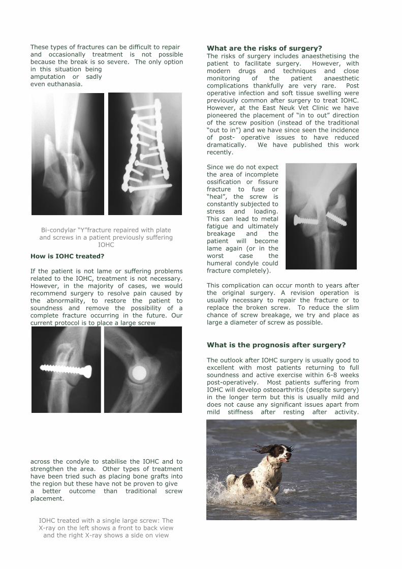
The outlook after IOHC surgery is usually good to
excellent with most patients returning to full
soundness and active exercise within 6-8 weeks
post-operatively. Most patients suffering from
IOHC will develop osteoarthritis (despite surgery)
in the longer term but this is usually mild and
does not cause any significant issues apart from
mild stiffness after resting after activity.
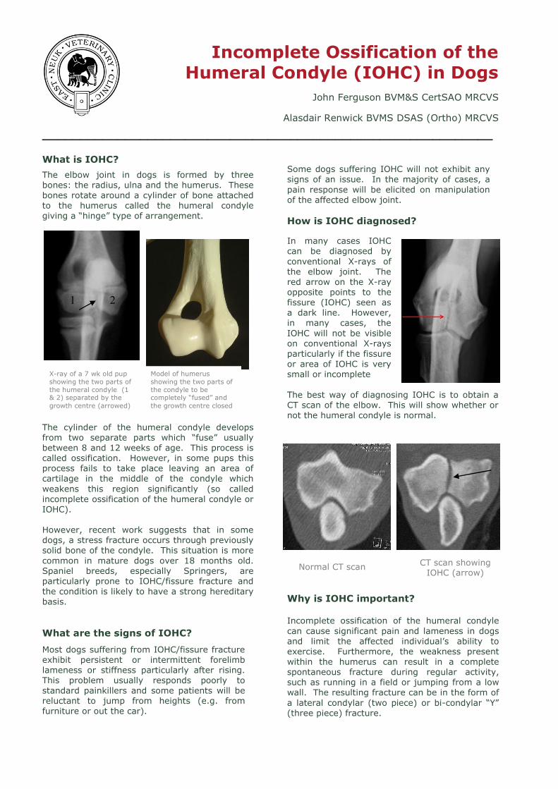
Bi-condylar “Y”fracture repaired with plate
and screws in a patient previously suffering IOHC
IOHC treated with a single large screw: The
X-ray on the left shows a front to back view and the right X-ray shows a side on view


