Ch 19
Infectious
Diseases
Affecting
the
Respiratory
System
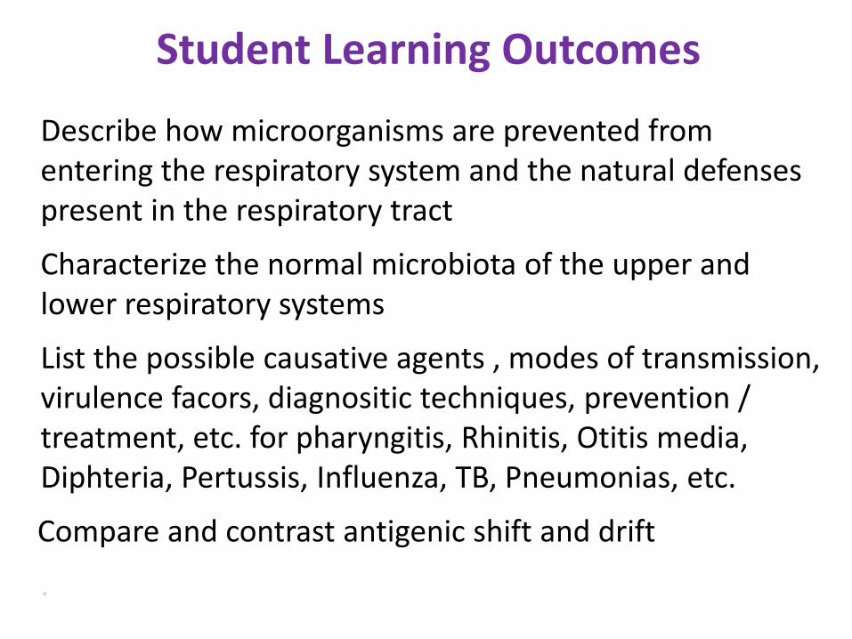
Student Learning Outcomes
Describe how microorganisms are prevented from entering the respiratory system and the natural defenses present in the respiratory tract
Characterize the normal microbiota of the upper and lower respiratory systems
List the possible causative agents , modes of transmission, virulence facors, diagnositic techniques, prevention / treatment, etc. for pharyngitis, Rhinitis, Otitis media, Diphteria, Pertussis, Influenza, TB, Pneumonias, etc.
Compare and contrast antigenic shift and drift
.
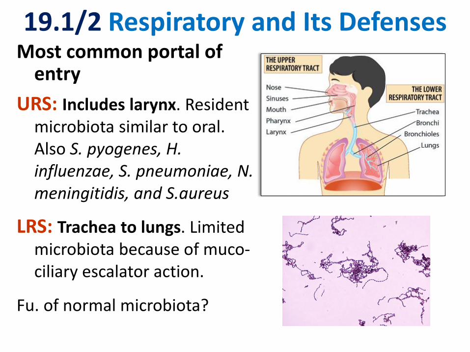
19.1/2 Respiratory and Its Defenses Most common portal of
entry
URS: Includes larynx. Resident microbiota similar to oral. Also S. pyogenes, H. influenzae, S. pneumoniae, N. meningitidis, and S.aureus
LRS: Trachea to lungs. Limited microbiota because of muco-ciliary escalator action.
Fu. of normal microbiota?

Fig 19.1

Pharyngitis Fusobacterium necrophorum children and young adults, S. pyogenes Droplet Transmission Symptoms: Sore throat, high fever, coughing, swollen LN, otitis media may also occur Both may be part of normal flora
Rapid Strep Test detects unique S.p. ags.
Penicillin works for both!
19.3 URS Diseases

Complications of Strep Throat S. pyogenes causes two major nonsuppurative
autoimmune complications (antibodies cross-react)
1. Acute rheumatic fever (see page 648):
Short period of arthritis and fever followed in 50% of affected by rheumatic heart disease heart valve damage chronic valvular disease (stenosis and/or incompetence) heart failure and/or subacute bacterial endocarditis
2. Acute poststreptococcal glomerulonephritis
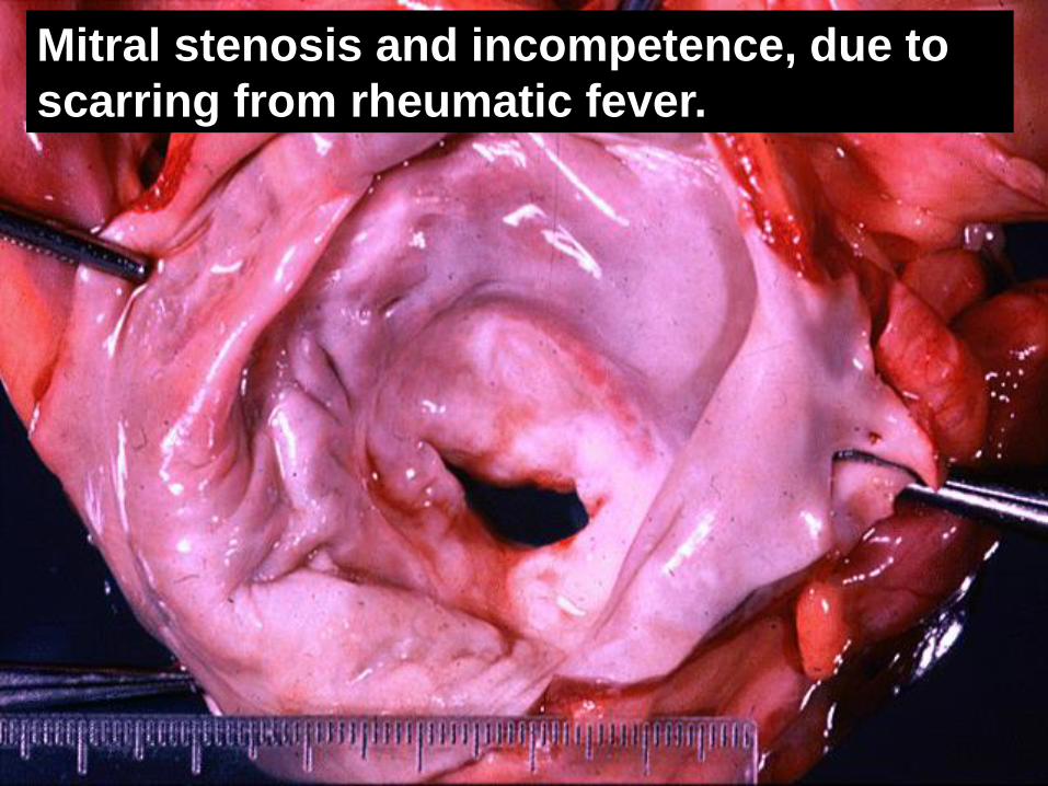
Mitral stenosis and incompetence, due to
scarring from rheumatic fever.
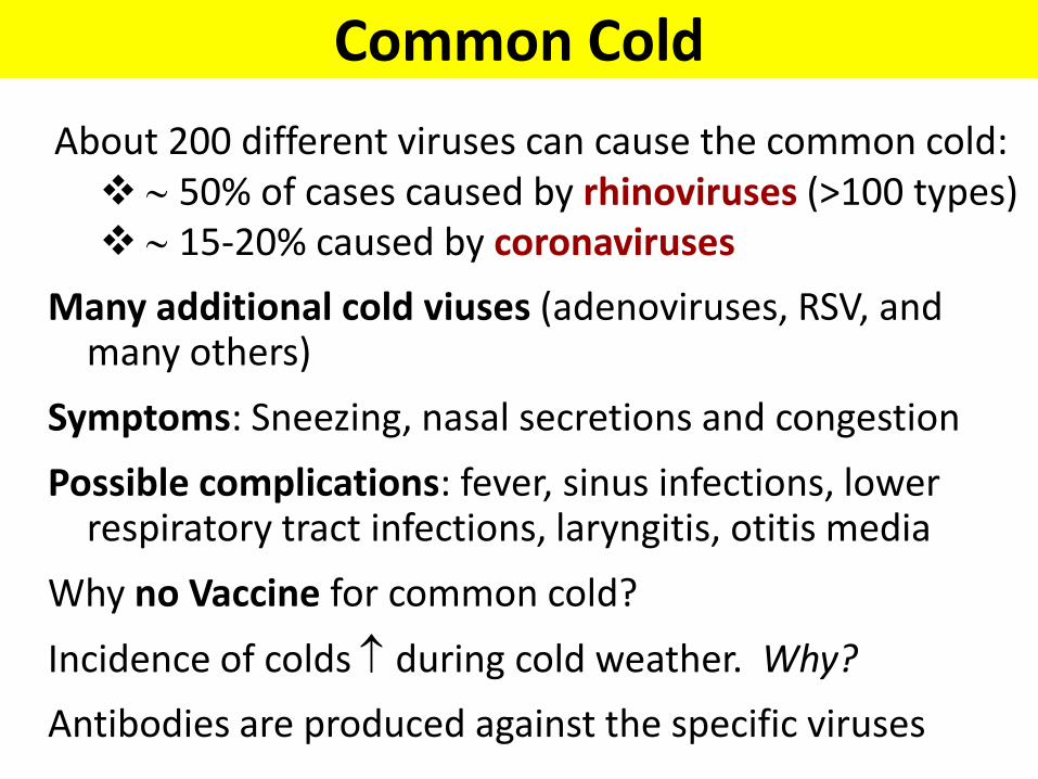
Common Cold
About 200 different viruses can cause the common cold: 50% of cases caused by rhinoviruses (>100 types) 15-20% caused by coronaviruses
Many additional cold viuses (adenoviruses, RSV, and many others)
Symptoms: Sneezing, nasal secretions and congestion
Possible complications: fever, sinus infections, lower respiratory tract infections, laryngitis, otitis media
Why no Vaccine for common cold?
Incidence of colds during cold weather. Why?
Antibodies are produced against the specific viruses

(Acute) Otitis Media
Complication of nose and throat infections
Pus accumulation causes pressure on the eardrum
Bacterial causes include
• S. pneumoniae (35%)
• H. influenzae (20-30%)
• M. catarrhalis (10-15%)
• S. pyogenes (8-10%)
• S. aureus (1-2%)
• Treated with broad-spectrum antibiotics
• Incidence of S. pneumoniae reduced by vaccine
Fig 19.5

Diphtheria
• Corynebacterium diphtheriae
• Pseudomembrane formation (fibrin, dead tissue and bacteria)
• Not very invasive, but prophage encoded exotoxin inhibits protein synthesis absorbed into blood heart, nerve and kidney damage. Use antitoxin early!
• DTaP
• Boosters: get Tdap once then Td boosters every 10 years
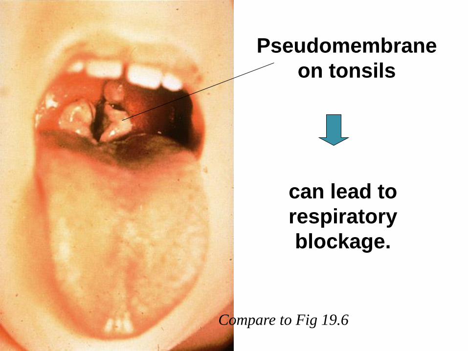
can lead to
respiratory
blockage.
Pseudomembrane
on tonsils
Compare to Fig 19.6

• Influenza: Influenzavirus; ssRNA, 8 segments
• Symptoms: Chills, fever, headache, muscle aches (no intestinal symptoms)
• Grouped according to antigenic differences in surface proteins:
– Type A → mammals and birds (most severe and extensive); currently most common antigenic variants of influenza A virus: H1N1 and H3N2
– Types B and C → humans only
• Viral isolates identified via serological testing
19.4 Microbial Diseases of Upper AND Lower Respiratory System
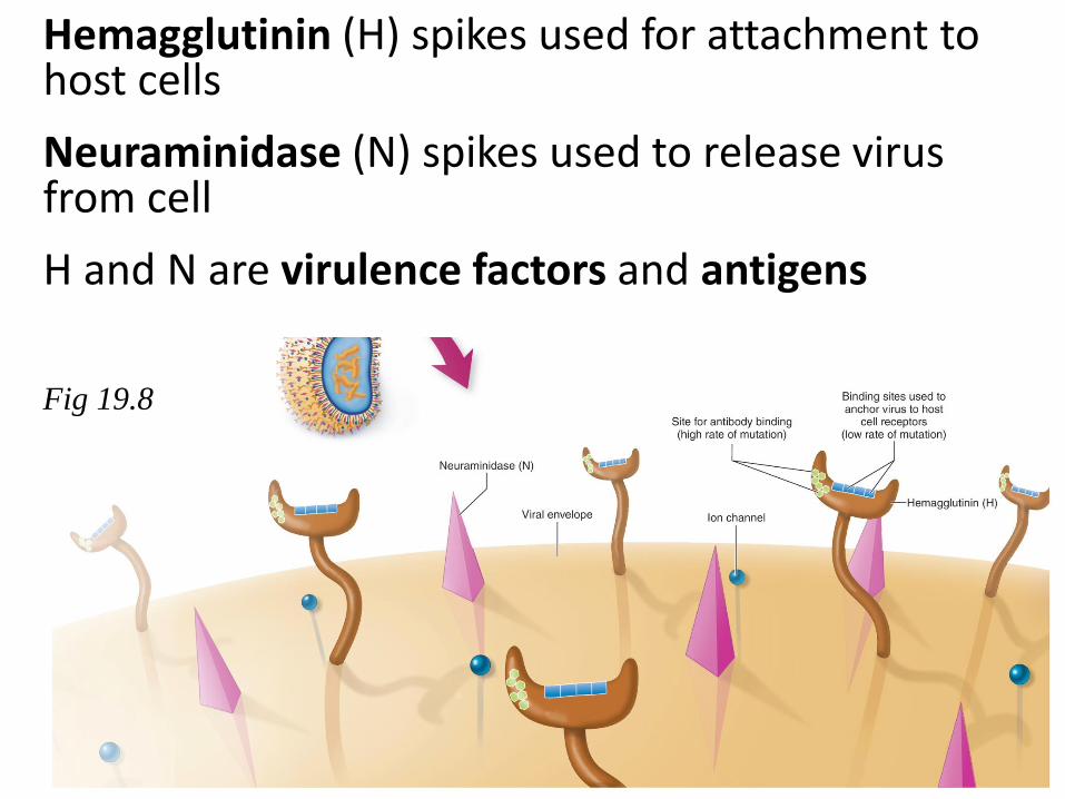
Hemagglutinin (H) spikes used for attachment to host cells
Neuraminidase (N) spikes used to release virus from cell
H and N are virulence factors and antigens
Fig 19.8

Mutations in H and N lead to
• antigenic shift (major changes only for type A) or
• antigenic drift (minor changes for all types)
natural immunity and vaccination obsolete
Compare to Fig 19.9
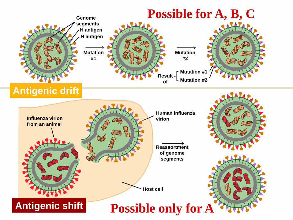
ANTIGENIC SHIFT
Reassortment
of genome
segments
Mutation
#2
Mutation
#1
Host cell
ANTIGENIC DRIFT
H antigen
N antigen
Human influenza
virion
Result
of
Genome
segments
Mutation #1
Mutation #2
Influenza virion
from an animal
Antigenic drift
Antigenic shift Possible only for A
Possible for A, B, C

Pandemics, Prevention and Treatment
• Wide spread epidemics due to antigenic shifts Pandemics
• 50,000 – 70,000 deaths/year in US - also Guillain-Barré and Reye’s syndrome
• Complications often due to bacterial secondary infections (??)
• Vaccine produced in chicken embryos
– flu shot (..... ) and
– nasal spray (LAIV)
• Effective antiviral drugs must be taken early on

Bordetella pertussis, highly contagious
Various toxins: •Tracheal cytotoxin damages ciliated cells •Pertussis toxin enters blood systemic symptoms
Three stages of disease
1. Catarrhal stage resembles a cold
2. Paroxysmal stage due to accumulation of mucus in trachea and bronchi deep paroxysmal coughs (brain and eye hemorrhage)
3. Convalescence stage can last for months
Laboratory diagnosis based on isolation of bacteria on enrichment and selective media, followed by serological tests
Whooping Cough Gram -, coccobacillus
Clinical presentation
DTaP

Bacteria, viruses, and fungi cause
–Bronchitis
–Bronchiolitis
–Pneumonia
What keeps LRS relatively sparsely colonized?
19.4 Microbial Diseases of the Lower Respiratory System
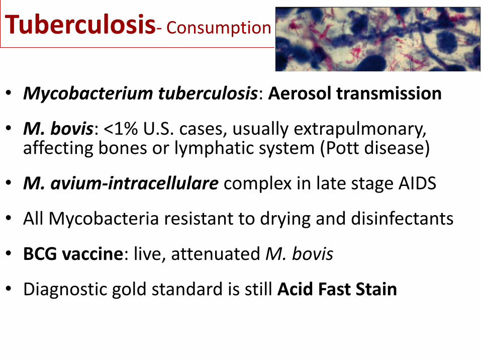
Tuberculosis- Consumption
• Mycobacterium tuberculosis: Aerosol transmission
• M. bovis: <1% U.S. cases, usually extrapulmonary, affecting bones or lymphatic system (Pott disease)
• M. avium-intracellulare complex in late stage AIDS
• All Mycobacteria resistant to drying and disinfectants
• BCG vaccine: live, attenuated M. bovis
• Diagnostic gold standard is still Acid Fast Stain

Tuberculin Sensitivity
(Mantoux) test: Inject
PPD,wait for delayed
hypersensitivity reaction (problem: BCG vaccination!)
Diagnostic tool for
presymptomatic
tuberculosis
Purified
protein
derivative induration

TB blood test as an alternative to Mantoux test: Interferon Gamma Release Assay or IGRA
Two types of TB blood tests:
• QuantiFERON®-TB
• T-SPOT®.TB
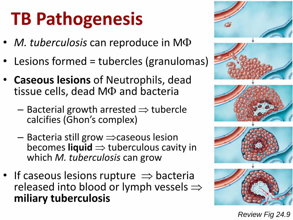
TB Pathogenesis • M. tuberculosis can reproduce in M
• Lesions formed = tubercles (granulomas)
• Caseous lesions of Neutrophils, dead tissue cells, dead M and bacteria
– Bacterial growth arrested tubercle calcifies (Ghon’s complex)
– Bacteria still grow caseous lesion becomes liquid tuberculous cavity in which M. tuberculosis can grow
• If caseous lesions rupture bacteria released into blood or lymph vessels miliary tuberculosis
Review Fig 24.9

Miliary tuberculosis due to massive lymphhaematogeneous dissemination. Due to impaired CMI potentially lethal. Weight loss, coughing of blood, loss of vigor
3 or 4 drugs taken for at least 6 months
MDR-TB becoming prevalent!

Globally, nearly 9 million new TB cases and approximately 1.5
million TB-related deaths each year.
MDR-TB (as high as 20%) resistance to rifampin and isoniazid
XDR-TB resistant to two additional drugs
Estimated Tuberculosis Incidence, 2014 (CDC)

Typical Pneumonia: Pneumococcal Pneumonia • Encapsulated S. pneumoniae
• Can be identified by production of -hemolysis and through serological tests
• Droplet transmission. Asymptomatic carriers illness in case of immune suppression, smoking, viral infection etc.
• Symptoms: fever, breathing difficulty, chest pain, rust-colored sputum
• Predominant in elderly
• Penicillin, but MDROs increasing
• Vaccine for 23 most common strains (> 90 strains)
Arrangement?
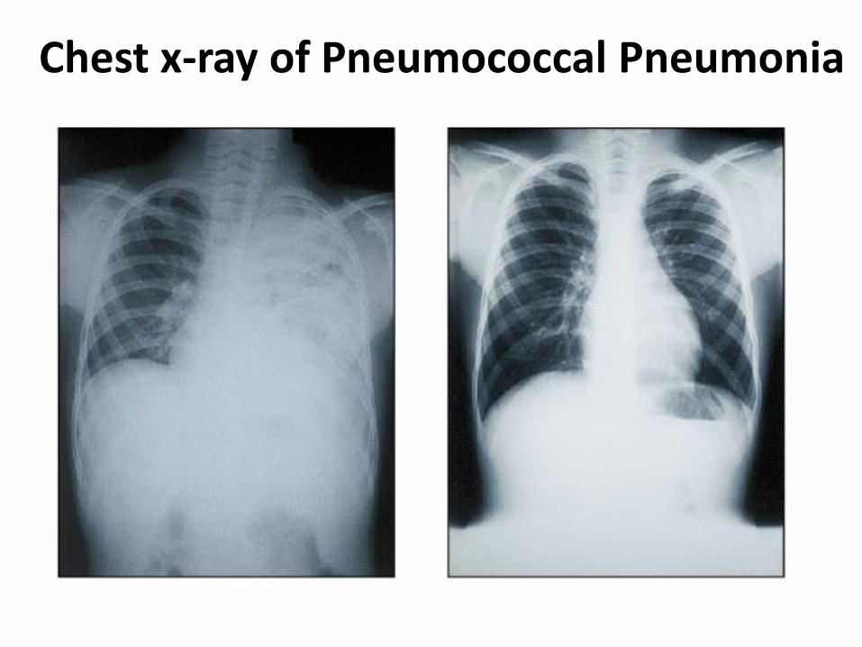
Chest x-ray of Pneumococcal Pneumonia
Legionellosis or Legionnaires’ disease
• Legionella pneumophila, Gram– rod
• Discovered among a group of elderly men attending an American Legion Convention in Philadelphia (1976 )
• Grow in water (pools, lakes, water systems of buildings air conditioning units, etc.), then disseminated in air
• Transmission by inhaling aerosols; no person to person transmission
• Diagnosis: Bacterial culture (selective charcoal- yeast extract medium, FA tests, DNA probes
• Pneumonia and pleurisy (15 - 20% mortality rate when hospitalized)

Mycoplasmal Pneumonia – also known as Primary Atypical Pneumonia or Walking Pneumonia
• Mycoplasma pneumoniae, pleomorphic, wall-less
• Produce small “fried-egg” colonies after two weeks’ incubation on enriched media containing horse serum and yeast extract
• Common in children and young adults – often mild enough to go undiagnosed for long periods of time
• Diagnosis: PCR or serological tests (IgM antibodies)

Hantavirus Pulmonary Syndrome (HPS)
Korean hemorrhagic fever caused by Hantaan virus of Bunyaviridae
HPS first reported in US in spring of 1993.
Transmission through urine, droppings, or saliva of infected rodents humans inhale aerosolized virus. No person to person transmission in US
Sudden respiratory failure
Mortality rate > 35%
2012 Yosemite

Hanta Virus Cases
Hantavirus Pulmonary Syndrome (HPS) Cases, by State of Residence Cumulative Case Count Per State Valid as of January 8, 2016

Fungal Diseases of the Lower Respiratory System (LRS)
• Fungal spores are easily inhaled; they may germinate in the lower respiratory tract
• The incidence of fungal diseases has been increasing in recent years
• Mycoses in the sections below can be treated with amphotericin B
–Coccidioidomycosis (see meningitis p.475)
–Pneumocystis Pneumonia

Coccidioidomycosis = Valley Fever
• Coccidioides immitis, dimorphic
• Airborne transmission in endemic areas
• Most cases subclinical, some respiratory infection with flu-like symptoms
• In < 1% of cases (due to predisposing factors, such as fatigue, poor nutrition, etc.): progressive, disseminated disease resembling TB; also meningitis (p. 475)
• Diagnosis: serological tests
• 97% of reported cases are from California and Arizona

Pneumocystis Pneumonia (PCP)
Pneumocystis jiroveci ( previously P. carinii) tiny fungus
Commonly found in nature, in healthy human lungs and animals Aerosol transmission
Illness and death in newly infected infants and immuno-suppressed individuals
Used to be leading cause of death in AIDS patients – now preventive special antifungal therapy
Diagnosis: detection of cysts in sputum samples
Healthcare-Associated Pneumonia (HAP)
In up to 1% of hospitalized or institutionalized
people due to abnormal breathing and aspiration:
– Common in stroke victims
– Mortality rate: 30 – 50%
– Most frequent causes:
• Pseudomonas aeruginosa
• Streptococcus pneumoniae
• Klebsiella pneumoniae
• S. aureus: HAP caused by MRSA
• Many are polymicrobial


