136
This article can be downloaded from http://www.ijlbpr.com/currentissue.php
Int. J. LifeSc. Bt & Pharm. Res. 2014 Abu T F A et al., 2014
PURIFICATION AND CHARACTERISATION OF-AMYLASE FROM BACILLUS SUBTILIS
ISOLATED FROM FERMENTED AFRICAN LOCUSTBEAN (PARKIA BIGLOBOSA) SEEDS
Abu T F A1*, Enujiugha V N1, Sanni D M2 and Bamidele O S2
Research Paper
-amylase was obtained from Bacillus subtilis isolated from fermented Parkia biglobosa seeds,purified and characterized. Purification was achieved using ion exchange DEAE column andgel filtration (Sephadex G-200) chromatography. Effects of temperature; pH and production timeon -amylase production were investigated. Also, physicochemical characteristics of the purifiedenzyme were investigated. The optimum production of -amylase was at temperature, pH andtime of 37oC, 7.0 and 24 h, respectively. The results showed that purified -amylase had moreenzymatic activity than crude samples from Bacillus subtilis whereby the activity of crude enzymewas 3.21 mM/min/mL while the purified enzyme had an improved activity of 21.46 mM/min/mL.Optimum temperature and pH values of the purified amylase were found to be 50°C and 5.0,respectively. pH stability of the enzyme ranged from 4.0- 9.0. At pH 5.0 and 7.0 it retained 70%and 60% of its activity after 5 h of incubation. Temperature stability ranged between 40oC and70oC but most stable at 50oC retaining 64% of its activity after 1 h of incubation. The enzymeexhibited maximum activity on soluble starch and sucrose, among other carbohydrate substrates.EDTA, Cu2+ and Fe2+ inhibited its activity while Ca2+ and K+ enhanced it up to 30%. The Lineweaver-Burke plot of the purified -amylase activity of B. subtilis indicates that the -amylase enzymehas apparent Km and Vmax values for the hydrolysis of soluble starch of 17.74 mg mL-1 and14.09U, respectively. The enzyme was purified 18.76 -fold and the molecular weight was 42.2kDa. The study revealed that -amylase from B. subtilis can be exploited for starch conversionbiotechnologies.
Keywords: Parkia biglobosa, Bacillus subtilis, -amylase, Purification, Characterization
*Corresponding Author:Abu T F A [email protected]
ISSN 2250-3137 www.ijlbpr.comVol. 3, No. 4, October 2014
© 2014 IJLBPR. All Rights Reserved
Int. J. LifeSc. Bt & Pharm. Res. 2014
1 Department of Food Science and Technology, 2 Department of Biochemistry; Federal University of Technology, P.M.B 704, Akure, Ondo State,Nigeria.
INTRODUCTIONAmylases are enzymes whose applications in the
industry have been increasing due to widespread
use such as in foods, brewing, textiles, adhesives,
detergents, pharmaceuticals, and sewage
treatments (Osaki and Yoshino, 1988). The
137
This article can be downloaded from http://www.ijlbpr.com/currentissue.php
Int. J. LifeSc. Bt & Pharm. Res. 2014 Abu T F A et al., 2014
enzymes involved are mainly -amylase (1,4-
glucan glucohydrolase, EC 3.2.1.1), – amylase
(1,4–glucan maltohydrolase, EC 3.2.1.2) and
glucoamylase (1,4-glucan glucohydrolase, EC
3.2.1.3) (Boldon and Effront, 2000). Though
amylases originate from different sources such
as plants, animals and microorganisms, the
microbial amylases are the most produced and
used in industry due to their productivity. Amylases
are among the most important industrial enzymes
that have a wide variety of applications ranging
from conversion of starch to sugar syrups, to the
production of cyclodextrins for the pharmaceutical
industry (Leveque et al., 2000). These enzymes
account for about 30% of the world’s enzyme
production. Unlike other members of the amylase
family, only a few attempts have been made to
study –amylases particularly of plant origin while
there is a dearth of information on –amylase from
microbial sources. Bacterial strains belonging to
the genera Bacillus, Pseudomonas, Clostridium
(Rani et al., 2007); and fungal strains belonging
to Rhizopus (Forgarty and Kelly, 1990) have been
reported to synthesize –amylase. The properties
of the -amylase varies from one source to the
other. Some of the microorganisms reported to
produce –amylases have employed starchy
wastes such as cassava, rice husk, potato rice
and maize as substrates for production (Forgarty
and Kelly, 1990). “Iru”, fermented locust beans
used as a condiment in cooking, similar to ogiri
and douchi made from fermentation of seeds of
the African locust bean tree; Parkia biglobosa a
perennial leguminous tree belonging to the sub-
family Mimosoideae and family leguminosae (now
family Fabaceae) Campbell-plat (1980). The
seeds are rich in protein and the food condiment
Iru is used as a flavor enhancer for soups, stews
and also adds protein to a poor-protein diet in the
developing countries (Dike and Odunfa, 2003).
Studies on the fermentation of African locust bean
seeds and other Nigerian condiments of protein
origin used for soups and sauces found that
fermentation was carried out by Bacillus subtilis
(Enujiugha, 2009). During fermentation, the
reducing sugar content increases, and the total
free amino acid content initially decreases; in the
end, however, there is a large increase in free
amino acid content. Processing of locust bean
fruits to food condiment, involves different unit
operations after harvesting, such unit operations
include de-podding, removal of the yellowish pulp
to produce locust bean seeds (Akande et al.,
2010). Other processing operations are cleaning,
boiling, de-hulling, washing, re-cooking, and then
fermentation to produce the food condiment
which is used as soup seasoning/spices (flavoring
agent) (Enujiugha, 2009). Due to the increasing
demand for these enzymes in various industries,
there is enormous interest in developing enzymes
with better properties; such as raw starch
degrading amylases suitable for industrial
applications and their cost effective production
techniques. The objectives of this project are to:
isolate Bacillus subtilis microorganism from IRU,
a local, cheap and ready legume, purify the -
amylase enzyme extracted from the isolated
Bacillus subtilis and determine the effects of some
metal ions, pH, and temperature on activity and
stability of the pure -amylase enzyme.
MATERIALS AND METHODSIsolation and Production of -amylase
Bacillus subtilis was isolated from fermented
African locust bean (Parkia biglobosa), samples
“iru” obtained from a local market in Akure, Ondo
State using the method reported by Yandri et al.
(2010). The colonies were picked up and streaked
138
This article can be downloaded from http://www.ijlbpr.com/currentissue.php
Int. J. LifeSc. Bt & Pharm. Res. 2014 Abu T F A et al., 2014
on nutrient agar plates to get pure culture and to
confirm zone formation. The culture was
characterised by Gram-staining and other
biochemical tests before storing on nutrient agar
slants for use. The inoculum was prepared by
the addition of sterile distilled water to the freshly
grown nutrient agar slants, from this 0.5 mL of
cell suspension was inoculated into 100 mL of
sterilized fermentation medium and incubated at
35°C for 10 h. The composition of the fermentation
medium was [g/L] 6.0 g Bacteriological peptone;
0.5 g MgSO4 .7H2O; 0.5 g NaCl; 1.0 g Starch; 1.0
g Yeast extract; 0.1% K2HPO4 and 0.02%
MgSO4.7H2O at pH 7 (Rehman et al., 2011) .
Inoculated flasks were maintained in water bath
shaker at 150 g for 48 h. Growth and enzyme
activity were determined from the aliquots (5 mL)
collected at every 6 h. Growth was estimated
turbidimetrically and the optical density of the
culture broth was measured at 660 nm in
spectrophotometer (Spectrophotometer Jenway,
6305).
To study the effect of temperature on amylase
production the submerged fermentation was
carried out at different temperatures (25°C, 30°C,
35°C and 40°C). About 100 mL of growth medium
was taken in one set consisting of 3 flasks,
autoclaved and then inoculated with 100 L of the
freshly prepared inocula. The cultures were
incubated at the above temperature range in an
incubator shaker at 150 rpm. An aliquot of culture
was taken at regular intervals (0, 4, 8, 12, 16, 20,
24 and 28 h) to measure absorbance at 600 nm
using a LAMBDA 650 UV/Vis Spectrophotometer
(PerkinElmer, USA).
Effect of pH
The fermentation medium was prepared by
varying the pH values (5.0, 6.0, 7.0 and 8.0) for
the production of amylase. Incubation was at
room temperature and aliquot of culture was taken
at regular intervals (0, 4, 8, 12, 16, 20, 24 and 28
h) to measure absorbance at 600 nm.
Effect of Starch Concentration onBacterial Growth
Growth curves of B. subtilis were determined in
1% soluble starch fermentation broth medium.
For bacterial isolate 100 mL medium was taken
in one set consisting of 3 flasks, autoclaved and
then inoculated with 100 L of the freshly prepared
inocula. The cultures were incubated at 45°C in
an incubator shaker at 150 rpm. An aliquot of
culture was taken at regular intervals (0, 4, 8, 12,
16, 20, 24 and 28 h) to measure absorbance at
600 nm
PURIFICATION OF -AMYLASEAMMONIUM SULPHATEFRACTIONATIONThe supernatant was gradually brought to 80%
saturation with ammonium sulphate with constant
gentle stirring for 1 h after centrifugation (Rehman,
2011). The precipitate was earlier removed after
centrifugation at 3,219 g for 30 min. Both enzyme
activity (Bernfield, 1955) and protein content
(Lowry, 1951) were determined for the separate
fractions. The obtained ammonium sulphate
precipitate (in solution) was introduced into
dialysis bag (Spectra/por standard grade
regenerated cellulose dialysis membrane)
followed by dialysis against 0.1 M phosphate
buffer at pH 6.2 for 48 h while replacing the buffer
thrice (Takasaki, 1976). The obtained amylase
enzyme preparation was concentrated against
crystals of sucrose to remove the remaining salt
and kept in the refrigerator at 5ºC for further
purification.

139
This article can be downloaded from http://www.ijlbpr.com/currentissue.php
Int. J. LifeSc. Bt & Pharm. Res. 2014 Abu T F A et al., 2014
Ion Exchange Chromatography
Further purification of amylase enzyme was
carried out using DEAE (Di-ethyl amino ethyl)
cellulose anion exchange chromatography. The
dialyzed sample was applied to a DEAE column.
The column was washed with 50 mM, Tris buffer
pH.8, and eluted with serially increasing
concentration of NaCl (0.1 M, 0.2 M). The eluted
fractions were monitored by UV absorption
spectrophotometer at 280 nm.
Gel Filtration Chromatography (UsingSephadex G-200)
Preparation of the gel column and the fractionation
procedures was determined as mentioned by
Ammar (1975). Sephadex G-200 (Pharmacia,
Upsalla, Sweden) was used, 0.1 M Tris-HCl buffer
of pH 8.0 was added and the slurry was allowed
to swell for 3 days at room temperature. Sodium
azide (0.02%) was added to prevent any microbial
growth. The enzyme solution was collected and
dissolved in Tris-HCl buffer 0.1 M; pH 8.0 and
fractionated through the Sephadex G-200 column
(2.6 x 7.0 cm) previously equilibrated with the
same buffer. Seven (7) mL of the enzyme
preparation sample was applied carefully to the
top of the gel and allowed to pass into the gel by
running the column. Buffer was added without
disturbing the gel surface and to the reservoir.
Elution was carried out with the respective buffer
at a flow rate of 20 mL/h. Fifty fractions (5 mL
each) were collected and absorbance read at 280
nm using spectrophotometer (Jenway, 6305).
Amylolytic activity and protein content were
carried out for each individual fraction. The eluted
enzymatically active fractions were pooled and
used as the purified enzyme.
AMYLASE ACTIVITYBeta-amylase Activity was estimated by the 3, 5
Dinitrosalicylic acid (DNSA) method of Bernfield
(1955). Appropriately diluted 0.5 mL of enzyme
was added to 0.5 mL of 1% (w/v) soluble starch
which was dissolved in appropriate buffer solution
(sodium acetate buffer, pH4.7). The reaction
mixture was prepared in triplicates. The reaction
tubes were incubated at room temperature for 3
min. Then 2 mL of color reagent (DNSA) was
added to the reaction mixture and placed in boiling
water bath (Gallenkamp) for 5 min. The tubes
were allowed to cool at room temperature. Then
10 mL of distilled water was further added to the
cooled tubes and absorbance at 540 nm was
measured using spectrophotometer (Jenway,
6305).
Control tube consisted of 0.5 mL buffer
solution plus 0.5 mL soluble starch solution. The
assay was also carried out as above. All assays
were carried out in triplicate. The amount of
maltose liberated was extrapolated from the
maltose standard curve. One unit of beta amylase
activity was taken as the amount of enzyme
required to produce one micromole of maltose
from starch under the assay condition. That is,
amount of the enzyme which released one
micromole of maltose from the starch in 5 min.
AMYLASE ASSAY USINGDIFFERENT SUBSTRATESThe amylase activity was also assayed by
measuring the reducing sugar released during
the reaction, using complex polysaccharide
substrates (soluble starch, carboxylmethyl-
cellulose (CMC), corn starch, cassava starch,
rice bran, lactose and sucrose). The reaction
mixture contained 0.5 mL of 1% solution of the
140
This article can be downloaded from http://www.ijlbpr.com/currentissue.php
Int. J. LifeSc. Bt & Pharm. Res. 2014 Abu T F A et al., 2014
substrate separately prepared in 2 mM sodium
acetate buffer of pH 4.7 and 0.5 mL of enzyme
solution.
Effect of pH on Purified -amylase Activity
The effect of pH on activity of -amylase was
determined by assaying for enzyme activity at
different pH values ranging from 3.0-9.0. Buffer
(0.05 M) of different pH ranging from 3.0 to 9.0
were prepared using different buffer systems,
Glycine-HCl, pH 3.0; acetate buffer, pH 4.0 and
5.0; phosphate buffer pH 6.0 and 7.0; Tris- HCl,
pH 8.0 and 9.0. Each of these buffer solutions
was used to prepare 1% soluble starch solution
used as substrate in assaying the enzyme. The
assay was carried out according to standard
assay procedure.
Effect of pH on Stability of the PurifiedEnzyme
The stability of purified enzyme was determined
by measuring the residual activity of the enzyme
being incubated for a specific period at different
pH values at room temperature based on the
method applied by Yang et al. (1996) and Rehman
et al. (2011). This was determined by mixing
aliquots of 1 mL enzyme with 2 mL buffer solution
earlier described. The mixture was incubated at
room temperature for 6 h. At one hour intervals,
an aliquot of 0.5 mL from the mixture was
assayed for residual activity under standard
assay condition except that each buffer solution
was used to prepare 1% soluble starch used as
substrate in assaying the enzyme activity.
Effect of Temperature on Purified -Amylase Activity
To find the optimum temperature, the variations
of temperature used were 30oC to 80oC. Beta
amylase activity was assayed by incubating the
enzyme reaction mixture of 0.5 mL enzyme and
1% soluble starch in sodium acetate buffer pH
5.0 at different temperatures (30oC to 80oC) for
10 min. After treatment the residual enzyme
activity was assayed and absorbance was
determined at 540 nm.
Effect of Temperature on -amylaseStability
The thermal stability of the enzyme was
determined by incubating about 4 mL of the pooled
enzyme fractions at temperatures ranging from
30oC to 80oC without the substrate (soluble
starch) for 1 h. At intervals of 10 min, aliquot of
0.5 mL of the incubated enzyme was assayed
for residual activity.
Determination of Kinetics Data of PurifiedEnzyme
The Michaelis constant (Km) and the maximum
reaction velocity (Vmax) of the amylase for starch
was determined at dif ferent substrate
concentrations. They were evaluated by plotting
the data on a Lineweaver-Burk double-reciprocal
graph (1/vi) versus (1/[S]) (Lineweaver and Burk,
1934).
Polyacrylamide Gel Electrophoresis
Sodium dodecyl sulphate-Polyacrylamide gel
electrophoresis (SDS-PAGE) at 12% was carriedout to determine the purity and molecular weightof the enzyme, as described by Laemmli (1970).The molecular weight of the enzyme wasestimated using a low molecular weightcalibration kit with markers consisting of (I)Phosphorylase b, 103.14 kDa; (II) Bovine serumalbumin, 81.35 kDa; (III) Ovalbumin, 47.05 kDa;(IV) Carbonic anhydrase, 34.17 kDa; (V) Soybeantrypsin inhibitor, 27.26 kDa and (VI) Lysozyme,17.67 kDa. The molecular weight was taken as ameasure of its purity.
141
This article can be downloaded from http://www.ijlbpr.com/currentissue.php
Int. J. LifeSc. Bt & Pharm. Res. 2014 Abu T F A et al., 2014
Effect of Heavy Metals and Cations onEnzyme Activity
A stock solution of 0.01 M of HgCl2 and
Ethylenediaminetetraacetic acid (EDTA) were
prepared. Two milliliter of each salt solution was
mixed with 2 mL of enzyme solution. The mixture
was incubated for 5 min at room temperature.
0.5 mL of the mixture was withdrawn and assayed
according to standard assay procedure. Also, a
stock solution of 0.01 M of each salt was
prepared. The salts used were NaCl, CaCl2, FeCl2and MgCl2. Two milliliters of salts solution was
mixed with 2 mL of enzyme solution, and the same
procedure for heavy metals was followed.
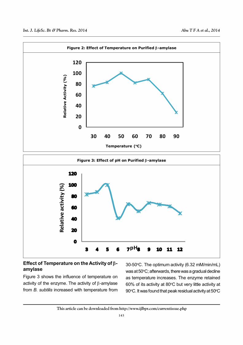
RESULT: SUMMARY OFPURIFICATION TABLEA summary of the results from purification of the
-amylase obtained from Bacillus subtilis isolated
from Parkia biglobosa seeds “iru” is presented in
Table 1. The table shows that specific activities
for crude extract, ammonium sulphate
precipitation, ion exchange chromatography, and
gel filtration were 0.19, 0.52, 0.79, and 3.56 (U/
mg) respectively. Purification (fold) for crude
extract, ammonium sulphate precipitation, ion
exchange, and gel filtration were 1, 2.75, 4.25,
and 18.74, respectively, an indication that
purification increased with each purification step
while percentage enzyme yield reduced with each
purification step.
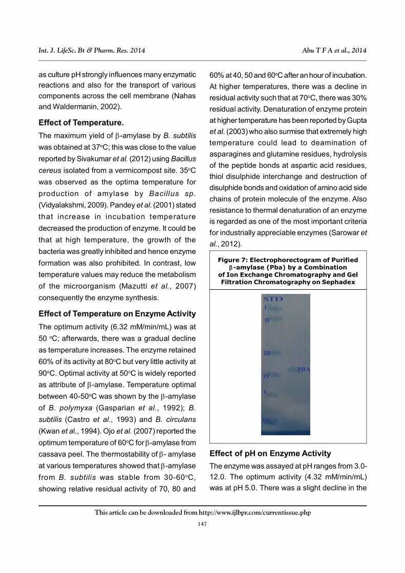
SDS PAGE Electrophoresis
From the electrophoretogram (Figure 7), the
protein band occurred as a single band and it was
spotted between protein standard iii and IV; which
are Ovalbumin and Carbonic anhydrase
respectively. The estimated molecular weight for
purified -amylase was 42.20 kDa.
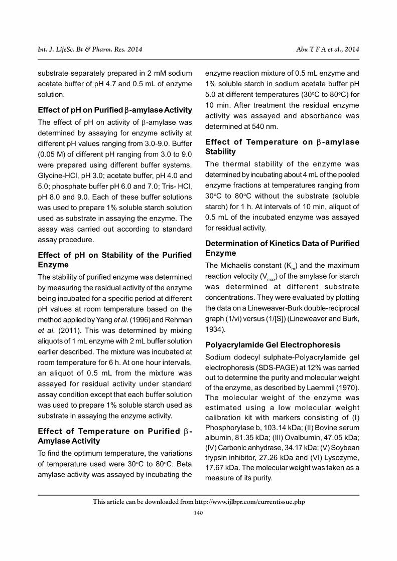
Effect of Starch Concentration onProduction of Bacillus subtilis
The effect of soluble starch concentration on
growth of B. subtilis from fermented African locust
bean seed shows that Log phase was from
0-5 h, lag phase continued from 5 h to 20 h while
stationary growth was at 20-25 h and decline in
growth started from 25 h. There was increase in
growth at 6 h of production steadily increasing
after 12 h and 18 h, at 18-24 h, 1% was found to
be higher than 0.5% by 19% and 2% by 12%,
respectively. Additionally bacterium achieved
optimum growth at 24 h incubation period but the
growth pattern and the growth curve of the 1%
starch was significantly different from those of
0.5% and 2%. There was a steady growth decline
after 25 h with a further 24 % drop in growth at
28 h incubation (Figure 1).
Table 1: Purification Table for -amylase from Bacillus subtilis
Purification Total protein Total enzyme activity Specific activity Purification YieldStep (mg) (U) (U/mg) (fold) (%)
Crude enzyme 68800 12840 0.19 1 100
NH4SO4 24325 12530 0.52 2.75 97.59
Precipitation DEAE Cellulose 234.4 186.08 0.79 4.25 1.45
Sephadex G-200 45.23 160.95 3.56 18.74 1.25
142
This article can be downloaded from http://www.ijlbpr.com/currentissue.php
Int. J. LifeSc. Bt & Pharm. Res. 2014 Abu T F A et al., 2014
Effect of Temperature on B. subtilisProduction
The optimum temperature for the growth of
Bacillus subtilis revealed that the optimum B.
subtilis production (14.42 µmole/ min/mL) was
recorded at 37oC. There was an increase (about
52%) in production from 30 h to 37 h, after which
a drop in growth was experienced from 38 h to
40 h. There was a growth decline at temperatures
above 40oC and the minimum growth (5.4 µmole/
min/mL) was recorded at 25oC.
Effect of pH on Growth of Bacillus subtilis
The effect of various pH on Bacillus subtilis growth
after 24 h incubation at 37oC shows that optimum
Bacillus subtilis production (19.78 µmole/min/mL)
was observed at pH 7.0. Growth was noticeable
from slightly acidic through neutral to the slightly
alkaline pH. With increase in pH above 7.0, there
was a decrease in yield. Minimum yield of growth
(6.0 µmole/min/ml) was recorded at pH 5.0. It was
observed that a slight change in pH of medium
adversely affected the growth of the bacterium.
Growth showed that at peak growth for pH 7.0
was about 59% higher than peak growth for pH
6.0 while decline in peak of 25% for pH 7.5 and
8.0, respectively.
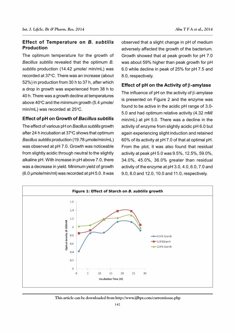
Effect of pH on the Activity of -amylase
The influence of pH on the activity of -amylase
is presented on Figure 2 and the enzyme was
found to be active in the acidic pH range of 3.0-
5.0 and had optimum relative activity (4.32 mM/
min/mL) at pH 5.0. There was a decline in the
activity of enzyme from slightly acidic pH 6.0 but
again experiencing slight induction and retained
60% of its activity at pH 7.0 of that at optimal pH.
From the plot, it was also found that residual
activity at peak pH 5.0 was 9.5%, 12.5%, 59.0%,
34.0%, 45.0%, 36.0% greater than residual
activity of the enzyme at pH 3.0, 4.0, 6.0, 7.0 and
9.0, 8.0 and 12.0, 10.0 and 11.0, respectively.
Figure 1: Effect of Starch on B. subtilis growth
143
This article can be downloaded from http://www.ijlbpr.com/currentissue.php
Int. J. LifeSc. Bt & Pharm. Res. 2014 Abu T F A et al., 2014
0
20
40
60
80
100
120
30 40 50 60 70 80 90
Temperature (� C)
Figure 2: Effect of Temperature on Purified -amylase
Temperature (oC)
Rela
tive A
ctiv
ity (
%)
Rela
tive
act
ivit
y (%
)
pH
Figure 3: Effect of pH on Purified -amylase
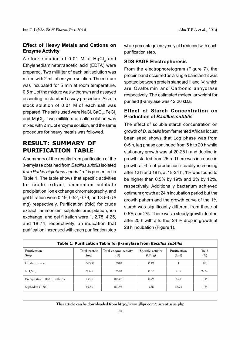
Effect of Temperature on the Activity of -amylase
Figure 3 shows the influence of temperature on
activity of the enzyme. The activity of -amylase
from B. subtilis increased with temperature from
30-50oC. The optimum activity (6.32 mM/min/mL)
was at 50oC; afterwards, there was a gradual decline
as temperature increases. The enzyme retained
60% of its activity at 80oC but very little activity at
90oC. It was found that peak residual activity at 50oC
144
This article can be downloaded from http://www.ijlbpr.com/currentissue.php
Int. J. LifeSc. Bt & Pharm. Res. 2014 Abu T F A et al., 2014
Rela
tive
act
ivit
y (%
)
Time (h)
Figure 4: Effect of pH on Stability of Purified -amylase
Figure 5: Effect of Temperature on Stability of Purified -amylase
Rela
tive
act
ivit
y (%
)
Time (H)
was greater than residual activity for 30oC, 40oC,
60oC, 70oC, 80oC and 90oC by 25%, 17%, 18%,
12%, 37%, and 72%.
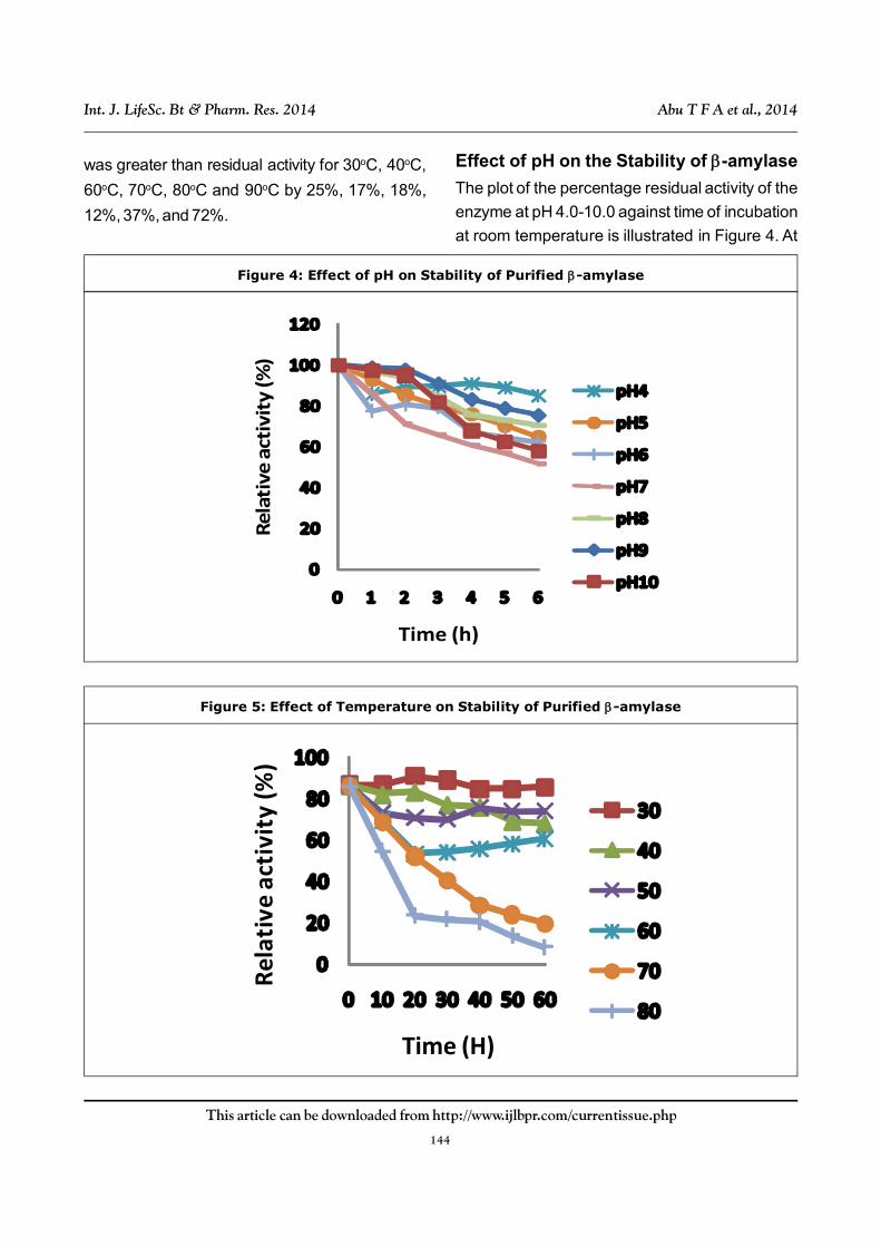
Effect of pH on the Stability of -amylase
The plot of the percentage residual activity of the
enzyme at pH 4.0-10.0 against time of incubation
at room temperature is illustrated in Figure 4. At
145
This article can be downloaded from http://www.ijlbpr.com/currentissue.php
Int. J. LifeSc. Bt & Pharm. Res. 2014 Abu T F A et al., 2014
all the pH examined, the enzyme was relatively
stable for 4 h. After 4 h, it was still able to retain
almost 70% of its activity except at pH 7.0 where
it dropped to 60%.
Effect of Temperature on Stability of -amylase
The thermostability of -amylase at various
temperatures is shown in Figure 5. -amylase
from B. subtilis was stable from 30 -60oC,
showing relative residual activity of 70, 80 and
60% at 40, 50 and 60oC after an hour of incubation.
At higher temperatures, there was a decline in
residual activity such that at 70oC, there was 30%
residual activity.
Effect of Different Carbohydrate Substrateon -amylase
The result revealed that maximum -amylase
activity was observed in 1% soluble starch
followed by sucrose. The least activity was
observed in 1% CMC (Carboxylmethylcellulose),
corn starch and cassava starch. Cassava starch
had a 34% residual activity in relation to soluble
starch (control), corn starch had 33% residual
activity, CMC was less at 27% in relation to
soluble starch, lactose 47% while sucrose
residual activity in relation to the control was at a
high by 92.0% and rice bran residual activity was
49% of the control.
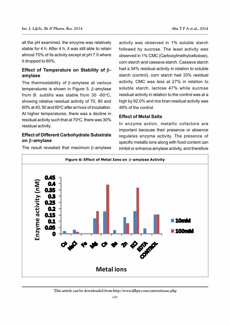
Effect of Metal Salts
In enzyme action, metallic cofactors are
important because their presence or absence
regulates enzyme activity. The presence of
specific metallic ions along with food content can
inhibit or enhance amylase activity, and therefore
Enzy
me
acti
vity
(nM
)
Metal ions
Figure 6: Effect of Metal Ions on -amylase Activity
146
This article can be downloaded from http://www.ijlbpr.com/currentissue.php
Int. J. LifeSc. Bt & Pharm. Res. 2014 Abu T F A et al., 2014
the rate of digestion (Mar et al., 2008, Kiran and
Chandra, 2008).
The effects of various metals ions at two
different concentrations on the activity of -
amylase are reported in Figure 6 where it showed
that Fe2+, Cu2+ as divalent ions almost or
completely inactivated the enzyme activity. Of the
monovalent ions, Na+ had inhibitory action on the
enzyme while K+ exhibited activation of the
enzyme at different concentrations. Also Ca2+ and
Mg2+ increased the activity of the enzyme
especially at higher concentrations of the metal
ions. Presumably because of its chelating
properties (Kiran and Chandra, 2008), from the
study, EDTA also inhibited the -amylase enzyme.
Effect of Substrate Concentration
The Lineweaver-Burke plot of the purified -
amylase activity of B. subtilis revealed that the -
amylase enzyme has apparent Km and Vmax
values for the hydrolysis of soluble starch of 17.74
mg mL–1and 14.09U respectively.
DISCUSSIONThe microbial growth: The features confirmed
from the growth was consistent with reported
literature Rehman et al. (2011), Ray and Nanda
(1996). Absence of lactose shows that the
fermentation of “iru” was not lact ic acid
fermentation and the presence of glucose
revealed that the organism broke down the
medium into simple sugars. The growth features
including the elevation, serrated edges and color
was consistent with B. subtilis growth reported
by Rehman et al. (2011) and Nouadri et al. (2010).
Optimal Growth Conditions
The study revealed that growth of B. subtilis was
at its peak when growth medium contained 1%
soluble starch, at pH 7.0, under temperature of
37oC and for 24 h. This was consistent with
findings by Rehman et al. (2011), Annamalai
(2011), Lin (1998), Sivakumar et al. (2012) and
Mazutti et al. (2007).
Effect of Starch Concentration on Growth
A growth curve often time consists of the Log
phase, Lag phase, Stationery phase and the
Logarithmic decline phase Fankhauser (2012). The
effect of starch concentration on production of B.
subtilis followed the production growth curve of
most microbial growth. The growth curve pattern
was studied by growing the bacterium in 0.5, 1
and 2% soluble starch. The bacterium achieved
optimum growth at 24 h incubation period during
the lag phase of growth as reported by
Rosenberger (2008) and Fankhauser (2012) but
the growth pattern of the 1% soluble starch was
significantly different from those of 0.5 and 2%.
Effect of pH on Growth
The optimum B. subtilis production (19.78 µmole/
min/mL) was observed at pH 7.0. At increase in
pH above 7.0, there was a decrease in B. subtilis
yield. Minimum yield of B. subtilis (6.0 µmole/min/
mL) was recorded at pH 5.0. This study has
recorded optimization of culture conditions for
growth and amylase production at pH, 7.0, 37oC,
24 h and 1% of substrate concentrations
consistent with (Annamalai, 2011 and Lin, 1998).Also Vijayalakshmi (2009) reported 35oC optima
temperature for amylase production by Bacillusspecies.
The effect of initial pH on SSF of amylaseproduction shows that the pH range of 5.0-7.0
produced more amount of amylase (Sivakumaret al., 2012). Amylase production by microbial
strains strongly depends on the extracellular pH,
147
This article can be downloaded from http://www.ijlbpr.com/currentissue.php
Int. J. LifeSc. Bt & Pharm. Res. 2014 Abu T F A et al., 2014
as culture pH strongly influences many enzymaticreactions and also for the transport of various
components across the cell membrane (Nahas
and Waldermanin, 2002).
Effect of Temperature.
The maximum yield of -amylase by B. subtilis
was obtained at 37oC; this was close to the value
reported by Sivakumar et al. (2012) using Bacillus
cereus isolated from a vermicompost site. 35oC
was observed as the optima temperature for
production of amylase by Bacillus sp.
(Vidyalakshmi, 2009). Pandey et al. (2001) stated
that increase in incubation temperature
decreased the production of enzyme. It could be
that at high temperature, the growth of the
bacteria was greatly inhibited and hence enzyme
formation was also prohibited. In contrast, low
temperature values may reduce the metabolism
of the microorganism (Mazutti et al., 2007)
consequently the enzyme synthesis.
Effect of Temperature on Enzyme Activity
The optimum activity (6.32 mM/min/mL) was at
50 oC; afterwards, there was a gradual decline
as temperature increases. The enzyme retained
60% of its activity at 80oC but very little activity at
90oC. Optimal activity at 50oC is widely reported
as attribute of -amylase. Temperature optimal
between 40-50oC was shown by the -amylase
of B. polymyxa (Gasparian et al., 1992); B.
subtilis (Castro et al., 1993) and B. circulans
(Kwan et al., 1994). Ojo et al. (2007) reported the
optimum temperature of 60oC for -amylase from
cassava peel. The thermostability of - amylase
at various temperatures showed that -amylase
from B. subtilis was stable from 30-60oC,
showing relative residual activity of 70, 80 and
60% at 40, 50 and 60oC after an hour of incubation.
At higher temperatures, there was a decline in
residual activity such that at 70oC, there was 30%
residual activity. Denaturation of enzyme protein
at higher temperature has been reported by Gupta
et al. (2003) who also surmise that extremely high
temperature could lead to deamination of
asparagines and glutamine residues, hydrolysis
of the peptide bonds at aspartic acid residues,
thiol disulphide interchange and destruction of
disulphide bonds and oxidation of amino acid side
chains of protein molecule of the enzyme. Also
resistance to thermal denaturation of an enzyme
is regarded as one of the most important criteria
for industrially appreciable enzymes (Sarowar et
al., 2012).
Figure 7: Electrophorectogram of Purified-amylase (Pba) by a Combination
of Ion Exchange Chromatography and GelFiltration Chromatography on Sephadex
Effect of pH on Enzyme Activity
The enzyme was assayed at pH ranges from 3.0-
12.0. The optimum activity (4.32 mM/min/mL)
was at pH 5.0. There was a slight decline in the
148
This article can be downloaded from http://www.ijlbpr.com/currentissue.php
Int. J. LifeSc. Bt & Pharm. Res. 2014 Abu T F A et al., 2014
enzyme activity from pH 6.0 but retained 60% of
its activity at pH 7.0. The optimum pH observed
agrees with those reported from purified
-amylase from different sources. Most of the
microbial - amylases have an optimum pH at
5.0-7.0 as reported from B. megaterium (Ray et
al., 1996); B. subtilis (Castro et al., 1992); while
Olaniyi et al. (2010) reported similar optimum pH
of 5.0 for the - amylase of Aspergillus niger. The
plot of the percentage residual activity of the
enzyme at pH 4.0-10.0 against time of incubation
at room temperature is illustrated in Figure 7. At
all the pH examined, the enzyme was relatively
stable for 4 h at almost all the pH. After 4 h, it was
still able to retain almost 70% of its activity except
at pH 7.0 where it dropped to 60%. The pH stability
is also a very important quality for continuous
production of enzymes because in large scale
fermenters, pH change is a usual and natural
occurrence (Sarowar et al., 2012).
Effect of Metals and Ions
In enzyme action, metallic cofactors are
important because their presence or absence
regulates enzyme activity. The presence of
specific metallic ions along with food content can
inhibit or enhance amylase activity, and therefore
the rate of digestion. The effects of various metals
ions is reported in Figure 6 where it showed that
Fe2+, Cu2+ as divalent ions almost or completely
inactivated the enzyme activity which is
consistent with the findings reported by Sarowar
et al. (2012), Ojo and Ajele (2011). Of the divalent
ions, Na+ had inhibitory action on the enzyme
while K+ exhibited activation of the enzyme at
different concentrations (Sarowar et al., 2012).
Also Ca2+ and Mg2+ increased the activity of the
enzyme especially at higher concentrations of the
metal ions. Usually, the role of Ca2+ in stability
and maintaining the structure of the -amylase
enzyme has been well documented (Parkin,
1993). It is well known that amylases contain Ca2+
as an essential component of the enzyme
molecule and are often inhibited by the chelating
reagent EDTA (Kiran and Chandra, 2008)
presumably because of its chelating properties,
from the study; EDTA also inhibited the -amylase
enzyme thereby confirming the above assertion.
It was observed that NaCl enhanced the activity
of the –amylase, while its activity was slightly
inhibited by salts such as CaCl2 and MgCl2. The
inhibition of the –amylase activity by EDTA may
suggest that the enzyme may contain inorganic
groups, which form inactive complexes with
EDTA. Fogarty and Kelly (1990) suggested that
EDTA acted by chelating Ca2+ and once the Ca2+
content of the enzyme was completely removed
by EDTA, there followed a quick loss in the
enzyme’s activity.
The -amylase was purified 18.7-fold. The
purification factor is a measure of purity of the -
amylase and the fact that it may be approaching
a homogenous state.
SDS PAGEELECTROPHORESISThe molecular weight of the -amylase was
estimated to be 42.2 kDa migrating as a single
protein band in SDS-PAGE 12% gel which
indicates its homogeneity. The estimated
molecular weight of 42.2 kDa from its mobility
relative to those of standard proteins on SDS-
PAGE indicated that the purified enzyme is a
monomer (Ray and Nanda, 1996).
This is lower than the molecular weight
reported in a wide variety of amylase isolated
from other sources. For example, 105 kDa was

149
This article can be downloaded from http://www.ijlbpr.com/currentissue.php
Int. J. LifeSc. Bt & Pharm. Res. 2014 Abu T F A et al., 2014
reported for amylase from Bacillus megaterium
(Ray, 2000), 69 kDa was reported by Olaniyi et
al. (2010) for amylase from Volvariella
volvacea. It was higher than 27.6 kDa reported
by Femi-Ola et al. (2013) for amylase from
Bacillus subtilis. Buonocore et al. (1976) has
suggested that the discrepancy observed
between the apparent molecular mass
determined by gel filtration may be due to the
interaction of the enzyme with the gel, resulting
in retardation of its mobility and thus an
underestimation of its molecular weight.
REFERENCES1. Akande F B, Adejumo O A, Adamade C A
and Bodunde J (2010), “Processing of locust
bean fruits: challenges and prospects”,
African Journal of agricultural research, Vol.
5, No. 17, pp.2268-2271
2. Annamalai N, Thavasi R, Vijayalakshmi S
and Balasubramanian T (2011), “Extraction,
purif ication and characterizat ion of
thermostable alkaline tolerant - amylase
from Bacillus cereus”, Ind. J. Microbiol., Vol.
51, No. 4, pp. 424-429.
3. Bernfield P (1955), “Amylases and
methods”, Enzymology, Vol. 1, pp. 147-154.
4. Bolden M and Effront P (2000), “Microbial
amylolytic enzymes”, Critical Review
Biochem. Molecular Biol., 24, 329-418.
5. Buonocore V C, Caporale M D and
Gambacorta A (1976), “Stable inducible
thermoacidiphilic -amylase from Bacillus
acidodurius”, Journal of Bacteriology, Vol.
128, pp. 515-521.
6. Campbell-platt G (1980), “African locust
bean (Parkia species) and its West African
fermented food product, dawadawa”, Ecol.
Food Nutr., Vol. 9, pp 123-132.
7. Castro G R, Ferrero MA, Mendez B S and
Sineriz F (1993), “Screening and selection
of bacteria with high amylolytic activity”, Acra
Biotech., Vol. 13, p. 197.
8. Dike E N and Odunfa S A (2003),
“Microbiological and biochemical evaluation
of a fermented soyabeans product-
Soyadawadawa”, J. Food Sci. Tech., Vol.
40, pp. 606-610.
9. Enujiugha V N (2009), “Major fermentative
organisms in some Nigeria soup
condiments”, Pakistan Journal of Nutrition,
Vol. 8, No. 3, pp. 279-283.
10. Femi-Ola T O, Oshokoya A H and Bamidele
O S (2013), “Kinetic Properties of Beta-
Amylase from Bacillus subtilis”, IOSR
Journal of Environmental Science,
Toxicology and Food Technology (IOSR-
JESTFT) ISSN: 2319-2402, ISBN: 2319-
2399, Vol. 2, Issue 4, pp. 19-23
11. Forgarty W M and Kelly C T (1990),
“Microbial Enzymes and Biotechnology”, 2nd
edition Elsevier Sci. publishers, London. pp.
71-132.
12. Gasparian A V, Ahelian V H and Afrikian E G
(1992), “Some characteristics of -amylase
produced by Bacillus polymyxa”, Biokhirniya
Agric. Biol. Chem., Vol. 39, pp. 856-860.
13. Gupta R, Gigras P, Mohapatra H, Goswami
V K and Chauhan B (2003), “Microbial -
amylases: A biotechnological perspective”,
Process Biochemistry, Vol. 38, pp. 1599–
1616.
14. Kiran K K and Chandra T S (2008),
150
This article can be downloaded from http://www.ijlbpr.com/currentissue.php
Int. J. LifeSc. Bt & Pharm. Res. 2014 Abu T F A et al., 2014
“Production of surfactant and detergent-
stable, halophilic and alkalitolerant alpha-
amylase by a moderately halophilic Bacillus
sp. Strain TSCVKK”, Applied Microbiol.
Biotechnol., Vol. 77, pp. 1023-1031.
15. Laemmli U K (1970), “Cleavage of structural
proteins during the assembly of the head of
bacteriophage T4”, Nature, Vol. 227, pp. 680-
685.
16. Leveque E, Janecek S, Haye B and Belarbi
A (2000), “Thermophilic archaeal amylolytic
enzymes”, Enzyme Microb. Technol., Vol.
26, pp. 3-14.
17. Line Weaver H and Burk D (1934), “The
determination of enzyme dissociation
constants”, J Am Chem Soc, Vol. 56, pp.
658–666.
18. Lowry O H, Rosebrough N J, Farr A L and
Randall R J (1951), “Protein measurement
with the Folin phenol reagent”, J. Bio. Chem.,
Vol. 193, pp. 265-275.
19. Mazutti M, Ceni G, Di Luccio M and Treichel
H (2007), “Production of inulinase by solid-
state fermentation: effect of process
parameters on production and preliminary
characterization of enzyme preparations”,
Bioprocess Biosyst. Eng., Vol. 30, pp. 297-
304.
20. Murao S, Ohyama K and Arai M (1979), -
amylase from Bacillus polymyxa No. 72”,
Agric. Biol. Chem., Vol. 43, No. 4, pp. 719-
726.
21. Nahas E and Waldermarin M M (2002),
“Control of amylase production and growth
characteristics of Aspergillus ocharaceus”,
Microbiological, Vol. 44, pp. 5-10.
22. Ojo O O and J O Ajele (2011), “Isolation,
Purification, Characterization and the
possible involvement of Histidine and
Cysteine in the catalytic mechanism of -
amylase source from Cassava (Manihot
esculenta) peel”, P. J. Nutri., Vol. 10, No. 9,
pp. 823-830.
23. Olaniyi O O, Akinyele B J and Arotupin D J
(2010), “Purification and Characterization of
-amylase from Volvariella volvacea”, Nig.
J. Microbio., Vol. 24, No. 1, pp. 76-82.
24. Osaki S and Yoshino Z (1988), “Manufacture
of oligosaccharides”, in Hand Book of
Amylases and Related Enzymes, The
Amylase Research Society of Japan,
Osaka.
25. Pandey A, Soccol C R and Soccol V T
(2000), “Biopotential of immobilized
enzymes”, Indian J. Microbiol. Vol. 40, pp.
1-14.
26. Parkin K L (1993), “Environmental Effects
on Enzyme Activity”, In Nagodawithana,
T.and Reed G (ed.), Enzymes in Food
Processing, 3rd Edn., Academic Press Inc.,
San Diego, pp. 480.
27. Rani T, Takagi T and Toda H (2007),
“Bacterial and mold amylases”, In: Boyer P
(ed.), The enzymes. Academic press Inc.,
New York, pp. 235-271.
28. Rao J L U M and Satyanarayana T (2003),
“Enhanced secretion and low temperature
stabilization of a hyperthermostable and Ca2+
independent a-amylase of Geobacillus
thermoleovorans by surfactants”, Lett. Appl.
Microbiol., Vol. 36, pp. 191–196.
29. Ray R R (2000), “Purif ication and
151
This article can be downloaded from http://www.ijlbpr.com/currentissue.php
Int. J. LifeSc. Bt & Pharm. Res. 2014 Abu T F A et al., 2014
characterization of extracellular amylase
of Bacillus megaterium B (6)”, Acta
Microbiology and Immunology, Vol. 47, pp.
29-40.
30. Ray R R, Jana S C and Nanda G (1996),
“Induction and catabolite repression in the
biosynthesis of -amylase by Bacillus
megatenurn”, Biochem. Mol. Biol. Int., Vol.
38, p. 223.
31. Rehman A, Shafaat S and Akram M (2011),
“Isolation and characterization of a
thermostable -amylase from Bacillus
subtilis”, African journal of Microbiology
Research, Vol. 5, No. 20, pp. 3334-3338.
32. Sarowar J, Shaela P, Shariar S, Dev
Sharma R and Habibur R (2012), “Effect of
Metal Ions, Chelating Agent and SH-
Reagents on Radish (Raphanus sativus L.)
Root -Amylase”, J. Stress Physio. &
Biochem., Vol. 8, No. 3, pp. 180-188.
33. Sivakumar T T, Shankar P, Vijayabaskar J,
Muthukumar and Nagendrakannan E (2012),
“Amylase Production Using Bacillus cereus
Isolated from a Vermicompost Site”,
International Journal of Microbiological
Research, Vol. 3, No. 2, pp. 117-123.
34. Takasaki Y (1976), “Purification and
Enzymatic properties of amylase from B.
cereus var mycoides”, Agric. Biol. Chem.,
Vol. 40, No. 8, pp. 1523-1530.
35. Yandri Suhartati T and Hadi S (2010),
“Purif ication and Characterization of
Extracellular -Amylase Enzyme from
Locale Bacteria Isolate Bacillus Subtilis”,
Europ. J. Sci. Res., Vol. 39, pp. 64-74.
36. Yang Z D, Michael A, Robert X Y, Fang and
Alan J R (1996), “Polyethylene glycol-
induced stabilization of subtilisin”, Enzyme
Microb. Technol., Vol. 18, pp. 82-89.