
The Neck
is the region of the body that lies
between the lower margin of the
mandible above and the suprasternal
notch and the upper border of the
clavicle below

Nerves of the neck

Cervical Plexus
Is formed by the
anterior rami of the first
four cervical nerves.
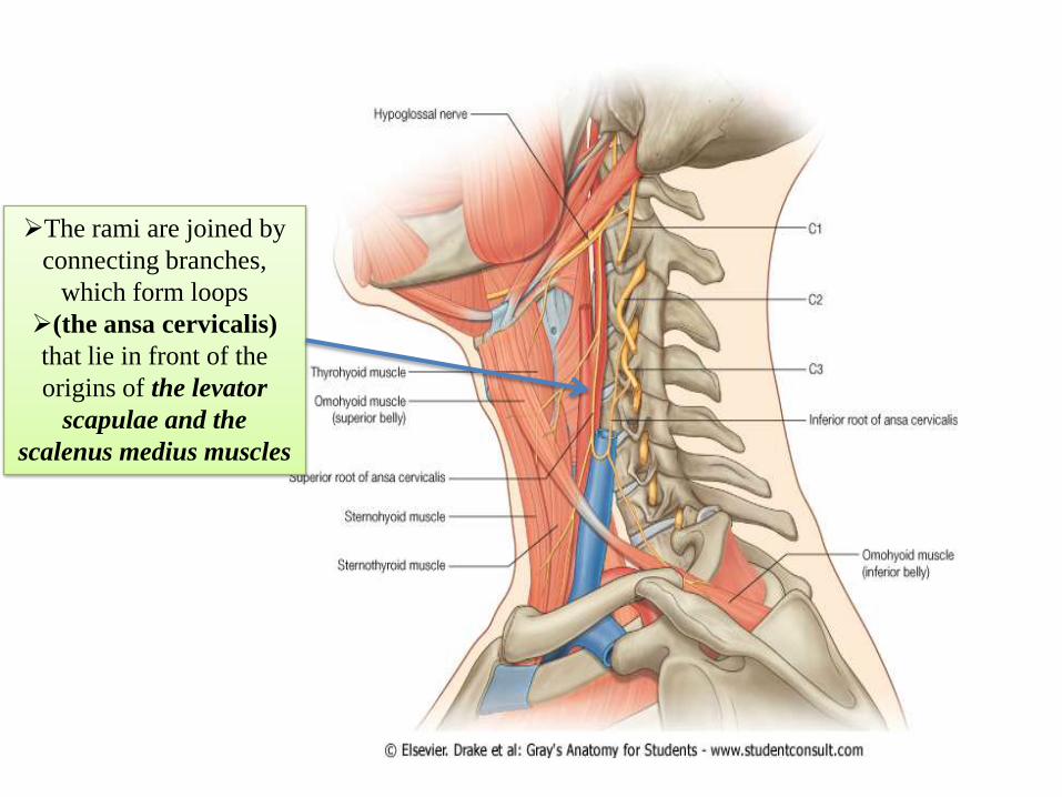
The rami are joined by
connecting branches,
which form loops
(the ansa cervicalis)
that lie in front of the
origins of the levator
scapulae and the
scalenus medius muscles
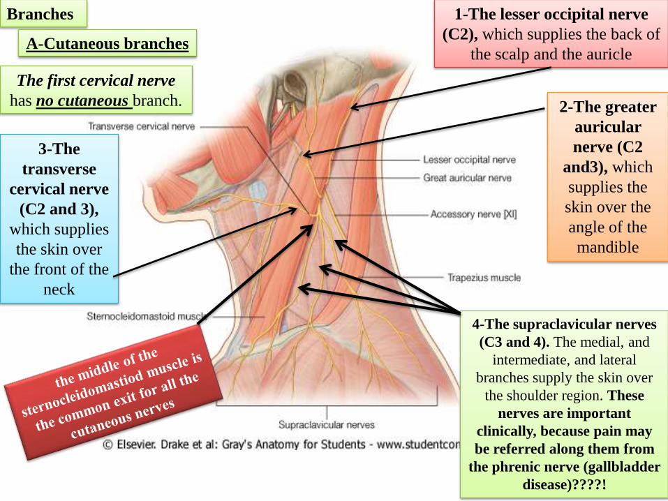
1-The lesser occipital nerve
(C2), which supplies the back of
the scalp and the auricle
Branches
The first cervical nerve
has no cutaneous branch. 2-The greater
auricular
nerve (C2
and3), which
supplies the
skin over the
angle of the
mandible
3-The
transverse
cervical nerve
(C2 and 3),
which supplies
the skin over
the front of the
neck
4-The supraclavicular nerves
(C3 and 4). The medial, and
intermediate, and lateral
branches supply the skin over
the shoulder region. These
nerves are important
clinically, because pain may
be referred along them from
the phrenic nerve (gallbladder
disease)????!
A-Cutaneous branches
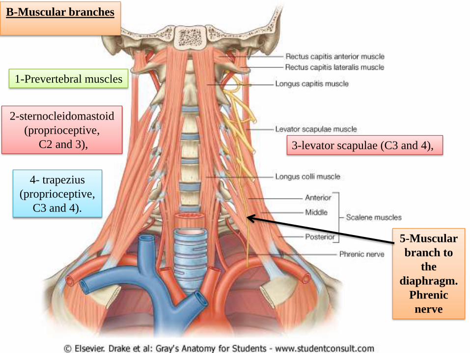
B-Muscular branches
5-Muscular
branch to
the
diaphragm.
Phrenic
nerve
3-levator scapulae (C3 and 4),
1-Prevertebral muscles
2-sternocleidomastoid
(proprioceptive,
C2 and 3),
4- trapezius
(proprioceptive,
C3 and 4).

6-A branch from C1 joins
the hypoglossal nerve.
Some of these C1 fibers later leave
the hypoglossal as the descending
branch
which unites with the descending
cervical nerve (C2 and 3)
to form the ansa cervicalis
The first, second, and third cervical
nerve fibers within the ansa
cervicalis supply :
1- omohyoid,
2-sternohyoid
3-sternothyroid muscles
Other C1 fibers within the hypoglossal
nerve leave it as the nerve to the
thyrohyoid and geniohyoid.

Superficial veins of the neck

SUPERFICIAL VEINS OF THE
NECK
1-External Jugular Vein
The external jugular vein
begins
just behind the angle of the
mandible
by the union of
the posterior auricular vein
with The posterior division of the
retromandibular vein
It descends obliquely across the
sternocleidomastoid muscle
Pierces the deep fascia and drains
into the subclavian vein
It varies considerably in size, and
its course extends from the angle of
the mandible to the middle of the
clavicle.
Tributaries:Transverse cervical
,Suprascapular,
posterior external jugular and
anterior jugular vein

Anterior Jugular Vein
The anterior jugular vein begins
just below the chin
It runs down the neck close to
the midline.
Just above the suprasternal
notch, the veins of the two sides
are united by a transverse trunk
called the jugular arch. Drains
into the external jugular vein.

Veins of the Face and the
Neck
Facial Vein
is joined by the anterior
division of the retromandibular
vein, and drains into the
internal jugular vein.
Superficial Temporal Vein
The superficial temporal vein is
formed on the side of the scalp
enters the parotid salivary
gland, where it joins the
maxillary vein to form the
retromandibular vein.
Maxillary Vein
The maxillary vein is formed in
the infratemporal fossa from the
pterygoid venous plexus
The maxillary vein joins the
superficial temporal vein to
form the retromandibular vein.

Retromandibular Vein
is formed by the union of
the superficial temporal
and the maxillary
On leaving the parotid
salivary gland, it divides
into:
an anterior branch, which
joins the facial vein,
a posterior branch, which
joins the posterior
auricular vein to form the
external jugular vein.

The neck is surrounded by
1-Skin
2-Superficial fascia
3-Deep fascia

1-The lesser occipital nerve
(C2), which supplies the back of
the scalp and the auricle
1-SKIN
The first cervical nerve
has no cutaneous branch. 2-The greater
auricular
nerve (C2
and3), which
supplies the
skin over the
angle of the
mandible
3-The
transverse
cervical nerve
(C2 and 3),
which supplies
the skin over
the front of the
neck
4-The supraclavicular nerves
(C3 and 4). The medial, and
intermediate, and lateral
branches supply the skin over
the shoulder region.
Cutaneous nerves of the neck

Superficial lymph nodes
of the face and scalp
1-Occipital nodes
2- Mastoid nodes
3-Pre-auricular and parotid nodes4-Submandibular nodes
5-Submental nodes
Five groups of superficial lymph nodes
Responsible for
the lymphatic
drainage of the
Face and scalp
form a ring around the head
2-Superficial cervical lymph nodes
The superficial cervical nodes are a collection of lymph nodes along the external jugular vein on
the superficial surface of the sternocleidomastoid muscle
2-Superficial fascia of the neck
Contains:

The deep cervical nodes are a collection of lymph
nodes that form a chain along the internal jugular
vein.
divided into
upper and lower
groups
BY the
intermediate
tendon of the
omohyoid
muscle
This large node is where the posterior belly of the digastric
muscle crosses the internal jugular vein and receives
lymphatic drainage from the tonsils and tonsillar region.
Two important
nodes in the
deep cervical
group
1-jugulodigastric
node
The 2-jugulo-omohyoid node
usually associated with the lower
deep cervical group
it is at or
just inferior
to the
intermediate
tendon of
the
omohyoid
muscle,
This node receives
lymphatic drainage
from the tongue
From the deep cervical nodes, lymphatic vessels form the
right and left jugular trunks, which empty into the right
lymphatic duct on the right side or the thoracic duct on the
left side.
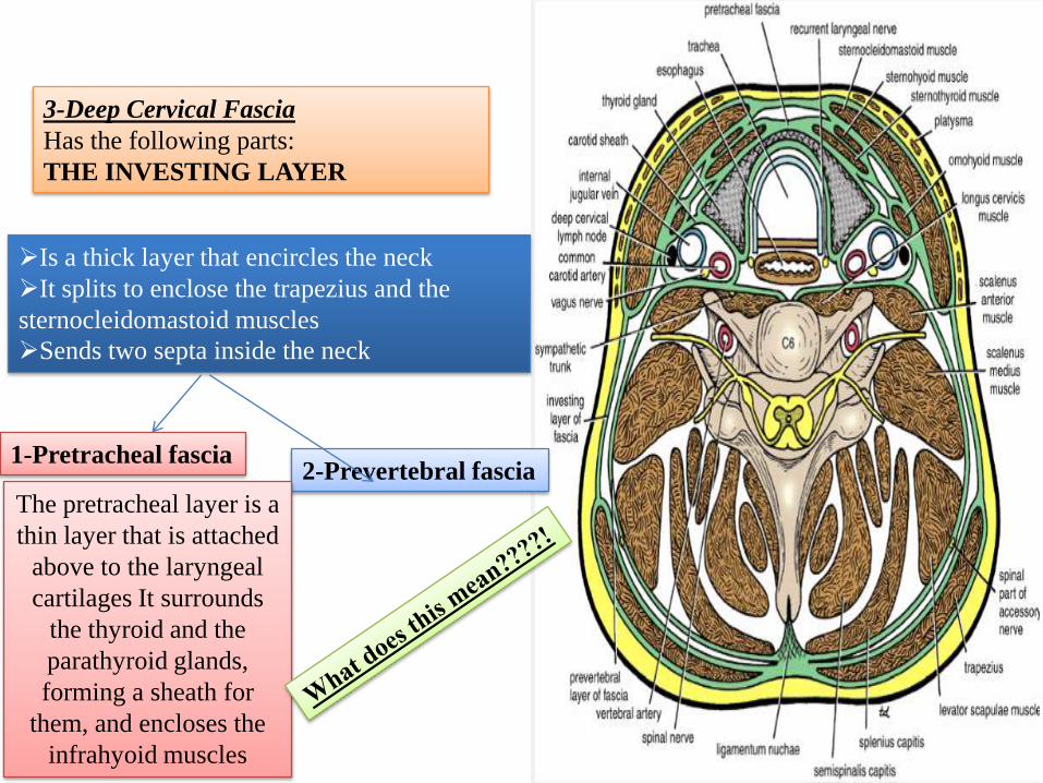
3-Deep Cervical Fascia
Has the following parts:
THE INVESTING LAYER
Is a thick layer that encircles the neck
It splits to enclose the trapezius and the
sternocleidomastoid muscles
Sends two septa inside the neck
2-Prevertebral fascia1-Pretracheal fascia
The pretracheal layer is a
thin layer that is attached
above to the laryngeal
cartilages It surrounds
the thyroid and the
parathyroid glands,
forming a sheath for
them, and encloses the
infrahyoid muscles

The two septa divide the neck
into three compartments
A- Muscular compartment (vertebral)
Located posterior to the prevertebral fascia
Contents:
2-Spinal nerves (that shear in the formation of
the brachial and cervical plexuses)
1-Prevertebral and postvertebral muscles

B- Visceral compartment
Located anterior to the Prevertebral fascia
And posterior to the pretracheal fascia
Contents:
1-Thyroid gland
2-Trachea
3-Oesophegus

C- Vascular compartment
Located Inside the carotid sheath
Contents:
Carotid sheath contents
"I See 10 CC's in the IV":
I See (I.C.)= Internal Carotid artery
10 = CN 10 (Vagus nerve)
CC = Common Carotid artery
IV = Internal Jugular Vein
1-Common carotid artery
2-Internal jugular vein
3- Vagus nerve

.
The fascial spaces: They are potential spaces filled
with loose connective tissue.
These are clinically important
because organisms originating in:
1-THE MOUTH
2-TEETH
3-PHARYNX
4-ESOPHAGUS
can spread among the fascial spaces,
and the fascia can determine the
direction of spread of infection
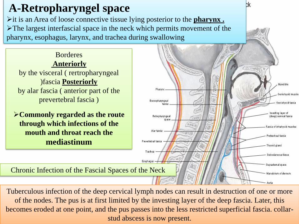
A-Retropharyngel spaceit is an Area of loose connective tissue lying posterior to the pharynx .
The largest interfascial space in the neck which permits movement of the
pharynx, esophagus, larynx, and trachea during swallowing
Borderes
Anteriorly
by the visceral ( rertropharyngeal
)fascia Posteriorly
by alar fascia ( anterior part of the
prevertebral fascia )
Commonly regarded as the route
through which infections of the
mouth and throat reach the
mediastinum
Tuberculous infection of the deep cervical lymph nodes can result in destruction of one or more
of the nodes. The pus is at first limited by the investing layer of the deep fascia. Later, this
becomes eroded at one point, and the pus passes into the less restricted superficial fascia. collar-
stud abscess is now present.
Chronic Infection of the Fascial Spaces of the Neck

B- Masticatory space• The masticatory space lies on either side of
the mandibular ramus and is formed by
cervical fascia, which ascends from the neck
and splits at the inferior mandibular border to
envelop
the ramus of the mandible, the masseter, the
medial and lateral pterygoid, and the lower
portion of the temporalis muscle.
It is traversed particularly by the
mandibular nerve (V3) and the maxillary
vessels
Infections from the 2nd or 3rd mandibular
molars may spread to these secondary sites
C-Submandibular space The submandibular space is composed of two spaces separated anteriorly by
the mylohyoid muscle: the sublingual space, which is superior, and the
submaxillary space, which is inferior.Ludwig's angina is an acute infection of the submandibular fascial space
and is commonly secondary to dental infection.
Read only
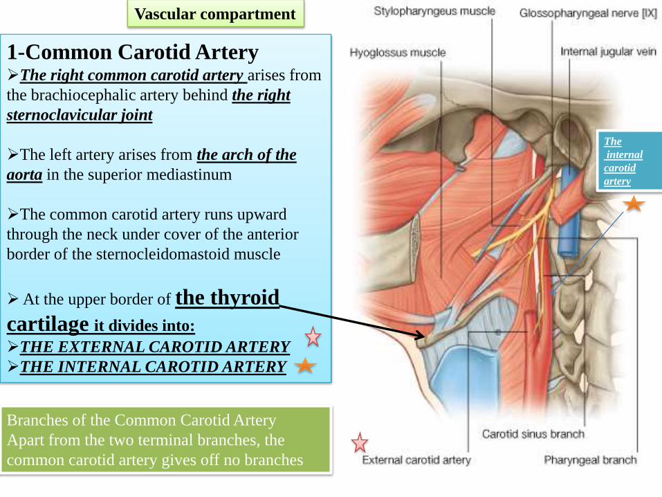
1-Common Carotid ArteryThe right common carotid artery arises from
the brachiocephalic artery behind the right
sternoclavicular joint
The left artery arises from the arch of the
aorta in the superior mediastinum
The common carotid artery runs upward
through the neck under cover of the anterior
border of the sternocleidomastoid muscle
At the upper border of the thyroid
cartilage it divides into:
THE EXTERNAL CAROTID ARTERY
THE INTERNAL CAROTID ARTERY
Vascular compartment
Branches of the Common Carotid Artery
Apart from the two terminal branches, the
common carotid artery gives off no branches
The
internal
carotid
artery

is one of the terminal branches of
the common carotid artery
It supplies structures in the neck
1-Face
2- scalp
3-the tongue and the maxilla
It lies outside the carotid sheathThe artery begins at the level of the
upper border of the thyroid cartilage
terminates in the substance of the
parotid gland behind the neck of the
mandible by dividing into the
superficial temporal and maxillary
arteries
External Carotid Artery
Its relation to the internal carotid artery
At first
it lies medial to the internal carotid artery
but as it ascends in the neck
it passes
backward and lateral to it

1-SUPERIOR THYROID
ARTERY
2-ASCENDING PHARYNGEAL
ARTERY
3-LINGUAL ARTERY
4-FACIAL ARTERY
5-OCCIPITAL ARTERY
6-POSTERIOR AURICULAR
ARTERY
7-SUPERFICIAL TEMPORAL
ARTERY
8-MAXILLARY ARTERY
Branches of the External Carotid Artery
Some American ladies find our Petra so magnificent


