
Joint Session with ACOFP and AAO: High Yield OMT for the Upper and Lower Extremities
Shawn R. Kerger, DO


10/2/2015
1
High-Yield OMT for the Upper & Lower Extremities
Shawn R. Kerger, DO, FAOASM
Associate Professor, OMM Dept, OU-HCOM – Dublin
Medical Director, OMM Department, Doctors Hospital
Medical Director, Peter E. Johnston, DO, Simulation & Education Center
© 2015 by Shawn Kerger, DO, FAOASM
• P. Gunnar Brolinson, DO, FAAFP, FAOASM
• Paul Tortland, DO, FAOASM
• Albert Kozar, DO, FAOASM
• Richard Schuster, DO© 2015 by Shawn Kerger, DO, FAOASM

10/2/2015
2
Osteopathic Principles• The Osteopathic principles proposed by AT
Still which most directly relate to our purposes here are:• “When all parts of the body are in line
we have health.”• “When complete, he is…in size & form to
suit the duties he may have to perform.”• “You as Osteopathic machinists …adjust
the abnormal condition, in which you find the afflicted. Nature will do the rest.” © 2015 by Shawn Kerger, DO, FAOASM
Osteopathic Principles
• Or as restated by the faculty of the Kirksville College of Osteopathy & Surgery in 1953:
• The body is a unit.• Structure & function are reciprocally
interrelated.• The body is self-healing.
© 2015 by Shawn Kerger, DO, FAOASM

10/2/2015
3
Tensegrity• Still in development
• A self-stabilizing system in which tension is continuously transmitted across all elements
• Stability from distribution & balancing of mechanical forces
• Triangulated structures form the basis for this system
• Tetrahedron• Octahedron• Icosahedron © 2015 by Shawn Kerger, DO, FAOASM
Functional Anatomic Concepts: Muscle
• Kinetic chain - the sequencing of individual body segments & joints to accomplish a task
• Generally functions from a base of support proximally & then proceeds distally, but this is entirely dependant on the task at hand:• a bench press would follow the aforementioned
path
• a pushup reverses the mechanics even though the muscles engaged are similar, if not identical
© 2015 by Shawn Kerger, DO, FAOASM
10/2/2015
4
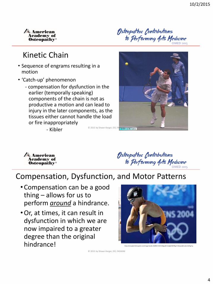
Kinetic Chain• Sequence of engrams resulting in a
motion
• ‘Catch-up’ phenomenon
- compensation for dysfunction in the earlier (temporally speaking) components of the chain is not as productive a motion and can lead to injury in the later components, as the tissues either cannot handle the load or fire inappropriately
- Kibler© 2015 by Shawn Kerger, DO, FAOASM
Compensation, Dysfunction, and Motor Patterns•Compensation can be a good
thing – allows for us to perform around a hindrance.
•Or, at times, it can result in dysfunction in which we are now impaired to a greater degree than the original hindrance! https://encrypted-tbn3.gstatic.com/images?q=tbn:ANd9GcTInDH-kMgLpNI-hLo8g5rMh9Mg-G-GkmpvejBS_NaLLJEeRog7-g
© 2015 by Shawn Kerger, DO, FAOASM

10/2/2015
5
Disturbed Motor Function
•Most important symptom ... PAIN!
•The area of the pain may not tell you where the problem is…
•Must learn to identify & treat underlying somatic dysfunction.
© 2015 by Shawn Kerger, DO, FAOASM
Somatic Dysfunction• Impaired or altered function of related components of the somatic (body framework) system: skeletal, arthrodial, & myofascial structures, & related vascular, lymphatic, & neural elements. © 2015 by Shawn Kerger, DO, FAOASM

10/2/2015
6
Dysfunction• Logically, lack of use of a tissue (either due to
injury, improper pain management, altered or improper technique, joint or soft tissue restrictions, etc.) will reverse the normal physiological processes.
• Bone will become less dense
• Joints will stiffen & ligaments will shorten
• Muscles will atrophy & neuromuscular control will be negatively altered
• Metabolic processes will revert to a lower energy (basal metabolic rate will drop), yet less exercise-tolerant, condition
© 2015 by Shawn Kerger, DO, FAOASM
© 2015 by Shawn Kerger, DO, FAOASM

10/2/2015
7
Functional Biomechanical Exam
•We’ll assume you know how to diagnose the “itis” pathologies
•Now that we know where the problem is, the issue becomes whyis it there?
© 2015 by Shawn Kerger, DO, FAOASM
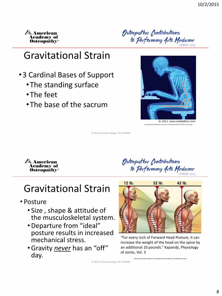
Gravitational Strain
•3 Cardinal Bases of Support•The standing surface•The feet•The base of the sacrum
© 2015 by Shawn Kerger, DO, FAOASM
10/2/2015
8
Gravitational Strain
•3 Cardinal Bases of Support•The standing surface•The feet•The base of the sacrum
http://themethuselahcenter.com/wp-content/uploads/2011/07/poor-posture.jpg
© 2015 by Shawn Kerger, DO, FAOASM
Gravitational Strain•Posture• Size , shape & attitude of
the musculoskeletal system.•Departure from “ideal”
posture results in increased mechanical stress.•Gravity never has an “off”
day.
“For every inch of Forward Head Posture, it can increase the weight of the head on the spine by an additional 10 pounds.” Kapandji, Physiology of Joints, Vol. 3
http://www.platinumphysio.co.uk/patient-info/conditions-treated/head-neck/
© 2015 by Shawn Kerger, DO, FAOASM

10/2/2015
9
Upper Extremity
© 2015 by Shawn Kerger, DO, FAOASM
SICK Scapula Syndrome (SSS)• So coined by Craig Morgan as “SICK (Scapula
Infera Coracoid dysKinesia) scapula syndrome”
• Collection of abnormal scapular mechanics that lead to repetitive microtrauma of the throwing motion, resulting in painful conditions of the shoulder due to muscle strain, fatigue and breakdown.
• Results in limited scapular retraction and acromial elevation with arm elevation, leading to subacromial impingement.
http://davidlasnier.com/2012/overhead-work-and-shoulder-flexion-limitation
© 2015 by Shawn Kerger, DO, FAOASM

10/2/2015
10
SICK Scapula Syndrome (SSS)
• Can develop from altered postures (see Upper Crossed Syndrome), repetitive microtrauma from poor technique, or atypical anatomy/firing patterns.
• Therefore, can be either causative or responsive to injury –so you need to be watchful both during diagnostic and treatment phases of patient care.
• If found, should be treated as it absolutely predisposes one to impingement syndrome – it no longer is a question of “If”, but “When”.
© 2015 by Shawn Kerger, DO, FAOASM
SICK Scapula Syndrome (SSS)• Hx: Symptoms above worsened by position or
activity usually involving the upper extremity. • Might have vague symptoms (burning/aching after
activity or prolonged postures) involving scapula, but these have usually been present for some time & the patient might not think of them as related.
• Usually not associated by patient with trauma.
• Pain is usually mild-moderate, but can be severe depending on structure under abnormal load (the SSS is usually asymptomatic itself).
• No central neurologic symptoms.
http://rpm-therapy.com/wp-content/uploads/2012/09/hairdresser-posture-bad3.jpg
http://rpm-therapy.com/wp-content/uploads/2012/09/hairdresser-posture-bad.jpg© 2015 by Shawn Kerger, DO, FAOASM

10/2/2015
11
Diagnose your partner’s scapular mechanics
Special Physical Examinations:
• Scapular resting position with arms:
• At the side
• Bent with hands on hips
• At or below 90° of abduction w/ shoulders IR & forearms pronated
• Wall push-up (long thoracic palsy)
• Scapular motion comparison for shoulder flexion & abduction in both concentric & eccentric phases
• Scapular muscle strength
• Scapular assistance (examiner pushes inferior & medial border of scapula laterally and superiorly – or anteriorly at inferior angle if winging present)
• Glenohumeral PROM and end-feel© 2015 by Shawn Kerger, DO, FAOASM
SICK Scapula Syndrome (SSS)
• Osteopathic: • Altered mechanics ANYWHERE in the kinetic chain for
throwing!
• Findings of elevated 1st or 2nd rib, cervical or thoracic vertebral or muscular dysfunction, clavicular or scapular dysfunction all can be primary or secondary to the problem developing.
• Frequently, tender/dysfunction levator scapula & upper traps – if lev scapula dysfunction recurrent, consider omohyoid TP.
Adapted from Thieme, Atlas of Anatomy Fig. 39.5 C
© 2015 by Shawn Kerger, DO, FAOASM

10/2/2015
12
Osteopathic Treatment
• If warm, wet/oily, swollen, painful/tender tissues with spongy or boggy end-points are encountered, consider indirect techniques (FPR, SCS, Still’s, OCF, etc.) as this is most likely an acute situation.
• If cool, dry, sore, ropy tissues with firmer/articular end-points are encountered, consider direct techniques (soft tissue, ME, HVLA/LVHA, etc.) as this is more of a chronic situation.
• Can always prep with indirect or soft tissue techniques before direct, too – you might just be wrong, and resolve the lesion with this!
© 2015 by Shawn Kerger, DO, FAOASM
Osteopathic Treatment
• When the somatic dysfunction is corrected, recheck any orthopedic or other findings to see what has changed – anything that’s left after correction of somatic dysfunction is concerning for tissue damage/inflammation.
• Pain is the LAST thing to go, usually – especially in the 8-72 hour period after an injury (or after you are poking and prodding the patient during physical examination) – so don’t get focused on pain response as your most important measure for success.
• Get things moving properly, & the pain will follow…
© 2015 by Shawn Kerger, DO, FAOASM

10/2/2015
13
We’re going to utilize this opportunity for upper quadrant treatment approach• First, treat in a gravity reactive (or kinetic
chain) pattern:
• Contralateral lower extremity
• Pelvis
• Sacrum
• Lumbar• Thorax
• Ribs
• Cervical
• Then, we’ll start with the upper extremity…
http://assets.nydailynews.com/polopoly_fs/1.474670!/img/httpImage/image.jpg_gen/derivatives/article_970/alg-rangers-lee-motion-jpg.jpg
© 2015 by Shawn Kerger, DO, FAOASM
We’re going to utilize this opportunity for upper quadrant treatment approach
• Work proximally to distally:• Scapulothoracic joint• Glenohumeral joint (see Spencer
Technique)• Elbow – both humeroulnar &
radial head• Wrist• Hand
http://assets.nydailynews.com/polopoly_fs/1.474670!/img/httpImage/image.jpg_gen/derivatives/article_970/alg-rangers-lee-motion-jpg.jpg
© 2015 by Shawn Kerger, DO, FAOASM

10/2/2015
14
Scapulothoracic Joint
© 2015 by Shawn Kerger, DO, FAOASM
Myofascial Release – Scapulothoracic lesion• Pt is in lateral recumbent with affected
side up
• Grasp scapula along medial and lateral borders.
• Can do:• Direct or indirect myofascial release
• Direct stretch against restriction
• Muscle Energy
© 2015 by Shawn Kerger, DO, FAOASM

10/2/2015
15
Omohyoid Tenderpoint – SCS
• Need to set up to treat C6 TP first –once in proper position for at least 70% pain relief (see picture 2, might need some slight refinement), then you set up for the omohyoid TP.
• With the flats of as many fingers as you can evenly distribute across the larynx and proximal trachea, deviate the laryngeal complex toward the side of the lesion.
• Follow typical SCS protocol from there.
Omohyoid TP
C6 TP
C6 TP SCS position
Omohyoid SCS position
© 2015 by Shawn Kerger, DO, FAOASM
Upper Crossed Syndrome (UCS)•Tight pectorals & upper traps inhibit
deep 4th layer cervical flexors & lower traps.
•Results in kyphotic thorax w/ internally rotated / protracted scapulae, & lordotic cervicals.
•Leads to impingement syndromes & cervical / upper thoracic complaints. http://www.muscleimbalancesyndromes.com/janda-syndromes/upper-crossed-syndrome/
© 2015 by Shawn Kerger, DO, FAOASM

10/2/2015
16
Upper Crossed Syndrome (UCS)
http://www.muscleimbalancesyndromes.com/janda-syndromes/upper-crossed-syndrome/
• In order to diagnose UCS, you find:
• Tight pairs of pectorals & upper traps /
suboccipital muscles
• Inhibited cervical flexors & lower traps /
rhomboids
• Altered scapular kinetics that are reversible with
correction of the above.
• You MAY also have a tight anterior G-H capsule,
setting up an arthrokinetic reflex. © 2015 by Shawn Kerger, DO, FAOASM
UCS – Arthrokinetic Reflex
•Capsule or ligament becomes stretched beyond what its programming allows for as a normal ROM (or if too rapid a motion occurs), inhibitory signals are sent to the agonist muscle responsible for loading the joint in the plane in question & stimulatory signals to the antagonist musculature. © 2015 by Shawn Kerger, DO, FAOASM

10/2/2015
17
UCS – Arthrokinetic Reflex• E.g., Anterior shoulder capsule is stretched
more than its programming allows:• Lower trap muscles receive an
inhibitory signal • Pectoralis muscles will be stimulated.
• This is the case in both healthy and dysfunctional states, with the difference being when this process is activated.
• Will prevent normal stretching…
© 2015 by Shawn Kerger, DO, FAOASM
Upper Crossed Syndrome - UCS
• Hx: Pain is rarely present with this – until something else breaks down…will present as RTC tendonitis, MTHA, periscapular pain, etc.
• Hx will be consistent with the presenting complaint, so you have to go looking for this – although it’s NOT hard to find.
• Also useful for athletes who are without complaint, but struggling to make an improvement or are having more general symptoms (“can’t move like I used to” or “I’m getting hurt all the time now with lots of little injuries”, etc.)
• This is absolutely a preventable situation and you should consider including in all your routine physicals…it’s osteopathic!
© 2015 by Shawn Kerger, DO, FAOASM

10/2/2015
18
Upper Crossed Syndrome (UCS) – Physical Exam
• Check scapular ROM to flexion and abduction – look for:– ROM– Congruity of motions b/n scapulae– Firing patterns of upper trap vs. lower and middle trap– Winging of scapula– Pain reports
• If abnormal, or your index of suspicion based on history is high – then check for anterior shoulder capsule restriction (arthrokinetic reflex early activation)© 2015 by Shawn Kerger, DO, FAOASM
UCS – Physical Exam
•Stabilize the scapula posteriorly•Horizontally abduct the arm at 90 degrees of abduction with the elbow extended•Should get 40-45 degrees of horizontal abduction, minimum•Also evaluate the end-feel…© 2015 by Shawn Kerger, DO, FAOASM

10/2/2015
19
Upper Crossed Syndrome - UCS
• Osteopathic: • Nearly always kyphotic in thorax, but might also have flattened
thoracic spine with more dramatic anterior head carriage.• If acute, usually painful ROM limitations are in 1-2 planes and
the midline may be shifted. Spinal/postural responses are large and rudimentary (vs. chronic in which multiple A-P and lateral curves have developed over time to accommodate).
• If chronic, variable postural or mechanical difficulties can be present and might even be maintaining the syndrome.
© 2015 by Shawn Kerger, DO, FAOASM
Treatment Order for UCS
Need to treat in the following sequence:1. Release ligamentous capsule with arthrokinetic technique (if
needed) - this allows the muscle to adapt it’s length from a neuromuscular standpoint (via ME or SCS)
2. Muscle energy (or SCS – but usually ME due to chronicity) to bypass any neuromuscular tone issues & allow stretch
3. Passively stretch any hypertonic muscles to address any remaining muscle fiber or fascial restrictions.
4. Now you can retrain the inhibited muscle…© 2015 by Shawn Kerger, DO, FAOASM

10/2/2015
20
UCS – Arthokinetic Tx• W/ pt prone and medial hand posterior to the
proximal humerus but off the scapula completely, horizontally abduct the shoulder until moderate resistance is encountered.
• With a moderate amount of force (10-20#), rhythmically apply a translatory force anteromedially through the glenohumeral joint without releasing initial tension point
• Abduct/adduct where resistance is met, & continue until loose.
• Recheck findings. © 2015 by Shawn Kerger, DO, FAOASM
http://karenoosterbaan.com/wp-content/uploads/2012/11/David_Yu_feature_alexander_technique_t6301.jpg
Exercise Rx – Postural Retraining• Imagine a cord coming up from the crown of your
head (not the very top/middle of your head, but slightly toward the back directly over the foramen magnum). Imagine someone pulling that cord upward – your chin drops toward your Adam’s apple & your spine elongates.
• Maintain the most upright of this posture as feels somewhat natural. Over weeks-months, your range of natural posture will move toward this ideal posture. Do NOT attempt to progress too quickly, you will make things worse!
• Use visual cues to remind yourself of this posture –use them anywhere you stand or sit for long periods of time (>10-15’). © 2015 by Shawn Kerger, DO, FAOASM

10/2/2015
21
Exercise Rx For UCS – Pectoralis Stretch
• Standing w/ affected limb at approximately shoulder height, same side foot against wall.
• Gently contract scapula inferomedially toward T10
• W/ opposite hand holding body away from wall, slowly lean into the wall until early/mild stretch in calf is felt. If chest touches wall before stretch is felt, rotate trunk away from affected side & repeat.
• Hold for 10-15 secs then move to new barrier, or do other side and repeat.
• Can add adduction at shoulder toward wall to engage desired stretch. Can also raise or lower arm to augment stretch in desired portion of pectoralis. © 2015 by Shawn Kerger, DO, FAOASM
Exercise Rx For UCS – Lev. Scapula Stretch
• Seated w/ affected limb holding bottom of chair or rung below.
• W/ opposite hand holding head flexed, sidebent & rotated away same side hand until early/mild stretch in levator is felt.
• Hold for 10-15 secs then move to new barrier, or do other side and repeat.
• Can add sidebend/rotate/extend neck toward affected side to engage desired stretch.
© 2015 by Shawn Kerger, DO, FAOASM

10/2/2015
22
Exercise Rx For UCS – Lower Trap Retraining - I
• With pt prone & arm hanging off table, ask patient to draw/pull the scapula inferiorly and toward the spine, using lower trap and avoiding contraction of lat or upper trapezius.
• Pt might need monitoring of, or even pressure on, muscle to help locate the appropriate portion of the trap.
• Hold for 5-7 seconds, then slowly let scapula return by releasing lower trap steadily. Rest for 4 seconds.
• Repeat as often as you can to perform a good, isolatedcontraction, up to 10.
• Then move on to Phase II training.© 2015 by Shawn Kerger, DO, FAOASM
Exercise Rx For UCS – Lower Trap Retraining - II
• Pt seated & forming a triangle with both thumbs and forefingers, ask patient to draw/pull the scapula & arms toward the spine, using lower trap and avoiding contraction of lat or upper trapezius.
• Hold for 5-7 seconds, then slowly return to starting position. Rest for 4 seconds.
• Repeat as often as you can to perform a good, isolated contraction, up to 10.
• Then move on to more standard lower trap and rhomboid strengthening (e.g., rows).© 2015 by Shawn Kerger, DO, FAOASM

10/2/2015
23
Glenohumeral Joint
© 2015 by Shawn Kerger, DO, FAOASM
Spencer Technique• A classically described shoulder technique,
originally used on baseball pitchers.
• This is a modification adding a muscle energy component that is very effective.
• Seven steps engaging motion around three axes, and along one axis, engaging all of the major muscles around the glenohumeraljoint.
• Properly done, very gentle and welltolerated.
Valley Morning Star article – Friday, July 29, 1938, pg.11© 2015 by Shawn Kerger, DO, FAOASM

10/2/2015
24
Spencer #s 1 & 2: Extension & Flexion
• Stabilize the scapula and clavicle with one hand.
• Engage the barrier to extension and ask patient to flex the arm while operator resists motion.
• Repeat 3-5 times, taking up slack and engaging new barrier after each repetition.
• Repeat this pattern engaging flexion barrier. © 2015 by Shawn Kerger, DO, FAOASM
Spencer #s 3 & 4: Internal & External rotation
• Stabilize scapula & clavicle with one hand.
• Engage the barrier to internal rotation and ask patient to externally rotate while the operator resists the motion
•Repeat 3-5 times, taking up slack & engaging new barrier after each rep.
•Repeat this pattern engaging external rotation barrier.
© 2015 by Shawn Kerger, DO, FAOASM

10/2/2015
25
Spencer #s 5 & 6: Abduction / Adduction
• Stabilize the scapula & clavicle w/ one hand.
• Patient’s hand is placed on operator’s forearm. Barrier to adduction is engaged, patient asked to abduct, while operator resists motion.
• Repeat 3-5 times, taking up slack and engaging new barrier after each repetition.
• Repeat this pattern engaging abduction rotation barrier.
• Notice that this is not true abduction/adduction, but all of the shoulder motions have now been engaged.
© 2015 by Shawn Kerger, DO, FAOASM
Spencer Step 7: Glenohumeral Pump
•Grasp humeral head w/ fingers around the greater tuberosity & thumbs in the axilla.
•Gentle distraction & compression is applied along the axis of humerus into glenoid.
•Repeat 3-5 times.© 2015 by Shawn Kerger, DO, FAOASM

10/2/2015
26
Radial Head
© 2015 by Shawn Kerger, DO, FAOASM
Still Technique – Ant. Radial Head
• Start in an indirect fashion (supinated, with the elbow extended) and add gentle(perhaps 0.5 kg) pressure toward the radial head
• Slowly move the arm toward the barrier (fully pronated, with the elbow flexed) as you maintain the gentle tension/pressure throughout the range
•Recheck© 2015 by Shawn Kerger, DO, FAOASM

10/2/2015
27
Still Technique – Post. Radial Head
•Start in an indirect fashion (pronated, with the elbow flexed) and add gentle(perhaps 1#) pressure toward the radial head
•Slowly move the arm toward the barrier (fully supinated, with the elbow extended) as you maintain the gentle tension/pressure throughout the range
•Recheck © 2015 by Shawn Kerger, DO, FAOASM
Wrist & Hand
© 2015 by Shawn Kerger, DO, FAOASM

10/2/2015
28
Carpal Mobilization
•General mobilization technique for wrist joint (radio/ulnocarpal)•Proximal row•Distal row
•Circumduction of the carpals
© 2015 by Shawn Kerger, DO, FAOASM
Decompress the Carpal Tunnel
• Pump technique
• Compress carpal tunnel firmly (but not to the point of strangulation!) between physician’s palms, fingers interlaced if possible
• Pt actively and fully flexes and extends fingers while physician maintains compression for several cycles
• Pt may notice temporary flare of symptoms during and very shortly after treatment, but has a long-term benefit overall.
© 2015 by Shawn Kerger, DO, FAOASM

10/2/2015
29
The Wrist & Hand
• Lymphatic Drainage•Physician begins with pt’s arm extended
in a relaxed fashion toward ceiling, allowing for gravity to assist with drainage• Starting with thumbs approximated at
distal carpal tunnel, physician spreads thumbs along the palmar fascia up to the MCP joints (as if “fanning” a hand of cards)•Perform several cycles of this
© 2015 by Shawn Kerger, DO, FAOASM
Lower Extremity
© 2015 by Shawn Kerger, DO, FAOASM

10/2/2015
30
Inversion Ankle Sprain
© 2015 by Shawn Kerger, DO, FAOASM
Inversion Ankle Sprain
• Typically with plantar flexion
• Thin posterior portion of talus offers little
ankle stability, relying primarily on soft
tissue support
• Peroneal muscles eccentrically loaded rapidly
• Weight of body coming down ‘jams’ talus into
the crural (distal tib/fib) articulation© 2015 by Shawn Kerger, DO, FAOASM

10/2/2015
31
Navicular Dysfunction• Peroneus longus tendon inserts on medial cuneiform bone - with
inversion, it pulls inferiorly & “collapses” the arch via the navicular-cuneiform ligament
• Can be acute or chronic
• Can also occur due to dysfunction elsewhere (hamstrings, sacrum, etc.)
Spring Ligament
Peroneus Longus Insertion
© 2015 by Shawn Kerger, DO, FAOASM
Navicular Dysfunction
•Palpation of arch reveals a more prominent (& usually tender) navicularbone in arch medially
•Pronation may be noticeable in standing examination
© 2015 by Shawn Kerger, DO, FAOASM

10/2/2015
32
Navicular Dysfunction - Articular•Restore arch by gapping superior aspects of navicular& cuneiform bones & applying plantar dorsal pressure• Can be done with one rapid action (more of HVLA) or with slow steady pressure
•Recheck findings© 2015 by Shawn Kerger, DO, FAOASM
Navicular Dysfunction - SCS• Find most tender point in tissues over
navicular
•With pt prone, greatly flex forefoot &invert/evert forefoot until tender point 70% (& can go for more) gone
•Maintain position with pt stabilized passively for 90 seconds
•Return (passively) to neutral
•Recheck findings© 2015 by Shawn Kerger, DO, FAOASM

10/2/2015
33
Navicular “Whip”
•With patient prone & leg relaxed, place thumbs over plantar aspect of navicular bone
•While plantar flexing the foot, apply a valgus motion to the ankle as you ‘snap’ or ‘whip’ the navicular bone dorsally
•Recheck your findings© 2015 by Shawn Kerger, DO, FAOASM
Navicular Dysfunction – “Molding”
© 2015 by Shawn Kerger, DO, FAOASM
10/2/2015
34
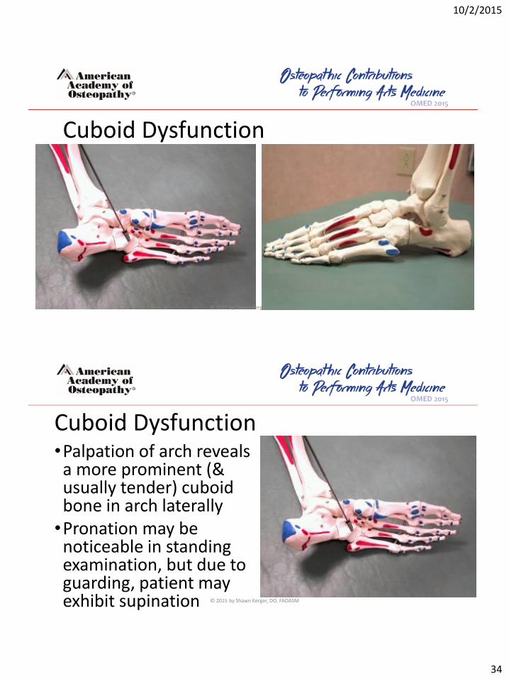
Cuboid Dysfunction
© 2015 by Shawn Kerger, DO, FAOASM
Cuboid Dysfunction•Palpation of arch reveals a more prominent (& usually tender) cuboidbone in arch laterally•Pronation may be noticeable in standing examination, but due to guarding, patient may exhibit supination © 2015 by Shawn Kerger, DO, FAOASM

10/2/2015
35
Cuboid Dysfunction –Articular / LVHA•Can be treated in a mirror fashion as navicular, but also may be addressed by grasping cuboid snugly & ‘chalking’ the 5th metatarsal head onto the cuboid gently, or the cuboidonto the calcaneus. © 2015 by Shawn Kerger, DO, FAOASM
Cuboid Dysfunction - SCS• Can also be treated successfully with strain-
counterstrain
• Find most tender point in tissues over cuboid
• With pt prone, greatly flex forefoot &invert/evert forefoot until tender point 70% (& can go for more) gone
•Maintain position with pt stabilized passively for 90 seconds
•Return (passively) to neutral
• Recheck findings© 2015 by Shawn Kerger, DO, FAOASM
10/2/2015
36
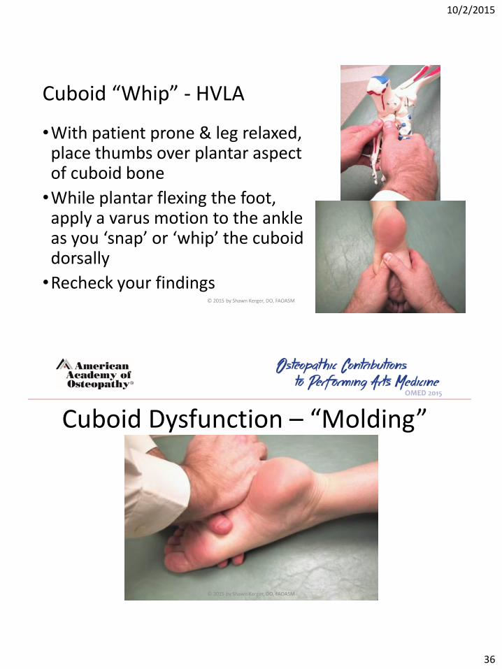
Cuboid “Whip” - HVLA
•With patient prone & leg relaxed, place thumbs over plantar aspect of cuboid bone
•While plantar flexing the foot, apply a varus motion to the ankle as you ‘snap’ or ‘whip’ the cuboiddorsally
•Recheck your findings© 2015 by Shawn Kerger, DO, FAOASM
Cuboid Dysfunction – “Molding”
© 2015 by Shawn Kerger, DO, FAOASM

10/2/2015
37
HVLA for plantarflexed 1st TMT Dysfunction - Dx
•With the 1st cuneiform stabilized, dorsiflex and plantarflex the 1st
TMT joint via the distal MT head
•Will find restricted dorsal motion at 1st MT head (compared to opposite side) and end-feel will be rigid/articular in nature
© 2015 by Shawn Kerger, DO, FAOASM
HVLA for plantarflexed 1st TMT Dysfunction - Tx•Dorsiflex the foot, using the TMT
joint as a fulcrum.•Use of other hand is optional,
but recommended.•With a straight traction force,
drive the hypothenar eminence of the dorsal hand distally along the plane of the tibia.•Recheck, regardless of audible
response…© 2015 by Shawn Kerger, DO, FAOASM

10/2/2015
38
Joint Play – Dx and Tx…
• This is about as basic a technique as you can get…
• Stabilize the proximal bone of the joint you wish to test and take the distal bone through all PROMs and articulate through any barriers.
• Many times will get an audible or palpable reduction, but recheck even if you don’t…
© 2015 by Shawn Kerger, DO, FAOASM
Articular Techniques for Talus•Commonly restricted anteriorly, or
impacted. May also present as an anterior fibular head…•Usually secondary to a traumatic
inversion mechanism at the ankle, but can also be due to chronically tight posterior calf muscles.•Can be associated with plantar fasciitis.•Pt will complain of anterior talar pain
or ‘jamming’ with attempted dorsiflexion, & possibly of reduced calf stretch when attempted.© 2015 by Shawn Kerger, DO, FAOASM

10/2/2015
39
Articular Techniques for Talus• Place ipsilateral middle or ring finger over
the superior aspect of the talus, below the tib-fib joint.
• Dorsiflex ankle to the barrier, while cradling the calcaneus with the contralateral hand. You may fine tune with inversion & eversion to maximize dorsiflexion.
• With the patient relaxed, either:• tug the foot quickly with a moderate
force in a caudal direction,• or with a traction force caudally, rock
the calcaneus & talus as a unit in an inversion/eversion plane.© 2015 by Shawn Kerger, DO, FAOASM
Talar Tug –Alternate Hold
•Need to pull & dorsiflex at the same time – makes a ‘J’ pattern movement when viewed this way
© 2015 by Shawn Kerger, DO, FAOASM

10/2/2015
40
Talar Release - Articular• Pt supine with knee & hip flexed to 90° &
hip slightly abducted, nestle your elbow against the mid-hamstring area while forming a ring with your thumbs & forefingers around the talus.
• Slowly, but firmly, flex the knee while maintaining the ring around the talus. You should feel a traction force building.
• Maintaining the tension, either exert a quick thrust with the talus or gently rock the talus into dorsiflexion with a little inversion/eversion until you feel a release, pop, or clunk.
© 2015 by Shawn Kerger, DO, FAOASM
Posterior Fibular Head - HVLA•Commonly restricted posteriorly.
•Usually secondary to a traumatic inversion mechanism at ankle.
•Can be associated w/ iliotibial tendonitis, and/or mimic lateral meniscal tears.
•Pt complains of lateral knee pain, usually w/ weightbearing & pivoting.
© 2015 by Shawn Kerger, DO, FAOASM

10/2/2015
41
Posterior Fibular Head –LVHA / HVLA
•Grasp affected extremity w/ contralateral hand at either distal tib/fib or at calcaneus• Ext. rotate tib to barrier (red
arrow)•Place ipsil. 2nd MCP jt behind
fibular head• Flex knee up to barrier• Either quickly flex knee over
2nd MCP joint (green arrow), or smoothly continue flexion
© 2015 by Shawn Kerger, DO, FAOASM
Plantar fasciitis - the problem:• Too much tension on the plantar fascia.
Why?
• Arches not able to support themselves:
• Navicular rotated
• Weak intrinsic foot muscles
• Weak or fatigued tibialis posterior, flexor digitorum longus, flexor hallucis longus
• Tightness of Achilles
© 2015 by Shawn Kerger, DO, FAOASM
10/2/2015
42
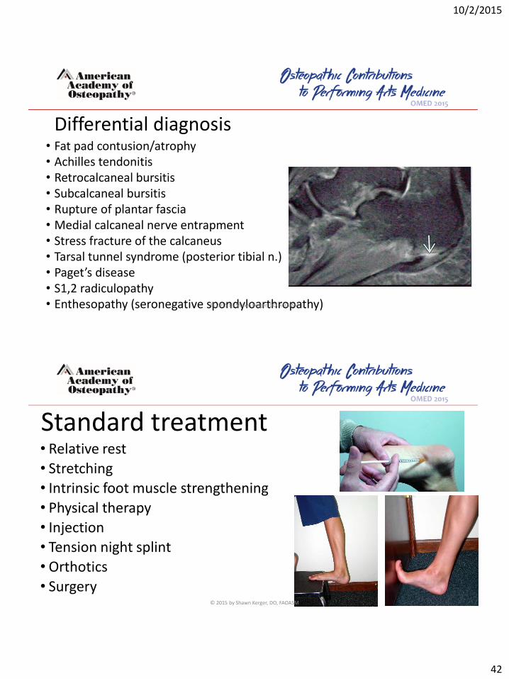
Differential diagnosis• Fat pad contusion/atrophy• Achilles tendonitis• Retrocalcaneal bursitis• Subcalcaneal bursitis• Rupture of plantar fascia• Medial calcaneal nerve entrapment• Stress fracture of the calcaneus• Tarsal tunnel syndrome (posterior tibial n.)• Paget’s disease• S1,2 radiculopathy• Enthesopathy (seronegative spondyloarthropathy)© 2015 by Shawn Kerger, DO, FAOASM
Standard treatment• Relative rest
• Stretching
• Intrinsic foot muscle strengthening
• Physical therapy
• Injection
• Tension night splint
• Orthotics
• Surgery © 2015 by Shawn Kerger, DO, FAOASM

10/2/2015
43
The osteopathic advantage• The goal of osteopathic approach must be to re-establish
normal function:• Maintenance of the medial arch
• Relieve pressure from the ligaments• OMT, arch support
• Improve strength of intrinsic foot muscles• exercises
• Correct tightness of the Achilles• OMT, stretching
• Improve proprioceptive function• OMT, specific proprioceptive retraining
• But don’t forget that there is still pathology that must heal!
© 2015 by Shawn Kerger, DO, FAOASM
Two arches to the foot
© 2015 by Shawn Kerger, DO, FAOASM

10/2/2015
44
Arches
© 2015 by Shawn Kerger, DO, FAOASM
Transverse Arch Supports
Passive at forefoot Active at midfootActive at midfoot
© 2015 by Shawn Kerger, DO, FAOASM

10/2/2015
45
Transverse Arch Supports
© 2015 by Shawn Kerger, DO, FAOASM
Tibialis posteriorFlexor hallucis longusFlexor digitorum longusFibularis brevisFibularis longus
© 2015 by Shawn Kerger, DO, FAOASM

10/2/2015
46
The treatment•Regardless of how you choose to affect the problem,
certain things must be consistently done in order to treat the problem, and prevent its recurrence.•Remember that there is pathology here.
• Inquire regarding changes in activity, footwear:•Often patients present after it has been present
for months—ask specifically. • Look at the shoes and the insoles.
© 2015 by Shawn Kerger, DO, FAOASM
Common aspects of treatment•You must take pressure off the plantar fascia:•Easiest way to do this is with a heel lift:•Typically 5-10mm is sufficient.
•Treat both sides.
•Stretch the Achilles tendon, both gastroc & soleus.
•Stretch the plantar fascia.
•Strengthen the intrinsic foot muscles.© 2015 by Shawn Kerger, DO, FAOASM

10/2/2015
47
Unload the plantar fascia• Critical to relieve tension on plantar fascia.
• Can be done multiple ways, but heel lift is often easiest:• This drops the forefoot during weight
bearing, shortening the distance between the metatarsals and calcaneus
• Secondarily relieves tension on Achilles
• Should be done from a horizontal, not sloping (such as a high-heeled shoe would do), position. © 2015 by Shawn Kerger, DO, FAOASM
Stretch the Achilles tendon•Possibly the most important aspect of treatment.
•Remember to stretch both gastroc (knee straight) & soleus (knee bent)
•Stretches should be held for 20-30s, repeated three times, both sides, regardless of symptoms.
•Consider using a step &/or activated stretching (muscle energy)
© 2015 by Shawn Kerger, DO, FAOASM

10/2/2015
48
Plantar fascia stretch•Direct stretching of plantar fascia is
often recommended
• I am not always sure how beneficial this is, or if the therapeutic benefit is really in stretching the fascia, or in some of the associated muscles supporting the arch.
•Stretch held same as previous ones.© 2015 by Shawn Kerger, DO, FAOASM
Strengthen intrinsic foot muscles
• I find this very helpful in reconditioning muscles to help support the arch.
•Does more than just intrinsic muscles, also includes the flexor hallucis longus, flexor digitorumlongus, and maybe tibialisposterior.
© 2015 by Shawn Kerger, DO, FAOASM

10/2/2015
49
OMT: navicular/cuboid•Correct dysfunction of the arch, especially the navicular, which tends to be rotated medially. •Functional approach:•Start from position of ease. •Add compressive force. •Take joint to, and through, the original barrier, maintaining the compressive force. © 2015 by Shawn Kerger, DO, FAOASM
OMT: tibiotalar joint – already did this• Often also restricted with talus held in relatively
valgus position.
• Many ways to do this: this is an articulatorytechnique:
• Contralateral elbow in popliteal fossa• Hand grasp calcaneus and anterior
process of talus. • Lean cephalad, elbow acting as fulcrum
to distract the talus from the mortise.• Gently rock the talus until articulation
and release occurs.© 2015 by Shawn Kerger, DO, FAOASM

10/2/2015
50
OMT: tibial torsion• Notice that we are working up the
kinetic chain. Obviously any somatic dysfunction should be treated, especially in lumbar, sacrum, & pelvis.
• Functional technique:• Start from position of ease, typically
ext rot.• Apply compressive force up to knee.• Move tibia to and through barrier
while extending the knee.© 2015 by Shawn Kerger, DO, FAOASM
Fascial stripping (Fascial Distortion Model Technique)
• This is something that has been modified from Steven Typaldos, DO. • It is very painful, but very effective, and they often stand
up feeling much better. • Treatment is done once per week, and typically takes ~6
treatments, sometimes less.
© 2015 by Shawn Kerger, DO, FAOASM

10/2/2015
51
Fascial stripping
© 2015 by Shawn Kerger, DO, FAOASM
So what do we do?•Make sure it is plantar fasciitis!• Treat the existing somatic dysfunction
on the first visit.• Heel lift.• HEP consisting of stretching &
strengthening as described.• Then either:• Fascial stripping protocol• Injection protocol
© 2015 by Shawn Kerger, DO, FAOASM
10/2/2015
52
References – including images1. http://themethuselahcenter.com/wp-content/uploads/2011/07/poor-posture.jpg
2. http://www.platinumphysio.co.uk/patient-info/conditions-treated/head-neck/
3. http://davidlasnier.com/2012/overhead-work-and-shoulder-flexion-limitation
4. http://rpm-therapy.com/wp-content/uploads/2012/09/hairdresser-posture-bad3.jpg
5. http://rpm-therapy.com/wp-content/uploads/2012/09/hairdresser-posture-bad.jpg
6. Thieme, Atlas of Anatomy, 2nd ed. Fig. 39.5 C.
7. http://www.muscleimbalancesyndromes.com/janda-syndromes/upper-crossed-syndrome/
8. http://karenoosterbaan.com/wp-content/uploads/2012/11/David_Yu_feature_alexander_technique_t6301.jpg
9. http://assets.nydailynews.com/polopoly_fs/1.474670!/img/httpImage/image.jpg_gen/derivatives/article_970/alg-rangers-lee-motion-jpg.jpg
10. Valley Morning Star article – Friday, July 29, 1938, pg.11
11. Karageanes, S & et al. Principles of Manual Sports Medicine. Lippincott, Williams & Wilkins; 2004. Multiple images used with written permission of editor - Steven Karageanes, DO, FAOASM.
12. DiGiovanna, Schiowitz, & Dowling. An Osteopathic Approach to Diagnosis & Treatment, 3rd ed. © 2015 by Shawn Kerger, DO, FAOASM
Credits
Special thanks to : • Beverly Goodwillie, RN• Debbie Verkin, LD, RD – Peds• Rhonda Price• Michael Sampson, DO,
FAOASM• Fran Adkins, OMS-IV• Bren Nolan, OMS-IV
• Abby Huck, OMS-IV• Zach Daniels, OMS-II• Mark Riley, OMS-II• Jesse Sheldon, OMS-II• Shannon Flahive, OMS-I
© 2015 by Shawn Kerger, DO, FAOASM


