Myc influences global chromatin structure
Paul S Knoepfler1, Xiao-yong Zhang2, Pei
Feng Cheng1, Philip R Gafken3, StevenB McMahon2 and Robert N Eisenman1,*1Division of Basic Sciences, Fred Hutchinson Cancer Research Center,Seattle, WA, USA, 2The Wistar Institute, Gene Expression andRegulation Program, Philadelphia, PA, USA and 3Proteomics Facility,Fred Hutchinson Cancer Research Center, Seattle, WA, USA
The family of myc proto-oncogenes encodes transcription
factors (c-, N-, and L-Myc) that regulate cell growth and
proliferation and are involved in the etiology of diverse
cancers. Myc proteins are thought to function by binding
and regulating specific target genes. Here we report that
Myc proteins are required for the widespread maintenance
of active chromatin. Disruption of N-myc in neuronal
progenitors and other cell types leads to nuclear conden-
sation accompanied by large-scale changes in histone
modifications associated with chromatin inactivation,
including hypoacetylation and altered methylation. These
effects are largely reversed by exogenous Myc as well as by
differentiation and are mimicked by the Myc antagonist
Mad1. The first chromatin changes are evident within 6h
of Myc loss and lead to changes in chromatin structure.
Myc widely influences chromatin in part through upregu-
lation of the histone acetyltransferase GCN5. This study
provides the first evidence for regulation of global chroma-
tin structure by an oncoprotein and may explain the broad
effects of Myc on cell behavior and tumorigenesis.
The EMBO Journal (2006) 25, 2723–2734. doi:10.1038/
sj.emboj.7601152; Published online 25 May 2006
Subject Categories: chromatin & transcription; development
Keywords: chromatin; epigenetics; histone modification;
Myc; stem and progenitor cells
Introduction
The members of the Myc/Mad/Mnt superfamily of basic
helix–loop–helix zipper (bHLHZ) transcription factors each
heterodimerize with the bHLHZ protein Max and bind the
E-box sequence CACGTG. Transcriptional activation by Myc
proteins and repression by Mad/Mnt proteins, at E-box bind-
ing sites, are involved in regulation of cell growth, prolifera-
tion, and apoptosis (Eisenman, 2001). Targeted disruption
of c-myc, N-myc, or max in the mouse leads to embryonic
lethality (Stanton et al, 1992; Davis et al, 1993; Shen-Li
et al, 2000), whereas overexpression of myc genes is strongly
associated with the genesis of diverse cancers in many
species (Lutz et al, 2002). Myc activates transcription through
recruitment of chromatin-modifying complexes. For example,
interaction with the coactivator TRRAP mediates Myc’s asso-
ciation with histone acetyltransferases (HATs) GCN5 and
Tip60 (McMahon et al, 1998, 2000; Frank et al, 2003). Myc
also interacts with CBP and the chromatin-remodeling com-
plex containing Ini1 (Cheng et al, 1999; Vervoorts et al, 2003).
By contrast, Mad proteins recruit histone deacetylases
(HDACs) via the corepressor mSin3 (Ayer, 1999; Knoepfler
and Eisenman, 1999). The complexes recruited by Myc–Max
and Mad–Max induce distinct chromatin modifications with-
in the regulatory regions of shared target genes, leading to
activation or repression (Eisenman, 2001; Frank et al, 2001;
Fernandez et al, 2003). The notion that Myc is a typical
transcription factor regulating the expression of a small
number of target genes has been challenged by recent find-
ings indicating that DNA binding and gene regulation by Myc
are both surprisingly widespread (Fernandez et al, 2003; Li
et al, 2003; Orian et al, 2003; Cawley et al, 2004; Patel et al,
2004). To study potential global gene regulatory functions of
Myc, we focused on myc loss-of-function mutations in cells
and tissues normally dependent on Myc activity.
Results
Altered nuclei and histone modifications in N-myc null
cells
We previously demonstrated that N-myc is essential for
normal nervous system development (Knoepfler et al, 2002)
by using nestin-cre to generate a nervous system-specific
conditional knockout of N-myc in the mouse (N-myc NS
null). In N-myc NS null E12.5 embryos, we observed that
neural stem and progenitor cell (NPC) nuclei were abnor-
mally small, round, and dark when stained with either H&E
or methyl green (compare control and N-myc null nuclei in
Figure 1Ai–ii and Supplementary Figure S1A). Both TUNEL
and caspase cleavage assays demonstrated that these changes
are not due to increased apoptosis in the N-myc null tissues
and cells (Knoepfler et al, 2002) (Supplementary Figure S2A
and B), and assays for senescence-associated b-gal activityindicated that they are not due to senescence (Supplementary
Figure S2C). We therefore examined whether the changes in
nuclear morphology might reflect alterations in chromatin.
Transcriptional activity of chromatin is associated with
specific histone modifications, including acetylation and
methylation (Strahl and Allis, 2000), implicated in local
gene-specific effects as well as global chromatin structure
(Vogelauer et al, 2000; Rea et al, 2000; Berger and Felsenfeld,
2001; Kurdistani et al, 2004; Schubeler et al, 2004). To
determine if disruption of Myc function induces changes in
histone modifications consistent with chromatin inactivation,
we first employed immunohistochemistry (IHC) to assess
global levels of acetylated histone H3 and H4 (AcH3, AcH4)
in the developing nervous system of control, N-myc NS null
(Knoepfler et al, 2002), and nestin-N-myc transgenic (Tg)
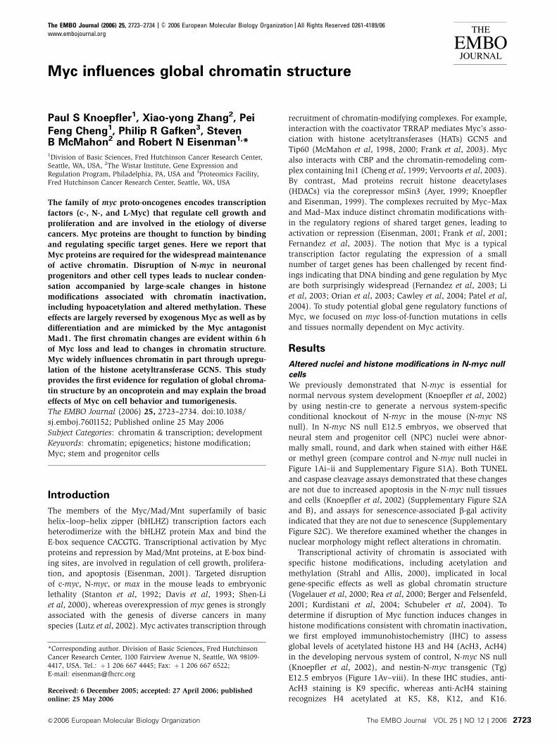
E12.5 embryos (Figure 1Av–viii). In these IHC studies, anti-
AcH3 staining is K9 specific, whereas anti-AcH4 staining
recognizes H4 acetylated at K5, K8, K12, and K16.Received: 6 December 2005; accepted: 27 April 2006; publishedonline: 25 May 2006
*Corresponding author. Division of Basic Sciences, Fred HutchinsonCancer Research Center, 1100 Fairview Avenue N, Seattle, WA 98109-4417, USA. Tel.: þ 1 206 667 4445; Fax: þ 1 206 667 6522;E-mail: [email protected]
The EMBO Journal (2006) 25, 2723–2734 | & 2006 European Molecular Biology Organization |All Rights Reserved 0261-4189/06
www.embojournal.org
&2006 European Molecular Biology Organization The EMBO Journal VOL 25 | NO 12 | 2006
EMBO
THE
EMBOJOURNAL
THE
EMBOJOURNAL
2723

Acetylation of each of these lysines is associated with active
chromatin (Turner et al, 1992; Jeppesen and Turner, 1993;
Braunstein et al, 1996). In the ventricular zone (VZ) of
control embryos, we noted a positive correlation between
nuclear size and the levels of N-Myc, AcH3, and AcH4 (Figure
1Ai and v and Supplementary Figure S1A and C). N-myc null
NPCs exhibited striking histone hypoacetylation (low/absent
brown stain) specifically associated with abnormally small
round nuclei that also counterstained darkly with the DNA
dye methyl green, suggesting chromatin condensation
(Figure 1Ai–ii and v–vi).
We next asked whether acute disruption of N-myc would
also alter histone acetylation. Using cultured N-mycflox/flox
cerebellar granule neural progenitors (CGNPs), N-myc was
acutely disrupted by infection with MSCV Cre-IRES-GFP (Cre-
GFP), a retroviral vector expressing Cre and GFP (Figure 1B).
Disruption of N-myc in a majority of GFPþ cells was verified
by immunofluorescence staining (IF) for endogenous nuclear
N-Myc protein (not shown), similar to the large fraction of
cells with N-myc loss in CGNPs with nestin-cre-driven knock-
out (Supplementary Figure S3A). N-Myc-deficient GFPþCGNPs exhibit dramatic changes in histone acetylation,
with the majority having an apparently complete loss of
detectable histone H3 and H4 acetylation (Figure 1B, white
arrows in column 3; data not shown). These changes are
associated with nuclear condensation and alterations in DAPI
staining (see below). Importantly, Cre-GFP virus had no
effect on histone acetylation in c-mycflox/flox (de Alboran
Figure 1 Analysis of N-Myc levels and histone acetylation. (A) E12.5 Sagittal sections from control (panels i and v), N-myc null (panels ii andvi), and N-myc transgenic embryos (Tg) expressing moderately (iii and vii) and highly elevated levels of N-Myc (iv and viii) under control ofthe nestin promoter/enhancer. Embryos were stained for N-Myc (brown stain, panels i–iv) and AcH3 (brown stain; panels v–viii) andcounterstained with methyl green (bluish). Nuclear and cytoplasmic N-Myc proteins are evident, as has been previously reported (Wakamatsuet al, 1993) (� 100 magnification). Boxed regions are shown at higher magnification in Supplementary Figure S1A. (B) CGNPs derived fromN-mycflox/flox (left three columns) or c-mycflox/flox (right column) embryos were grown in medium containing Shh (3 ng/ml), infected with GFPor Cre-IRES-GFP retroviruses, and stained with DAPI and anti-AcH3. White arrows denote infected cells. (C–F) Control and nestin-cre-derivedN-myc null CGNPs stained for DAPI (blue) and histone H3 diMeK9 (C), triMeK4 (D), and triMeK9 (E) (red). (F) Control and N-myc null CGNPsstained for DAPI (blue) and HP1a (monochrome). (G) Cultured E12.5-derived control and N-myc null neurospheres stained for DNA (DAPI),N-Myc (green), and AcH3 (red). Columns 3 and 4 represent null cells transfected with N-Myc. White arrow indicates untransfected cell. Yellowarrows indicate NPCs with supraphysiological levels of N-Myc. Control (flox/flox) and null (flox/flox nestin-cre) cultures were grown for atleast 1 month before these analyses and exhibited a high degree of stability in culture composition and properties (extremely low, but similarrates of spontaneous differentiation, stable proliferation rates, stable neurosphere morphology and size, and stable cellular morphology upongrowth as a monolayer). We consistently observed that 80–90% of the N-mycflox/flox nestin-creþ neurosphere cells exhibited a complete loss ofdetectable N-Myc protein.
Myc oncoprotein influence on global chromatinPS Knoepfler et al
The EMBO Journal VOL 25 | NO 12 | 2006 &2006 European Molecular Biology Organization2724
et al, 2001) CGNPs, consistent with the report that c-myc
is not expressed in CGNPs (Kenney et al, 2003) (Figure 1B).
Further, infection of N-mycflox/flox CGNPs with MSCV IRES-
GFP virus, which only expresses GFP, had no effect on
acetylation or nuclear structure (Figure 1B, column 1).
Disruption of N-myc also does not appear to influence
CGNP identity or culture composition. Control and null
CGNP cultures exhibit essentially identical fractions (84–87%)
of cells staining with the CGNP-specific marker Zic1 (Aruga
et al, 1994) (Supplementary Figure S3B). Our data indicate that
loss of Myc from neuronal progenitors is associated with
significantly decreased levels of H3 and H4 acetylation.
Myc is required for maintenance of normal histone
methylation patterns
To determine whether the decreased histone acetylation
observed in N-myc null cells correlates with altered histone
methylation patterns (Rea et al, 2000), we began by staining
CGNPs for methylated H3-K9. Control (N-mycflox/flox) CGNPs
displayed only faint speckled staining for both H3-diMeK9
(Figure 3A, top panel of column 8, and Figure 1C) and H3-
triMeK9 (Figure 1E), marks of repressive chromatin. In con-
trast, we found that N-myc null (N-mycflox/flox nestin-cre)
CGNPs exhibited high levels of H3-diMeK9 and H3-triMeK9
(Figures 1C, E, and 3A, column 8). Moreover, N-myc null
CGNPs show a dramatic reduction in H3-triMeK4, a modifi-
cation strongly associated with active chromatin (Strahl and
Allis, 2000) (Figure 1D). We see the same general pattern
of histone methylation changes in Tet-Off Myc B cells
(Supplementary Figure S4) and, to a lesser extent, in c-myc
null fibroblasts (not shown). The heterochromatin binding
protein HP1a, which has been shown to directly interact with
H3-MeK9 (Bannister et al, 2001; Lachner et al, 2001), exhibits
a focal nuclear staining pattern evident in control CGNPs
(Figure 1F) similar to that reported in other studies (Lachner
et al, 2001). Consistent with their high levels of H3-di-MeK9
and H3-tri-MeK9, N-myc null CGNPs exhibit unusually
intense and abundant HP1a foci (Figure 1F), presumably
reflecting abnormal spreading of heterochromatin (see
below). In summary, our targeted deletion experiments in-
dicate that an apparently general loss of histone acetylation,
increased histone methylation, and chromatin condensation
in the N-myc null CGNPs are associated with loss of N-myc.
Reintroduction of Myc restores altered histone
acetylation in N-myc null cells
To ascertain if the decreased levels of histone acetylation
represent an irreversible cellular response to N-Myc loss,
we examined N-myc null (N-mycflox/flox nestin-creþ ) neuro-
sphere cultures derived from E12.5 whole embryonic brains.
Overall IF analysis indicated that such N-myc null neuro-
sphere cultures exhibited very low or undetectable nuclear
histone acetylation compared to N-mycflox/flox controls
(Figure 1G, columns 1 and 2). Introduction of N-Myc
(Figure 1G, columns 3 and 4, and Figure 5G) or c-Myc
(data not shown) into null cells resulted in markedly
increased acetylation within 2 days of transfection. In the
subset of N-myc null neurospheres, which expressed supra-
physiological levels of introduced N-Myc, histone acetylation
increased to substantially above normal (Figure 1G, yellow
arrows). In addition, VZ cells in N-myc Tg mice displayed
above-normal H3 and H4 acetylation (Figure 1Aiii, iv, vii, and
viii and Supplementary Figure S1B) consistent with the
notion that Myc levels are linked to the extent of histone
acetylation. Such N-myc overexpression and hyperacetylation
is also associated with VZ hyperplasia (Supplementary Figure
S1B). Restoration of histone acetylation is strongly attenuated
in N-myc null neurospheres transfected with N-myc mutants
lacking Myc Box II (MBII), a highly conserved transactivation
domain that associates with the HAT-binding coactivator
TRRAP (McMahon et al, 2000), or lacking the C-terminal
basic region, which is required for DNA binding
(Supplementary Figure S10). We note that overexpression of
the transcription factor E2F had no apparent effect on wide-
spread histone acetylation (Supplementary Figure S9).
Quantitative analysis of chromatin changes
Because quantitative analyses require more chromatin than
can be readily obtained from our primary murine neuronal
cell cultures, we turned to the well-characterized c-myc null
rat fibroblast cell line, HO15.19 (Mateyak et al, 1997). As
in the neuronal cells, HO15.19 cells lacking c-myc (hereafter
‘c-myc null’ and which do not express N- or L-myc) (Mateyak
et al, 1997) are hypoacetylated at H3 and H4 compared to the
TGR wild-type (WT) parental control line when assayed by IF
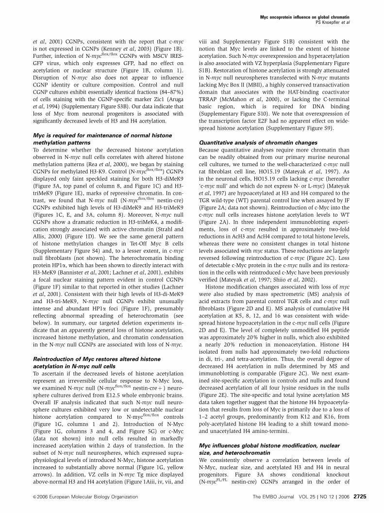
(Figure 2A; data not shown). Reintroduction of c-Myc into the
c-myc null cells increases histone acetylation levels to WT
(Figure 2A). In three independent immunoblotting experi-
ments, loss of c-myc resulted in approximately two-fold
reductions in AcH3 and AcH4 compared to total histone levels,
whereas there were no consistent changes in total histone
levels associated withmyc status. These reductions are largely
reversed following reintroduction of c-myc (Figure 2C). Loss
of detectable c-Myc protein in the c-myc nulls and its restora-
tion in the cells with reintroduced c-Myc have been previously
verified (Mateyak et al, 1997; Shiio et al, 2002).
Histone modification changes associated with loss of myc
were also studied by mass spectrometric (MS) analysis of
acid extracts from parental control TGR cells and c-myc null
fibroblasts (Figure 2D and E). MS analysis of cumulative H4
acetylation at K5, 8, 12, and 16 was consistent with wide-
spread histone hypoacetylation in the c-myc null cells (Figure
2D and E). The level of completely unmodified H4 peptide
was approximately 20% higher in nulls, which also exhibited
a nearly 20% reduction in monoacetylation. Histone H4
isolated from nulls had approximately two-fold reductions
in di, tri-, and tetra-acetylation. Thus, the overall degree of
decreased H4 acetylation in nulls determined by MS and
immunoblotting is comparable (Figure 2C). We next exam-
ined site-specific acetylation in controls and nulls and found
decreased acetylation of all four lysine residues in the nulls
(Figure 2E). The site-specific and total lysine acetylation MS
data taken together suggest that the histone H4 hypoacetyla-
tion that results from loss of Myc is primarily due to a loss of
1–2 acetyl groups, predominantly from K12 and K16, from
poly-acetylated histone H4 leading to a shift toward mono-
and unacetylated H4 amino-termini.
Myc influences global histone modification, nuclear
size, and heterochromatin
We consistently observe a correlation between levels of
N-Myc, nuclear size, and acetylated H3 and H4 in neural
progenitors. Figure 3A shows conditional knockout
(N-mycFL/FL nestin-cre) CGNPs arranged in the order of
Myc oncoprotein influence on global chromatinPS Knoepfler et al
&2006 European Molecular Biology Organization The EMBO Journal VOL 25 | NO 12 | 2006 2725
decreasing nuclear size. Varying levels of residual N-Myc
protein remain in a small subset of these conditionally null
CGNPs and N-Myc level correlates with nuclear size and
histone acetylation (Figure 3A, columns 1–6; data not
shown). These findings suggest that loss of histone acetylation
parallels decreasing levels of Myc (column 6). This notion is
also supported by the observation that in cultures of condition-
ally null CGNPs, the small subset with residual N-Myc levels
are the only ones with remaining detectable, albeit low,
levels of histone acetylation (Supplementary Figure S3A).
Interestingly, the subnuclear localization pattern of N-Myc
broadly overlaps with regions of anti-AcH3 and anti-AcH4 IF,
all of which are excluded from islands of intense DAPI
staining (Figure 3A, column 6), characteristic of heterochro-
matin (Bickmore and Craig, 1997). Also evident is a correla-
tion between H3-diMeK9 levels, decreased nuclear size, and
the extent of heterochromatic regions (Figure 3A, columns 7
and 8). A more detailed analysis of nuclear and DNA struc-
ture in control and N-myc null CGNPs was conducted using
transmission electron microscopy (EM) of uranyl acetate-
stained cells. As shown in Figure 3B, nuclei from control
CGNPs are approximately 5–10 mm in diameter composed
predominantly of lightly stained euchromatic regions, with
the exception of 3–5 darkly staining heterochromatic regions
(Busch, 1974) (arrows in Figure 3Bi). These darkly staining
regions, each approximately 0.5–1 mm across, are frequently
associated with the nuclear lamina, as expected for hetero-
chromatin (Cohen et al, 2001). They are similar in size,
location, and appearance to the intense DAPI foci in CGNPs
(Figure 3B, white arrows in panel ii), which have been
established to be heterochromatin in murine cells
(Bickmore and Craig, 1997). In N-myc null CGNPs, the
majority of nuclei are several fold smaller in area compared
to controls and the heterochromatic regions are greatly
expanded (Figure 3Biii–v). Thus, loss of Myc results in a
decrease in nuclear volume and a striking spreading of
heterochromatin, no longer limited to foci, throughout the
nuclei of null cells.
Loss of Myc leads to decreased DNA accessibility
To address whether myc levels influence chromatin structure,
we conducted micrococcal nuclease (MNase) accessibility
assays (Weintraub and Groudine, 1976) using the well-estab-
lished Tet-Off Myc B (P493-6) cell system (Schuhmacher et al,
1999) in which Myc can be reproducibly turned off by the
addition of tetracycline. The P493-6 cells exhibit the same
type of chromatin changes upon Myc downregulation as
observed in myc-deficient neuronal cells and fibroblasts
(see below). Intact living cells were permeabilized so as to
minimize effects on chromatin structure (Zaret, 1999) and
cells were treated with increasing amounts of MNase
(Figure 3C). In the absence of MNase, neither Myc-Off nor
Figure 2 Histone modifications in c-myc null,mad1–transfected, and WT fibroblasts. (A) Staining for AcH4 (red) in parental (TGR), c-myc null(HO15.19), and c-myc null rat fibroblasts stably transfected with c-myc. (B) Mad1 overexpression in WT murine fibroblasts partiallyphenocopies loss of Myc. (C) Immunoblotting for the indicated acetylated and methylated histones H3 and H4 comparing levels in TGR, c-mycnull, and c-myc null rat fibroblasts transfected with c-myc. Values in the graphs represent the ratio of arbitrary values measured by Odysseysystem for bands representing modified/total histones from three independent biological repeats. (D, E) Mass spectrometric data on total andK-specific histone H4 N-terminal acetylation from three independent experiments. The relative fraction of N-terminal peptides containing 0–4acetyl groups was determined as well as the fraction of specific K residues that were acetylated. All error bars in this figure are s.e.m.
Myc oncoprotein influence on global chromatinPS Knoepfler et al
The EMBO Journal VOL 25 | NO 12 | 2006 &2006 European Molecular Biology Organization2726

Myc-On cells exhibited evidence of endogenous nuclease
activity. However, at increasing concentrations of MNase,
Myc-Off cells exhibited a strongly enhanced resistance to
MNase, indicative of a more closed chromatin structural
state (Shogren-Knaak et al, 2006). We have observed de-
creased accessibility following Myc loss in four independent
experiments in these cells. Thus, Myc appears to influence
DNA accessibility, consistent with the histone modifications
described above. These data support the notion that Myc
has a widespread influence on chromatin structure.
Loss of Myc rapidly alters histone modifications in a cell
cycle- and differentiation-independent manner
To assess the kinetics of chromatin changes associated with
loss of Myc, we analyzed Tet-Off Myc B cells (Schuhmacher
et al, 1999) in which introduction of tetracycline shuts down
c-Myc expression. Expression of endogenous Myc proteins is
undetectable in these cells, and introduction of tetracycline
rapidly (within 16 h) leads to strong downregulation of the
c-Myc transgene (Grandori et al, 2003). Introduction of tetra-
cycline for 72 h in a serum-free context resulted in loss of Myc
(data not shown) as well as the same general pattern of
changes we observed in neuronal cells and fibroblasts upon
Myc disruption: decreased histone H3 K9 acetylation and K4
methylation as well as increased levels of H3-diMeK9, and
nuclear condensation (Supplementary Figure S4; not shown).
Initial changes were detectable as early as 6 h after introduc-
tion of tetracycline (Supplementary Figure S4) and down-
regulation of Myc (data not shown), whereas more
substantial changes were evident after 24 and 72 h. Thus,
changes in histone modifications occur rapidly following
alterations in Myc levels. Further, the changes in chromatin
do not appear to be secondary to changes in cell cycle status
because the chromatin alterations are observed with loss of
Figure 3 Histone modifications, heterochromatin, and DNA accessibility in control and null N-myc CGNPs. (A) N-mycflox/flox nestin-cre CGNPswere visually sorted by nuclear size following staining with the indicated antibodies. Each color intensity in column 6 was uniformly increasedin each panel to maximize detection of potential colocalization of signal. (B) Control (top) and N-myc null (bottom) CGNPs were analyzed byEM following uranyl acetate staining (panels i and iii–vi) or by DAPI (panel ii). Arrows indicate heterochromatic regions. Note: Although thescale bar in panel (i) is 2 mm and the bars in the other panels are 1mm, the bar in (i) is also twice as long as the bars in the other panels,indicating that all images are exactly of the same magnification. (C) MNase assay on Tet-off Myc B cells with and without 72 h of tetracyclinetreatment.
Myc oncoprotein influence on global chromatinPS Knoepfler et al
&2006 European Molecular Biology Organization The EMBO Journal VOL 25 | NO 12 | 2006 2727
Myc in a system in which there are no cycling cells (serum-
free conditions) (Schuhmacher et al, 1999).
Several additional lines of evidence argue against the
possibility that the changes in histone modifications are
secondary consequences of cell cycle arrest upon Myc loss.
The c-myc null HO15.19 fibroblasts, which exhibit decreased
acetylation (Figure 2), proliferate, albeit at a lower rate
(Mateyak et al, 1997). Similarly, nestin-cre-derived N-myc
null and WT CGNPs express Ki67 (Supplementary Figure
S5C), a nuclear antigen present in cycling but not quiescent
cells (Gerdes et al, 1991). Furthermore, we found that a
subset of N-myc null CGNPs, even those with the most
extreme nuclear condensation and histone hypoacetylation,
nonetheless exhibited anti-BrdU and anti-phosphoH3 staining
(Supplementary Figure S5A and B; data not shown). We have
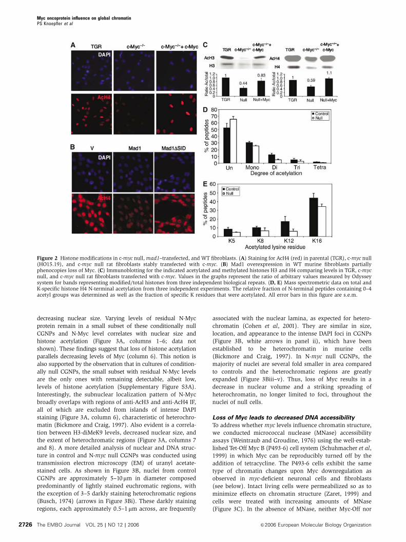
also observed that differentiation of neurospheres and
CGNPs, associated with terminal cell cycle arrest, leads to
increased histone acetylation and decreased H3-K9 methyla-
tion (Figure 4; not shown). Furthermore, a recent study
indicates that quiescent lymphocytes exhibit a striking de-
crease in repressive histone methylation marks compared
to activated, proliferating cells (Baxter et al, 2004). Taken
together, these data argue that the changes observed upon
Myc loss of function are not simply a consequence of
proliferation arrest.
The Myc antagonist Mad1 suppresses widespread
histone acetylation: a role for the global balance of
HDACs and HATs
Mad-related proteins exhibit widespread genomic binding in
Drosophila overlapping with dMyc binding sites (Orian et al,
2003) and antagonize some Myc functions through shared
target genes in mammalian cells (Iritani et al, 2002). We
asked whether Mad1 also influences widespread chromatin
modification. Mad1 overexpression in control neurospheres
(not shown) and murine fibroblasts resulted in a pronounced
reduction in global AcH4 (Figure 2B) and AcH3 (not shown)
levels in both cell types. Furthermore, deletion of the Mad1
SID domain (Mad1DSID), which constitutes the binding site
for the mSin3–HDAC corepressor complex, largely abrogated
Mad1-induced suppression of histone acetylation (Figure 2B).
In contrast to Mad1, overexpression of Max had no discern-
able effects on global chromatin in either fibroblasts or in WT
neurospheres and Max was also unable to reverse the histone
hypoacetylation in myc null neurospheres (data not shown).
These findings were expected given that Max is required for
the opposing activities of Myc as well as Mad proteins
(Eisenman, 2001).
We hypothesized that the widespread alterations in chro-
matin owing to changes in Mad or Myc could be due to a
large-scale imbalance in the overall levels of HATs and
HDACs. The HDAC inhibitor TSA reversed histone hypo-
acetylation in N-myc null neurospheres (Supplementary
Figure S6A), suggesting that loss of Myc may cause
chromatin changes in part by shifting the balance of HDACs
and HATs toward HDACs. This notion is also supported
by our observation that overexpression of HDAC1 in fibro-
blasts phenocopies loss of Myc (Supplementary Figure S6B)
in terms of nuclear condensation as well as histone
hypoacetylation. Furthermore, introduction of the HATs
GCN5, MOF, or TIP60 reverses the histone hypoacetylation
observed in N-myc null neurospheres (Figure 5G; not
shown).
GCN5 is a direct Myc target gene
One mechanism by which Myc could control the overall
equilibrium of histone-modifying enzymes is by regulation
of their expression. In order to address this possibility, levels
Figure 4 Differentiation of control and N-myc null neurospheres influences histone modifications. IF staining of control and N-myc nullneurospheres induced to differentiate for 7 days by growth factor withdrawal and retinoic acid treatment.
Myc oncoprotein influence on global chromatinPS Knoepfler et al
The EMBO Journal VOL 25 | NO 12 | 2006 &2006 European Molecular Biology Organization2728

of chromatin-modifying enzymes in Tet-Myc cells or in
primary cells with and without Myc were analyzed by IF,
immunoblotting, and RT–PCR (Figure 5). Levels of six HDACs
(HDACs 1–6), two HATs (TIP60 and CBP), and the two
histone methyl transferases (Set9 and G9a) that target H3-
K4 and H3-K9 respectively (Peterson and Laniel, 2004) (the
methyl marks affected by loss of Myc) were not affected by
Myc status (Figure 5A and B). However, the expression of one
HAT, GCN5, was strikingly reduced upon loss of Myc at both
RNA and protein levels in every system tested. For example,
loss of Myc in Tet-off Myc B cells led to strong reductions
in GCN5 levels (Figure 5A and B). Similarly, in N-myc null
neurospheres and CGNPs, levels of GCN5 were strongly
reduced (Supplementary Figure S7), whereas levels of other
histone-modifying enzymes such as the HATs TIP60, CBP,
and p300 were not reduced in Tet-off B cells or in primary
N-myc null CGNPs. Further, RNAi-mediated knockdown (KD)
of c-Myc (Zhang et al, 2005) in human cells also caused
consistent reductions in GCN5 RNA and protein levels
(Figure 5C and D), whereas the levels of another HAT,
Figure 5 GCN5 is a direct Myc target gene and can reverse histone hypoacetylation in N-myc null cells. (A, B) Shutting off Myc in the Tet-repressor Myc B cells leads to a reduction in GCN5 levels, but has no effect on the levels of other HATs, HDACs, and HMTs as indicated by IF(monochrome) and immunoblot with antisera against the indicated protein. (C, D) shRNA-mediated KD of endogenous Myc results indecreased hGCN5 mRNA levels. Treatment of the human lung cancer cell line H1299 for 48 h with shRNA directed against Myc (shMyc) resultsin decreased levels of Myc mRNA and protein, whereas a scrambled shRNA (shScr) had no effect. Concomitant with the loss of Myc expressionis the loss of hGCN5 at both the protein and mRNA level. (E) Myc activates transcription of the hGCN5 gene in primary human cells. Thehuman diploid fibroblast strain IMR90 was stably transduced with a retrovirus directing expression of the Myc-ER protein. Myc-ER-expressingcells were treated with 4-OHT (or EtOH as a negative control) to activate c-Myc for the times indicated. mRNAwas harvested and analyzed byquantitative RT–PCR. Actin mRNA levels were determined simultaneously and used to normalize mRNA levels for the other genes. hGCN5levels are increased by Myc activation even more dramatically than those of the known Myc targets CAD and cyclin D2. Furthermore, mRNAfor the other TRRAP-associated HATs, PCAF and TIP60, were not responsive to Myc activation. The non-Myc responsive gene ELF1a was usedas a negative control. Values are expressed as fold induction. (F) Direct binding of endogenous Myc to the hGCN5 locus in vivo. The humanGCN5 locus contains two matches to the CACGTG consensus Myc binding site as indicated. In addition, several matches to the non-canonicalsite bound by Myc in the cytochrome c gene (CATGCG) are present. To examine the binding of Myc to these sites, we utilized human diploidfibroblasts that had been either serum deprived or re-fed with 10% FCS for 2 h. Binding of endogenous Myc to three sites within the hGCN5locus was then assessed. Inducible binding of Myc to site 2, which is adjacent to the transcriptional start site, was evident. (G) Overexpressionof hGCN5 in N-myc null neurospheres reverses histone hypoacetylation as effectively as reintroduction of N-myc. Null neurospheres weretransfected with N-myc or hGCN5 (red), then IF stained for acetylation of histone H3 (green).
Myc oncoprotein influence on global chromatinPS Knoepfler et al
&2006 European Molecular Biology Organization The EMBO Journal VOL 25 | NO 12 | 2006 2729

PCAF, were unaffected. Several lines of evidence indicate
that GCN5 is a direct Myc target gene. Induction of Myc
activity by administration of tamoxifen to primary human
fibroblasts stably expressing MycER strongly induced GCN5
expression (Figure 5E), but not expression of other HATs
(Tip60, PCAF). Furthermore, chromatin immunoprecipitation
(ChIP) assay (Zhang et al, 2005) indicates that endogenous
Myc directly binds two E-boxes in the GCN5 promoter
displaying a 5- to 10-fold increase in binding upon addition
of serum (Figure 5F). Serum-inducible Myc occupancy of the
GCN5 promoter also correlates with binding of RNA poly-
merase II as well as histone H3 and H4 acetylation, all signs
of gene activation (Supplementary Figure S11). Myc binding
to GCN5 is fairly specific as Myc does not detectably bind
to HDAC1, HDAC2, Set9, nor PCAF by ChIP assay
(Supplementary Figure S11). We did find evidence of Myc
binding to TIP60, which contains two E-boxes in its promoter
as well; however, because there was no evidence of a link
between Myc and TIP60 mRNA or protein expression levels,
it remains unclear if Myc regulates TIP60 in this biological
setting.
Reduction of endogenous GCN5 levels interferes with
Myc-induced hyperacetylation
To more directly assess a potential functional role for GCN5 in
Myc’s global regulation of chromatin, we employed RNAi to
KD endogenous GCN5 utilizing a set of five independent
unique GCN5 shRNA constructs (sequence search verified
no off-site targets for any of the five constructs), along with a
nonspecific control shRNA. The five independent GCN5 RNAi
constructs exhibited a range of inhibitory activity that corre-
lated well with reduction in endogenous GCN5 by IF and by
Western blot (Figures 6A and Supplementary Figure S8),
a target of GCN5’s HAT activity, further evidence of the
functional specificity of the shRNAs.
If GCN5 is required for Myc’s ability to regulate global
chromatin, then KD of endogenous GCN5 should block the
ability of reintroduced N-Myc to restore the histone acetyla-
tion in N-myc null neurospheres. Consistent with a critical
role for endogenous GCN5 in Myc’s global chromatin func-
tion, the five GCN5 RNAi constructs interfered with the
ability of reintroduced N-Myc to reverse histone hypoacetyla-
tion in proportion to their GCN5 KD effectiveness (Figure 6B
Figure 6 A critical role for GCN5 in Myc’s regulation of chromatin. (A) RNAi-mediated KD of endogenous GCN5 in N-myc null neurospheres.IF analysis with GCN5 antisera (red) indicating variably reduced GCN5 protein levels with five RNAi constructs targeted against GCN5. Column1, no RNAi treatment; column 2, control RNAi treatment; columns 3–7, treatment with indicated GCN5 RNAi. Columns 1–7 are N-myc nullneurospheres, whereas column 8 is control neurospheres with no RNAi treatment. (B) RNAi KD of endogenous GCN5 blocks the ability ofexogenous N-Myc to rescue histone hypoacetylation in N-myc null neurospheres. Representative IF images are shown. Columns 1–7 are cellstreated with the same RNAi constructs as in (A) but also followed 48 h later by transfection with exogenous N-Myc (red) and stained foracetylated histone H3 (green) as well as DAPI (blue). Cells expressing exogenous N-Myc are indicated by white arrows. The N-Myc IF panel incolumn 8 for the Fl/Fl cre� control is from a separate experiment but is included to show representative control levels of endogenous N-Mycprotein for comparison. (C) Quantitation of the ability of GCN5 RNAi to block rescue. Mean values from four independent groups of 10 N-Myc-transfected cells that were also previously transfected with each type of RNAi are shown with error bars of s.e.m. The differences between thevalues for the control RNAi and the three strongest RNAi (RNAi #2, #3, and #4) exhibit P-values o0.0002, 0.00005, and 0.0009, respectively.
Myc oncoprotein influence on global chromatinPS Knoepfler et al
The EMBO Journal VOL 25 | NO 12 | 2006 &2006 European Molecular Biology Organization2730
and C), whereas the nonspecific RNAi control had no effect.
Thus, endogenous GCN5 plays a critical role in Myc’s regula-
tion of widespread histone modifications.
Discussion
The notion that Myc is a general chromatin regulator, while to
our knowledge unprecedented for an oncoprotein, is none-
theless consistent with several recent observations concern-
ing Myc function. First, a series of independent expression
microarray studies have collectively identified an unexpect-
edly large group of potential genes (representing about 5% of
all genes) that are transcriptionally regulated by Myc (Zeller
et al, 2003). Second, recent experiments directly assessing
genomic binding by Myc suggest binding to thousands of
sites throughout the genome encompassing approximately
15% of genes as well as intergenic regions (Fernandez et al,
2003; Li et al, 2003; Orian et al, 2003; Cawley et al, 2004; Patel
et al, 2004). Finally, although many Myc target genes are
transcribed by RNA polymerase II, Myc has also been shown
to directly stimulate both RNA polymerase III and RNA
polymerase I transcription (Gomez-Roman et al, 2003;
Arabi et al, 2005; Grandori et al, 2005). Thus, the widespread
binding of Myc complexes to DNA appears to be linked to
pervasive effects on gene expression.
The data presented in this report demonstrate that both
loss and gain of Myc function substantially influence wide-
spread histone modifications. Disruption or downregulation
of myc expression leads to decreased active and increased
repressive chromatin marks, an effect that appears to be
reversible by overexpression of myc. The changes in histone
modifications upon loss of myc correlate with decreased
accessibility of DNA, increases in heterochromatic regions,
and decreased nuclear size. We show that these reversible
effects are unlikely to be secondary consequences of apopto-
sis, senescence, differentiation, or loss of proliferative
capacity.
How does Myc regulate chromatin on a broad scale?
The widespread binding of Myc to genomic DNA and Myc’s
recruitment of chromatin-modifying complexes to bound loci
are likely to contribute to the observed activity. However,
widespread binding by Myc is unlikely to fully account for the
large-scale effects we observe on chromatin and we believe
that additional mechanisms must come into play.
Importantly, we have shown that the gene encoding the
HAT GCN5 is transcriptionally regulated by Myc and that
GCN5 expression is required for introduced Myc to fully
reverse the loss of acetylation observed in myc null cells.
Myc itself recruits GCN5 to its binding sites (McMahon et al,
2000); however, we have demonstrated that increased levels
of GCN5 alone can strongly augment acetylation in myc null
cells (Figure 5G) indicating that GCN5 has widespread effects
on chromatin independent of its recruitment by Myc. Indeed,
studies in yeast have shown that GCN5 can drive global
histone acetylation (Vogelauer et al, 2000). We favor the
possibility that targeted induction of GCN5 represents a
feed-forward mechanism by which Myc augments expression
and recruitment of its own HATwhile simultaneously permit-
ting additional widespread effects of GCN5 on chromatin.
Although additional chromatin-associated factors, including
other histone-modifying enzymes as well as those that result
in a spreading of chromatin states, may also be recruited by
Myc and contribute to its effects on chromatin, GCN5 alone
could mediate the effects on acetylation as it has been linked
to acetylation of both H3 and H4 in yeast (Kuo et al, 1996;
Zhang et al, 1998). We propose that Myc influences global
chromatin structure through both direct (i.e. widespread
binding and recruitment of chromatin-modifying activities)
and indirect (i.e. induction of GCN5) mechanisms.
Although we also do not know the precise temporal order
of the changes we observe, we hypothesize that loss of Myc
induces a widespread state of histone hypoacetylation fol-
lowed by increases in repressive methylation and ultimately
nuclear condensation. As recent studies indicate that H3-K4
methylation may direct subsequent histone acetylation, the
loss of H3-K4 methylation we observe with disruption of Myc
could precede decreased histone acetylation as well (Dou
et al, 2005; Pray-Grant et al, 2005; Wysocka et al, 2005)—in
this regard, it will be interesting to determine whether Myc
recruits histone methyl transferases.
There is considerable interest in possible chromatin-based
therapies for cancer (Egger et al, 2004) and two recent papers
have demonstrated substantial changes in histone modifica-
tions associated with specific tumors (Fraga et al, 2005;
Seligson et al, 2005). Because Myc deregulation is linked to
the etiology of many different types of tumors, our data
suggest a mechanism by which Myc may drive initial changes
in chromatin during tumorigenesis. There is currently no
evidence that other oncoproteins or transcription factors
similarly influence large-scale chromatin structure; however,
we would expect that a subset of regulatory proteins with
ubiquitous binding sites on DNA might behave like Myc.
Thus, our studies provide an example of how other transcrip-
tion factors and oncoproteins may regulate chromatin on
a global scale.
Materials and methods
IHCStaining of tissue sections was conducted as described (Knoepfleret al, 2002). A 1:200 dilution of all antibodies was used.
Immunofluorescence studiesStaining of cultured cells was conducted as described (Knoepfleret al, 2002) except that cells were blocked in 5% BSA, 3% NGS, and0.3% Triton X-100; antibody incubations were conducted in 3%NGS and 0.3% Triton X-100 in PBS. All antibodies were from USB(AcH3: 06-942, AcH4: 06-866, diMeK9: 07-212, triMeK9: 07-422,triMeK4: 07-473, HP1a: 05-689, p300: 05-257, TIP60: 07-389, CBP:06-294), except N-Myc (Santa Cruz; SC-791 and SC-142), GCN5(Abcam 18381), and mAb AcH3 (Abcam 12179). A 1:500 dilutionwas used in each case. Mean fluorescence intensity was determinedusing Photoshop by subtracting the value of background fluores-cence (areas with no nuclei) from fluorescence from nuclei.
Electron microscopyCultured CGNPs were embedded in Epon. Processing and imagingwas conducted as described (Morrish et al, 2003).
Preparation, culture, and transfection/infection of cellsCGNPs were isolated and cultured as described (Kenney et al,2003). Neurospheres were isolated and cultured as described(Knoepfler et al, 2002). Virus was produced as described (Knoepfleret al, 2002) except that the helper plasmid was VSV-G and the viruswas concentrated by centrifugation at 30 000 g for 30min. Neuro-spheres were transfected with Fugene-6. In the rescue experiment inneurospheres, N-MycER and N-MycERDMBII were used withtamoxifen treatment or WT N-Myc was used. TSA treatment ofcells was at 100 ng/ml for 20h. In the experiments looking at
Myc oncoprotein influence on global chromatinPS Knoepfler et al
&2006 European Molecular Biology Organization The EMBO Journal VOL 25 | NO 12 | 2006 2731

induction of GCN5 by Myc, c-MycER was used as described (Zhanget al, 2005).
Knockout and transgenic miceThe production and use of the N-myc and c-myc conditionalknockout mice have been described (de Alboran et al, 2001;Knoepfler et al, 2002). Although derived from the same ES cell line,the N-mycflox/flox mice used in the current study do not retain a neocassette. The same nestin-cre Tg mice were used as before(Knoepfler et al, 2002). As the nestin-cre Tg activity is moderatelyleaky in gametes, some mice used in these studies are flox/flox andsome are flox/null, but there is no consistent phenotypic differencebetween flox/flox and flox/null mice. The N-myc Tg mice wereproduced by pronuclear injection of a Tg vector designed to expressN-MycER-IRES-GFP. Nine founder strains were established; data arefrom Tg embryos from two founders.
Immunoblotting/ChIPEqual amounts of total protein from acid-extracted histones,prepared as described (McKittrick et al, 2004), were used. Blotswere probed with the indicated antibodies and analyzed using theOdyssey system as directed by the manufacturer (LI-COR).Quantitative data for relative histone acetylation are the meanfrom two separate experiments on unique extracts, whereas data formethylation are from one experiment. Antibody dilutions were1:1000 for all antibodies with the exception of 1:5000 for triMeK9and triMeK4. ChIP was conducted as follows. NHDF (2091) cellswere plated on 15-cm dishes, incubated for 24 h, and then deprivedof growth factors for a subsequent 24 h by incubation in 0.1%serum-containing medium. After 0 or 2 h of serum stimulation(10%), cells were fixed in 1% formaldehyde. Chromatin wassheared to an average size of 500–1000 bp by sonication (6–8 timeswith 10-s pulses, 30% output on a Branson Model 250). Lysatescorresponding to 5–10 million cells were rotated at 41C overnightwith 2mg of polyclonal antibodies specific for c-MYC (sc-764, SantaCruz Biotechnology). Precipitated DNA fragments were quantifiedby using qPCR. Experiments were performed in triplicate, andnormalized by input DNA.
RNAiFive independent shRNA expression plasmids targeted againstmGCN5 were used according to the manufacturer’s instructions(Sigma). RNAi constructs #1–5 are shRNAs with a 21 bp stem (6 bploop) with homology against mGCN5 sequences beginning at thefollowing base-pairs of the coding region: (1) 280, (2) 841, (3) 941,(4) 1770, and (5) 1996. For specific sequences of each construct andother details, see http://www.sigmaaldrich.com/catalog/search/ProductDetail/SIGMA/SHDNA-NM_020004. Verification of the ab-sence of off-site targets was conducted by blastn search of the non-redundant database (Altschul et al, 1990). The control RNAi was anshRNA against the empty vector pBS. The plasmids were transientlytransfected into N-myc null neurospheres using Fugene-6. After24 h, cells were transfected with either empty vector or N-Myc, andthen 48h after the second transfection, cells were harvested.Effectiveness of KD of GCN5 was analyzed by IF staining for GCN5,
whereas blockage of rescue was gauged by double IF staining forAcH3 and N-Myc. Four randomly selected sets of 10 clearly N-Myc-transfected cells (strongly N-Myc positive N-myc null cells) of eachtype were analyzed by AcH3 levels and scored as rescued if theyexhibited AcH3 levels clearly above the surrounding untransfectedcells. RNAi against c-Myc was conducted as described (Zhang et al,2005).
MNase accessibility assayAssays were conducted as described (Zaret, 1999). Briefly, livingcells were permeabilized on ice with lysolecithin and then treatedwith various concentrations of MNase for 5min. DNA was purifiedby phenol/chloroform extraction and 10 mg was loaded on 1.2%agarose gels. Only 2.5mg of DNA from the 0 MNase samples wasloaded to avoid smearing of the highly viscous undigested DNA;however, at 10 or even 20 mg of DNA, there was no evidence ofendogenous nuclease activity in either sample despite smearing.
HPLC and MSIsolated histone mixtures were adjusted to 0.1% trifluoroaceticacid and 30% acetonitrile and separated by HPLC as described(McKittrick et al, 2004). Analysis of histone H4 used an establishedderivitization-based MS technique that combines isotopic labelingwith tandem mass spectrometry to determine the percentage ofacetylation at each lysine within the amino-terminal peptide4-GKGGKGLGKGGAKR-17 of H4 (Smith et al, 2003). Mass spectro-metry analyses were performed on an LTQ-FT (ThermoElectron)hybrid mass spectrometer configured for microcapillary LC-MS(Gatlin et al, 1998). High-resolution MS was conducted in the FTICRportion of the instrument to determine the proportion of unacetyla-tion, mono-, di-, tri-, and tetra-acetylation on the above H4 peptide.Measurements to determine the distribution of acetylation on thelysines in the H4 peptide were conducted by MS/MS in the ion trapportion of the instrument.
Supplementary dataSupplementary data are available at The EMBO Journal Online.
Acknowledgements
We thank Ignacio Moreno de Alboran for the c-myc flox/flox mice,Tina Xu for excellent technical assistance, Anna Kenney and DavidRowitch for teaching us CGNP culture and for reagents, Amir Orianfor sharing unpublished data, John Sedivy and Yuzuru Shiio for thec-myc null rat fibroblasts, and Bobbie Schneider and the FHCRC EMstaff for excellent technical help. We are indebted to Samir Hanashfor access to the LTQ-FT and to Hong Wang and Doug Phanstiel forcollection of the mass spectrometry data. We also thank SteveHenikoff, Mark Groudine, Susan Mendrysa, Julie Secombe, andAmir Orian for critical reading of the manuscript. We also thankSanta Cruz Biotechnology for help with antibodies. The authorshave no competing interests. This work was supported by NIH/NCIgrant CA20525 to RNE and KOICA114400-01 to PSK. RNE is anAmerican Cancer Society Professor.
References
Altschul SF, Gish W, Miller W, Myers EW, Lipman DJ (1990) Basiclocal alignment search tool. J Mol Biol 215: 403–410
Arabi A, Wu S, Ridderstrale K, Bierhoff H, Shiue C, Fatyol K,Fahlen S, Hydbring P, Soderberg O, Grummt I, Larsson LG,Wright AP (2005) c-Myc associates with ribosomal DNAand activates RNA polymerase I transcription. Nat Cell Biol 7:303–310
Aruga J, Yokota N, Hashimoto M, Furuichi T, Fukuda M, MikoshibaK (1994) A novel zinc finger protein, zic, is involved in neurogen-esis, especially in the cell lineage of cerebellar granule cells.J Neurochem 63: 1880–1890
Ayer DE (1999) Histone deacetylases: transcriptional repressionwith siners and nurds. Trends Cell Biol 9: 193–198
Bannister AJ, Zegerman P, Partridge JF, Miska EA, Thomas JO,Allshire RC, Kouzarides T (2001) Selective recognition of methy-lated lysine 9 on histone H3 by the HP1 chromo domain. Nature410: 120
Baxter J, Sauer S, Peters A, John R, Williams R, Caparros ML, ArneyK, Otte A, Jenuwein T, Merkenschlager M, Fisher AG (2004)Histone hypomethylation is an indicator of epigenetic plasticity inquiescent lymphocytes. EMBO J 23: 4462–4472
Berger SL, Felsenfeld G (2001) Chromatin goes global. Mol Cell 8:263
Bickmore WA, Craig JM (1997) Chromosome Bands: Patterns in theGenome. Heidelberg: Springer
Braunstein M, Sobel RE, Allis CD, Turner BM, Broach JR (1996)Efficient transcriptional silencing in Saccharomyces cerevisiaerequires a heterochromatin histone acetylation pattern. Mol CellBiol 16: 4349
Busch H (1974) The Cell Nucleus. New York: Academic PressCawley S, Bekiranov S, Ng HH, Kapranov P, Gingeras TR (2004)Unbiased mapping of transcription factor binding sites alonghuman chromosomes 21 and 22 points to widespread regulationof noncoding RNAs. Cell 116: 499–509
Myc oncoprotein influence on global chromatinPS Knoepfler et al
The EMBO Journal VOL 25 | NO 12 | 2006 &2006 European Molecular Biology Organization2732
Cheng S-WG, Davies KP, Yung E, Beltran RJ, Yu J, Kalpana GV(1999) c-MYC interacts with INI1/hSNF5 and requires the SWI/SNF complex for activation function. Nat Genet 22: 102–105
Cohen M, Lee KK, Wilson KL, Gruenbaum Y (2001) Transcriptionalrepression, apoptosis, human disease and the functional evolu-tion of the nuclear lamina. Trends Biochem Sci 26: 41–47
Davis AC, Wims M, Spotts GD, Hann SR, Bradley A (1993) A nullc-myc mutation causes lethality before 10.5 days of gestation inhomozygous and reduced fertility in heterozygous female mice.Genes Dev 7: 671–682
de Alboran IM, O’Hagan RC, Gartner F, Malynn B, Davidson L,Rickert R, Rajewsky K, DePinho RA, Alt FW (2001) Analysis ofC-MYC function in normal cells via conditional gene-targetedmutation. Immunity 14: 45–55
Dou Y, Milne TA, Tackett AJ, Smith ER, Fukuda A, Wysocka J, AllisCD, Chait BT, Hess JL, Roeder RG (2005) Physical association andcoordinate function of the H3 K4 methyltransferase MLL1 and theH4 K16 acetyltransferase MOF. Cell 121: 873–885
Egger G, Liang G, Aparicio A, Jones PA (2004) Epigenetics in humandisease and prospects for epigenetic therapy. Nature 429:457–463
Eisenman RN (2001) Deconstructing myc. Genes Dev 15: 2023–2030Fernandez PC, Frank SR, Wang L, Schroeder M, Liu S, Greene J,
Cocito A, Amati B (2003) Genomic targets of the human c-Mycprotein. Genes Dev 17: 1115–1129
Fraga MF, Ballestar E, Villar-Garea A, Boix-Chornet M, Espada J,Schotta G, Bonaldi T, Haydon C, Ropero S, Petrie K, Iyer NG,Perez-Rosado A, Calvo E, Lopez JA, Cano A, Calasanz MJ,Colomer D, Piris MA, Ahn N, Imhof A, Caldas C, Jenuwein T,Esteller M (2005) Loss of acetylation at Lys16 and trimethylationat Lys20 of histone H4 is a common hallmark of human cancer.Nat Genet 37: 391–400
Frank SR, Parisi T, Taubert S, Fernandez P, Fuchs M, Chan HM,Livingston DM, Amati B (2003) MYC recruits the TIP60histone acetyltransferase complex to chromatin. EMBO Rep 4:575–580
Frank SR, Schroeder M, Fernandez P, Taubert S, Amati B (2001)Binding of c-Myc to chromatin mediates mitogen-induced actyla-tion of histone H4 and gene activation. Genes Dev 15: 2069
Gatlin CL, Kleemann GR, Hays LG, Link AJ, Yates III JR (1998)Protein identification at the low femtomole level from silver-stained gels using a new fritless electrospray interface for liquidchromatography-microspray and nanospray mass spectrometry.Anal Biochem 263: 93–101
Gerdes J, Li L, Schlueter C, Duchrow M, Wohlenberg C, Gerlach C,Stahmer I, Kloth S, Brandt E, Flad HD (1991) Immunobiochemicaland molecular biologic characterization of the cell proliferation-associated nuclear antigen that is defined by monoclonal anti-body Ki-67. Am J Pathol 138: 867–873
Gomez-Roman N, Grandori C, Eisenman RN, White RJ (2003)Direct activation of RNA polymerase III transcription by c-Myc.Nature 421: 290–294
Grandori C, Gomez-Roman N, Felton-Edkins ZA, Ngouenet C,Galloway DA, Eisenman RN, White RJ (2005) c-Myc binds tohuman ribosomal DNA and stimulates transcription of rRNAgenes by RNA polymerase I. Nat Cell Biol 7: 311–318
Grandori C, Wu KJ, Fernandez P, Ngouenet C, Grim J, Clurman BE,Moser MJ, Oshima J, Russell DW, Swisshelm K, Frank S, Amati B,Dalla-Favera R, Monnat Jr RJ (2003) Werner syndromeprotein limits MYC-induced cellular senescence. Genes Dev 17:1569–1574
Iritani BM, Delrow J, Grandori C, Gomez I, Klacking M, Carlos LS,Eisenman RN (2002) Modulation of T lymphocyte development,growth, and cell size by the Myc-antagonist Mad1 transcriptionalrepressor. EMBO J 21: 4820–4830
Jeppesen P, Turner BM (1993) The inactive X chromosome in femalemammals is distinguished by a lack of histone H4 acetylation, acytogenetic marker for gene expression. Cell 74: 281
Kenney AM, Cole MD, Rowitch DH (2003) Nmyc upregulation bysonic hedgehog signaling promotes proliferation in developingcerebellar granule neuron precursors. Development 130: 15–28
Knoepfler PS, Cheng PF, Eisenman RN (2002) N-myc is essentialduring neurogenesis for the rapid expansion of progenitor cellpopulations and the inhibition of neuronal differentiation. GenesDev 16: 2699–2712
Knoepfler PS, Eisenman RN (1999) Sin meets NuRD and other tailsof repression. Cell 99: 447–450
Kuo MH, Brownell JE, Sobel RE, Ranalli TA, Cook RG, EdmondsonDG, Roth SY, Allis CD (1996) Transcription-linked acetylationby Gcn5p of histones H3 and H4 at specific lysines. Nature 383:269–272
Kurdistani SK, Tavazoie S, Grunstein M (2004) Mapping globalhistone acetylation patterns to gene expression. Cell 117: 721
Lachner M, O’Carroll D, Rea S, Mechtler K, Jenuwein T (2001)Methylation of histone H3 lysine 9 creates a binding site for HP1proteins. Nature 410: 116
Li Z, Van Calcar S, Qu C, Cavenee WK, Zhang MQ, Ren B (2003)A global transcriptional regulatory role for c-Myc in Burkitt’slymphoma cells. Proc Natl Acad Sci USA 100: 8164–8169
Lutz W, Leon J, Eilers M (2002) Contributions of Myc to tumorigen-esis. Biochim Biophys Acta 1602: 61–71
Mateyak MK, Obaya AJ, Adachi S, Sedivy JM (1997) Phenotypes ofc-myc-deficient rat fibroblasts isolated by targeted homologousrecombination. Cell Growth Differ 8: 1039–1048
McKittrick E, Gafken PR, Ahmad K, Henikoff S (2004) Histone H3.3is enriched in covalent modifications associated with activechromatin. Proc Natl Acad Sci USA 101: 1525–1530
McMahon SB, Van Buskirk HA, Dugan KA, Copeland TD,Cole MD (1998) The novel ATM-related protein TRRAP is anessential cofactor for the c-Myc and E2F oncoproteins. Cell 94:363–374
McMahon SB, Wood MA, Cole MD (2000) The essential cofactorTRRAP recruits the histone acetyltransferase hGCN5 to c-Myc.Mol Cell Biol 20: 556–562
Morrish F, Giedt C, Hockenbery D (2003) c-MYC apoptotic functionis mediated by NRF-1 target genes. Genes Dev 17: 240–255
Orian A, van Steensel B, Delrow J, Bussemaker HJ, Li L, Sawado T,Williams E, Loo LM, Cowley SM, Yost C, Pierce S, Edgar BA,Parkhurst SM, Eisenman RN (2003) Genomic binding by theDrosophila Myc, Max, Mad. Mnt transcription factor network.Genes Dev 17: 1101–1114
Patel JH, Loboda AP, Showe MK, Showe LC, McMahon SB (2004)Analysis of genomic targets reveals complex functions of MYC.Nat Rev Cancer 4: 562
Peterson CL, Laniel MA (2004) Histones and histone modifications.Curr Biol 14: R546–R551
Pray-Grant MG, Daniel JA, Schieltz D, Yates 3rd JR, Grant PA (2005)Chd1 chromodomain links histone H3 methylation with SAGA-and SLIK-dependent acetylation. Nature 433: 434–438
Rea S, Eisenhaber F, O’Carroll D, Strahl BD, Sun ZW, Schmid M,Opravil S, Mechtler K, Ponting CP, Allis CD, Jenuwein T (2000)Regulation of chromatin structure by site-specific histone h3methyltransferases. Nature 406: 593–599
Schubeler D, MacAlpine DM, Scalzo D, Wirbelauer C, KooperbergC, van Leeuwen F, Gottschling DE, O’Neill LP, Turner BM, DelrowJ, Bell SP, Groudine M (2004) The histone modification pattern ofactive genes revealed through genome-wide chromatin analysisof a higher eukaryote. Genes Dev 18: 1263–1271
Schuhmacher M, Staege MS, Pajic A, Polack A, Weidle UH,Bornkamm GW, Eick D, Kohlhuber F (1999) Control of cellgrowth by c-Myc in the absence of cell division. Curr Biol 9:1255–1258
Seligson DB, Horvath S, Shi T, Yu H, Tze S, Grunstein M, KurdistaniSK (2005) Global histone modification patterns predict risk ofprostate cancer recurrence. Nature 435: 1262–1266
Shen-Li H, O’Hagan RC, Hou HJ, Horner JW, Lee HW, DePinho RA(2000) Essential role for Max in early embryonic growth anddevelopment. Genes Dev 14: 17–22
Shiio Y, Donohoe S, Yi EC, Goodlett DR, Aebersold R, Eisenman RN(2002) Quantitative proteomic analysis of Myc oncoprotein func-tion. EMBO J 21: 5088–5096
Shogren-Knaak M, Ishii H, Sun JM, Pazin MJ, Davie JR, Peterson CL(2006) Histone H4-K16 acetylation controls chromatin structureand protein interactions. Science 311: 844–847
Smith CM, Gafken PR, Zhang Z, Gottschling DE, Smith JB, Smith DL(2003) Mass spectrometric quantification of acetylation at specificlysines within the amino-terminal tail of histone H4. AnalBiochem 316: 23–33
Stanton BR, Perkins AS, Tessarollo L, Sassoon DA, Parada LF (1992)Loss of N-myc function results in embryonic lethality and failureof the epithelial component of the embryo to develop. Gene Dev6: 2235–2247
Strahl BD, Allis CD (2000) The language of covalent histonemodifications. Nature 403: 41–45
Myc oncoprotein influence on global chromatinPS Knoepfler et al
&2006 European Molecular Biology Organization The EMBO Journal VOL 25 | NO 12 | 2006 2733
Turner BM, Birley AJ, Lavender J (1992) Histone H4 isoformsacetylated at specific lysine residues define individual chromo-somes and chromatin domains in Drosophila polytene nuclei. Cell69: 375
Vervoorts J, Luscher-Firzlaff JM, Rottmann S, Lilischkis R,Walsemann G, Dohmann K, Austen M, Luscher B (2003)Stimulation of c-MYC transcriptional activity and acetylation byrecruitment of the cofactor CBP. EMBO Rep 4: 1–7
Vogelauer MWJ, Suka N, Grunstein M (2000) Global histoneacetylation and deacetylation in yeast. Nature 408: 495
Wakamatsu Y, Watanabe Y, Shimono A, Kondoh H (1993) Transitionof localization of the N-Myc protein from nucleus to cytoplasm indifferentiating neurons. Neuron 10: 1–9
Weintraub H, Groudine M (1976) Chromosomal subunitsin active genes have an altered conformation. Science 193:848–856
Wysocka J, Swigut T, Milne TA, Dou Y, Zhang X, Burlingame AL,Roeder RG, Brivanlou AH, Allis CD (2005) WDR5 associates with
histone H3 methylated at K4 and is essential for H3 K4 methyla-tion and vertebrate development. Cell 121: 859–872
Zaret K (1999) Micrococcal nuclease analysis of chromatin struc-ture. In Current Protocols in Molecular biology, Ausubel FM,Kingston RBRE, Moore DD, Seidman JG, Smith JA, Struhl K(eds) Vol. 3, pp 21.1.1–21.1.17. New York: Wiley
Zeller KI, Jegga AG, Aronow BJ, O’Donnell KA, Dang CV (2003) Anintegrated database of genes responsive to the Myc oncogenictranscription factor: identification of direct genomic targets.Genome Biol 4: R69
Zhang W, Bone JR, Edmondson DG, Turner BM, Roth SY (1998)Essential and redundant functions of histone acetylation revealedby mutation of target lysines and loss of the Gcn5p acetyltrans-ferase. EMBO J 17: 3155–3167
Zhang XY, Desalle LM, Patel JH, Capobianco AJ, Yu D, Thomas-Tikhonenko A, McMahon SB (2005) Metastasis-associated pro-tein 1 (MTA1) is an essential downstream effector of the c-MYConcoprotein. Proc Natl Acad Sci USA 102: 13968–13973
Myc oncoprotein influence on global chromatinPS Knoepfler et al
The EMBO Journal VOL 25 | NO 12 | 2006 &2006 European Molecular Biology Organization2734


