
KSHV Reactivation from Latency Requires Pim-1 andPim-3 Kinases to Inactivate the Latency-AssociatedNuclear Antigen LANAFang Cheng1, Magdalena Weidner-Glunde2., Markku Varjosalo1,3., Eeva-Marja Rainio4,5, Anne
Lehtonen1, Thomas F. Schulz2, Paivi J. Koskinen4,5, Jussi Taipale1,3, Paivi M. Ojala1,6*
1 Genome-Scale Biology Program, Institute of Biomedicine, Biomedicum Helsinki, University of Helsinki, Helsinki, Finland, 2 Institute of Virology, Hannover Medical School,
Hannover, Germany, 3 Department of Molecular Medicine, National Public Health Institute (KTL), Helsinki, Finland, 4 Turku Centre for Biotechnology, BioCity, Turku,
Finland, 5 Department of Biology, University of Turku, Turku, Finland, 6 The Foundation for the Finnish Cancer Institute, Finland
Abstract
Host signal-transduction pathways are intimately involved in the switch between latency and productive infection of herpesviruses. As with other herpes viruses, infection by Kaposi’s sarcoma herpesvirus (KSHV) displays these two phases. Duringlatency only few viral genes are expressed, while in the productive infection the virus is reactivated with initiation ofextensive viral DNA replication and gene expression, resulting in production of new viral particles. Viral reactivation is crucialfor KSHV pathogenesis and contributes to the progression of KS. We have recently identified Pim-1 as a kinase reactivatingKSHV upon over-expression. Here we show that another Pim family kinase, Pim-3, also induces viral reactivation. Wedemonstrate that expression of both Pim-1 and Pim-3 is induced in response to physiological and chemical reactivation innaturally KSHV-infected cells, and we show that they are required for KSHV reactivation under these conditions.Furthermore, our data indicate that Pim-1 and Pim-3 contribute to viral reactivation by phosphorylating the KSHV latency-associated nuclear antigen (LANA) on serine residues 205 and 206. This counteracts the LANA–mediated repression of theKSHV lytic gene transcription. The identification of Pim family kinases as novel cellular regulators of the gammaherpesviruslife cycle facilitates a deeper understanding of virus–host interactions during reactivation and may represent potential noveltargets for therapeutic intervention.
Citation: Cheng F, Weidner-Glunde M, Varjosalo M, Rainio E-M, Lehtonen A, et al. (2009) KSHV Reactivation from Latency Requires Pim-1 and Pim-3 Kinases toInactivate the Latency-Associated Nuclear Antigen LANA. PLoS Pathog 5(3): e1000324. doi:10.1371/journal.ppat.1000324
Editor: Klaus Fruh, Oregon Health & Science University, United States of America
Received July 23, 2008; Accepted February 3, 2009; Pulblished March 6, 2009
Copyright: � 2009 Cheng et al. This is an open-access article distributed under the terms of the Creative Commons Attribution License, which permitsunrestricted use, distribution, and reproduction in any medium, provided the original author and source are credited.
Funding: This work was supported by grants from the Academy of Finland (P.M.O., P.J.K.), including also the Centre of Excellence in Translational Genome-ScaleBiology (P.M.O., J.T.). Additional funds were obtained from Finnish Cancer Foundations (P.M.O., J.T.), Sigrid Juselius Foundation (P.M.O., J.T.), and the EuropeanUnion (FP6 INCA project LSHC-CT-2005-018704; P.M.O., T.F.S.). Support for M.V. was also provided by Paulo, Finnish Cultural, Emil Aaltonen, Wihuri, andBiomedicum Helsinki Foundations. F.C. and M.V. were supported by the Helsinki Biomedical Graduate School and Helsinki Graduate School of Biotechnology andMolecular Biology, respectively.
Competing Interests: The authors have declared that no competing interests exist.
* E-mail: [email protected]
. These authors contributed equally to this work.
Introduction
Kaposi’s sarcoma herpesvirus (KSHV) is an etiological agent of
three types of malignancies: Kaposi’s sarcoma (KS), multicentric
Castleman disease, and primary effusion lymphoma (PEL) [1].
The KSHV genome encodes homologs of cellular proteins, which
deregulate signaling pathways, govern cell proliferation, and
apoptosis [2]. KSHV infection displays two different phases:
latent and lytic. Most tumor cells are latently infected [3,4], and
the viral genome remains episomal with only few viral genes
expressed [5]. Upon induction of the lytic phase, extensive viral
DNA replication is initiated leading to expression of viral lytic
genes (reactivation), and production of new infectious viral
particles [6]. Viral reactivation is important for spreading of
progeny virions between cells and hosts, and is critical for the
progression to KS [7,8].
Latency-associated nuclear antigen (LANA) encoded by the
open reading frame 73 (ORF73) of the KSHV genome is
expressed in all KSHV-infected cells [9,10]. During latency,
LANA tethers the viral episomal DNA to the host chromosomes
upon cell division [11,12]. Many gammaherpesviruses such as
KSHV, Epstein-Barr virus (EBV), rhesus rhadinovirus, herpesvirus
saimiri, and murine herpesvirus 68 encode a replication and
transcription activator protein (RTA) which plays a critical role in
the initiation of viral lytic gene expression (reviewed in [13]). RTA
is a transcription factor that activates expression of multiple
downstream target genes through the RTA-responsive elements
and also autoregulates its own promoter. Expression of RTA is
necessary and sufficient to disrupt latency and trigger the complete
lytic cascade. LANA also represses expression of RTA and other
RTA-responsive lytic genes [14].
Multiple cellular signaling pathways have been shown to
regulate KSHV reactivation. Hypoxia and inflammatory cytokines
including interferon-c and oncostatin M [15,16,17,18] induce
KSHV reactivation, but the underlying mechanisms remain
undefined. The primary target of the Notch signaling pathway,
RBP-Jk, mediates RTA-dependent activation of KSHV lytic genes
[19], and constitutive activation of Notch1 via over-expression of
PLoS Pathogens | www.plospathogens.org 1 March 2009 | Volume 5 | Issue 3 | e1000324

its intracellular domain is sufficient to reactivate KSHV from
latency in PEL cells [20]. Moreover, recent reports further imply
that Ras/Raf/MEK/ERK/Ets-1, JNK and p38 MAPK pathways
mediate TPA-induced KSHV reactivation [21,22,23]. Many
chemicals can also induce reactivation in cell culture. These
include 12-O-tetradecanoyl-phorbol-13-acetate (TPA) and histone
deacetylase inhibitors such as sodium butyrate (NaB) or trichos-
tatin A (reviewed in [24]).
To allow unbiased genome-wide analysis of cross-talk between
cellular kinase pathways and KSHV reactivation, we recently
carried out a gain-of-function screen utilizing a novel expression
library for human protein kinase cDNAs [25]. The screen assessed
the ability of 480 individual, ectopically expressed human kinases
to induce KSHV reactivation, and identified Pim-1 as a novel
kinase involved in KSHV reactivation.
Pim-1 belongs to an oncogenic serine/threonine kinase family
with two other members, Pim-2 and Pim-3, sharing significant
sequence similarities and largely overlapping functions with Pim-1
(reviewed in [26,27]). Pim kinases are overexpressed in various
lymphomas and leukemias [28,29] as well as in prostate cancer
[30]. Recent results have implicated Pim kinases in regulation of
herpesviral oncogenesis. Expression levels of Pim-1 and Pim-2 are
up-regulated upon EBV infection and they in turn enhance the
activity of the viral nuclear antigen EBNA2, suggesting roles in
EBV-induced immortalization and tumorigenesis [31]. In addi-
tion, KSHV infection has been shown to enhance expression of
Pim-2 in CD34+ bone marrow cells [32].
In this report, we have analyzed the requirement of Pim family
kinases in the induction of viral reactivation and investigated the
underlying molecular mechanism. Our results demonstrate that
Pim-1 and -3 are required for KSHV reactivation, and that
phosphorylation of LANA by Pim-1 and -3 counteracts LANA-
dependent repression of viral transcription. These results implicate
Pim-1 and -3 as critical regulators of KSHV reactivation.
Results
Ectopic expression of Pim kinases induces KSHVreactivation
Our recent gain-of function kinome screen identified Pim-1 as a
novel kinase inducing KSHV reactivation upon over-expression
[25]. In the screen, a genome-wide collection of protein-kinase
cDNAs was transfected to Vero cells latently infected with a
recombinant KSHV (rKSHV.219 [33]). The rKSHV.219 is a
double-reporter virus, which expresses the green fluorescent
protein (GFP) from the constitutively active EF-1a-promoter,
and can be induced to express the red fluorescent protein (RFP)
from the KSHV transactivator protein (RTA)-responsive lytic
promoter for polyadenylated nuclear RNA (PAN). PAN is the
most abundant transcript made during the lytic phase [34]. Viral
reactivation was screened by analysis of RFP-positive cells using an
automated high-content microscope. As Pim-1 is not the only
representative of its kinase family, we decided to examine the roles
of other Pim kinase family members in viral reactivation using the
rKSHV.219-Vero cells. The experiments were also performed in
rKSHV.219-infected EA.hy926 endothelial cells, which represent
a biologically relevant KSHV infection model. To this end,
latently infected rKSHV.219-Vero and -EA.hy926 cells were
transiently transfected with V5-tagged Pim-1, -2 or -3 cDNAs or
with an empty vector control. After 48 h, basal reactivation was
induced using a low-titer recombinant baculovirus encoding RTA
(BacK50) as described in Materials and Methods, and RFP
expression was monitored 30 h later (Figure 1A and 1D). The
basal reactivation induces low level of RFP expression (2.5% in
rKSHV Vero, and 1.9% in rKSHV- EA.hy926) in the infected
cells (negative controls), but is necessary for priming the cells for
reactivation by transfected Pim-1 [25]. For positive controls,
maximal viral reactivation was induced by treatment with high-
titer RTA-encoding baculovirus (BacK50) and sodium butyrate
(NaB). Interestingly, all Pim kinases induced RFP expression to at
least 3-fold more as compared to the level obtained by basal
reactivation (negative controls), with Pim-1 inducing the strongest
(4- to 7-fold) increase in both cell lines. Reactivation by Pim
kinases was stronger in Vero than in EA.hy926 cells, which is in
accordance with the ability to obtain higher maximal reactivation
efficiency in these cells by BacK50 and NaB (Figure S1A). Over-
expression of irrelevant kinases CDK7 or LKB1, did not induce
any significant reactivation over the negative control (Figure S1A).
To study the requirement of kinase activity for reactivation, we
used mutant cDNAs of the Pim kinases from the kinome library
[25]. The ATP binding site of the kinases was disrupted by a single
point mutation (K67M in Pim-1, K61M in Pim-2, and K69M in
Pim-3), rendering the kinases inactive. After transfection of the
individual kinase-deficient (KD) mutants, reactivation levels were
close to that of the negative controls (Figure 1A and 1D),
suggesting that kinase activity was required for reactivation.
To analyze whether expression of Pim kinases was sufficient to
trigger the complete lytic replication cascade, we analyzed the
production of progeny virions to the supernatant of the transfected
rKSHV-Vero or -EA.hy926 cells by monitoring GFP expression in
naıve human osteosarcoma (U2OS) cells infected with the
supernatants as described in Materials and Methods (Figure 1B
and 1E). The U2OS cells were chosen as target cells due to their
good susceptibility to KSHV infection, and suitable morphology
for automated microcopy (our unpublished results). All wild-type
(wt) Pim kinases induced a 2- to 5-fold increase in GFP in the
U2OS cells infected with supernatants from the rKSHV-Vero or
-EA.hy926 cell lines when compared to the negative controls,
suggesting that the expression of Pim kinases induced completion
of the full lytic cascade. Importantly, production of infectious
virions was also dependent on kinase activity as expression of the
V5-tagged kinase-deficient Pim kinases, the Pim-1KD, -2KD and
-3KD, in both cell lines led to a clear reduction (maximally 10-
fold) in GFP expression as compared to their wt counterparts.
Over-expression of irrelevant kinases CDK7 or LKB1, did not
induce increase in GFP expression over the negative control in
U2OS target cells (Figure S1B).
To further confirm activation of the full lytic program,
expression of the late lytic protein K8.1 was analyzed in
Author Summary
The switch from latency to productive viral replication(reactivation) is a crucial decision in the viral life cycle, andrecent clinico-epidemiological studies support the impor-tance of lytic replication in the development andprogression of Kaposi’s sarcoma. Hence, cellular signalingpathways operative during viral reactivation could repre-sent potential novel targets for therapeutic intervention.Our work identifies Pim-1 and Pim-3 kinases as essentialkey regulators of the gammaherpesvirus life cycle. Thesekinases target the hallmark of KSHV latency, the LANAprotein, by phosphorylation, which abolishes its ability toact as a transcriptional suppressor of viral lytic replication.This study facilitates a deeper understanding of virus–hostinteractions during reactivation and provides novel op-portunities for pharmacological control and interventionalso in virus-associated cancers.
Pim-1 and -3 Counteract LANA in KSHV Reactivation
PLoS Pathogens | www.plospathogens.org 2 March 2009 | Volume 5 | Issue 3 | e1000324
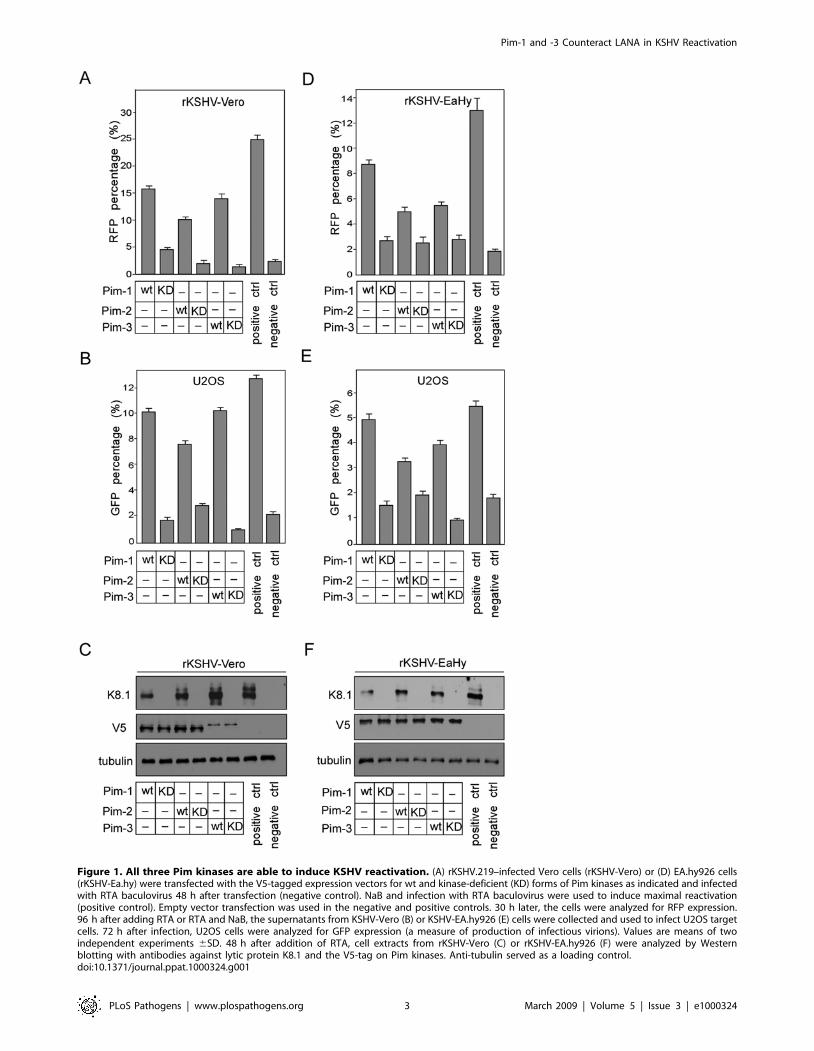
Figure 1. All three Pim kinases are able to induce KSHV reactivation. (A) rKSHV.219–infected Vero cells (rKSHV-Vero) or (D) EA.hy926 cells(rKSHV-Ea.hy) were transfected with the V5-tagged expression vectors for wt and kinase-deficient (KD) forms of Pim kinases as indicated and infectedwith RTA baculovirus 48 h after transfection (negative control). NaB and infection with RTA baculovirus were used to induce maximal reactivation(positive control). Empty vector transfection was used in the negative and positive controls. 30 h later, the cells were analyzed for RFP expression.96 h after adding RTA or RTA and NaB, the supernatants from KSHV-Vero (B) or KSHV-EA.hy926 (E) cells were collected and used to infect U2OS targetcells. 72 h after infection, U2OS cells were analyzed for GFP expression (a measure of production of infectious virions). Values are means of twoindependent experiments 6SD. 48 h after addition of RTA, cell extracts from rKSHV-Vero (C) or rKSHV-EA.hy926 (F) were analyzed by Westernblotting with antibodies against lytic protein K8.1 and the V5-tag on Pim kinases. Anti-tubulin served as a loading control.doi:10.1371/journal.ppat.1000324.g001
Pim-1 and -3 Counteract LANA in KSHV Reactivation
PLoS Pathogens | www.plospathogens.org 3 March 2009 | Volume 5 | Issue 3 | e1000324
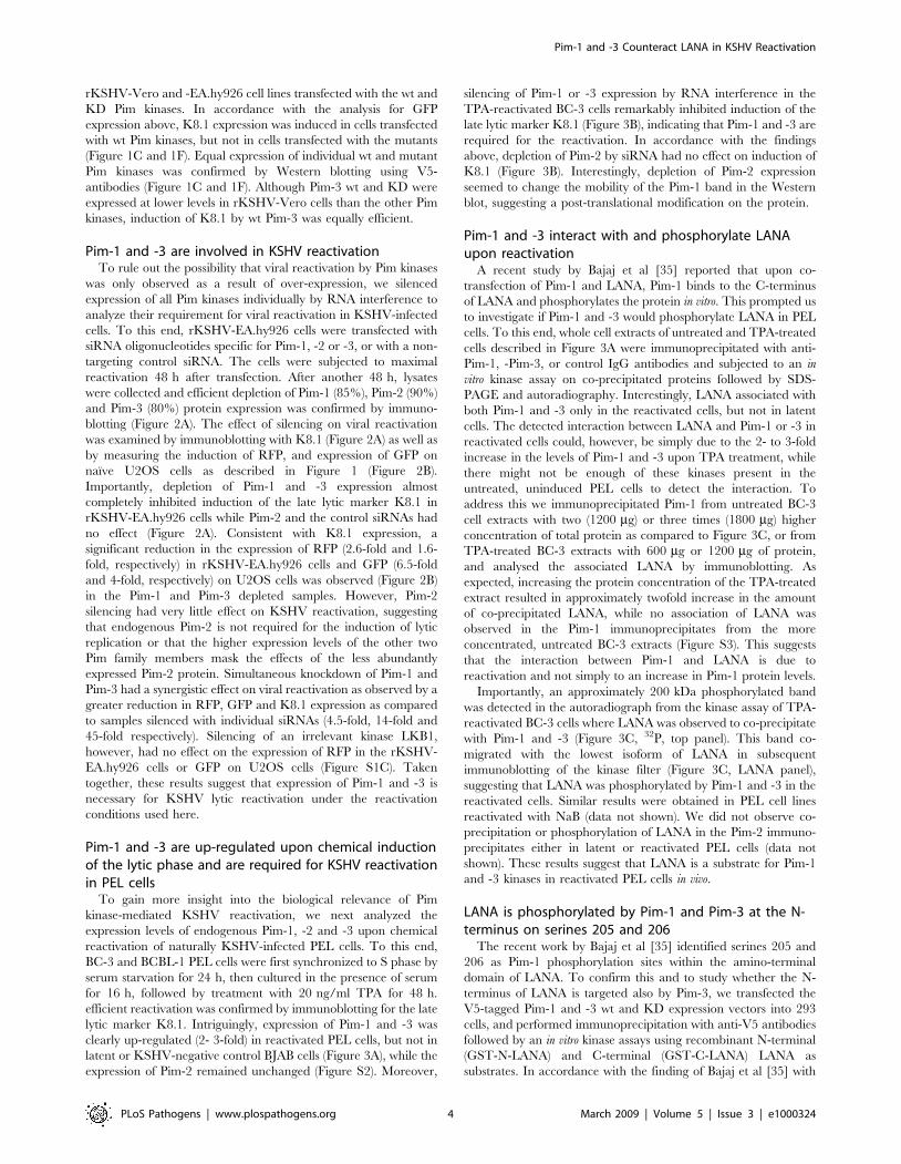
rKSHV-Vero and -EA.hy926 cell lines transfected with the wt and
KD Pim kinases. In accordance with the analysis for GFP
expression above, K8.1 expression was induced in cells transfected
with wt Pim kinases, but not in cells transfected with the mutants
(Figure 1C and 1F). Equal expression of individual wt and mutant
Pim kinases was confirmed by Western blotting using V5-
antibodies (Figure 1C and 1F). Although Pim-3 wt and KD were
expressed at lower levels in rKSHV-Vero cells than the other Pim
kinases, induction of K8.1 by wt Pim-3 was equally efficient.
Pim-1 and -3 are involved in KSHV reactivationTo rule out the possibility that viral reactivation by Pim kinases
was only observed as a result of over-expression, we silenced
expression of all Pim kinases individually by RNA interference to
analyze their requirement for viral reactivation in KSHV-infected
cells. To this end, rKSHV-EA.hy926 cells were transfected with
siRNA oligonucleotides specific for Pim-1, -2 or -3, or with a non-
targeting control siRNA. The cells were subjected to maximal
reactivation 48 h after transfection. After another 48 h, lysates
were collected and efficient depletion of Pim-1 (85%), Pim-2 (90%)
and Pim-3 (80%) protein expression was confirmed by immuno-
blotting (Figure 2A). The effect of silencing on viral reactivation
was examined by immunoblotting with K8.1 (Figure 2A) as well as
by measuring the induction of RFP, and expression of GFP on
naıve U2OS cells as described in Figure 1 (Figure 2B).
Importantly, depletion of Pim-1 and -3 expression almost
completely inhibited induction of the late lytic marker K8.1 in
rKSHV-EA.hy926 cells while Pim-2 and the control siRNAs had
no effect (Figure 2A). Consistent with K8.1 expression, a
significant reduction in the expression of RFP (2.6-fold and 1.6-
fold, respectively) in rKSHV-EA.hy926 cells and GFP (6.5-fold
and 4-fold, respectively) on U2OS cells was observed (Figure 2B)
in the Pim-1 and Pim-3 depleted samples. However, Pim-2
silencing had very little effect on KSHV reactivation, suggesting
that endogenous Pim-2 is not required for the induction of lytic
replication or that the higher expression levels of the other two
Pim family members mask the effects of the less abundantly
expressed Pim-2 protein. Simultaneous knockdown of Pim-1 and
Pim-3 had a synergistic effect on viral reactivation as observed by a
greater reduction in RFP, GFP and K8.1 expression as compared
to samples silenced with individual siRNAs (4.5-fold, 14-fold and
45-fold respectively). Silencing of an irrelevant kinase LKB1,
however, had no effect on the expression of RFP in the rKSHV-
EA.hy926 cells or GFP on U2OS cells (Figure S1C). Taken
together, these results suggest that expression of Pim-1 and -3 is
necessary for KSHV lytic reactivation under the reactivation
conditions used here.
Pim-1 and -3 are up-regulated upon chemical inductionof the lytic phase and are required for KSHV reactivationin PEL cells
To gain more insight into the biological relevance of Pim
kinase-mediated KSHV reactivation, we next analyzed the
expression levels of endogenous Pim-1, -2 and -3 upon chemical
reactivation of naturally KSHV-infected PEL cells. To this end,
BC-3 and BCBL-1 PEL cells were first synchronized to S phase by
serum starvation for 24 h, then cultured in the presence of serum
for 16 h, followed by treatment with 20 ng/ml TPA for 48 h.
efficient reactivation was confirmed by immunoblotting for the late
lytic marker K8.1. Intriguingly, expression of Pim-1 and -3 was
clearly up-regulated (2- 3-fold) in reactivated PEL cells, but not in
latent or KSHV-negative control BJAB cells (Figure 3A), while the
expression of Pim-2 remained unchanged (Figure S2). Moreover,
silencing of Pim-1 or -3 expression by RNA interference in the
TPA-reactivated BC-3 cells remarkably inhibited induction of the
late lytic marker K8.1 (Figure 3B), indicating that Pim-1 and -3 are
required for the reactivation. In accordance with the findings
above, depletion of Pim-2 by siRNA had no effect on induction of
K8.1 (Figure 3B). Interestingly, depletion of Pim-2 expression
seemed to change the mobility of the Pim-1 band in the Western
blot, suggesting a post-translational modification on the protein.
Pim-1 and -3 interact with and phosphorylate LANAupon reactivation
A recent study by Bajaj et al [35] reported that upon co-
transfection of Pim-1 and LANA, Pim-1 binds to the C-terminus
of LANA and phosphorylates the protein in vitro. This prompted us
to investigate if Pim-1 and -3 would phosphorylate LANA in PEL
cells. To this end, whole cell extracts of untreated and TPA-treated
cells described in Figure 3A were immunoprecipitated with anti-
Pim-1, -Pim-3, or control IgG antibodies and subjected to an in
vitro kinase assay on co-precipitated proteins followed by SDS-
PAGE and autoradiography. Interestingly, LANA associated with
both Pim-1 and -3 only in the reactivated cells, but not in latent
cells. The detected interaction between LANA and Pim-1 or -3 in
reactivated cells could, however, be simply due to the 2- to 3-fold
increase in the levels of Pim-1 and -3 upon TPA treatment, while
there might not be enough of these kinases present in the
untreated, uninduced PEL cells to detect the interaction. To
address this we immunoprecipitated Pim-1 from untreated BC-3
cell extracts with two (1200 mg) or three times (1800 mg) higher
concentration of total protein as compared to Figure 3C, or from
TPA-treated BC-3 extracts with 600 mg or 1200 mg of protein,
and analysed the associated LANA by immunoblotting. As
expected, increasing the protein concentration of the TPA-treated
extract resulted in approximately twofold increase in the amount
of co-precipitated LANA, while no association of LANA was
observed in the Pim-1 immunoprecipitates from the more
concentrated, untreated BC-3 extracts (Figure S3). This suggests
that the interaction between Pim-1 and LANA is due to
reactivation and not simply to an increase in Pim-1 protein levels.
Importantly, an approximately 200 kDa phosphorylated band
was detected in the autoradiograph from the kinase assay of TPA-
reactivated BC-3 cells where LANA was observed to co-precipitate
with Pim-1 and -3 (Figure 3C, 32P, top panel). This band co-
migrated with the lowest isoform of LANA in subsequent
immunoblotting of the kinase filter (Figure 3C, LANA panel),
suggesting that LANA was phosphorylated by Pim-1 and -3 in the
reactivated cells. Similar results were obtained in PEL cell lines
reactivated with NaB (data not shown). We did not observe co-
precipitation or phosphorylation of LANA in the Pim-2 immuno-
precipitates either in latent or reactivated PEL cells (data not
shown). These results suggest that LANA is a substrate for Pim-1
and -3 kinases in reactivated PEL cells in vivo.
LANA is phosphorylated by Pim-1 and Pim-3 at the N-terminus on serines 205 and 206
The recent work by Bajaj et al [35] identified serines 205 and
206 as Pim-1 phosphorylation sites within the amino-terminal
domain of LANA. To confirm this and to study whether the N-
terminus of LANA is targeted also by Pim-3, we transfected the
V5-tagged Pim-1 and -3 wt and KD expression vectors into 293
cells, and performed immunoprecipitation with anti-V5 antibodies
followed by an in vitro kinase assays using recombinant N-terminal
(GST-N-LANA) and C-terminal (GST-C-LANA) LANA as
substrates. In accordance with the finding of Bajaj et al [35] with
Pim-1 and -3 Counteract LANA in KSHV Reactivation
PLoS Pathogens | www.plospathogens.org 4 March 2009 | Volume 5 | Issue 3 | e1000324

Pim-1, both Pim-1 and -3 phosphorylated GST-N-LANA while no
phosphorylation was observed on GST-C-LANA or with Pim-
1KD or Pim-3KD (Figure 4A). To further map the site of Pim-1
and -3 phosphorylation on the LANA N-terminus, we prepared
truncated versions of the GST-N-LANA as described in the
Materials and methods, and used them as substrates in the in vitro
kinase assay as described above. The results demonstrated that the
critical residues for phosphorylation by Pim-1 and -3 were located
between the amino acids 200 and 340 (Figure 4B). To define the
specific phosphorylation sites on LANA, we prepared a site-
specific LANA phosphomutant identical to the one described
earlier by Bajaj et al [35] where the two critical serines at positions
205 and 206 were mutated into arginines (SS205/206RR). The wt
and two different clones of the SS205/206RR mutant of LANA
(SS205/206RR (1) and SS205/206RR (2)) were then transfected
in the presence or absence of V5-tagged Pim-1 or -3, and cell
extracts were subjected to immunoprecipitation with anti-V5
antibodies. When in vitro kinase assays were performed on the co-
precipitated LANA proteins, we observed that wt LANA was
phosphorylated by both Pim-1 and -3, while the SS205/206RR
mutant of LANA failed to be phosphorylated by neither of them
(Figure 4C). Neither of the LANA proteins was phosphorylated in
the absence of transfected Pim kinases. These data suggest that
serines 205 and 206 on the LANA N-terminus are specifically
phosphorylated by Pim-1 and Pim-3.
To provide additional evidence that the 200-kDa band co-
precipitated and phosphorylated by Pim-1 from TPA-induced BC-
3 cells (Figure 3C) is indeed LANA we performed a competition
experiment where GST-N-LANA and/or GST-C-LANA were
added as competing substrates into the cell extract prior to Pim-1
immunoprecipitation from BC-3 cells that had been treated with
TPA for 48 h. The immunoprecipitates were then subjected to an
in vitro kinase assay on co-precipitated proteins. Purified GST was
used as a negative control. As shown in Figure 4D, the
phosphorylation signal on the 200- kDa band was dramatically
reduced in samples containing GST-N-LANA while no effect was
observed with GST-C-LANA or GST alone. This suggests that the
phosphorylated 200-kDa band is indeed LANA. To obtain further
support that the kinase responsible for the phosphorylation of
LANA is Pim-1, instead of another kinase co-precipitating with
Pim-1, we performed an in vitro kinase assay with purified wt and
KD Pim-1 proteins and used GST-N-LANA, GST-C-LANA or
GST as substrates. Phosphorylation was detected on GST-N-
LANA, but not on GST-C-LANA or GST, only in the presence of
the purified wt Pim-1 kinase (Figure 4E), demonstrating direct
phosphorylation of the LANA N-terminus by Pim-1. Phosphor-
ylation of GST-N-LANA on both Ser 205 and 206 was further
confirmed by mass spectrometry (data not shown). These data
confirm that serines 205 and 206 on the LANA N-terminus are the
specific residues phosphorylated by Pim-1.
Phosphorylation of LANA by Pim-1 and -3 counteractsthe ability of LANA to inhibit transcription from theterminal repeat region
A previous study has reported that the C-terminus of LANA
binds specifically to sequences within the terminal repeat (TR)
Figure 2. Pim-1 and -3 are required for KSHV reactivation. (A)rKSHV-EA.hy926 cells (rKSHV-Ea.hy) were transfected with siRNAoligonucleotides specific for Pim-1, -2, or -3, or with control siRNA (sictrl), and subjected to maximal reactivation (RTA+NaB) 48 h aftertransfection. After another 48 h, whole cell extracts were collected andanalyzed by Western blotting with anti-Pim-1, -2, -3, and -K8.1antibodies. Quantification of K8.1 protein level is shown below thepanel. Anti-tubulin served as a loading control. (B) Cells were treated asin (A), and 30 h after maximal reactivation, the cells were analyzed for
RFP expression. 72 h after reactivation, supernatants were collected andused to infect naive U2OS cells. (C) 72 h after infection, the U2OS cellswere analyzed for GFP expression (a measure of production ofinfectious virions). Values are means of two independent experiments6SD.doi:10.1371/journal.ppat.1000324.g002
Pim-1 and -3 Counteract LANA in KSHV Reactivation
PLoS Pathogens | www.plospathogens.org 5 March 2009 | Volume 5 | Issue 3 | e1000324

regions of the KSHV genome [36]. Furthermore, by using a
reporter construct consisting of multimerized TR repeats linked to
a heterologous promoter for luciferase gene expression (pGL3-
7xTR), this and other studies [36] showed that transcription was
suppressed up to 10-fold in the presence of LANA. We next
addressed the effect of Pim-1 or -3 over-expression on the LANA-
mediated repression of the pGL3-7xTR reporter. To this end,
increasing amounts of Pim-1 or -3 were co-transfected with LANA
and pGL3-7xTR, and the cells were subjected to luciferase
reporter assays 48 h after transfection. Equal expression of LANA
was confirmed by immunoblotting (data not shown). Ectopic
expression of increasing amounts of Pim-1 or -3 counteracted
LANA-mediated transcriptional repression of the TR-containing
reporters (Figure 5A). Expression of Pim-1 or -3 did not influence
transcription from TR-containing reporters in the absence of
LANA, suggesting that Pim-1 or -3 does not directly enhance
transcription from the reporter.
To test the requirement of kinase activity for the de-repression,
we transfected increasing amounts of Pim-1KD and -3KD together
with constant levels of LANA, wt Pim-1 and -3, as well as pGL3-
7xTR. The samples were subjected to the luciferase reporter assays
48 h later. Expression of increasing amounts of Pim-1KD and
-3KD neutralized the effect of Pim-1 and Pim-3 on LANA-
mediated repression and resulted in a dose-dependent suppression
of transcription from the TR-containing reporters (Figure 5B). To
address whether phosphorylation of LANA at SS205/206 was
necessary for the de-repression, we co-transfected cells with pGL3-
7xTR, wt or SS205/206RR mutant of LANA (SS205/206RR) with
Figure 3. Pim-1 and -3 interact with and phosphorylate LANA upon reactivation of PEL cells. (A) BCBL-1, BC-3, as well as a KSHV–negativecontrol cell line BJAB, were treated with TPA (+) or solvent (DMSO; 2) for 48 h. Whole cell extracts were immunoblotted with anti-Pim-1, -3, and -K8.1antibodies. Anti-tubulin served as a loading control. (B) BC-3 cells were transfected with siRNAs specific for Pim-1, -2, -3, or control siRNA (si ctrl), andtreated with TPA 48 h after transfection. After another 48 h, whole cell extracts were collected and analyzed by Western blotting with anti-Pim-1, -2,-3, and -K8.1 antibodies. Anti-tubulin served as a loading control. (C) Whole cell extracts from (A) were immunoprecipitated with anti-Pim-1 and -3 orcontrol IgG antibodies and subjected to an in vitro kinase assay towards co-precipitated proteins. The samples were resolved by SDS-PAGE (8%)followed by autoradiography. The kinase filter was immunoblotted with anti-LANA, -Pim-1, and -3 antibodies.doi:10.1371/journal.ppat.1000324.g003
Pim-1 and -3 Counteract LANA in KSHV Reactivation
PLoS Pathogens | www.plospathogens.org 6 March 2009 | Volume 5 | Issue 3 | e1000324

Figure 4. LANA is phosphorylated by Pim-1 and Pim-3 at the N-terminus on serines 205 and 206. (A) 293 cells were transfected with V5-tagged expression constructs for either wt or kinase-deficient (KD) Pim-1 or -3. After 48 h, cell extracts were immunoprecipitated by anti-V5antibodies and subjected for in vitro kinase assay on GST-C-LANA or GST-N-LANA. The samples were resolved by SDS-PAGE (8%), and followed byautoradiography. The kinase filter was blotted with anti-GST antibody and anti-V5 antibodies. (B) The truncated fragments of the LANA N-terminusare illustrated in the upper part of the panel. 293 cells were transfected with V5-tagged expression constructs for either Pim-1 or -3. After 48 h, the invitro kinase assay was performed essentially as described in (A) on GST-fusions of N-terminal truncations of LANA as indicated. Coomassie stainingwas used to detect the substrates. (C) 293 cells were transfected with wt (LANA wt) or two different clones of the SS205/206RR mutant of LANA(SS205/206RR (1), and SS205/206RR (2)) with and without V5-tagged expression constructs for Pim-1 or -3 as indicated. After 48 h, cell extracts were
Pim-1 and -3 Counteract LANA in KSHV Reactivation
PLoS Pathogens | www.plospathogens.org 7 March 2009 | Volume 5 | Issue 3 | e1000324

and without Pim-1 or Pim-3. The samples were subjected to
luciferase reporter assays 48 h after transfection. Expression of the
LANA phosphosite mutant without the Pim kinases induced similar
repression of luciferase activity as the wt LANA, but co-expressed
Pim-1 and -3 were unable to relieve this repression (Figure 5C).
These results indicate that phosphorylation of LANA at serines 205
and 206 by Pim-1 or -3 is needed to counteract LANA-mediated
suppression of transcription from the TR.
Pim-1 and -3 counteract the ability of LANA to inhibitRTA autoactivation
Induction of K8.1 upon Pim-1 and -3 over-expression or in
response to TPA-induced reactivation suggests that RTA responsive
lytic genes are also activated by these kinases in KSHV-infected
cells. Since RTA is not located in the proximity of the TR region in
the KSHV genome we wanted to investigate whether co-expression
of Pim-1 or -3 could affect the ability of LANA to inhibit
transcription from the RTA promoter. Previous studies have
demonstrated that RTA can auto-activate its own viral promoter
[37], and this was shown to be partially repressed by LANA [38].
We therefore addressed the ability of LANA to repress transcription
of luciferase from a RTA-Luc reporter (pGL2-ORF50p) upon co-
expression of RTA, wt Pim-1 or -3. In accordance with Lan et al
[38] expression of LANA led to a partial repression of the RTA-Luc
reporter activity. Moreover, Pim-1 and -3 wt were both able to de-
repress the RTA-Luc activity (Figure 5D). Equal expression of
LANA was confirmed by immunoblotting (data not shown). To
address whether phosphorylation of LANA at SS205/206 was
necessary for the de-repression, we co-transfected cells with pGL2-
ORF50p, wt or SS205/206RR mutant of LANA (SS205/206RR)
with and without Pim-1 or Pim-3. Expression of the LANA
phosphosite mutant without the Pim kinases was able to repress the
RTA-luciferase activity but to a lesser extent as the wt LANA. Yet,
co-expressed Pim-1 and -3 were unable to relieve this repression
(Figure 5D). Taken together, this suggests that expression of Pim-1
and -3 kinases can antagonize the ability of LANA to inhibit
autoactivation of the RTA promoter.
IFN-gamma increases Pim-1 levels and inducesreactivation in PEL cells
To further assess the role of Pim kinases in KSHV reactivation
upon physiological instead of chemical induction, we next analyzed
the expression levels of endogenous Pim-1 in PEL cells upon
cytokine stimulation. To this end, BC-3 cells were first synchronized
as described in Figure 3 and treated with increasing amounts of
interferon-c (IFN-c) for 3 or 16 h. Expression of Pim-1 was
moderately up-regulated in BC-3 cells already 3 h after IFN-ctreatment (Figure 6A). In addition, a slower migrating form of Pim-1
appeared in samples treated with 5–10 IU/ml or 1–10 IU/ml for
3 h or 16 h, respectively, suggesting a post-translational modification
on the protein. Next we examined the effects of IFN-c on the
expression of KSHV lytic genes by quantitative real-time PCR
(qRT-PCR). At 16 h IFN-c treatment with 0.1–10 IU/ml induced
the expression of ORF50/RTA (immediate-early transcript) up to
16.5-fold and ORF57 (delayed-early transcript) up to 19-fold in a
dose-dependent manner (Figure 6D), implying that cytokine
treatment lead to expression of viral lytic genes. We also addressed
if Pim-1 phosphorylated LANA in IFN-c stimulated PEL cells. To
this end, whole cell extracts of untreated, IFN-c- or TPA-treated BC-
3 (16 h stimulation) or control BJAB cells were immunoprecipitated
with anti-Pim-1 antibodies and subjected to in vitro kinase assays on
co-precipitated proteins followed by SDS-PAGE and autoradiogra-
phy. In accordance with the induction of lytic gene expression, an
approximately 200 kDa band co-migrating with LANA in subse-
quent immunoblotting was detected in the autoradiograph of Pim-1
immunoprecipitates after treatment of BC-3 cells with either IFN-cor TPA (Figure 6B, 32P, top panel), further supporting the
physiological role for Pim-1 in KSHV reactivation.
To study the requirement of Pim-1 for the IFN-c induced
KSHV reactivation, we silenced Pim-1 expression by RNA
interference in BC-3 cells as described for Figure 3, and confirmed
the depletion by Western blotting (Figure 6C). 48 h after the cells
were treated with 1 or 5 U/ml of IFN-c for 16 h, and viral
reactivation was analysed by qRT-PCR for the expression of lytic
genes ORF50 and ORF57. Silencing of Pim-1 expression led to a
significant decrease in the expression of lytic genes (66% and 77%
reduction for ORF50, and 38% and 81% reduction for ORF57
with 1 and 5 U/ml IFN-c treatment, respectively) as compared to
cells treated with the control siRNA (Figure 6D and not shown).
Taken together, this data suggests that IFN-c-induced KSHV
reactivation occurs via activation of Pim-1.
Discussion
In this report, we have identified two cellular kinases, Pim-1 and
Pim-3, as critical regulators of KSHV viral reactivation. During
reactivation, Pim-1 and -3 interact with and phosphorylate LANA
in both de novo and naturally KSHV-infected cells. LANA can
repress expression of a subset of lytic genes by specifically binding
to the LANA binding sites 1 and 2 (LBS1 and LBS2) within the
TR region of the KSHV genome [39,40]. Here we demonstrate
that phosphorylation by Pim-1 and -3 counteracts this LANA-
mediated inhibition of transcription from the TR region. Besides
inhibiting the TR-enhancer dependent transcription LANA can
down-modulate the transcriptional activity of the RTA gene
promoter as well as the ability of RTA to autoactivate its own
promoter [38]. Our data shows that co-expression of Pim-1 and -3
with LANA was able to de-repress the LANA-induced partial
inhibition of autoactivation of the RTA-promoter (Figure 5).
However, this Pim kinase-mediated de-repression or the inhibitory
effect of LANA phosphosite mutant was not as dramatic as those
seen in the TR-enhancer dependent transcription assay. This
variation could be due to different mechanisms behind the
repression. While the LANA-induced inhibition of transcription
from the TR region depends on direct binding of LANA to DNA
at LBS1 and LBS2 [39,40], the suppression of RTA autoactivation
occurs through direct binding of LANA to RTA [38]. Taken
collected and subjected to an in vitro kinase assay essentially as described in (A) on co-precipitated proteins. The samples were resolved by SDS-PAGE(8%) followed by autoradiography. The kinase filter was immunoblotted with anti–LANA, and anti-V5 antibodies to ensure the efficiency ofimmunoprecipitation. An arrow indicates phosphorylated LANA band on the right. (D) BC-3 cells were treated with TPA for 48 h. Whole cell extractswere incubated with GST-N-LANA and GST-C-LANA alone or in combination for 6 hrs and subjected to immunoprecipitation with anti-Pim-1antibody, followed by in vitro kinase assay on co-precipitated proteins. GST alone (GST) was used as a negative control. The kinase filter wasimmunoblotted with anti–LANA, –GST, and –Pim-1 antibodies. The arrows indicate phosphorylated LANA and GST-N-LANA on the right. (E)Recombinant GST-fusion proteins (GST, GST-N-LANA, or GST-C-LANA) were incubated with purified wild-type (wt) or kinase-dead (KD) Pim-1 proteinsand subjected to an in vitro kinase assay. Samples were resolved by SDS-PAGE and autoradiographed. The substrates indicated on the right werevisualized by Coomassie staining.doi:10.1371/journal.ppat.1000324.g004
Pim-1 and -3 Counteract LANA in KSHV Reactivation
PLoS Pathogens | www.plospathogens.org 8 March 2009 | Volume 5 | Issue 3 | e1000324

together, our results suggest that phosphorylation of LANA by Pim
kinases can modulate its ability to interact with the enhancer
elements within the TR region and with the RTA protein itself,
and thereby lead to viral reactivation (Figure 7).
Our KSHV-infected cell models manifest a very low level of
spontaneous lytic replication. However, upon stimulation by low
level of exogenous RTA (using a low-titer BacK50) and co-
expression of Pim kinases, a relatively large population of cells
undergoes reactivation. There are several possible reasons to
explain why priming with exogenous RTA is needed to induce the
Pim kinase-mediated lytic replication. One explanation is that as
the latency program in these infected cell models is very tight, a
threshold level of RTA might be required for the activation of viral
lytic cascade by Pim. Alternatively, Pim kinases may also have an
effect on RTA itself.
Bajaj et al [35] recently demonstrated that Pim-1 phosphory-
lates LANA at either one or both of the serines at positions 205
and 206. We identified Pim-3 as a novel kinase phosphorylating
Figure 5. Phosphorylation of LANA by Pim-1 and -3 antagonizes LANA–mediated transcription inhibition. (A) The reporter plasmidpGL3-7XTR (0.1 mg) was co-transfected with expression vectors for LANA (0.5 mg) with and without increasing concentrations of V5-tagged vectorsfor Pim-1 or -3 (indicated below the graph) into 293 cells. The total transfected DNA concentrations (mg) were kept constant by including an emptycontrol vector. 48 h after transfection, cell extracts were collected for the luciferase assay. (B) Increasing amounts of V5-tagged Pim-1KD or -3KDexpression vectors were co-transfected with constant amount of pGL3-7XTR (0.1 mg), LANA (0.5 mg), and V5-Pim-1 or -3 (0.5 mg each) wt vectors into293 cells. The DNA concentrations (mg) for Pim-1KD and -3KD cDNAs used are indicated below the graph, while the total DNA concentrations (mg)were kept constant by including an empty control vector. Cell extracts were analyzed as in (A). (C) The pGL3-7XTR (0.1 mg), V5-tagged Pim-1 or -3(0.5 mg each) wt vectors were co-transfected with 0.5 mg of wt LANA or SS205/206RR mutant of LANA into 293 cells. At 48 h after transfection, dualluciferase assays were performed with extracts of transfected cells. An empty pcDNA3 was used as a filler plasmid to keep the total DNA constant ineach transfection. Values represent an average of two independent experiments 6SD. (D) The reporter plasmid pGL2-ORF50p (0.1 mg), as well asexpression vectors for RTA (0.5 mg) and wt LANA or SS205/206RR mutant of LANA (0.5 mg) were co-transfected with V5-tagged cDNAs for Pim-1 or -3(indicated below the graph) into 293 cells. The transfected DNA concentrations (mg) were normalized by including an empty control plasmid. 48 hafter transfection, cell extracts were collected for the luciferase assay. Values represent an average of two independent experiments 6SD.doi:10.1371/journal.ppat.1000324.g005
Pim-1 and -3 Counteract LANA in KSHV Reactivation
PLoS Pathogens | www.plospathogens.org 9 March 2009 | Volume 5 | Issue 3 | e1000324

LANA in naturally infected PEL cells, and demonstrated that the
phosphorylation takes place only upon viral reactivation. Our data
confirms Ser 205/206 as the target sites for Pim-1 and
demonstrate that these sites are phosphorylated also by Pim-3.
Pim kinases are involved in partially overlapping survival pathways
and are implicated in a wide variety of human diseases including
cancer, inflammatory disorders, and ischemic diseases [26,41].
Moreover, they have previously been linked to regulation of viral
oncogenesis [31,32,35]. Our data extend these observations by
suggesting a novel link between Pim kinases and KSHV, and
establishes Pim-1 and -3 as key factors in KSHV reactivation.
Our findings provide compelling evidence that Pim-1 and -3, but
not Pim-2, are essential for KSHV reactivation both in de novo
infected endothelial cells and in the naturally infected PEL cells.
Figure 6. IFNc induces reactivation via Pim-1. BC-3 cells were first synchronized to S (Pim-1 si)-phase and treated with increasing amount of IFN-cfor 3 or 16 h. TPA treatment was used as a positive control. (A) Cell extracts were analyzed for the expression of Pim-1 by Western blot, and (D) forexpression of ORF50 and ORF57 lytic transcripts by qRT-PCR. (B) BC-3 and BJAB cells were treated with 1 or 5 U/ml of IFN-c for 16 h, and whole cellextracts were immunoprecipitated with anti-Pim-1 antibodies and subjected to an in vitro kinase assay on co-precipitated proteins. The samples wereresolved by SDS-PAGE (8%) followed by autoradiography. The kinase filter was immunoblotted with anti–LANA and anti–Pim-1 antibodies. Anti-tubulinserved as a loading control. (C) BC-3 cells were transfected with siRNAs specific for Pim-1 (Pim-1 si) or control siRNA (ctrl si) or left untreated (2). 48 hafter transfection, cells were treated for 16 h with IFN-c as described above. Cell extracts were analyzed for the silencing of Pim-1 expression by Westernblot with anti-Pim-1 antibodies, and (D) for expression of ORF50 and ORF57 lytic transcripts by qRT-PCR. Anti-tubulin served as a loading control in C.doi:10.1371/journal.ppat.1000324.g006
Figure 7. A model for Pim-1 and Pim-3 function in KSHV reactivation. In latency, binding of LANA to the LANA binding sites 1 and 2 (LBS 1and LBS2) at the terminal repeat (TR) region represses transcription from the TR region. LANA can also repress transcription from the RTA promoterand inhibit RTA to autoactivate its own promoter. Upon stimulation by cytokines, TPA or NaB, Pim-1 and -3 are up-regulated, and they interact withand phosphorylate LANA. The phosphorylation by Pim-1 and -3 counteracts the LANA–mediated inhibition of transcription from the TR region anddown-modulates autoactivation of the RTA-promoter, leading to viral reactivation.doi:10.1371/journal.ppat.1000324.g007
Pim-1 and -3 Counteract LANA in KSHV Reactivation
PLoS Pathogens | www.plospathogens.org 10 March 2009 | Volume 5 | Issue 3 | e1000324
Interestingly, we observe a surprising additive effect of Pim-1 and
Pim-3 as depletion of one of them results in almost equal level of
inhibition of reactivation. One possible explanation could be that
the KSHV-infected cells require a threshold level of active Pim
kinases to reactivate, and that endogenous levels of Pim-2 would be
below the limit, and thus its depletion does not affect reactivation.
When expression of Pim-1 or -3 is efficiently silenced, the total Pim
kinase activity may fall below the required threshold, leading to
inhibition of reactivation upon depletion of either of them.
A number of inflammatory and angiogenic cytokines including
IL-6, tumor necrosis factor-a, IFN-c, platelet-derived growth
factor, oncostatin M and interleukin-1b are expressed in KS
tumors [42]. Some of these cytokines have been suggested to
modulate KSHV reactivation [43]. Intriguingly, expression of
Pim-1 is induced by a number of cytokines, such as IFN-a, IFN-c,
IL-3, and IL-6 [26]. It is therefore plausible that the presence of a
variety of cytokines in the microenvironment of KSHV-associated
tumors may induce Pim kinase expression and/or activation and
lead to viral reactivation in affected tissues. Our data demonstrat-
ing involvement of Pim-1 in IFN-c treated PEL cells on activation
of ORF50 and ORF57 lytic gene transcription supports a
physiological role of Pim-1 in KSHV reactivation.
The switch from latency to lytic replication is an elementary
decision in the viral life cycle and several lines of evidence support
the importance of viral lytic replication in herpesviral tumorigen-
esis [7,8]. A sustained, although low level of reactivation can
contribute to progression of KS by not only producing new virions
but also by contributing to other aspects of KS pathogenesis, such
as the production of inflammatory and angiogenic cytokines.
Hence, cellular signaling pathways operative during viral reacti-
vation could represent potential novel targets for therapeutic
intervention. Accordingly, inhibitors specific for either Pim-1 or -3
or a broad spectrum Pim inhibitor could be developed as antiviral
agents, as inhibition of either Pim-1 or -3 results in a significant
decrease in reactivation. These inhibitors could also be tested for
their anti-tumor activity in PEL mouse models to explore a novel
therapeutic strategy to treat KSHV-malignancies. As animals
lacking all three Pim genes are viable with a mild phenotype [44],
one could anticipate that small-molecule inhibitors targeting Pim-1
and -3 might be tolerated without significant side effects.
Materials and Methods
CellsVero cells latently infected with a recombinant GFP-expressing
KSHV (rKSHV.219) were a kind gift from J. Vieira (Univ. of
Washington). EA.hy926 cells infected with rKSHV.219 were
prepared as described in [45]. The BC-3 and BCBL-1 cell lines
were kindly provided by E. Cesarman (Cornell Medical College)
and and the BJAB B-lymphoblastoid cell line was a gift from J.
Salonen (Univ. of Tampere). BC-3, BCBL-1 and BJAB cells were
cultured in a humidified 5% CO2 atmosphere at 37uC in RPMI
1640 medium supplemented with 10% fetal calf serum (FCS;
Invitrogen, CA), 100 U/ml penicillin G, and 100 mg/ml strepto-
mycin. 293 and U2OS human osteosarcoma cell lines (ATCC;
Manassas, VA), rKSHV-Vero, and -EA.hy926 cells were cultured
similarly, but with Dulbecco modified Eagle’s medium (DMEM). In
addition, 5 ug/ml or 1 ug/ml of puromycin was added to the
medium for rKSHV-Vero or rKSHV-EA.hy926 cells, respectively.
PlasmidsWild-type and kinase-deficient (KD) versions of human Pim-1,
Pim-2 and Pim-3 expression vectors were generated by PCR
amplification and cloning into the pcDNA3.1/V5-HisC vector
[25]. The GST-tagged Pim-1 and Pim-1KD constructs were
generated by PCR subcloning into the pGEX-6P-1 vector (GE
Healthcare). pGEX-2T plasmid (GE Healthcare) was used to
produce GST in control samples. The serine to arginine (SS205/
206RR) mutations on LANA were generated using pCDNA3.1-
LANA [46] as a template by site-directed mutagenesis. The GST-
tagged carboxy-terminal LANA (GST-C-LANA; amino acids (aa)
972-1162) and amino-terminal LANA (GST-N-LANA; aa 1–340
(N-LANA) [47] were expressed from the pGEX4T1-LANA
construct. The full length and truncated versions of the N-
terminal LANA (aa 1–340 (N), aa 100–340 (N6), aa 24–100 (N11),
and aa 75–200 (N17)) were generated by PCR amplification from
the pCDNA3-LANA and fused to GST expression cassette by
ligation to pGEX-4T-1 (GE Healthcare). The luciferase reporter
construct in the pGL2-ORF50p backbone containing the RTA
promoter (3 kb sequence upstream of the RTA translation
initiation codon) was prepared as described before [37] and the
luciferase reporter containing seven terminal repeats (TR) of
KSHV genome, pGL3-7xTR, was a kind gift from Dr. Rolf
Renne (University of Florida) [36].
Viral reactivation assayViral reactivation was addressed in Vero and EA.hy926 cells
stably infected with recombinant KSHV (rKSHV.219) [33]. The
rKSHV.219 is a double-reporter virus, which expresses the green
fluorescent protein (GFP) from a constitutively active EF-1-
promoter, and can be induced to express the red fluorescent
protein (RFP) from the RTA-responsive KSHV lytic promoter for
polyadenylated nuclear RNA (PAN) [34]. To address the effect of
Pim kinases or their mutants on reactivation, rKSHV.219-Vero
and -EA.hy926 cells were seeded on 6- or 24-well dishes and
transiently transfected with the empty vector pcDNA3.1 or vectors
expressing wt or kinase-dead Pim family members, as described
previously [25]. In brief, 26105 rKSHV.219-Vero and -EA.hy926
cells per well were plated on a 6-well plate. On the following day,
1 mg aliquots of DNA samples were mixed with serum-free
medium containing 3 ml FuGENE HD (Roche, Nutley, NJ)
according to the manufacturer’s protocol. One day after
transfection, the media was changed into fresh DMEM media.
To monitor transfection efficiency, pcDNA/DsRed plasmid was
transfected into duplicate wells and the number of dsRed-positive
cells was determined by fluorescence microscopy. The typical
efficiency of transfection was between 15–20%. After 48 h, cells
were treated for 2 h with a low-titer baculovirus (BacK50)
expressing the KSHV lytic activator ORF 50/RTA (a kind gift
from Dr. J. Vieira; Univ. of Washington). This triggered basal
reactivation but led to a low level of RFP induction (2.5% in
rKSHV-Vero, and 1.9% in rKSHV- EA.hy926) over the non-
treated control (0.4% in rKSHV-Vero, and 0.05% in rKSHV-
EA.hy926). Cells transfected with the control vector and treated as
above served as negative controls, while positive controls were
prepared by treating these cells for 2 h with a high-titer BacK50,
followed by replacement with media containing 1.25 mM sodium
butyrate (NaB, Sigma) to induce maximal reactivation (25% in
rKSHV.219-Vero and 13% in rKSHV.219-EA.hy926 cells). 30 h
after baculovirus infection, cells were fixed with 4% paraformal-
dehyde (PFA) and red fluorescent protein (RFP) expression was
monitored using an automated high-content fluorescence micro-
scope Arrayscan 4.5 (Cellomics). To measure completion of the
lytic cascade and production of progeny virions, naıve target cells
(U2OS) were infected 72 h later with 500 ml of the supernatant
from transfected/reactivated cells in the presence of 8 ug/ml
polybrene to enhance the infectivity. Plates were spin-transduced
by centrifugation at 1,050 g for 30 minutes at room temperature
Pim-1 and -3 Counteract LANA in KSHV Reactivation
PLoS Pathogens | www.plospathogens.org 11 March 2009 | Volume 5 | Issue 3 | e1000324

and returned to 37uC, 5% CO2 for 2 h, after which the
supernatant was replaced with complete media. 72 h after
infection, target U2OS cells were fixed by 4% PFA and green
fluorescent protein (GFP) intensity was analyzed by Cellomics
Arrayscan 4.5.
Antibodies, reagents, and siRNA transfectionsThe following antibodies were used for Western blotting or
immunoprecipitation: anti–Pim-1 (12H8, Santa Cruz, CA), anti–
Pim-2 (Atlas, Stockholm, Sweden), anti-Pim-3 (C-term, Abgent,
San Diego, CA), anti-LANA, anti-K8.1 (ABI Biotechnologies,
Columbia, MD), anti-V5-epitope (Invitrogen, Carlsbad, CA), anti-
tubulin (5H1) (BD Biosciences, San Jose, CA). Protein-G
Sepharose, Bisbenzimide Hoechst 33342, TPA and NaB were
obtained from Sigma-Aldrich Chemical (St Louis, MO). IFN-cwas kindly provided by I. Julkunen (National Public Health
Institute, Helsinki). The siRNA oligonucleotides targeting Pim-1,
-2, -3 (ON-TARGETplus siRNAs) and a non-target control siRNA
were obtained from Dharmacon (Chicago, IL). The rKSHV-
EA.hy926 cells were seeded on 6- or 24-well dishes and transfected
with 100 ng (per well on a 6-well dish) or 50 ng (per well on a 24-
well dish) of the siRNA oligonucleotides targeting Pim-1, -2, -3 or
a control non-targeting siRNA using DharmaFECT 1 (Dharma-
con, Chicago, IL) according to manufacturer’s instructions. 26106
BC-3 cells per well were seeded on a 6-well plate and transfected
with 300 ng siRNA oligonucleotides per well using the Oligofec-
tamine reagent (Invitrogen, Carlsbad, CA) according to manufac-
turer’s instructions.
Purification of the GST proteinsGST-proteins were produced in E. coli BL21 (GST-Pim-1 and
-Pim-1KD) or DH5a cells (the GST-LANA proteins). Briefly,
bacteria were grown at 30uC for the GST-Pim-1, -Pim-1KD, and
for all the GST-N-LANA forms (full length and truncations), or at
25uC for the GST-C-LANA. The proteins were induced with
200 nM IPTG for the GST-Pim-1, -Pim-1KD, 100 mM for all the
GST-N-LANA forms for 6 h, or with 100 mM for the GST-C-
LANA for 16 h before the cells were harvested. The bacteria were
lysed in PBS containing 0.5% Triton X-100 and protease
inhibitors (Complete Mini EDTA- free, Roche) and sonicated.
Solubilized GST-proteins were bound to glutathione sepharose
beads (GE Healthcare) and eluted with buffer containing 30 mM
glutathione in 75 mM Tris, pH 8.0. To cleave off the GST-tag
from the Pim-kinases PreScission protease was used according to
the manufacturer’s instructions (GE Heatlhcare).
Western blotting and quantitative image analysisCells were lysed in ELB lysis buffer (150 mM NaCl; 50 mM
HEPES, pH 7.4; 0.1% Igepal; 5 mM EDTA; 2 mM DTT; 1 mM
phenylmethylsulfonyl fluoride [PMSF]; 2 mg/ml leupeptin; 2 mg/
ml pepstatin; and 1.5 mg/ml aprotinin) and lysates were then
clarified by centrifugation at 20 800 g for 15 minutes at 4uC.
Western blotting analysis was carried out as described previously
[47,48]. Quantitative protein analysis was performed using Single
Dimensional Electrophoretic Gel Analysis program from the
ImageJ software package, version 1.38 (NIH).
Immunoprecipitation and kinase assayTo induce KSHV lytic replication, BC-3, BCBL-1 and control
BJAB cells were treated with either 20 ng/ml TPA or 2 mM NaB
for 48 h prior to lysis into the ELB lysis buffer (150 mM NaCl;
50 mM HEPES, pH 7.4; 0.1% Igepal; 5 mM EDTA; 2 mM
DTT; 1 mM phenylmethylsulfonyl fluoride [PMSF]; 2 mg/ml
leupeptin; 2 mg/ml pepstatin; and 1.5 mg/ml aprotinin). 600 mg of
the whole-cell extracts were incubated with anti-Pim-1 or -3
antibodies for 2 h at +4uC. Immunocomplexes were coupled to
protein-A or G Sepharose beads for an additional 1.5 hour at 4uCand washed 3 times with the lysis buffer followed by one wash with
the kinase buffer (20 mM Tris-HCl, pH 7.5; 50 mM KCl;
7.5 mM MgCl2; 1 mM DTT; 25 mM ß-glycerophosphate;
leupeptin 2 mg/ml; pepstatin 2 mg/ml; and aprotinin 1.5 mg/
ml). Complexes bound to protein-A/G beads were either directly
subjected to a kinase reaction towards co-precipitated proteins or
were supplemented with exogenous GST-fusion proteins as
substrates. For in vitro kinase assays, wild-type and kinase-dead
Pim-1 proteins produced in bacteria as GST-fusion proteins were
purified and cleaved by Precission protease (GE Healthcare).
Kinase reactions were performed in the presence of 2 mCi (0.074
MBq) of [32P] adenosine triphosphate (ATP) for 30 minutes at
30uC and stopped by boiling in Laemmli buffer for 10 min.
Phosphorylated proteins were resolved in 8% SDS-PAGE,
transferred onto nitrocellulose membranes (Schleicher and
Schuell, Dassel, Germany) and analysed by autoradiography and
Western blotting when needed. Equal loading of proteins was
confirmed with Colloidal Coomassie staining or Ponceau.
Reporter assaysThe luciferase reporter plasmids were transfected with
FuGENE HD (Roche, Nutley, NJ) into HEK-293 cells according
to the manufacturer’s protocol. All transfections were performed
with equal amounts of DNA by normalization with empty vector.
The pRL-CMV plasmid that constitutively expresses Renilla
luciferase was included as an internal control. Relative luciferase
activities were calculated by dividing the normalized firefly
luciferase activity of each reporter by that of pGL3 or pGL2
plasmid in transfected cells. 24 h or 48 h after the transfection,
cells were harvested, washed in PBS and lysed into the cell lysis
buffer provided by the manufacturer of the Dual-Luciferase
Reporter Assay System kit (Promega). 100 ml of the cell lysate was
used for the reporter assay using a DCR-1 luminometer
(DIGENE, Gaithersburg, MD). An aliquot of the cell lysate was
used for Western blotting to ensure equal expression of the
transfected cDNAs.
Quantitative real-time PCRTotal RNA was prepared by using the RNAeasy Mini-kit with
removal of genomic DNA (Qiagen, Valencia, CA). Reverse
Transcription (RT) was performed with TaqMan RT Reagents
kit (Roche Diagnostics, Indianapolis, IN) according to manufac-
turer’s protocol. Quantitative real-time PCR was performed with
SYBRGreen chemistry, PCR conditions and sets of primers for
ORF50, ORF57 and human beta-actin were essentially as
described previously [48].
Accession numbersThe Genbank (http://www.ncbi.nlm.nih.gov/Genbank/) ac-
cession numbers for the genes and gene products discussed in this
paper are Pim-1 (NM_002648), Pim-2 (NM_006875), Pim-3
(NM_001001852), LANA (AF305694), TR (U75700), RTA
(4961526), PAN (4961489), CDK7 (1022), LKB1 (6794).
Supporting Information
Figure S1 Overexpression of irrelevant kinases does not lead to
KSHV reactivation. (A) Vero or EA.hy926 cells latently infected
with rKSHV were transfected with expression vectors of CDK7 or
LKB1 kinase. Basal reactivation of cells was induced by infection
Pim-1 and -3 Counteract LANA in KSHV Reactivation
PLoS Pathogens | www.plospathogens.org 12 March 2009 | Volume 5 | Issue 3 | e1000324

with RTA baculovirus 48 h after transfection (negative control), and
NaB and RTA baculovirus were used to induce maximal
reactivation (positive control). 30 h later, the cells were analyzed
for RFP expression. (B) 96 h after adding the RTA baculovirus, the
supernatants from rKSHV-Vero or rKSHV-EA.hy926 were
collected and used to infect U2OS target cells. 72 h after infection,
the U2OS cells were analyzed for GFP expression. (C) rKSHV-
EA.hy926 cells were transfected with siRNA specific for LKB1, a
control siRNA (si ctrl), or left untreated (mock), and subjected to
maximal reactivation (RTA+NaB) 48 h after transfection. 30 h
after reactivation, the cells were analyzed for RFP expression (three
bars on the left). 72 h after reactivation, supernatants were collected
and used to infect naive U2OS cells. 72 h after infection, the U2OS
cells were analyzed for GFP expression (three bars on the right).
Values are means of two independent experiments 6SD.
Found at: doi:10.1371/journal.ppat.1000324.s001 (0.10 MB
DOC)
Figure S2 Pim-2 expression levels do not increase upon TPA
treatment. BC-3, BCBL-1 as well as a KSHV-negative control cell
line BJAB were treated with TPA (+) or solvent (DMSO; 2) for
48 h. Whole cell extracts were immunoblotted with anti-Pim-2
antibody. Anti-tubulin served as a loading control.
Found at: doi:10.1371/journal.ppat.1000324.s002 (0.05 MB
DOC)
Figure S3 Interaction of LANA and Pim-1 occurs upon
induction of viral reactivation. BC-3 cells were treated with TPA
(+) or solvent (DMSO; 2) for 48 h. The indicated amounts of cell
extracts were immunoprecipitated with anti-Pim-1 antibody and
subjected to an in vitro kinase assay towards co-precipitated
proteins. Samples were resolved by SDS-PAGE (8%) followed by
autoradiography. The kinase filter was immunoblotted with anti-
LANA and -Pim-1 antibodies. The inputs (10%) are shown on the
right.
Found at: doi:10.1371/journal.ppat.1000324.s003 (0.15 MB
DOC)
Acknowledgments
The Ojala group and D. Ganem are acknowledged for discussions, N.
Tiensuu for preparation of GST-Pim-1 constructs, and J. Barlund, J.
Rukert, and R. Andersen for excellent technical assistance. The Molecular
Imaging Unit of the Faculty of Medicine at the University of Helsinki is
thanked for assistance in imaging analysis, and CBT Proteomics Facility at
the Turku Centre for Biotechnology is acknowledged for providing the
instruments and expert technical assistance. I. Julkunen, T. Makela, R.
Renne, J. Salonen, and J. Vieira are warmly thanked for reagents.
Author Contributions
Conceived and designed the experiments: FC MWG MV EMR AL TFS
PJK JT PMO. Performed the experiments: FC MWG MV EMR.
Analyzed the data: FC MWG EMR TFS PJK PMO. Contributed
reagents/materials/analysis tools: MWG MV EMR AL TFS PJK JT.
Wrote the paper: FC TFS PJK JT PMO. Provided the funding: PMO.
References
1. Ablashi DV, Chatlynne LG, Whitman JE Jr, Cesarman E (2002) Spectrum of
Kaposi’s sarcoma-associated herpesvirus, or human herpesvirus 8, diseases. Clin
Microbiol Rev 15: 439–464.
2. Jarviluoma A, Ojala PM (2006) Cell signaling pathways engaged by KSHV.
Biochim Biophys Acta 1766: 140–158.
3. Dittmer DP (2003) Transcription profile of Kaposi’s sarcoma-associatedherpesvirus in primary Kaposi’s sarcoma lesions as determined by real-time
PCR arrays. Cancer Res 63: 2010–2015.
4. Parravicini C, Chandran B, Corbellino M, Berti E, Paulli M, et al. (2000)
Differential viral protein expression in Kaposi’s sarcoma-associated herpesvirus-infected diseases: Kaposi’s sarcoma, primary effusion lymphoma, and multicen-
tric Castleman’s disease. Am J Pathol 156: 743–749.
5. Dittmer D, Lagunoff M, Renne R, Staskus K, Haase A, et al. (1998) A cluster oflatently expressed genes in Kaposi’s sarcoma-associated herpesvirus. J Virol 72:
8309–8315.
6. Deng H, Liang Y, Sun R (2007) Regulation of KSHV lytic gene expression.
Curr Top Microbiol Immunol 312: 157–183.
7. Grundhoff A, Ganem D (2004) Inefficient establishment of KSHV latency
suggests an additional role for continued lytic replication in Kaposi sarcoma
pathogenesis. J Clin Invest 113: 124–136.
8. Song J, Yoshida A, Yamamoto Y, Katano H, Hagihara K, et al. (2004) Viral
load of human herpesvirus 8 (HHV-8) in the circulatory blood cells correlates
with clinical progression in a patient with HHV-8-associated solid lymphoma
with aids-associated Kaposi’s sarcoma. Leuk Lymphoma 45: 2343–2347.
9. Kedes DH, Lagunoff M, Renne R, Ganem D (1997) Identification of the gene
encoding the major latency-associated nuclear antigen of the Kaposi’s sarcoma-
associated herpesvirus. J Clin Invest 100: 2606–2610.
10. Rainbow L, Platt GM, Simpson GR, Sarid R, Gao SJ, et al. (1997) The 222- to
234-kilodalton latent nuclear protein (LNA) of Kaposi’s sarcoma-associated
herpesvirus (human herpesvirus 8) is encoded by orf73 and is a component of the
latency-associated nuclear antigen. J Virol 71: 5915–5921.
11. Ballestas ME, Chatis PA, Kaye KM (1999) Efficient persistence of extrachro-
mosomal KSHV DNA mediated by latency-associated nuclear antigen. Science
284: 641–644.
12. Ye FC, Zhou FC, Yoo SM, Xie JP, Browning PJ, et al. (2004) Disruption of
Kaposi’s sarcoma-associated herpesvirus latent nuclear antigen leads to abortive
episome persistence. J Virol 78: 11121–11129.
13. Staudt MR, Dittmer DP (2007) The Rta/Orf50 transactivator proteins of the
gamma-herpesviridae. Curr Top Microbiol Immunol 312: 71–100.
14. Lu F, Day L, Gao SJ, Lieberman PM (2006) Acetylation of the latency-
associated nuclear antigen regulates repression of Kaposi’s sarcoma-associatedherpesvirus lytic transcription. J Virol 80: 5273–5282.
15. Blackbourn DJ, Fujimura S, Kutzkey T, Levy JA (2000) Induction of human
herpesvirus-8 gene expression by recombinant interferon gamma. AIDS 14:98–99.
16. Chang J, Renne R, Dittmer D, Ganem D (2000) Inflammatory cytokines and the
reactivation of Kaposi’s sarcoma-associated herpesvirus lytic replication.
Virology 266: 17–25.
17. Mercader M, Taddeo B, Panella JR, Chandran B, Nickoloff BJ, et al. (2000)
Induction of HHV-8 lytic cycle replication by inflammatory cytokines produced
by HIV-1-infected T cells. Am J Pathol 156: 1961–1971.
18. Monini P, Colombini S, Sturzl M, Goletti D, Cafaro A, et al. (1999)
Reactivation and persistence of human herpesvirus-8 infection in B cells and
monocytes by Th-1 cytokines increased in Kaposi’s sarcoma. Blood 93:
4044–4058.
19. Liang Y, Chang J, Lynch SJ, Lukac DM, Ganem D (2002) The lytic switch
protein of KSHV activates gene expression via functional interaction with RBP-
Jkappa (CSL), the target of the Notch signaling pathway. Genes Dev 16:
1977–1989.
20. Lan K, Murakami M, Choudhuri T, Kuppers DA, Robertson ES (2006)
Intracellular-activated Notch1 can reactivate Kaposi’s sarcoma-associated
herpesvirus from latency. Virology 351: 393–403.
21. Cohen A, Brodie C, Sarid R (2006) An essential role of ERK signaling in TPA-
induced reactivation of Kaposi’s sarcoma-associated herpesvirus. J Gen Virol 87:
795–802.
22. Xie J, Ajibade AO, Ye F, Kuhne K, Gao SJ (2008) Reactivation of Kaposi’s
sarcoma-associated herpesvirus from latency requires MEK/ERK, JNK and
p38 multiple mitogen-activated protein kinase pathways. Virology 371:
139–154.
23. Yu F, Harada JN, Brown HJ, Deng H, Song MJ, et al. (2007) Systematic
identification of cellular signals reactivating Kaposi sarcoma-associated herpes-
virus. PLoS Pathog 3: e44. doi:10.1371/journal.ppat.0030044.
24. Miller G, ElGuindy A, Countryman J, Ye J, Gradoville L, et al. (2007) Lytic
Cycle Switches of Oncogenic Human Gammaherpesviruses1. Advances in
Cancer Research: Academic Press. pp 81–109.
25. Varjosalo M, Bjorklund M, Cheng F, Syvanen H, Kivioja T, et al. (2008)Application of active and kinase-deficient kinome collection for identification of
kinases regulating hedgehog signaling. Cell 133: 537–548.
26. Bachmann M, Moroy T (2005) The serine/threonine kinase Pim-1. Int J BiochemCell Biol 37: 726–730.
27. Bullock AN, Debreczeni J, Amos AL, Knapp S, Turk BE (2005) Structure and
substrate specificity of the Pim-1 kinase. J Biol Chem 280: 41675–41682.
28. Amson R, Sigaux F, Przedborski S, Flandrin G, Givol D, et al. (1989) The
human protooncogene product p33pim is expressed during fetal hematopoiesis
and in diverse leukemias. Proc Natl Acad Sci U S A 86: 8857–8861.
29. Hoefnagel JJ, Dijkman R, Basso K, Jansen PM, Hallermann C, et al. (2005)Distinct types of primary cutaneous large B-cell lymphoma identified by gene
expression profiling. Blood 105: 3671–3678.
30. Dhanasekaran SM, Barrette TR, Ghosh D, Shah R, Varambally S, et al. (2001)Delineation of prognostic biomarkers in prostate cancer. Nature 412: 822–826.
Pim-1 and -3 Counteract LANA in KSHV Reactivation
PLoS Pathogens | www.plospathogens.org 13 March 2009 | Volume 5 | Issue 3 | e1000324
31. Rainio EM, Ahlfors H, Carter KL, Ruuska M, Matikainen S, et al. (2005) Pim
kinases are up-regulated during Epstein-Barr virus infection and enhance
EBNA2 activity. Virology 333: 201–206.
32. Mikovits J, Ruscetti F, Zhu W, Bagni R, Dorjsuren D, et al. (2001) Potential
cellular signatures of viral infections in human hematopoietic cells. Dis Markers
17: 173–178.
33. Vieira J, O’Hearn PM (2004) Use of the red fluorescent protein as a marker of
Kaposi’s sarcoma-associated herpesvirus lytic gene expression. Virology 325:
225–240.
34. Song MJ, Brown HJ, Wu TT, Sun R (2001) Transcription activation of
polyadenylated nuclear rna by rta in human herpesvirus 8/Kaposi’s sarcoma-
associated herpesvirus. J Virol 75: 3129–3140.
35. Bajaj BG, Verma SC, Lan K, Cotter MA, Woodman ZL, et al. (2006) KSHV
encoded LANA upregulates Pim-1 and is a substrate for its kinase activity.
Virology 351: 18–28.
36. Garber AC, Shu MA, Hu J, Renne R (2001) DNA binding and modulation of
gene expression by the latency-associated nuclear antigen of Kaposi’s sarcoma-
associated herpesvirus. J Virol 75: 7882–7892.
37. Deng H, Young A, Sun R (2000) Auto-activation of the rta gene of human
herpesvirus-8/Kaposi’s sarcoma-associated herpesvirus. J Gen Virol 81:
3043–3048.
38. Lan K, Kuppers DA, Verma SC, Robertson ES (2004) Kaposi’s sarcoma-
associated herpesvirus-encoded latency-associated nuclear antigen inhibits lytic
replication by targeting Rta: a potential mechanism for virus-mediated control of
latency. J Virol 78: 6585–6594.
39. Garber AC, Hu J, Renne R (2002) Latency-associated nuclear antigen (LANA)
cooperatively binds to two sites within the terminal repeat, and both sites
contribute to the ability of LANA to suppress transcription and to facilitate DNA
replication. J Biol Chem 277: 27401–27411.40. Verma SC, Choudhuri T, Kaul R, Robertson ES (2006) Latency-associated
nuclear antigen (LANA) of Kaposi’s sarcoma-associated herpesvirus interacts
with origin recognition complexes at the LANA binding sequence within theterminal repeats. J Virol 80: 2243–2256.
41. Amaravadi R, Thompson CB (2005) The survival kinases Akt and Pim aspotential pharmacological targets. J Clin Invest 115: 2618–2624.
42. Ensoli B, Sirianni MC (1998) Kaposi’s sarcoma pathogenesis: a link between
immunology and tumor biology. Crit Rev Oncog 9: 107–124.43. Ensoli B, Sturzl M, Monini P (2001) Reactivation and role of HHV-8 in Kaposi’s
sarcoma initiation. Adv Cancer Res 81: 161–200.44. Mikkers H, Nawijn M, Allen J, Brouwers C, Verhoeven E, et al. (2004) Mice
deficient for all PIM kinases display reduced body size and impaired responses tohematopoietic growth factors. Mol Cell Biol 24: 6104–6115.
45. Koopal S, Furuhjelm JH, Jarviluoma A, Jaamaa S, Pyakurel P, et al. (2007) Viral
oncogene-induced DNA damage response is activated in Kaposi sarcomatumorigenesis. PLoS Pathog 3: e140. doi:10.1371/journal.ppat.0030140.
46. Viejo-Borbolla A, Ottinger M, Bruning E, Burger A, Konig R, et al. (2005)Brd2/RING3 interacts with a chromatin-binding domain in the Kaposi’s
Sarcoma-associated herpesvirus latency-associated nuclear antigen 1 (LANA-1)
that is required for multiple functions of LANA-1. J Virol 79: 13618–13629.47. Viejo-Borbolla A, Ottinger M, Schulz TF (2003) Human Herpesvirus 8: Biology
and Role in the Pathogenesis of Kaposi’s Sarcoma and Other AIDS-relatedMalignancies. Curr Infect Dis Rep 5: 169–175.
48. Fakhari FD, Dittmer DP (2002) Charting latency transcripts in Kaposi’ssarcoma-associated herpesvirus by whole-genome real-time quantitative PCR.
J Virol 76: 6213–6223.
Pim-1 and -3 Counteract LANA in KSHV Reactivation
PLoS Pathogens | www.plospathogens.org 14 March 2009 | Volume 5 | Issue 3 | e1000324


