
Marine Biotechnology
Student name: Hagay Livne
I.D: 300171113
03.06.2013
www.providencecanhelp.com

Duchenne muscular dystrophy (DMD) Giovanni Semmola in 1834 and Gaetano
Conte in 1836 named after the French neurologist
Guillaume Benjamin Amand Duchenne (1806–1875)
The first who did a biopsy to obtain tissue from a living patient for microscopic examination
at first the muscle forms normally but then degenerates faster than it can be repaired.
Fatal ,life expectancy is around 25 years (a frequency of about 1 in 3,500 new-born males)
www.wellcomeimages.org
www.joiningjack.org

www.riversideonline.com

Duchenne muscular dystrophy (DMD)
caused by a mutation of the dystrophin gene at locus Xp21 (nonsense or frame shift mutations)[2]
DMD is inherited in an X-linked recessive pattern
www.dababolabs.comwww.magazine.ayurvediccure.com

Deutekom et al, 2003.

Dystrophin
The largest gene found in nature (2.4 MB) Highly complex Large, rod-like cytoskeletal protein Found at the inner surface of muscle fibers Part of the dystrophin-glycoprotein complex (DGC) Bridges the inner cytoskeleton (F-actin) and the
extra-cellular matrix
http://www.ncbi.nlm.nih.gov/gene/1756

www.humgen.nl

The zebra-fish (Danio Rerio)
Small, cheap and easy to grow Penetrable for small compounds suitable for chemical screens Easily mutated in a large scale Orthologs for the Dystrophin
gene (DGC) Follow the Formation of the
muscle fibers

The research goal is to perform a chemical
screen in zebra fish dystrophin mutants
That might correct the pathology of the
Muscle structure

Materials and Methods Fish Cultures – zebrafish mutants
sapje (stop codon in exon 4) sapje-like (splice site mutation in exon 62)
The Prestwick chemical library (Harvard Institute of Chemistry and Cell Biology)
Birefringence Assay Genotyping Histology and Immunohistochemistry Antisense MO Injection Western Blotting PKA Assay

methodology
First-Round Screen (Pooled Compounds)
Second-Round Screen Using Individual Compounds
Kawahara et al, 2011.
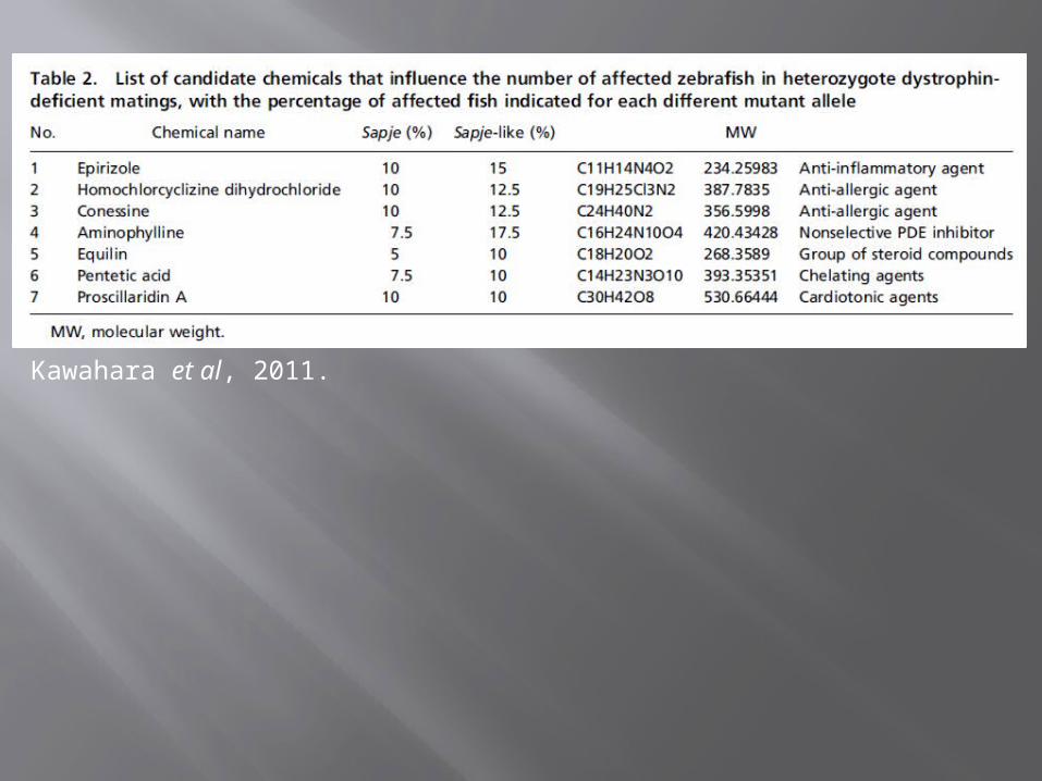
Kawahara et al, 2011.
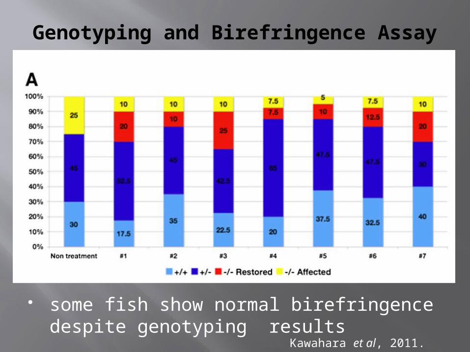
Genotyping and Birefringence Assay
some fish show normal birefringence despite genotyping results
Kawahara et al, 2011.

Immunostaining - an anti-dystrophin antibody.
restored muscle structure
no expression of dystrophin
Kawahara et al, 2011.

Dystrophin Morphants (MO)
(A and B) Normal light image (C and D) Birefringence image (E and G) Immunostaining - anti-dystrophin antibody (F and H) Immunostaining - anti-laminin antibody
WT
WT
WT
WT
Dys-mut
Dys-mut
Dys-mut
Dys-mut
Kawahara et al, 2011.
one to two cell stage WT embryos
4 dpf

www.humgen.nl

Testing 7 Candidate Chemicals (MO)
For each chemical treatment, the percentage of affected fish is reduced (4 dpf)
Kawahara et al, 2011.

Long-Term Culture Fish with 7 Candidate Chemicals (4-30 dpf)
chemicals 2,3,7 proved toxic to zebrafish the average number of surviving fish is greater in
chemical 4 Kawahara et al, 2011.

Long-Term Culture Fish with 7 Candidate Chemicals (1-30 dpf)
Red – survivorsLight Blue – controlBlue – WTGreen - untreated fish
Kawahara et al, 2011.

Chemical 4 - Aminophylline Surviving fish (30 dpf) were sectioned
skeletal muscle structure restored
Kawahara et al, 2011.

Chemical 4 - Aminophylline
Kawahara et al, 2011.

Treatment with chemical 4 restored the muscle structure of these
dystrophin-null fish

Aminophylline A nonselective PDE5 inhibitor Increases the levels of intercellular cAMP Activation of cAMP-dependant PKA Anti-inflammatory effects:
inhibition of inflammatory mediators activation of NF-κB
PDE5 inhibitor restores mdx mouse muscle to normal
The expression, phosphorylation, and activation of PKA were examined

PKA Expression and Activity
Immunoblot (C) pPKA/PKA Ratio (D) pProteins/proteins (E)
Kawahara et al, 2011.

Activated phosphorylated PKA and the activity
of PKA were increased in aminophylline-treated
fish
Intracellular cAMP is increased with
Aminophylline treatment
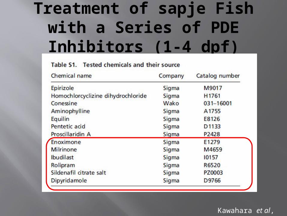
Treatment of sapje Fish with a Series of PDE Inhibitors (1-4 dpf)
Kawahara et al, 2011.

birefringence assay
sildenafil citrate and aminophylline decreased the percentage of fish showing abnormal birefringence
Kawahara et al, 2011.

Discussion
The muscle structure of aminophylline-treated dystrophin-null fish appeared normal
The activity of PKA is clearly up-regulated in aminophylline-treated dystrophin-null fish
Each of the seven chemicals increased the percentage of fish with normal birefringence
The chemical treatment did not restore dystrophin expression

Discussion
A two-tiered screening strategy PDE inhibitors cause an increase in intracellular
cAMP and/or cGMP Mutations in the zebrafish dystrophin gene (sapje
and sapje-like mutants): good models for studies of DMD ideally suited for use in chemical screens easily detectable by a highly accurate birefringence assay

Discussion
The zebrafish: small enough to be permeable to small molecules can be assayed in large numbers
Sildenafil (viagra ©) and Tadalafil (Cialis ©) have been independently identified by others
Thousands more compounds are now available for further screening

Thank you for listening!
Questions?

References1. Kawahara, Genri, et al. "Drug screening in a zebrafish model of
Duchenne muscular dystrophy." Proceedings of the National Academy of Sciences 108.13 (2011): 5331-5336.
2. Koenig, Michel, et al. "Complete cloning of the Duchenne muscular dystrophy (DMD) cDNA and preliminary genomic organization of the DMD gene in normal and affected individuals." Cell 50.3 (1987): 509-517.
3. Bassett, David I., and Peter D. Currie. "The zebrafish as a model for muscular dystrophy and congenital myopathy." Human molecular genetics 12.suppl 2 (2003): R265-R270.
4. Van Deutekom, Judith CT, and Gert-Jan B. Van Ommen. "Advances in Duchenne muscular dystrophy gene therapy." Nature Reviews Genetics 4.10 (2003): 774-783.


