
MATERNAL METABOLIC DISORDERS IDENTIFIED
THROUGH NEWBORN SCREENING
Marzia Pasquali PhD, FACMGProfessor of Pathology, University of Utah
Medical Director, Biochemical Genetics and Supplemental Newborn ScreeningARUP Laboratories
3rd Annual Winter Update in Laboratory and Clinical Medicine
Park City, Utah1 – 5 March 2010
OBJECTIVES
• Discuss metabolic disorders and newborn screening
• Review maternal diseases identified by newborn screening
• Discuss clinical implications

NORMAL METABOLISM• Our body assimilates
and transforms nutrients for growing and producing energy
• Metabolic disorders affect the transformation of nutrients to obtain energy and other products
• In most cases, these disorders are caused by mutations in enzymes which control the rate at which chemical reactions occur
http://www.accessexcellence.org/RC/VL/GG/

METABOLIC DISORDERS• The lack of an enzyme causes the accumulation of
toxic metabolites proximal to the metabolic block, byproducts not normally present, and the lack of required products
• This results in morbidity and mortality characteristic of each disease
Metabolic blockSubstrate
accumulationProduct
deficiency
Accumulation of byproducts
DFE
A B C

METABOLIC DISORDERS• Most metabolic disorders are inherited as recessive
traits• Heterozygotes do not show any clinical
manifestations
http://www.accessexcellence.org/RC/VL/GG/

FREQUENCY OF INHERITEDMETABOLIC DISORDERS
While the frequency of individual metabolic disorders is rare, their cumulative frequency is high (more than 1:3,000).PKU (phenylketonuria) 1:12,000MCAD deficiency 1:12,000Glutaric Acidemia Type 1 1:30,000Primary Carnitine Deficiency 1:40,000Propionic Acidemia 1:50,000Biotinidase deficiency 1:60,000Galactosemia 1:60,000Tyrosinemia Type I 1:100,000Methylmalonic acidemia 1:100,000Homocystinuria 1:120,000Maple Syrup Urine Disease 1:180,000
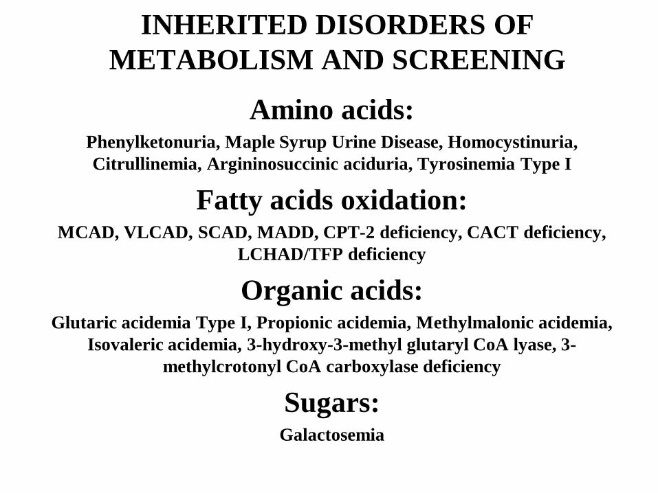
INHERITED DISORDERS OF METABOLISM AND SCREENING
Amino acids:Phenylketonuria, Maple Syrup Urine Disease, Homocystinuria, Citrullinemia, Argininosuccinic aciduria, Tyrosinemia Type I
Fatty acids oxidation:MCAD, VLCAD, SCAD, MADD, CPT-2 deficiency, CACT deficiency,
LCHAD/TFP deficiency
Organic acids:Glutaric acidemia Type I, Propionic acidemia, Methylmalonic acidemia,
Isovaleric acidemia, 3-hydroxy-3-methyl glutaryl CoA lyase, 3-methylcrotonyl CoA carboxylase deficiency
Sugars:Galactosemia
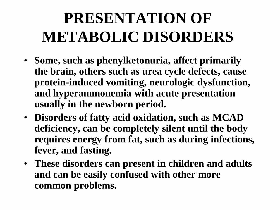
PRESENTATION OF METABOLIC DISORDERS
• Some, such as phenylketonuria, affect primarily the brain, others such as urea cycle defects, cause protein-induced vomiting, neurologic dysfunction, and hyperammonemia with acute presentation usually in the newborn period.
• Disorders of fatty acid oxidation, such as MCAD deficiency, can be completely silent until the body requires energy from fat, such as during infections, fever, and fasting.
• These disorders can present in children and adults and can be easily confused with other more common problems.

DIAGNOSIS OF METABOLIC DISORDERS
• Metabolic disorders can be suspected from clinical presentation and routine laboratory testing: metabolic acidosis, hyperammonemia, hypoglycemia, ketonuria
• Metabolic disorders require specific “routine” tests: plasma amino acids, urine organic acids, plasma carnitine, plasma acylcarnitine profile, urine acylglycine analysis
• The diagnosis is usually confirmed by DNA testing or enzyme/transporter/receptor assay

CURRENT THERAPY OF METABOLIC DISORDERS
• Restriction of toxic substrates (examples Phe in PKU, Phe+Tyr in tyrosinemia, milk in galactosemia, etc.)
• Provision of products (arginine in urea cycle defects, tyrosine in PKU)
• Inhibitors of the formation of toxic products/byproducts (NTBC in tyrosinemia type 1, allopurinol in gout, statins in hypercholesterolemia)
• Drugs to bypass or reduce the effects of the metabolic block (phenylbutyrate/benzoate in urea cycle defects, carnitine in fatty acid oxidation defects, glycine in isovaleric acidemia, etc)
• Pharmacologic amounts of vitamins to stabilize or bypass mutant enzymes (Thiamine in MSUD, B12 in MMA, biotin in multiple carboxylase deficiency, B6 in homocystinuria, biotin in biotinidase deficiency, etc.)
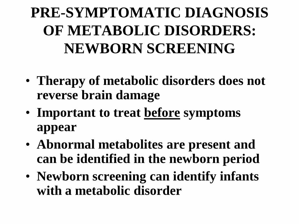
PRE-SYMPTOMATIC DIAGNOSIS OF METABOLIC DISORDERS:
NEWBORN SCREENING
• Therapy of metabolic disorders does not reverse brain damage
• Important to treat before symptoms appear
• Abnormal metabolites are present and can be identified in the newborn period
• Newborn screening can identify infants with a metabolic disorder

NEWBORN SCREENING
• Newborn screening is a public health activity aimed at the early identification
of conditions for which timely intervention can lead to the elimination
or reduction of mortality, morbidity, and disabilities associated with these
conditions.

NEWBORN SCREENING TODAY
• Mandated in all states in the United States
• Primarily performed by state public health laboratories
– Some contract with private laboratories– Some contract with other states– Some states are combined into regional
programs
• Newborn screening is the largest genetic testing effort in the nation

HOW IS NEWBORN SCREENING DONE?
Blood is collected from each newborn at time of discharge from the hospital by heel stick and
spotted on filter paper. Blood spots are sent to a centralized laboratory for analysis. Positive or suspicious results are followed up with a repeat newborn screen or with more definitive tests.
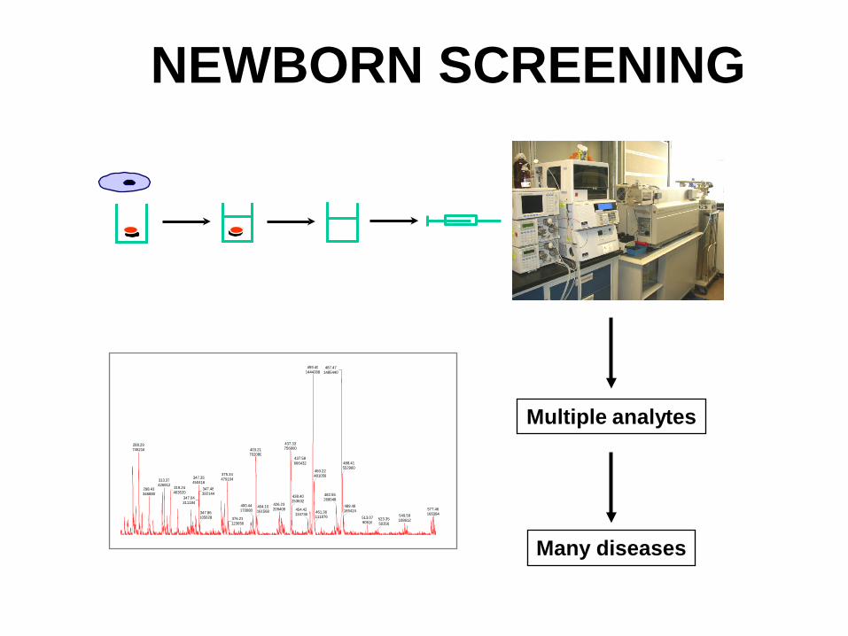
NEWBORN SCREENING
Multiple analytes
Many diseases
487.47 1485440 459.40
1444288
437.33 756800 288.29
745216 403.21 702080
375.34 479104 347.35
450416 313.37 426912
298.43 346880 319.24
403520 347.04 311184
347.48 392144
347.86 105628
400.44 170080
376.23 123656
426.23 209408 404.10
161568
437.58 666432
438.40 253632
454.42 134736
460.22 481056
482.55 268048
461.36 111876
488.41 552960
577.46 166384
489.48 165424
549.59 108612 513.07
90616 523.35 50356
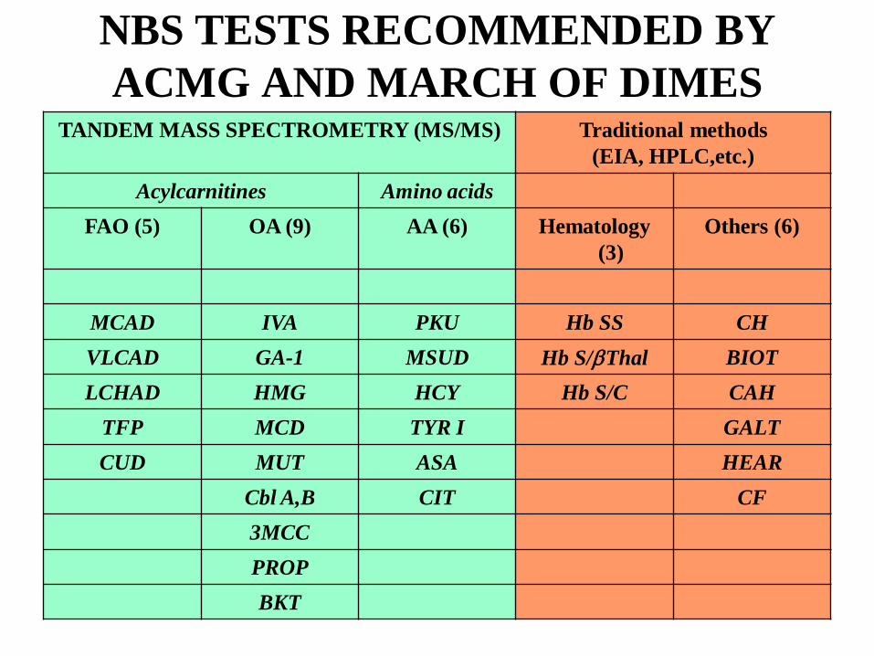
NBS TESTS RECOMMENDED BY ACMG AND MARCH OF DIMES
TANDEM MASS SPECTROMETRY (MS/MS) Traditional methods (EIA, HPLC,etc.)
Acylcarnitines Amino acidsFAO (5) OA (9) AA (6) Hematology
(3)Others (6)
MCAD IVA PKU Hb SS CHVLCAD GA-1 MSUD Hb S/βThal BIOTLCHAD HMG HCY Hb S/C CAH
TFP MCD TYR I GALTCUD MUT ASA HEAR
Cbl A,B CIT CF3MCCPROPBKT
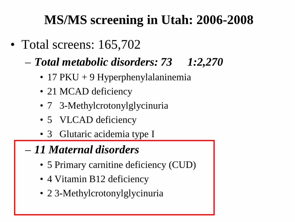
MS/MS screening in Utah: 2006-2008
• Total screens: 165,702– Total metabolic disorders: 73 1:2,270
• 17 PKU + 9 Hyperphenylalaninemia• 21 MCAD deficiency• 7 3-Methylcrotonylglycinuria• 5 VLCAD deficiency• 3 Glutaric acidemia type I
– 11 Maternal disorders• 5 Primary carnitine deficiency (CUD)• 4 Vitamin B12 deficiency• 2 3-Methylcrotonylglycinuria
MOTHERS WITH METABOLIC DISORDERS
• Several infants identified through newborn screening with a possible metabolic disorder, had completely normal confirmatory test results.
• This led to laboratory investigation of the mothers of these infants and their subsequent diagnosis.
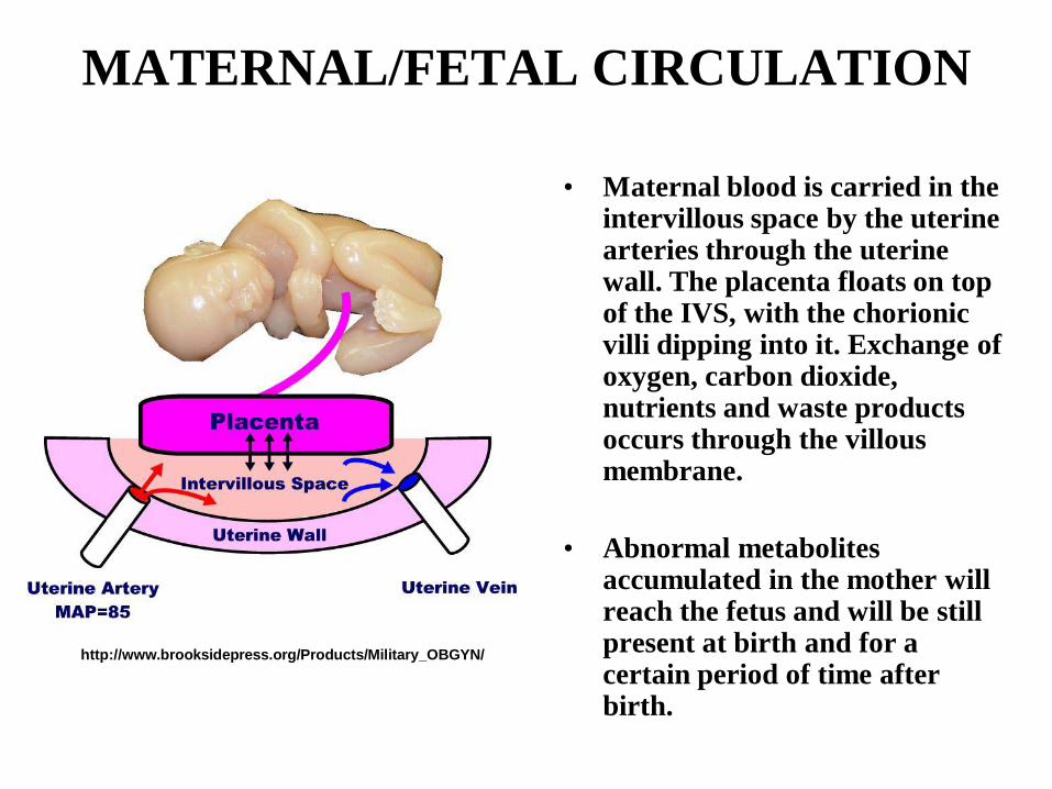
MATERNAL/FETAL CIRCULATION
• Maternal blood is carried in the intervillous space by the uterine arteries through the uterine wall. The placenta floats on top of the IVS, with the chorionic villi dipping into it. Exchange of oxygen, carbon dioxide, nutrients and waste products occurs through the villous membrane.
• Abnormal metabolites accumulated in the mother will reach the fetus and will be still present at birth and for a certain period of time after birth.
http://www.brooksidepress.org/Products/Military_OBGYN/

DISORDERS OF METABOLISMAmino acids/Fatty acids
Short, medium, long chainorganic acids/fatty acids
Energy production
Carnitine
Short, medium, long chain acylcarnitines

MARKERS OF MATERNAL DISEASE
• Primary markers:– Primary analyte, immediately upstream of
the metabolic block (amino acids, acylcarnitines)
• Secondary marker:– Carnitine (low concentrations resulting from
chronic depletion due to undiagnosed condition)
FEDA B C

PRIMARY MARKERS of MATERNAL DISEASES
• Amino acids: usually patients have already been identified – Phenylalanine (PKU); Citrulline
(Citrullinemia)• Acylcarnitines: the most frequently
identified maternal conditions are the following– C5OH-carnitine: 3-methylcrotonylglycinuria,
3-methylglutaconic aciduria– C3-carnitine: vitamin B12 deficiency
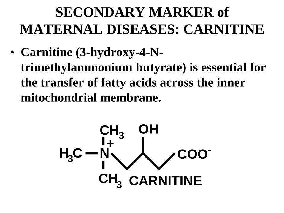
SECONDARY MARKER of MATERNAL DISEASES: CARNITINE
• Carnitine (3-hydroxy-4-N-trimethylammonium butyrate) is essential for the transfer of fatty acids across the inner mitochondrial membrane.
H CCH
CH
N COO-OH
+
3
3
3
CARNITINE

SOURCES OF CARNITINESynthesized by liver and kidneys, but not in heart or skeletal muscle, which depend on carnitine transport for fatty acid oxidation.
In the diet, most carnitine is supplied by red meat and dairy products, while fruits and vegetables contain insignificant amounts. About 75% of carnitine is provided by the diet in normal adults.
Carnitine is lost in the urine and secreted in the bile. Acute renal failure can result in high levels of plasma carnitine (100-300 µM). By contrast, chronic renal failure and dialysis can cause carnitine deficiency.
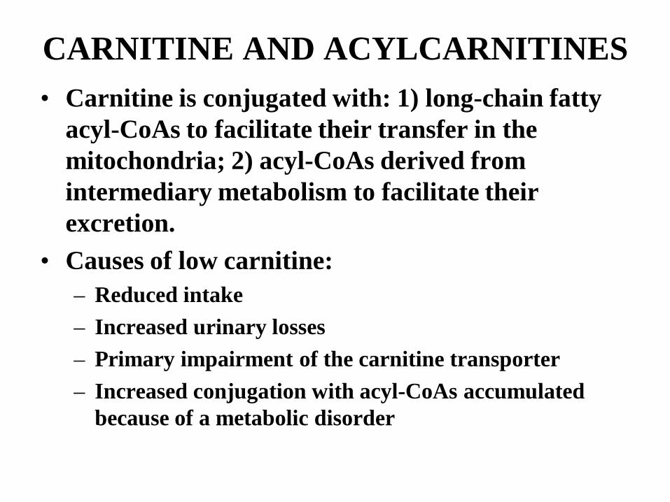
CARNITINE AND ACYLCARNITINES• Carnitine is conjugated with: 1) long-chain fatty
acyl-CoAs to facilitate their transfer in the mitochondria; 2) acyl-CoAs derived from intermediary metabolism to facilitate their excretion.
• Causes of low carnitine:– Reduced intake– Increased urinary losses– Primary impairment of the carnitine transporter – Increased conjugation with acyl-CoAs accumulated
because of a metabolic disorder

CPT-1
CACT
PlasmaMembraneOUT
IN
MITOCHONDRION
Acyl-S-CoA
ß-OXIDATION
PEROXISOMES
MEDIUM CHAIN DICARBOXYLIC ACIDS
MICROSOMESω , ω-1 oxidation
COOH
FATTY ACID
FATPCARNITINE
OHCOO
CH3
CH3
NH3C
OHCOO
CH3
CH3
NH3C
CoASHAcylCoA Synthase
OCOO
CH3
CH3
NH3C
Acyl Acyl-S-CoA
OCOO
CH3
CH3
NH3C
Acyl
COOH
FA
OCTN2
CPT-2OHCOO
CH3
CH3
NH3C
OHCOO
CH3
CH3
NH3C
Longo et al (2006)Am J Med Genet
THE CARNITINE CYCLE IN FATTY ACID OXIDATION

CARNITINE UPTAKE DEFECT(PRIMARY CARNITINE DEFICIENCY)
• Carnitine derives from diet and endogenous synthesis• Frequency 1:40,000 (1% are carriers)• Cause: Carnitine transporter (OCTN2)• Pathogenesis: Loss of carnitine in urine reduces
availability of carnitine in liver, muscle and heart, impairing FAO
• Presentation: Reye syndrome, sudden death, cardiomyopathy
• Diagnosis: Plasma carnitine levels (very low, usually <5 uM), confirmed by transport studies in fibroblasts
• Therapy: carnitine 100-300 mg/kg per day PO div. TID• Prognosis: excellent (with treatment)

FAROE ISLANDSSmall archipelago in the North Atlantic located between Scotland and Island. The Faroe Islands are a part of the Kingdom of Denmark, along with Denmark proper and Greenland. Population about 50,000 with another 20,000 living abroad, mostly in Denmark. They have a very high incidence of several IEMs. Carnitine uptake defect: 1:1,300 Founder mutation: N32S Young adults (age 25-30 years) died from ventricular fibrillation (no cardiomyopathy). Some complain of lassitude and weakness which improves with carnitine supplementation.

NEWBORN SCREENING FOR PRIMARY CARNITINE DEFICIENCY
Age (days)0 10 20 30 40 50
Free
Car
nitin
e ( µ
M)
0
10
20
30
40
50First Newborn screening
Second Newborn screening
NormalRange
Cutoff
After supplementation
Infant with primary carnitine deficiency Infant of mother with primary carnitine deficiency

NEWBORN SCREENING FOR PRIMARY CARNITINE DEFICIENCY• Carnitine is transferred from the mother to the
fetus during pregnancy.• Babies with primary carnitine deficiency usually
do not have extremely low concentrations of carnitine at birth; however their carnitine concentration decreases with time.
• Often, a second screen at 7-28 days of life identifies infants with primary carnitine deficiency.

NEWBORN SCREENING FOR PRIMARY CARNITINE DEFICIENCY• Extremely low concentrations of free carnitine in the
first screen (24-48 hours of life) of an infant are often associated with a maternal disease:– Maternal primary carnitine deficiency– Maternal Glutaric acidemia type I– Maternal MCAD deficiency– Maternal 3-methylcrotonylglycinuria
• Often, low carnitine is the only abnormal finding in the newborn screen of an infant with a mother with undiagnosed metabolic disorder.
• Appropriate follow-up includes evaluation of plasma free and total carnitine, plasma acylcarnitine profile and/or urine organic acids in both, mother and infant.
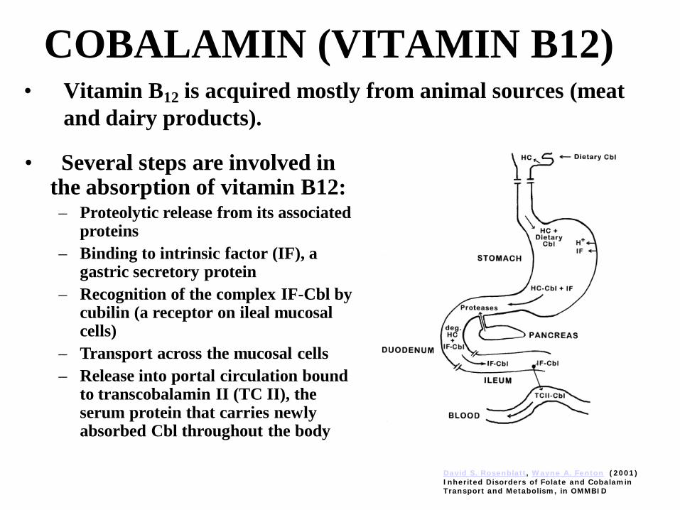
COBALAMIN (VITAMIN B12)• Vitamin B12 is acquired mostly from animal sources (meat
and dairy products).
• Several steps are involved in the absorption of vitamin B12:– Proteolytic release from its associated
proteins– Binding to intrinsic factor (IF), a
gastric secretory protein– Recognition of the complex IF-Cbl by
cubilin (a receptor on ileal mucosal cells)
– Transport across the mucosal cells– Release into portal circulation bound
to transcobalamin II (TC II), the serum protein that carries newly absorbed Cbl throughout the body
David S. Rosenblatt, Wayne A. Fenton (2001) Inherited Disorders of Folate and Cobalamin Transport and Metabolism, in OMMBID

COBALAMIN (VITAMIN B12)• Higher animals convert the vitamin into the two required
coenzyme forms, adenosylcobalamin (AdoCbl) and methylcobalamin (MeCbl).
• AdoCbl and MeCbl are co-factor of, respectively, methylmalonyl-CoA mutase (elevated methylmalonic acid) and methionine synthase (elevated homocysteine).
David S. Rosenblatt, Wayne A. Fenton (2001) Inherited Disorders of Folate and Cobalamin Transport and Metabolism, in OMMBID

MATERNAL VITAMIN B12 DEFICIENCY
• One of the most frequent maternal diseases identified is vitamin B12 deficiency.
• Infants of mothers with vitamin B12 deficiency show elevated C3-(propionyl-) carnitine in the newborn screen.
• Confirmatory tests performed on these infants (urine organic acids, plasma amino acids, total plasma homocysteine) will show elevated excretion of methylmalonic acid and possibly elevated total plasma homocysteine.
• After administration of vitamin B12, usually, laboratory tests normalize in these infants.
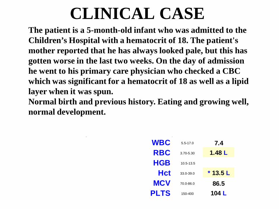
CLINICAL CASE
WBC 5.5-17.0 7.4RBC 3.70-5.30 1.48 L HGB 10.5-13.5
Hct 33.0-39.0 * 13.5 L MCV 70.0-86.0 86.5
PLTS 150-400 104 L
The patient is a 5-month-old infant who was admitted to the Children’s Hospital with a hematocrit of 18. The patient's mother reported that he has always looked pale, but this has gotten worse in the last two weeks. On the day of admission he went to his primary care physician who checked a CBC which was significant for a hematocrit of 18 as well as a lipid layer when it was spun. Normal birth and previous history. Eating and growing well, normal development.
B12 DEFICIENCY
1/14/2004 1/7/2004
5:45 22:15
Test Status Final Final
Amino Acid Interp., Ur * SEE NOTE * SEE NOTE
Alanine 200-600 umol/L 244 288Arginine 20-160 umol/L 35 20
Aspartic Acid 0-40 umol/L 5 6Citrulline Jun-60 umol/L 11 6
Cystine Jul-70 umol/L 28 18Glutamate 10-190 umol/L 80 54Glutamine 410-960 umol/L 819 703
Glycine 220-520 umol/L 210 L 165 L Histidine 40-120 umol/L 59 61
Homocystine NDT umol/L * NOT DET * PRESENT Hydroxyproline Jun-90 umol/L 19 8
Isoleucine 20-130 umol/L 27 35Allo-Isoleucine NDT umol/L * NOT DET * NOT DET
Leucine 40-230 umol/L 38 L 75Lysine 60-250 umol/L 109 105
Methionine Oct-60 umol/L 11 3 L Ornithine 20-135 umol/L 60 36
Phenylalanine 30-100 umol/L 13 L 35Proline 110-500 umol/L 164 204
Amino Acids, Plasma Quant. Show more...
Last Ref. Range Units
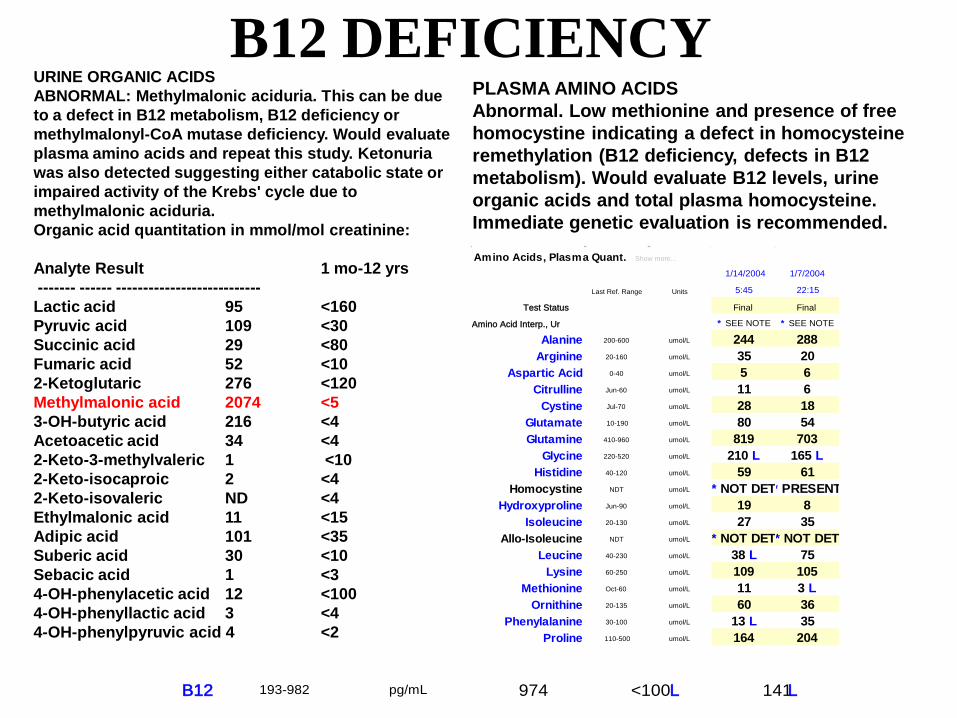
PLASMA AMINO ACIDSAbnormal. Low methionine and presence of free homocystine indicating a defect in homocysteine remethylation (B12 deficiency, defects in B12 metabolism). Would evaluate B12 levels, urine organic acids and total plasma homocysteine. Immediate genetic evaluation is recommended.
URINE ORGANIC ACIDSABNORMAL: Methylmalonic aciduria. This can be dueto a defect in B12 metabolism, B12 deficiency or methylmalonyl-CoA mutase deficiency. Would evaluate plasma amino acids and repeat this study. Ketonuria was also detected suggesting either catabolic state or impaired activity of the Krebs' cycle due to methylmalonic aciduria.Organic acid quantitation in mmol/mol creatinine:
Analyte Result 1 mo-12 yrs------- ------ ---------------------------Lactic acid 95 <160 Pyruvic acid 109 <30 Succinic acid 29 <80 Fumaric acid 52 <10 2-Ketoglutaric 276 <120 Methylmalonic acid 2074 <53-OH-butyric acid 216 <4 Acetoacetic acid 34 <42-Keto-3-methylvaleric 1 <10 2-Keto-isocaproic 2 <42-Keto-isovaleric ND <4 Ethylmalonic acid 11 <15Adipic acid 101 <35Suberic acid 30 <10Sebacic acid 1 <3 4-OH-phenylacetic acid 12 <1004-OH-phenyllactic acid 3 <44-OH-phenylpyruvic acid 4 <2
B12 193-982 pg/mL 974 <100 L 141 L

SUMMARY• Newborn screening for metabolic disorders can
identify effectively infants with a metabolic condition and, often, even mothers with undiagnosed diseases.
• The most frequent maternal diseases identified by newborn screening are primary carnitine deficiency (carnitine uptake defect) and vitamin B12 deficiency.
• Appropriate follow-up of abnormal newborn screen results may include evaluation of the mother.


