EVALUATION OF LOW BACK PAIN
Michael A. Mitchell, PA-CBone and Joints Sports Medicine

INTRODUCTION


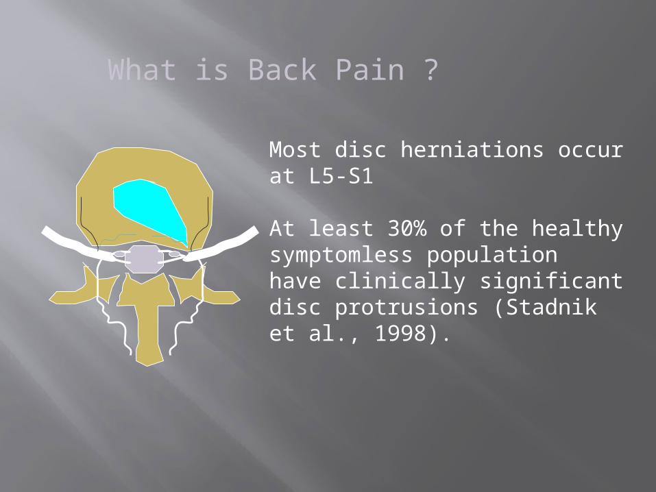
Most disc herniations occur at L5-S1
At least 30% of the healthy symptomless population have clinically significant disc protrusions (Stadnik et al., 1998).
What is Back Pain ?

What is Back Pain ?
Several studies have shown that there is no correlation between MRI findings and patients’ low back symptoms.
1. Wittenberg et al., 19982. Smith et al., 1998
3. Savage et al., 1997

What is Back Pain ?
There are many more joints in the back than discs.
There are many more muscles than joints.
The most common cause of low back pain is when one or more muscles “forget” to relax. We call this a somatic dysfunction.

Common Sources of LBPSomatic dysfunction
Muscle in “spasm”
Nerve root
In somatic dysfunction, some muscles become overactive (“spasm”)and other muscles become inactive.

Common Sources of LBP
Any dysfunction involving the thoracic or lumbarspine, the sacroiliac joint or the hip can createlow back pain.

Common Sources of LBP
L2L3L4L5
S1S2

Long dorsal si ligament
sacrotuberous ligament
sacrospinous ligament
sciatic nerve
piriformis
Common Sources of LBP

Role of the sacroiliac jointThe coxal bones consist of a thin shell of
cortical bone (1-2 mm) over trabecular bone.
Muscles play an important role in helping the pelvis resist stress.When muscles can’t work due to pain, the risk of injury increases.
BACK FACTS

Introduction
COMMON, 2ND only to URTI Tx is symptomatic HISTORY is critical to ruling out serious
issues. Conduct a Physical Exam to confirm and
assess functional status

What Causes Acute Low Back Pain Muscle strain? DJD or OA? Disc disease? Who cares?
Initially they are all treated same for the most part.
Most all get better with conservative treatment.
Beware of the serious causes!

Evaluate for “Red Flags”: May Signal Serious Causes of LBP
Cancer Infection Fracture Sciatica Cauda Equina syndrome Ankylosing spondylitis

Sciatica
The sciatic nerve is the longest nerve in your body. It runs from your spinal cord to your buttock and hip area and down the back of each leg. The term "sciatica" refers to pain that radiates along the path of this nerve — from your back down your buttock and leg. Source: Mayoclinic.com

Cauda Equina Syndrome:
Caused by massive midline disc herniation or mass compressing cord or cauda equina. Rare (<.04% of LBP patients). Needs emergent surgical referral.
Symptoms: bilateral lower extremity weakness, numbness, or progressive neurological deficit.
Ask about: Recent urinary retention (most common) or
incontinence? Fecal incontinence?
Ankylosing spondylitis
Ankylosing spondylitis is one of many forms of inflammatory arthritis, the most common of which is rheumatoid arthritis. Ankylosing spondylitis primarily causes inflammation of the joints between the vertebrae of your spine and the joints between your spine and pelvis (sacroiliac joints). Source: Mayoclinic.com

Evaluation of the Patient With LBP Start with a detailed history – your best
diagnostic tool. Get an idea of the severity. Look for the “red flags” of serious causes.
Use the physical exam to confirm what you suspect based on history.
Keep in mind: Most of the time you won’t have a definitive
diagnosis. Imaging rarely changes initial treatment. Most patients get better with conservative TX.

What Was the Mechanism of Injury or Overuse?
Was there an acute trauma or injury? Sudden severe pain with bending. Motor vehicle accident or fall.
Was there a recent history of excessive lifting or bending?
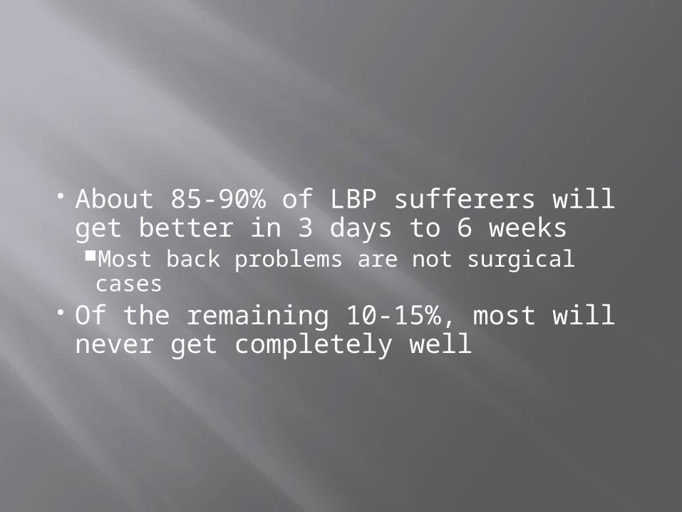
About 85-90% of LBP sufferers will get better in 3 days to 6 weeks Most back problems are not surgical cases
Of the remaining 10-15%, most will never get completely well

0 <3 >30
20
40
60
80
100
Succ
ess
Rate
(%
)
Risk Factors
Spine Surgery Outcomes
Treatment ApproachesSurgery

CAUSES/EXACERBATING
FACTORS

Mechanisms of Injury
Congenital abnormalities Poor body mechanics Back trauma
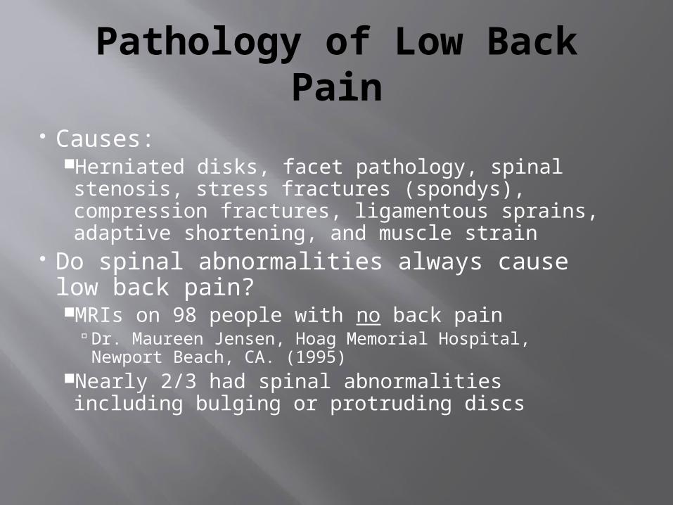
Pathology of Low Back Pain
Causes:Herniated disks, facet pathology, spinal stenosis, stress fractures (spondys), compression fractures, ligamentous sprains, adaptive shortening, and muscle strain
Do spinal abnormalities always cause low back pain?MRIs on 98 people with no back pain
Dr. Maureen Jensen, Hoag Memorial Hospital, Newport Beach, CA. (1995)
Nearly 2/3 had spinal abnormalities including bulging or protruding discs

Intervertebral Discs

THE KEY PLAYERS

Trunk Musculature
Musculature Superficial
Thoracic group Abdominal group Erector Spinae group
Spinalis Longissimus Iliocostalis
Deep Transversospinal group
Multifidus Rotatores Intertransversarius

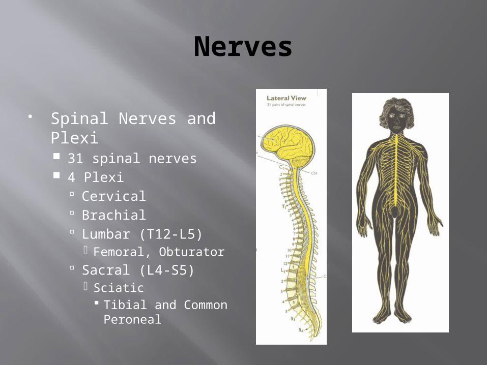
Nerves
Spinal Nerves and Plexi 31 spinal nerves 4 Plexi
Cervical Brachial Lumbar (T12-L5)
Femoral, Obturator Sacral (L4-S5)
Sciatic Tibial and Common
Peroneal

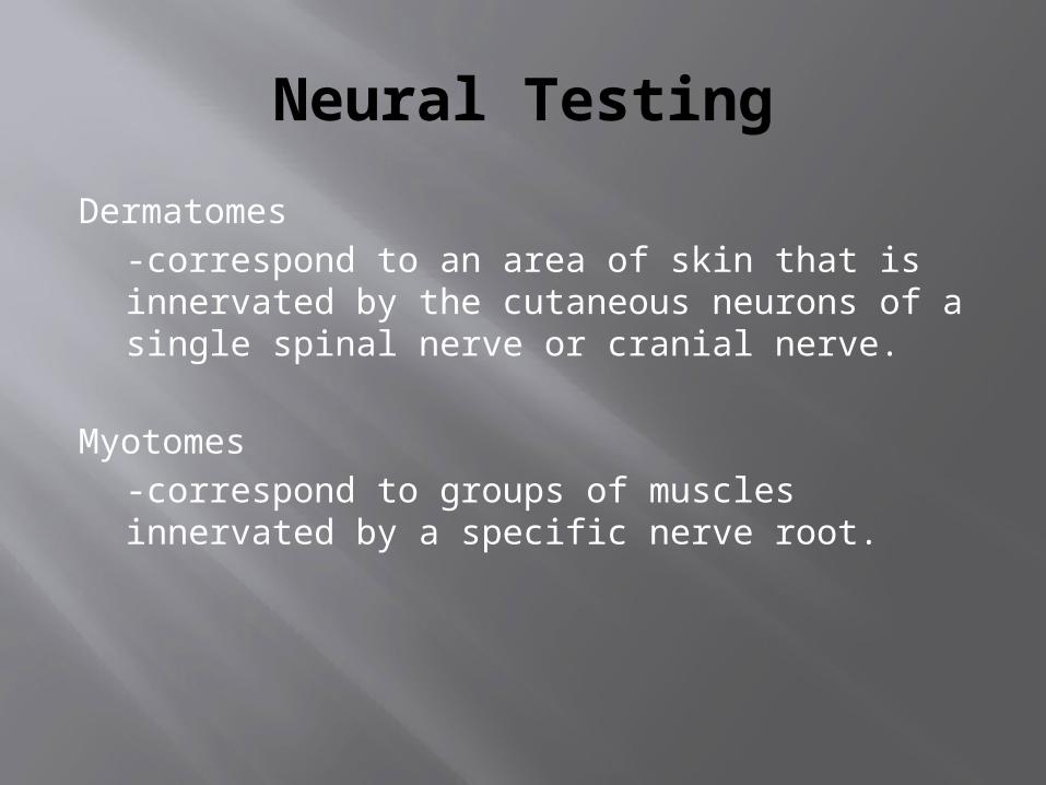
Neural Testing
Dermatomes-correspond to an area of skin that is innervated by the cutaneous neurons of a single spinal nerve or cranial nerve.
Myotomes-correspond to groups of muscles innervated by a specific nerve root.


CLASSIFICATION
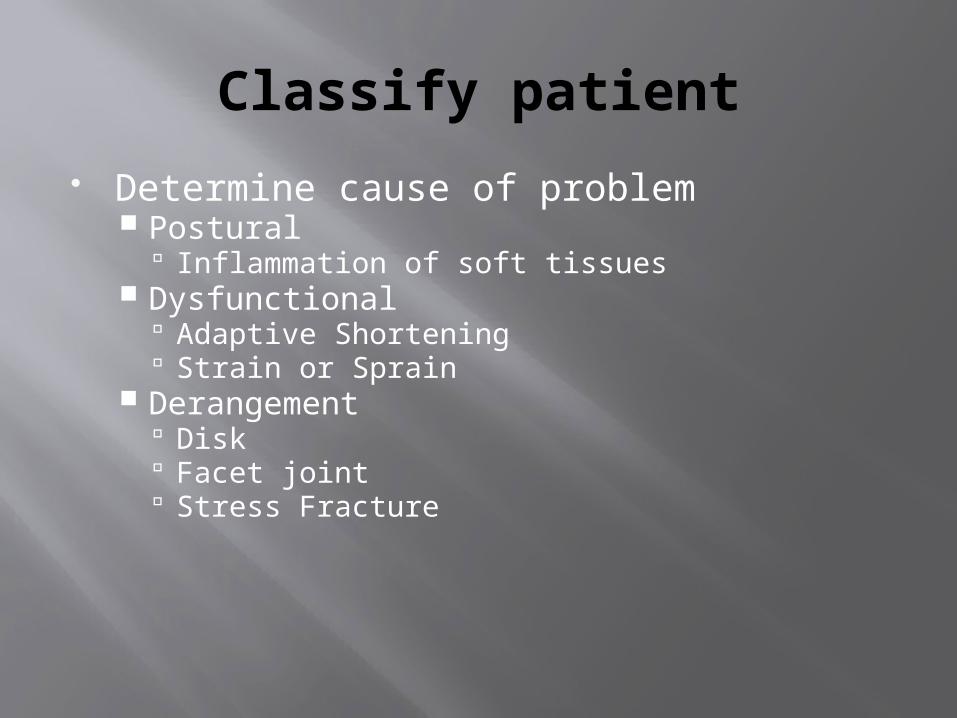
Classify patient
Determine cause of problem Postural
Inflammation of soft tissues Dysfunctional
Adaptive Shortening Strain or Sprain
Derangement Disk Facet joint Stress Fracture
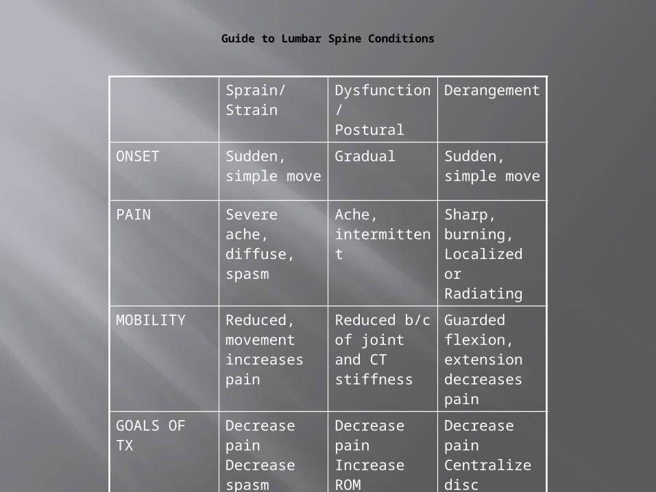
Sprain/Strain Dysfunction/Postural
Derangement
ONSET Sudden, simple move
Gradual Sudden, simple move
PAIN Severe ache, diffuse, spasm
Ache, intermittent
Sharp, burning, Localized or Radiating
MOBILITY Reduced, movement increases pain
Reduced b/c of joint and CT stiffness
Guarded flexion, extension decreases pain
GOALS OFTX
Decrease painDecrease spasmRestore ROM
Decrease pain Increase ROMPosture Strength/Flex
Decrease painCentralize discPrevention
Guide to Lumbar Spine Conditions

Lumbar Spine Conditions Low Back Muscle Strain
Acute (Overextension) and Chronic (Faulty posture) Facet Joint Dysfunction
Dislocation or Subluxation (Acute or Chronic) Low Back fracture
Compression, Stress, or Spinous and Transverse Processes
Herniated Disc Protrusion, Prolapse, Extrusion, and Sequestration Local and Radiating Pain
Classic term “Sciatica”
Lumbar Spine Conditions
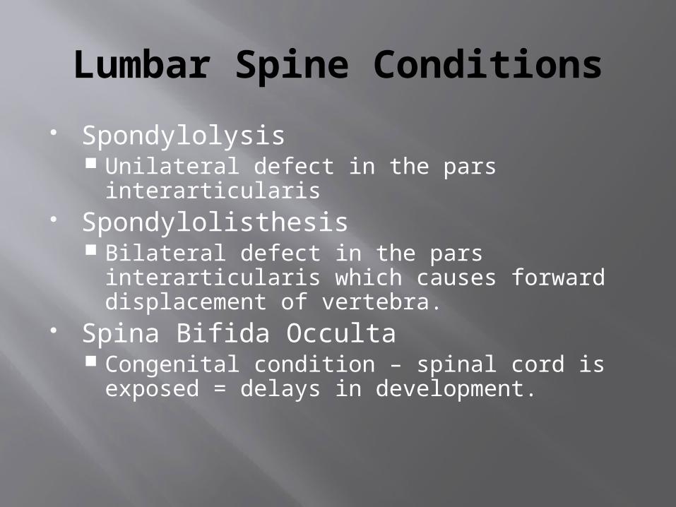
Spondylolysis Unilateral defect in the pars interarticularis
Spondylolisthesis Bilateral defect in the pars interarticularis
which causes forward displacement of vertebra.
Spina Bifida Occulta Congenital condition – spinal cord is exposed
= delays in development.

Sacroiliac Joint Conditions(note this is advanced)
Sacral torsion Forward or Backward torsion
Ilium torsion, upslip, downslip, outflare, inflare
Piriformis strain/trigger points

WALK THROUGH IT…WHAT YOU ARE THINKING.

Unique risk factors for athletes
High impact trauma: football, rugby
End range loading: gymnastics, diving
Overuse trauma: impact loading: distance running rotational loading: golf, baseball prolonged sitting: travel

Evaluation Techniques
HOPS/HIPS History, Observation/Inspection, Palpation,
Special Tests Your first priority!
Establish the integrity of the spinal cord and nerve roots
History and several specific tests provide information (Dermatomes, Myotomes, Reflexes)
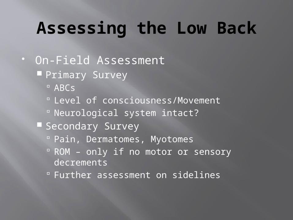
Assessing the Low Back
On-Field Assessment Primary Survey
ABCs Level of consciousness/Movement Neurological system intact?
Secondary Survey Pain, Dermatomes, Myotomes ROM – only if no motor or sensory decrements Further assessment on sidelines

Assessing the Low Back
Off-Field Assessment HISTORY!!!! Observation and Palpation
The Triad of Assessment Asymmetry, ROM alteration, Tissue texture
Special Tests Begin to be selective in your choices. Classify tests as to their main findings Use results of key tests to determine further
testing
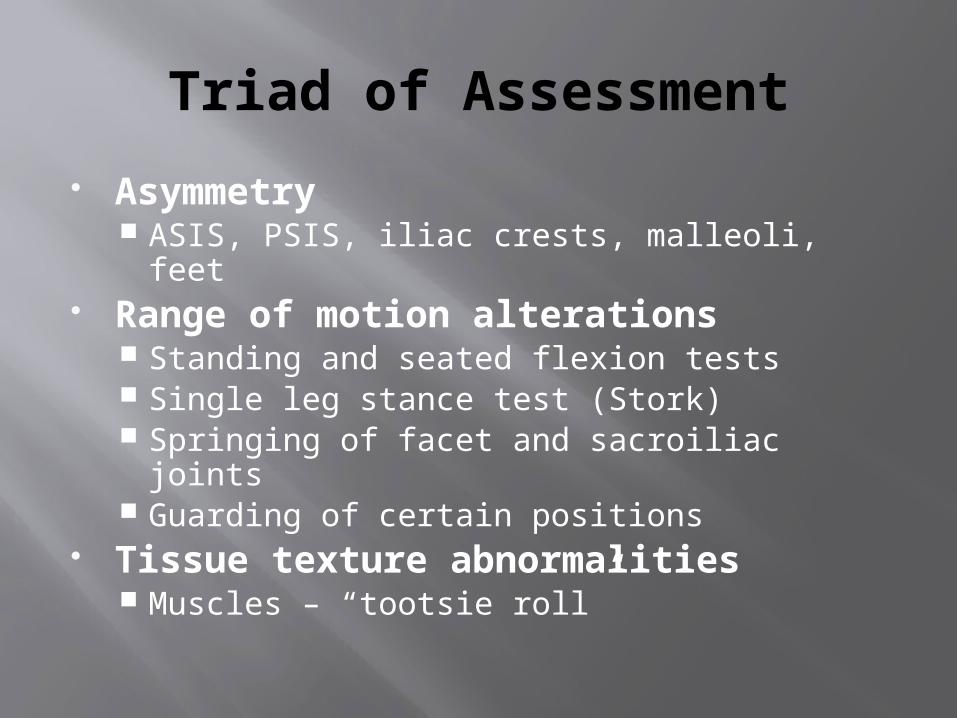
Triad of Assessment
Asymmetry ASIS, PSIS, iliac crests, malleoli, feet
Range of motion alterations Standing and seated flexion tests Single leg stance test (Stork) Springing of facet and sacroiliac joints Guarding of certain positions
Tissue texture abnormalities Muscles – “tootsie roll”

Kinetic Chain
Why do we need to assess the pelvis, hip and lower extremity?
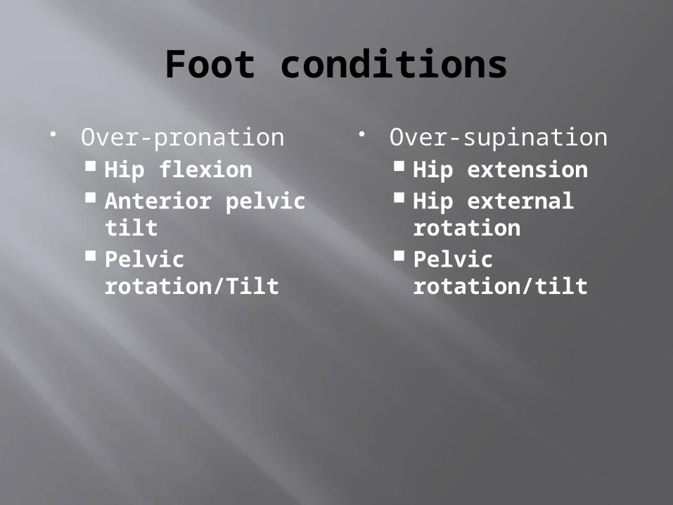
Foot conditions
Over-pronation Hip flexion Anterior pelvic
tilt Pelvic
rotation/Tilt
Over-supination Hip extension Hip external
rotation Pelvic
rotation/tilt

Specific evaluation techniques
1. HISTORY!!!!2. Alignment and
symmetry3. Lumbar spine
active movements4. Neurological
Testing5. Disc Pathology
Tests6. Extension
mechanics Prone assessment
7. Sacroiliac tests 8. Sitting forward
flexion and hip flexion
9. Standing forward flexion and hip flexion
10. Flexibility testing11. Feet alignment

History
Location of pain Onset of pain
Acute, chronic, or insidious Mechanism of Injury (MOI) Consistency of the pain
Constant vs. Intermittent pain Bowel and Bladder signs Changes in activity, surface, or
equipment

What positions bother you?
Bending Sitting Rising from sitting Standing Walking Lying prone Lying supine
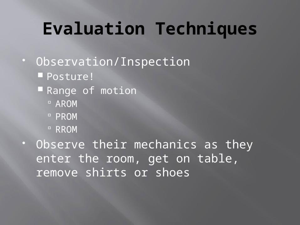
Evaluation Techniques
Observation/Inspection Posture! Range of motion
AROM PROM RROM
Observe their mechanics as they enter the room, get on table, remove shirts or shoes

Evaluation Techniques
Palpation This is your chance to “contain” the injury to
specific structures. Also allows for natural comparison of “normal”
landmarks Muscular Tension
“Tootsie Roll Test” Ligamentous Tests
Spring Test
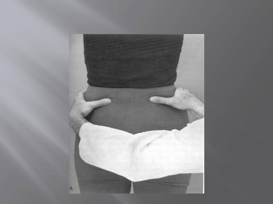
Special Tests
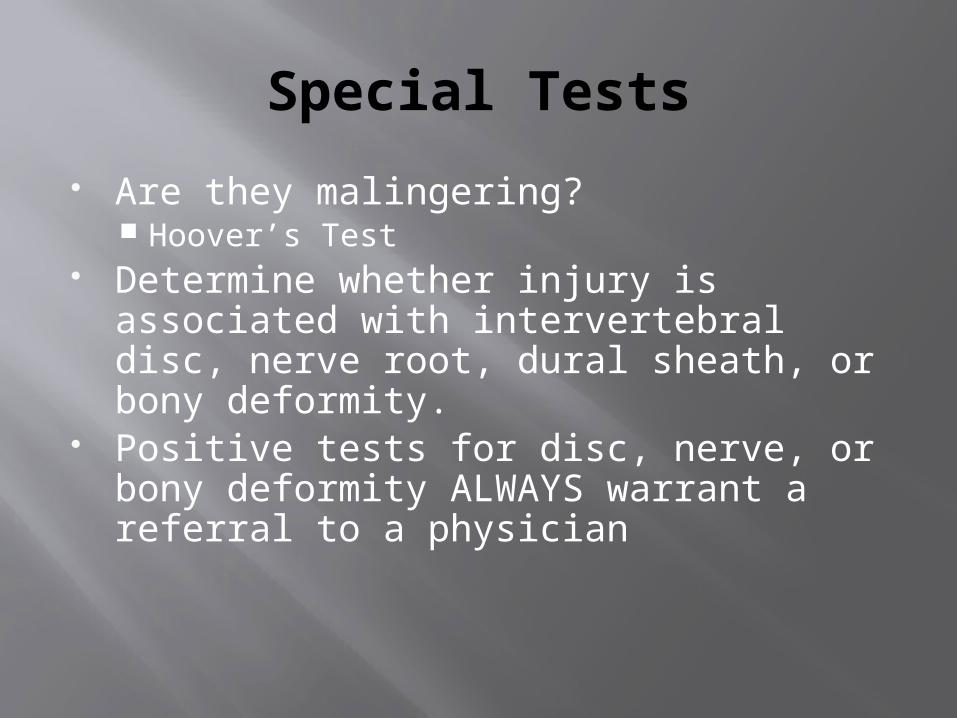
Are they malingering? Hoover’s Test
Determine whether injury is associated with intervertebral disc, nerve root, dural sheath, or bony deformity.
Positive tests for disc, nerve, or bony deformity ALWAYS warrant a referral to a physician

Tests for Nerve Root Impingement
Valsalva test Milgram test Kernigs/Brudzinski’s test Straight Leg Raise – Affected and Well Quadrant test Slump test

Lumbar Spine Conditions Low Back Muscle Strain
Very common and self-limiting Acute (Overextension) and Chronic (Faulty
posture) Pain increases with passive and active
flexion and resisted extension Key Evaluative techniques:
History and Palpation Rule out neural involvement Test PROM, AROM, and RROM

Lumbar Spine Conditions
Low Back fracture Compression or Stress Body, Spinous Process, and Transverse
Processes Localized or diffuse pain Treatment doesn’t relieve symptoms X-ray and MRI are definitive diagnoses

Lumbar Spine Conditions
Facet Joint Dysfunction Inflammation, sprain, degeneration Dislocation or Subluxation (Acute or Chronic)
“stuck open” or “stuck closed”
Usually localized but may involve several segments
May be associated with nerve root impingement
Often times pain decreases with activity

Facet Joint Dysfunction
AROM Flexion = “opening” and Extension = “closing” Lumbar facet joints “open” on right side with
left lateral flexion and left rotation Lumbar facet joints “close” on right side with
right lateral flexion and right rotation Prone assessment – elbows to hands Spring test Quadrant test


Lumbar Spine Conditions
Herniated Discs MOI: Overload (Direct or Indirect) or faulty
biomechanics (or both) Protrusion, Prolapse, Extrusion, and
Sequestration Pain usually aggravated by activity Prolonged body position often increases
symptoms Patient may choose a position that relieves pain
Local and Radiating Pain Reflexes and Sensory/Motor screening is essential
Definitive diagnosis comes from MRI
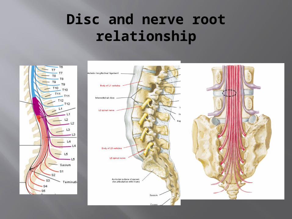
Disc and nerve root relationship


Neural Testing
Dermatomes Myotomes L1/L2 – Hip flexion L3/L4 – Knee
extension L4 – Ankle
dorsiflexion L5 – Great toe
extension S1 – Eversion S2 – Knee flexion

Observation Posture
Plum line Motions
Flexion Extension Lateral flexion Rotation


Back Malalignments

Discogenic Pain
Special Tests: Lower and Upper quarter screening
Dermatomes and Myotomes Valsalva test Milgram test Well straight leg raise Kernig’s/Brudzinski test Quadrant test


Lumbar Spine Conditions
Sciatica General term for inflammation of sciatic
nerve Sciatica is a result and NOT an injury in and
of itself Need to find what has caused the irritation
Disc, Muscle, Spondylopathy
Special tests: Straight leg raise Tension sign (Bowstrings) Slump Test

Lumbar Spine Conditions
Nerve Root Impingement/Dural Sheath Impingement Special Tests:
Quadrant test Femoral nerve stretch test Kernig’s/Brudzinski test Slump test

Lumbar Spine Conditions
Spondylopathies Mechanisms – Hyperextension
Onset – Insidious Muscular imbalances
Pain usually localized (may radiate) Increased during and after activity
Single leg stork stand Unilateral – Pain with opposite leg
MRI or X-ray are definitive diagnoses

Spondylosis Spondylolysis
generally mean changes in the vertebral joint characterized by increasing degeneration of the intervertebral disc with subsequent changes in the bones and soft tissues.
Unilateral or bilateral stable defect in the pars interarticularis
“Collared Scottie dog” deformity
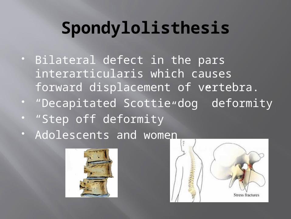
Spondylolisthesis
Bilateral defect in the pars interarticularis which causes forward displacement of vertebra.
“Decapitated Scottie dog” deformity “Step off deformity” Adolescents and women

Spondys
Treatment: REST and ice Flexion is best. Reduce extension moments. Bracing sometimes a solution.
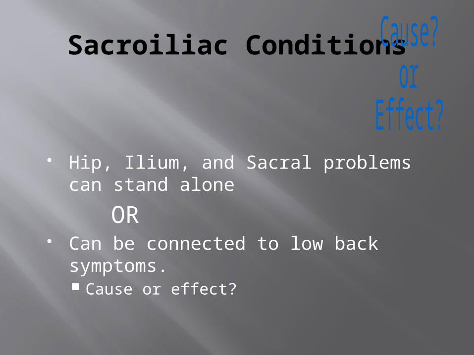
Sacroiliac Conditions
Hip, Ilium, and Sacral problems can stand alone
OR Can be connected to low back
symptoms. Cause or effect?

CAUSE or EFFECT?
Pelvis or Sacral alignment Hamstring Tightness
Straight Leg Raise 90/90 test
Hip Flexor tightness Thomas Test Trigger points
Piriformis tightness IR of hip is limited Trigger points

Special Tests for Pelvis and Sacrum
Alignment Supine and prone Prone extension
Sitting forward flexion and hip flexion
Monitoring PSIS Monitoring low back
Standing forward flexion and hip flexion
Monitoring PSIS Monitoring low back
Long Sitting Test Pen Dot Test FABERE Gaenslen’s Compression/
Distraction Outflare/Inflare

Pelvis and Sacral Conditions
PELVIS Upslip
ASIS and PSIS higher Anterior Rotation
ASIS lower, PSIS higher Tight hip flexor, weak
gluteus
Posterior Rotation ASIS higher, PSIS lower
Tight piriformis/gluteus and weak hip flexor
SACRUM Flexion – sulcus is
deep Extension – sulcus is
shallow Forward Torsion Backward Torsion

Questions?


